Station II - aacmyrms.org · Egyptian embalming General historical landmarks before Renaissence of...
13
Transcript of Station II - aacmyrms.org · Egyptian embalming General historical landmarks before Renaissence of...

Station II
Herodotus (484 B.C)Historian Greek.First description of Egyptian embalming
General historical landmarks before Renaissence of human anatomy
Hippocrates (460 - 377 B.C.)Anatomy science founder .The earliest description of
structures of the human body.
Papyrus of Edwin Smith (1600 BC)
First written evidenceswere found in Egyptiancivilization
Papyrus of Ebers (1550 BC)
Aristotle (384 - 322 B.C.) First to use the word
"anatomy“, derives from the Greek word "ana
temnein,"which means "to cut up."
Mondino de Luzzi (1270 - 1326 ) Italian physician. He published one of the �rst texts of human anatomy withouth drawings “Anatomia corporis humani”(1316). It is consideredthe �rst example of a modern dissection manual and as the �rst true anatomical text . He introducedthe anatomical dissection in medical courses.
Herophilus (335 - 280 B.C.) Greek MD of Medical School of Alexandria . First anatomical dissectionsin public (300 B.C).
Erasístrato Kea (304 - 250 B. C.) Cofounder and disector of
Medical School of Alexandria.
Galen of Pergamun (130 - 200 A.C)Greek physician. He developmentthe Modern anatomy. His ideas
dominated European medicine forover a thousand years. Because in
ancient Rome the dissection ofcorpses was forbidden by law, Galen
conducted studies dissecting animalssuch as pigs and monkeys.
Guido da Vigevano (1280 - 1350), disciple of Mondino, was an italian physician and inventor and he was also one of the �rst to add drawings of organs to his anatomical descriptions. In his book “Anathomia Designata per Figures”, written in 1345.
1
Station II “The great anatomists of the hand” in the Renaissance
Leonardo Da Vinci 1452 - 1519
Andrea Vesalio1514 - 1564
Bartolome Eustaquio1500 - 1574
Juan Valverde de Hamusco1525 - 1588
Giovanni B. Canano1515 - 1579
Gabrielle Fallopio1523 - 1562
Realdo Colombo 1516 - 1564
2
Station II “The great anatomists of the hand” in the Renaissance
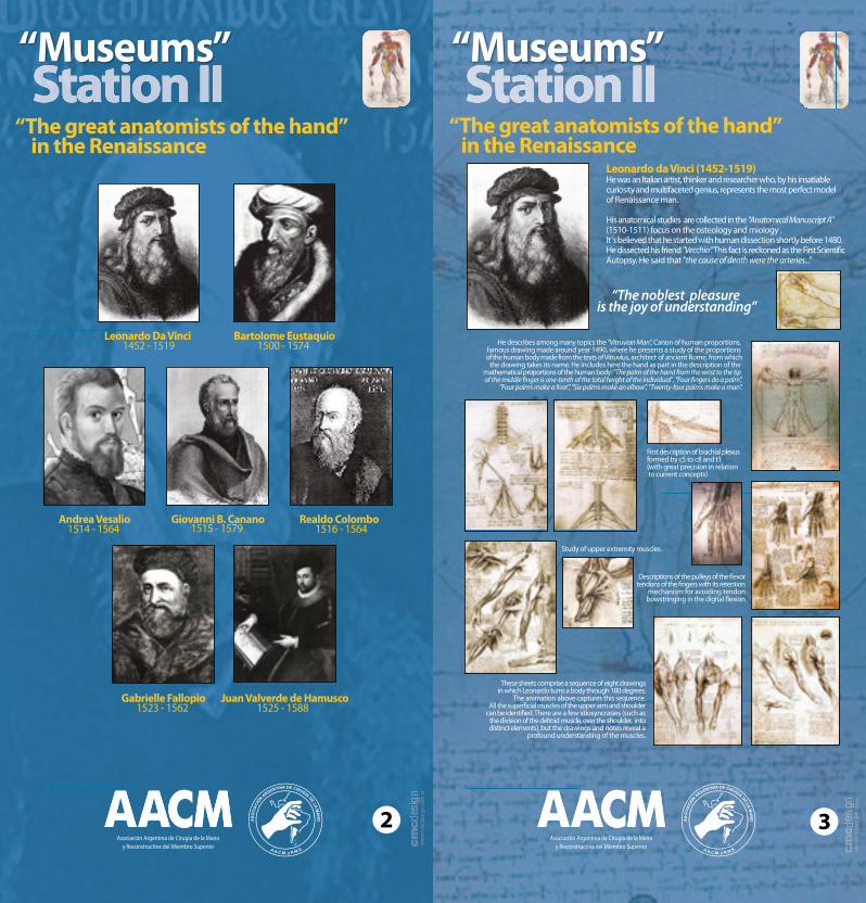
Leonardo da Vinci (1452-1519)He was an Italian artist, thinker and researcher who, by his insatiablecuriosity and multifaceted genius, represents the most perfect model of Renaissance man.
His anatomical studies are collected in the "Anatomical Manuscript A" (1510-1511) focus on the osteology and miology . It́ s believed that he started with human dissection shortly before 1480.He dissected his friend "Vecchio“. This fact is reckoned as the First Scienti�c Autopsy. He said that "the cause of death were the arteries..."
“The noblest pleasure is the joy of understanding”
He describes among many topics the "Vitruvian Man", Canon of human proportions, famous drawing made around year 1490, where he presents a study of the proportions of the human body made from the texts of Vitruvius, architect of ancient Rome, from which
the drawing takes its name. He Includes here the hand as part in the description of the mathematical proportions of the human body: "The palm of the hand from the wrist to the tip of the middle �nger is one-tenth of the total height of the individual", "Four �ngers do a palm”,
"Four palms make a foot”, "Six palms make an elbow”, "Twenty-four palms make a man”.
First description of brachial plexusformed by c5 to c8 and t1 (with great precision in relation to current concepts)
Study of upper extremity muscles.
Descriptions of the pulleys of the �exortendons of the �ngers with its retention
mechanism for avoiding tendon bowstringing in the digital �exion
These sheets comprise a sequence of eight drawings in which Leonardo turns a body through 180 degrees.
The animation above captures this sequence. All the super�cial muscles of the upper arm and shoulder
can be identi�ed. There are a few idiosyncrasies (such as the division of the deltoid muscle, over the shoulder, into distinct elements), but the drawings and notes reveal a
profound understanding of the muscles.
3

Station II Giovanni Battista Canano was born in Italy in 1515 and his work is mostly unknown. His only published work was “Musculorum humani corporis picturata dissection” in 1543, a small book but of outstandingimportance for its originality
It contains the �rst anatomical drawings of the lumbrical and interosseus muscles of the hand, and the �rst descriptionand drawing of the short palmar muscle (palmaris brevis)and of the thumb adductor, which Vesalius did never observed and which was unknown to Galeno. Also he described the extensor tendons in detail.
One of Valverde's most striking original plates is that of a muscle �gure holding his own skin in one hand and a knife in the other one, which hasbeen compared to St Bartholomew´s in The Last Judgment (Michelangelo) of the Sixtina Chapel. It was a major step in the use of spanish as the language of science, as it increased the anatomical vocabulary in this language.
Juan Valverde de Amusco (or "de Hamusco") (1525-1588)was born in the Kingdom of León (Spain) and studied medicine in Padua and Rome. He published "Historia de la Composición del Cuerpo Humano” (1556). All but four of its 42 engraved copperplate illustrationswere taken almost directly from Vesalius's Book, however Valverde corrected some Vesalius' images
He describes the function of cutaneous �xing of the palmar aponeurosis: "The palmar aponeurosisprovides, because of its many insertions, a strong and
stable hand grip" , and made the �rst descriptions of the extensor apparatus of the �ngers (single central tendon).
4
“The great anatomists of the hand” in the Renaissance
Station II
The renaissance descriptive anatomy acquires its apogee with Andreas Vesalius (1514-1564) born in a Bravantian (in modern-day Belgium). In 1537 he goes to Padua where he became, with only 23 years, professor of anatomy and surgery.
He published in 1543 "De humani corporis fabrica" consisting of 7 volumes with 300illustrations by E. Calcar, disciple of Tiziano (Italian painter).He describes the huge disparity that existed between what it can be read in the works ofGalen and the reality of dissection. He is considered to be "the founder of modern anatomy". He was the �rst to give each muscle a name according to the movement it performed.
He de�ned hand as the main part of the human body as among its functions he described: "to carry hand near the mouth to maintain whole body", "to exercise all the mechanical arts", "to perceive and recognize all objects by touch sensitivity, which is most perfect in the hand than in the rest of the body ", " they are useful for defending the body but also can attack holding all kinds of weapons”. For this last reason he called hands "weapons above all the weapons"
Hand’s intrinsic muscles and extensor tendons descriptions.
Hand’s bones and joints descriptions
Palmaris brevis anatomical details.Hand’s vascularization
5
“The great anatomists of the hand” in the Renaissance
Andreas Vesalius (1514-1564)
Station II
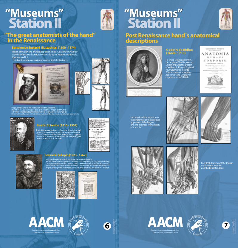
Italian physician and anatomist published his “Opuscula anatomica”(1563) in Venice with annotations made by his relative and disciple,Pier Matteo Pini. This book contains a series of anatomical illustrations.
The Italian anatomist born in Cremona, �rst disciple and then successor of Vesalius, with the support of his work "De re anatomica" (Venice 1559) is one of the undisputedreferences of anatomy. He described the morphology ofthe lumbrical muscles in the hand.
He gave the name to the "lumbrical" hand muscles and described the extensor apparatus of the last four �ngers and thumb with their interdigital connections with its approximate morphology. He drew the distal ends of the intrinsic muscles of the thumb on the extensor mechanism.
was another physician in�uenced by the work of Vesalius. He teached in Padua surgery and anatomy at the beginning of 1551, and publishing “Anatomical observations" (Venice, 1561); review of the Fabrica of Vesalius, without illustrations. In conjunction with Vesalius he described the extension of the IFD �ngers through the interosseous muscles and mentions the functions thereof.
“The great anatomists of the hand” in the Renaissance
6
Realdo Colombo (1516 - 1554)
Gabrielle Fallopio (1523 - 1562)
Bartolomeo Eustachi (Eustachius) (1500 - 1574)
Station II
He described the inclusion inthe phalanges of the extensor apparatus of the �ngersand the extensor retinaculum of the wrist
Post Renaissance hand´s anatomical descriptions
Excellent drawings of the thenar and intrinsic muscles and the �exor tendons
He was a Dutch anatomist. He taught at The Hague andLeiden and was the Doctor of William III, King of England. His major works include "Variae anatomiae medicae positiones" and "Anatomia corporis humani" (1685)
Godofredo Bidloo (1649 - 1713)
7

Station II Post Renaissance hand´s anatomicaldescriptions
Was a French physician and anatomist (Danish origin). His major work was "Exposition Anatomique de la Structure du Corps Humaine" in 1732. He describes the extensor tendinous rhombus, the extensor tendinous rhombus’s triangular ligament, the sagittal band (lateral expansion of the extensor tendons), the trapeziometacarpal joint as "double ginglymus", and recognizes the actual longitudinal rotation of the thumb. He is the �rst one to describe that some muscle �bers of the thenar muscles(which in the present are recognized aspart of the short �exor of the thumb) end up on the lateral or palmar sesamoid bone.
was a Dutch anatomist of German origin. He taught at the University of Leiden, best known for the exalted prints ofhis book "Tables of the skeleton and muscles of the human body" (1734)
In the hand he described: the transverse �bers of themiddle palmar fascia, the bifurcated �bers of pretendinosas bands ("two prolongued"), the current terminology of the thenar muscles; he gave the name to the "interosseous muscles" and classi�ed them in dorsal and palmar and �nallygave name also to the paratendinous vertical partitions
8
Jacques Benigne Winslow (1669 - 1760)
Bernhard Siegfried Albinus (1697 - 1770)
Station II Post Renaissance hand’s anatomical descriptions Anatomist’s French School. Nineteenth century
Portal 1742-1832 Bichat 1771-1802Desault 1744-1795
9
Dupuytren 1777-1835 Cloquet 1790 -1883
Bourgery 1797-1849
Testut 1849-1925 Rouvière 1876-1952 Poirier and Charpy 1899
Cruveilhier 1791-1874
Farabeuf 1841-1910
Latarjet 1877-1947
Duchenne 1806 -1875
Velpeau 1795-1867
Ranvier 1835-1922
Station II Post Renaissance hand’s anatomical descriptions
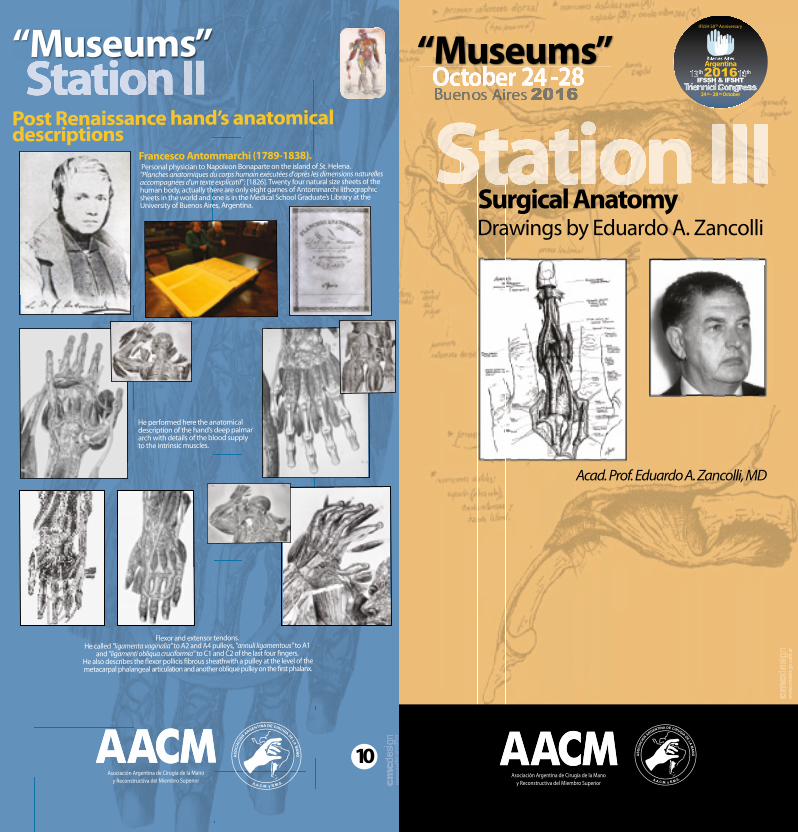
He performed here the anatomical description of the hand’s deep palmar arch with details of the blood supply to the intrinsic muscles.
Personal physician to Napoleon Bonaparte on the island of St. Helena. "Planches anatomiques du corps humain exécutées d'après les dimensions naturelles accompagnées d'un texte explicatif"; [1826]. Twenty four natural size sheets of thehuman body, actually there are only eight games of Antommarchi lithographicsheets in the world and one is in the Medical School Graduate’s Library at the University of Buenos Aires, Argentina.
Flexor and extensor tendons. He called "ligamenta vaginalia" to A2 and A4 pulleys, "annuli ligamentous" to A1
and "ligamenti obliqua cruciformia" to C1 and C2 of the last four �ngers. He also describes the �exor pollicis �brous sheathwith a pulley at the level of themetacarpal phalangeal articulation and another oblique pulley on the �rst phalanx.
10
Francesco Antommarchi (1789-1838).
Station III Station III Station III Station III Station III Station III Station III Station III Station III Station III Station III Station III Station III Station III Station III
Ed. Med. Pan.
Ed. Med. Pan.
Ed. Med. Pan.
Ed. Med. Pan.
Surgical AnatomyDrawings by EA Zancolli
Station III Surgical AnatomyDrawings by EA Zancolli
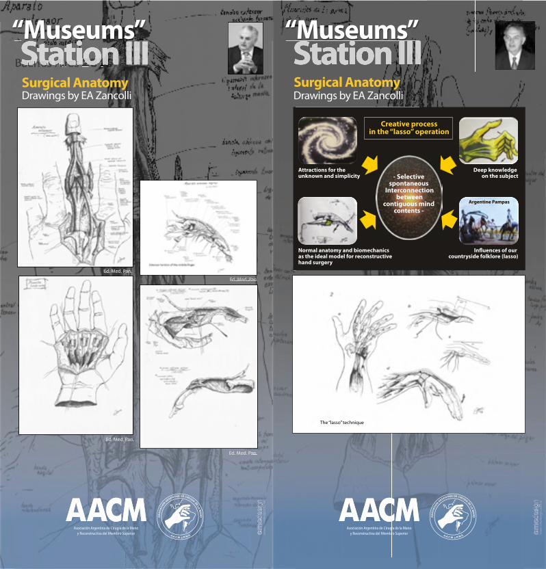
- Selective spontaneous
Interconnection between
contiguous mind contents -
Creative processin the “lasso” operation
Normal anatomy and biomechanics as the ideal model for reconstructive hand surgery
In�uences of our countryside folklore (lasso)
Deep knowledge on the subject
Attractions for the unknown and simplicity
Argentine PampasArgentine Pampas
The “lasso” technique

Station III Station III
Ed. Med. Pan.
Ed. Med. Pan.
Ed. Med. Pan.
Surgical AnatomyDrawings by EA Zancolli
Station III
Ed. Med. Pan.
Surgical AnatomyDrawings by EA Zancolli
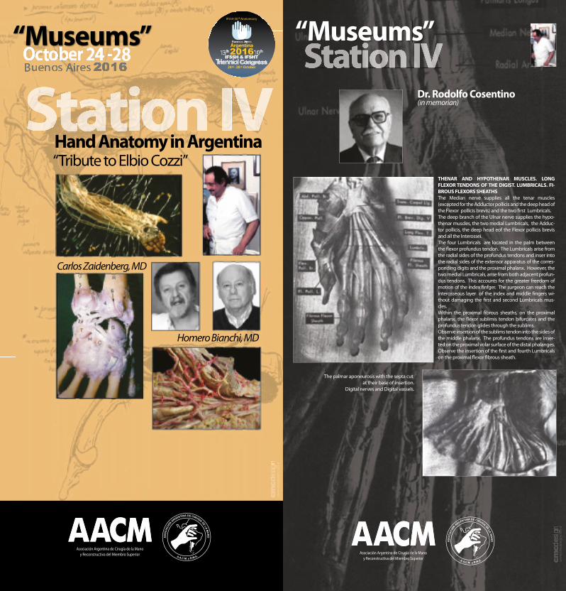
Station IV Dr. Rodolfo Cosentino(in memorian)
THENAR AND HYPOTHENAR MUSCLES. LONG FLEXOR TENDONS OF THE DIGIST. LUMBRICALS. FI-BROUS FLEXORS SHEATHSThe Median nerve supplies all the tenar muscles (excepted for the Adductor pollicis and the deep head of the Flexor pollicis brevis) and the two �rst Lumbricals.The deep branch of the Ulnar nerve supplies the hypo-thenar muscles, the two medial Lumbricals, the Adduc-tor pollicis, the deep head eof the Flexor pollicis brevis and all the Interossei.The four Lumbricals are located in the palm between the �exor profundus tendon. The Lumbricals arise from the radial sides of the profundus tendons and inser into the radial sides of the extensor apparatus of the corres-ponding digits and the proximal phalanx. However, the two medial Lumbricals, arise from both adjacent profun-dus tendons. This accounts for the greater freedom of motion of the index �nfger. The surgeon can reach the interosseous layer of the index and middle �ngers wi-thout damaging the �rst and second Lumbricals mus-cles.Within the proximal �brous sheaths, on the proximal phalanx, the �exor sublimis tendon bifurcates and the profundus tendon glides through the sublims.Observe insertion of the sublims tendon into the sides of the middle phalanx. The profundus tendons are inser-ted on the proximal volar surface of the distal phalanges. Observe the insertion of the �rst and fourth Lumbricals on the proximal �exor �brous sheath.
The palmar aponeurosis with the septa cut at their base of insertion.
Digital nerves and Digital vassels.

Station IV Dr. Roque Nigro
Plexus Brachialis Vascularizations
Station IV
Dr. Luciano Poitevin
Blood Supply to the Fingertip
Distal Interosseous Membrane of the forearm
Blood Supply to the PIP Jointand �exor tendons
Scalene Muscle Complex
First Rib. Costo-Septo-Costal Ligament
T1 Buttonhole
Radio-Scapholunate-Ligament (Testut-Kuenz)

Station IV
Braquial plexus
Supraescapular and frenic nerves
Cervical’s roots
Braquial plexus vascularization
Symphatetic �bers braquial plexus
Latisimus dorsi fascicle
Original Dissections by Elbio CozziStation IV
Original Dissections by Elbio Cozzi
Braquial plexus
Ulnar nerve fasciculation
Median nerve
Super�cial palmar arch