Starch Biosynthesis - Plant Cell · Starch is the most significant form of carbon reserve in plants...
16
The Plant Cell, Vol. 7, 971-985, July 1995 O 1995 American Society of Plant Physiologists Starch Biosynthesis Cathie Martinl and Alison M. Smith John lnnes Centre, Norwich Research Park, Colney, Norwich NR4 7UH, United Kingdom INTRODUCTION Starch is the most significantform of carbon reserve in plants in terms of the amount made, the universalityof its distribution among different plant species, and its commercial importance. It consists of different glucose polymers arranged into a three- dimensional,semicrystallinestructure-the starch granule. The biosynthesis of starch involves not only the production of the composite glucans but also their arrangement into an orga- nized form within the starch granule. The formationof the starch granule can be viewed as a simple model for the forma- tion of ordered three-dimensional polysaccharide structures in plants. Understanding the biochemical basis for the as- sembly of the granule could provide a conceptual basis for understanding other higher order biosynthetic systems such as cellulose biosynthesis (see Delmer and Amor, 1995, this issue). For example, one emerging concept is that structure within the granule itself may determine or influence the way in which starch polymers are synthesized. Starch is synthesized in leaves during the day from pho- tosynthetically fixed carbon and is mobilized at night. It is also synthesized transiently in other organs, such as meristems and root cap cells, but its major site of accumulation is in stor- age organs, including seeds, fruits, tubers, and storage roots. Almost all structural studies have used starch from storage organs because it is readily available and commercially im- portant; we therefore focus on starch biosynthesis in storage organs. However, where aspects of transient biosynthesis are clearly different from long-term reserve synthesis, reference is made to biosynthesis in nonstorage tissues. Starch is synthesized in plastids, which in storage organs committed primarily to starch production are called amylo- plasts. These develop directly from proplastids and have little interna1lamellar structure. Starch may also be synthesized in plastids that have other specialized functions, such as chlo- roplasts (photosynthetic carbon fixation), plastids of oilseed (fatty acid biosynthesis), and chromoplasts of roots such as carrot (carotenoid biosynthesis). In some cases, for example, in the storage cotyledonsof some legumes, amyloplasts in stor- age organs develop from chloroplasts.These amyloplasts may maintain considerable amounts of stacked lamellar material from the thylakoids and, in cells receivingsufficient light, may undertakesome photosynthetic carbon fixationfor use in starch To whom correspondence should be addressed. biosynthesis and thus supplement the sucrose imported for starch biosynthesis from the rest of the plant (Smith and Denyer, 1992). THE STRUCTURE OF STARCH AND THE STARCH GRANULE Starch can be chemically fractionated into two types of glu- can polymer: amylose and amylopectin. Amylose consists of predominantlylinear chains of a(l4)-linked glucose residues, each -1000 residues long. Amylose is usually branched at a low leve1(approximately one branch per 1000 residues) by a(1-6) linkages and makes up -30% of starch. This propor- tion, however, may vary considerably with the plant species (a range of 11 to 35% was found in a survey of 51 species; Detherage et al., 1955) and variety (a range of 20 to 36% was found in a survey of 399 maize varieties; Detherageet al., 1955) and also with the plant organ, the developmental age of that organ, and, to some extent, the growth conditionsof the plant (Shannon and Garwood, 1984). Once extracted from plants and in solution, amylose forms hydrogen bonds between mol- ecules, resulting in rigid gels. However, depending on the concentration, degree of polymerization, and temperature, it may crystallize and shrink (retrogradation) after heating (Shewmaker and Stalker, 1992). Amylopectin, which consists of highly branched glucan chains, makes up -70% of starch. Chains of roughly 20 a(l4)-linked glucose residues are joined by a(1-6) linkages to other branches. The branches themselves form an orga- nized structure (Figure 1A). Some are not substituted on the six positions and are called A chains. These chains are a(1-6) linked to inner branches (B chains), which may be branched at one or severa1points. A single chain per amylopectin mole- cule has a free reducingend (the C chain). The branches are not randomly arranged but are clustered at 7- to 10-nm inter- vals (Figure 1). An averageamylopectin molecule is 200 to 400 nm long (20 to 40 clusters) and -15 nm wide (for review, see Kainuma, 1988; Smith and Martin, 1993). After extraction, amylopectin has more limited hydrogen bonding than amy- lose in solutionand is more stable, remaining fluid and giving high viscosity and elasticity to pastes and thickeners. Some '
Transcript of Starch Biosynthesis - Plant Cell · Starch is the most significant form of carbon reserve in plants...

The Plant Cell, Vol. 7, 971-985, July 1995 O 1995 American Society of Plant Physiologists
Starch Biosynthesis
Cathie Martinl and Alison M. Smith John lnnes Centre, Norwich Research Park, Colney, Norwich NR4 7UH, United Kingdom
INTRODUCTION
Starch is the most significant form of carbon reserve in plants in terms of the amount made, the universality of its distribution among different plant species, and its commercial importance. It consists of different glucose polymers arranged into a three- dimensional, semicrystalline structure-the starch granule. The biosynthesis of starch involves not only the production of the composite glucans but also their arrangement into an orga- nized form within the starch granule. The formation of the starch granule can be viewed as a simple model for the forma- tion of ordered three-dimensional polysaccharide structures in plants. Understanding the biochemical basis for the as- sembly of the granule could provide a conceptual basis for understanding other higher order biosynthetic systems such as cellulose biosynthesis (see Delmer and Amor, 1995, this issue). For example, one emerging concept is that structure within the granule itself may determine or influence the way in which starch polymers are synthesized.
Starch is synthesized in leaves during the day from pho- tosynthetically fixed carbon and is mobilized at night. It is also synthesized transiently in other organs, such as meristems and root cap cells, but its major site of accumulation is in stor- age organs, including seeds, fruits, tubers, and storage roots. Almost all structural studies have used starch from storage organs because it is readily available and commercially im- portant; we therefore focus on starch biosynthesis in storage organs. However, where aspects of transient biosynthesis are clearly different from long-term reserve synthesis, reference is made to biosynthesis in nonstorage tissues.
Starch is synthesized in plastids, which in storage organs committed primarily to starch production are called amylo- plasts. These develop directly from proplastids and have little interna1 lamellar structure. Starch may also be synthesized in plastids that have other specialized functions, such as chlo- roplasts (photosynthetic carbon fixation), plastids of oilseed (fatty acid biosynthesis), and chromoplasts of roots such as carrot (carotenoid biosynthesis). In some cases, for example, in the storage cotyledons of some legumes, amyloplasts in stor- age organs develop from chloroplasts. These amyloplasts may maintain considerable amounts of stacked lamellar material from the thylakoids and, in cells receiving sufficient light, may undertake some photosynthetic carbon fixation for use in starch
To whom correspondence should be addressed.
biosynthesis and thus supplement the sucrose imported for starch biosynthesis from the rest of the plant (Smith and Denyer, 1992).
THE STRUCTURE OF STARCH AND THE STARCH GRANULE
Starch can be chemically fractionated into two types of glu- can polymer: amylose and amylopectin. Amylose consists of predominantly linear chains of a(l4)-linked glucose residues, each -1000 residues long. Amylose is usually branched at a low leve1 (approximately one branch per 1000 residues) by a(1-6) linkages and makes up -30% of starch. This propor- tion, however, may vary considerably with the plant species (a range of 11 to 35% was found in a survey of 51 species; Detherage et al., 1955) and variety (a range of 20 to 36% was found in a survey of 399 maize varieties; Detherage et al., 1955) and also with the plant organ, the developmental age of that organ, and, to some extent, the growth conditions of the plant (Shannon and Garwood, 1984). Once extracted from plants and in solution, amylose forms hydrogen bonds between mol- ecules, resulting in rigid gels. However, depending on the concentration, degree of polymerization, and temperature, it may crystallize and shrink (retrogradation) after heating (Shewmaker and Stalker, 1992).
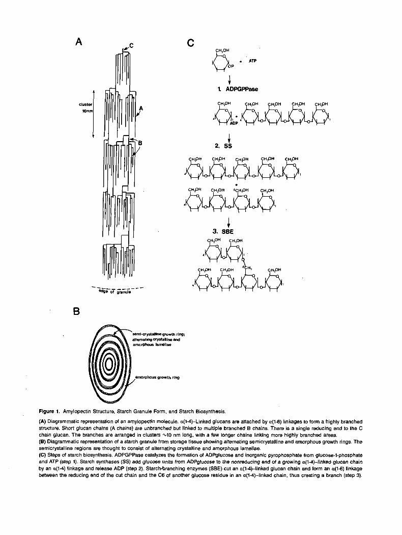
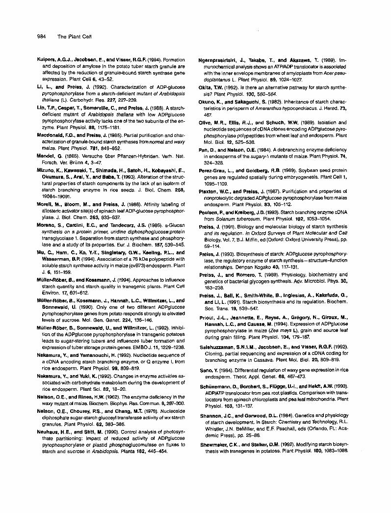
Amylopectin, which consists of highly branched glucan chains, makes up -70% of starch. Chains of roughly 20 a(l4)-linked glucose residues are joined by a(1-6) linkages to other branches. The branches themselves form an orga- nized structure (Figure 1A). Some are not substituted on the six positions and are called A chains. These chains are a(1-6) linked to inner branches (B chains), which may be branched at one or severa1 points. A single chain per amylopectin mole- cule has a free reducing end (the C chain). The branches are not randomly arranged but are clustered at 7- to 10-nm inter- vals (Figure 1). An average amylopectin molecule is 200 to 400 nm long (20 to 40 clusters) and -15 nm wide (for review, see Kainuma, 1988; Smith and Martin, 1993). After extraction, amylopectin has more limited hydrogen bonding than amy- lose in solution and is more stable, remaining fluid and giving high viscosity and elasticity to pastes and thickeners. Some
'
A
cluster
lonm
C r Y
- ---_ _ _ _ -- - adge ot granule
CH,OH C oop + ATp
4 1. ADPGPPase
-c 2. ss
1 I
4 1
.c 3. SBE
B
semi-aystallime growih ring; altemating Crystalline md amorphous lamellae
amorphous growth ring
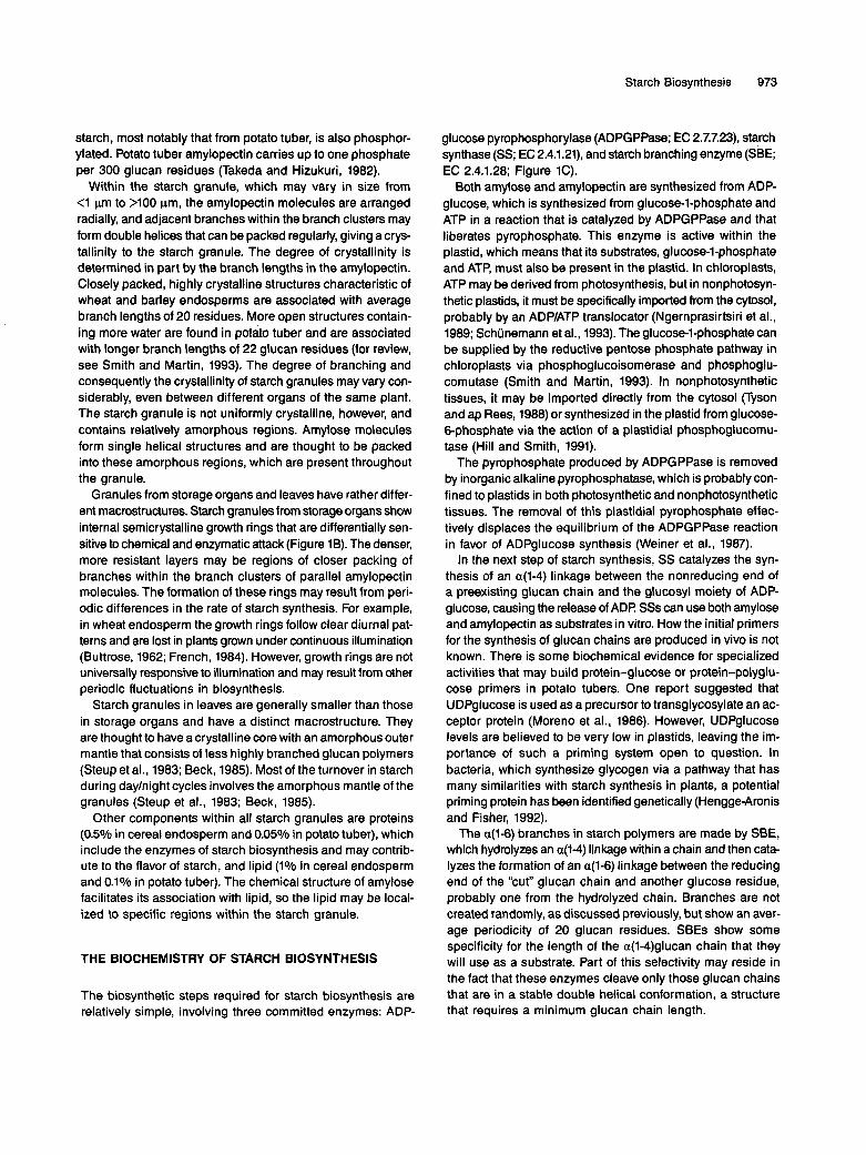
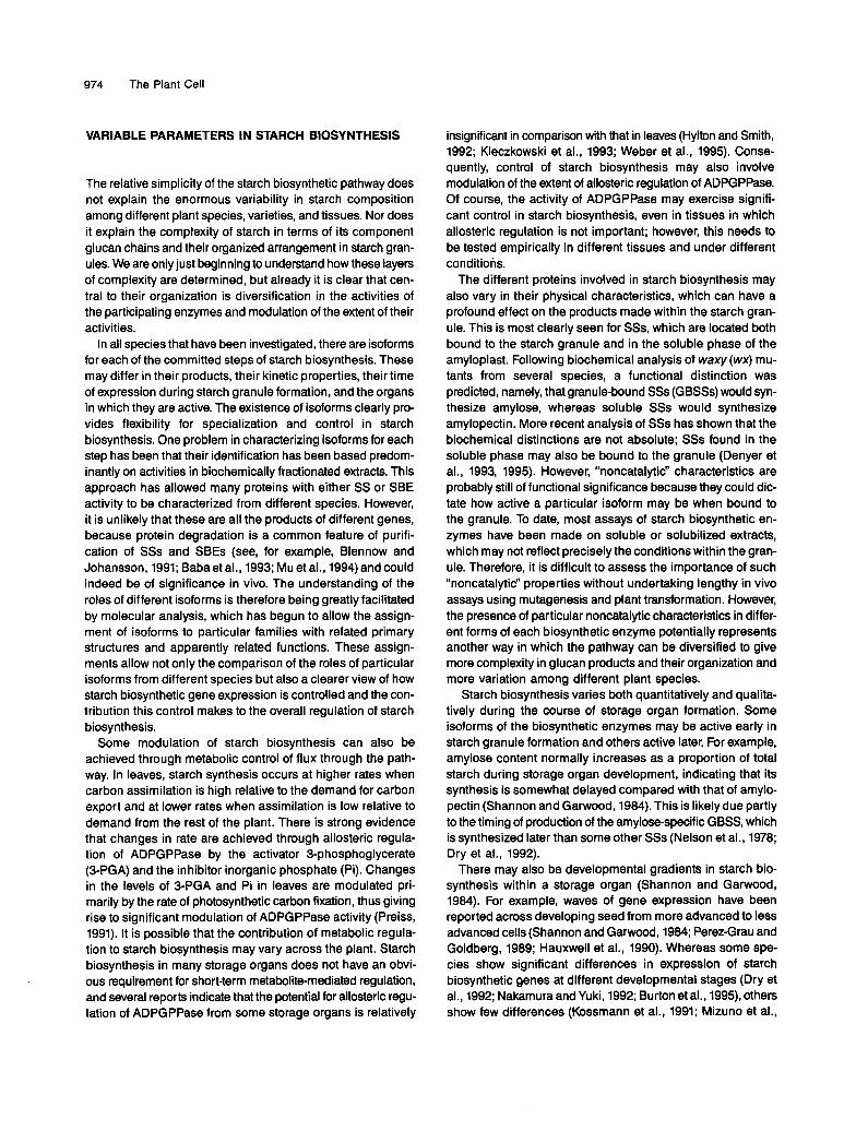
Figure 1. Amylopectin Structure, Starch Granule Form, and Starch Biosynthesis.
(A) Diagrammatic representation of an amylopectin molecule. a(M)-Linked glucans are attached by a(1-6) linkages to form a highly branched structure. Short glucan chains (A chains) are unbranched but linked to multiple branched B chains. There is a single reducing end to the C chain glucan. The branches are arranged in clusters 4 0 nm long, with a few longer chains linking more highly branched areas. (8) Diagrammatic representation of a starch granule from storage tissue showing alternating semicrystalline and amorphous growth rings. The semicrystalline regions are thought to consist of alternating crystalline and amorphous lamellae. (C) Steps of starch biosynthesis. ADPGPPase catalyzes the formation of ADPglucose and inorganic pyrophosphate from glucose-I-phosphate and ATP (step 1). Starch synthases (SS) add glucose units from ADPglucose to the nonreducing end of a growing a(l+linked glucan chain by an a(l-4) linkage and release ADP (step 2). Starch-branching enzymes (SBE) cut an a(l-4)-linked glucan chain and form an a(1-6) linkage between the reducing end of the cut chain and the C6 of another glucose residue in an a(l-r()-linked chain, thus creating a branch (step 3).
Starch Biosynthesis 973
starch, most notably that from potato tuber, is also phosphor- ylated. Potato tuber amylopectin carries up to one phosphate per 300 glucan residues (Takeda and Hizukuri, 1982).
Within the starch granule, which may vary in size from <1 pm to >100 pm, the amylopectin molecules are arranged radially, and adjacent branches within the branch clusters may form double helices that can be packed regularly, giving a crys- tallinity to the starch granule. The degree of crystallinity is determined in part by the branch lengths in the amylopectin. Closely packed, highly crystalline structures characteristic of wheat and barley endosperms are associated with average branch lengths of 20 residues. More open structures contain- ing more water are found in potato tuber and are associated with longer branch lengths of 22 glucan residues (for review, see Smith and Martin, 1993). The degree of branching and consequently the crystallinity of starch granules may vary con- siderably, even between different organs of the same plant. The starch granule is not uniformly crystalline, however, and contains relatively amorphous regions. Amylose molecules form single helical structures and are thought to be packed into these amorphous regions, which are present throughout the granule.
Granules from storage organs and leaves have rather differ- ent macrostructures. Starch granules from storage organs show interna1 semicrystalline growth rings that are differentially sen- sitive to chemical and enzymatic attack (Figure lB). The denser, more resistant layers may be regions of closer packing of branches within the branch clusters of parallel amylopectin molecules. The formation of these rings may result from peri- odic differences in the rate of starch synthesis. For example, in wheat endosperm the growth rings follow clear diurna1 pat- terns and are lost in plants grown under continuous illumination (Buttrose, 1962; French, 1984). However, growth rings are not universally responsive to illumination and may result from other periodic fluctuations in biosynthesis.
Starch granules in leaves are generally smaller than those in storage organs and have a distinct macrostructure. They are thought to have acrystalline core with an amorphous outer mantle that consists of less highly branched glucan polymers (Steup et al., 1983; Beck, 1985). Most of the turnover in starch during dayhight cycles involves the amorphous mantle of the granules (Steup et al., 1983; Beck, 1985).
Other components within all starch granules are proteins (0.5% in cereal endosperm and 0.05% in potato tuber), which include the enzymes of starch biosynthesis and may contrib- ute to the flavor of starch, and lipid (1% in cereal endosperm and 0.1% in potato tuber). The chemical structure of amylose facilitates its association with lipid, so the lipid may be local- ized to specific regions within the starch granule.
THE BIOCHEMISTRY OF STARCH BIOSYNTHESIS
The biosynthetic steps required for starch biosynthesis are relatively simple, involving three committed enzymes: ADP-
glucose pyrophosphorylase (ADPGPPase; EC 2.7.7.23), starch synthase (SS; EC 2.4.1.21), and starch branching enzyme (SBE; EC 2.4.1.28; Figure 1C).
60th amylose and amylopectin are synthesized from ADP- glucose, which is synthesized from glucose-1-phosphate and ATP in a reaction that is catalyzed by ADPGPPase and that liberates pyrophosphate. This enzyme is active within the plastid, which means that its substrates, glucose-1-phosphate and ATP, must also be present in the plastid. In chloroplasts, ATP may be derived from photosynthesis, but in nonphotosyn- thetic plastids, it must be specifically imported from the cytosol, probably by an ADP/ATP translocator (Ngernprasirtsiri et al., 1989; Schünemann et al., 1993). The glucose-1-phosphate can be supplied by the reductive pentose phosphate pathway in chloroplasts via phosphoglucoisomerase and phosphoglu- comutase (Smith and Martin, 1993). In nonphotosynthetic tissues, it may be imported directly from the cytosol (Tyson and ap Rees, 1988) or synthesized in the plastid from glucose- 6-phosphate via the action of a plastidial phosphoglucomu- tase (Hill and Smith, 1991).
The pyrophosphate produced by ADPGPPase is removed by inorganic alkaline pyrophosphatase, which is probably con- fined to plastids in both photosynthetic and nonphotosynthetic tissues. The removal of this plastidial pyrophosphate effec- tively displaces the equilibrium of the ADPGPPase reaction in favor of ADPglucose synthesis (weiner et al., 1987).
In the next step of starch synthesis, SS catalyzes the syn- thesis of an a(1-4) linkage between the nonreducing end of a preexisting glucan chain and the glucosyl moiety of ADP- glucose, causing the release of ADP. SSs can use both amylose and amylopectin as substrates in vitro. How the initial primers for the synthesis of glucan chains are produced in vivo is not known. There is some biochemical evidence for specialized activities that may build protein-glucose or protein-polyglu- cose primers in potato tubers. One report suggested that UDPglucose is used as a precursor to transglycosylate an ac- ceptor protein (Moreno et al., 1986). However, UDPglucose levels are believed to be very low in plastids, leaving the im- portance of such a priming system open to question. In bacteria, which synthesize glycogen via a pathway that has many similarities with starch synthesis in plants, a potential priming protein has been identified genetically (HenggeAronis and Fisher, 1992).
The a(1-6) branches in starch polymers are made by SBE, which hydrolyzes an a(l-4) linkage within a chain and then cata- lyzes the formation of an a(1-6) linkage between the reducing end of the “cut” glucan chain and another glucose residue, probably one from the hydrolyzed chain. Branches are not created randomly, as discussed previously, but show an aver- age periodicity of 20 glucan residues. SBEs show some specificity for the length of the a(1-4)glucan chain that they will use as a substrate. Part of this selectivity may reside in the fact that these enzymes cleave only those glucan chains that are in a stable double helical conformation, a structure that requires a minimum glucan chain length.
974 The Plant Cell
VARIABLE PARAMETERS IN STARCH BIOSYNTHESIS
The relative simplicity of the starch biosynthetic pathway does not explain the enormous variability in starch composition among different plant species, varieties, and tissues. Nor does it explain the complexity of starch in terms of its component glucan chains and their organized arrangement in starch gran- ules. We are only just beginning to understand how these layers of complexity are determined, but already it is clear that cen- tral to their organization is diversification in the activities of the participating enzymes and modulation of the extent of their activities.
In all species that have been investigated, there are isoforms for each of the committed steps of starch biosynthesis. These may differ in their products, their kinetic properties, their time of expression during starch granule formation, and the organs in which they are active. The existence of isoforms clearly pro- vides flexibility for specialization and control in starch biosynthesis. One problem in characterizing isoforms for each step has been that their identification has been based predom- inantly on activities in biochemically fractionated extracts. This approach has allowed many proteins with either SS or SBE activity to be characterized from different species. However, it is unlikely that these are all the products of different genes, because protein degradation is a common feature of purifi- cation of SSs and SBEs (see, for example, Blennow and Johansson, 1991; Baba et al., 1993; Mu et al., 1994) and could indeed be of significance in vivo. The understanding of the roles of different isoforms is therefore being greatly facilitated by molecular analysis, which has begun to allow the assign- ment of isoforms to particular families with related primary structures and apparently related functions. These assign- ments allow not only the comparison of the roles of particular isoforms from different species but also a clearer view of how starch biosynthetic gene expression is controlled and the con- tribution this control makes to the overall regulation of starch biosynthesis.
Some modulation of starch biosynthesis can also be achieved through metabolic control of flux through the path- way. In leaves, starch synthesis occurs at higher rates when carbon assimilation is high relative to the demand for carbon export and at lower rates when assimilation is low relative to demand from the rest of the plant. There is strong evidence that changes in rate are achieved through allosteric regula- tion of ADPGPPase by the activator 3-phosphoglycerate (3-PGA) and the inhibitor inorganic phosphate (Pi). Changes in the levels of 3-PGA and Pi in leaves are modulated pri- marily by the rate of photosynthetic carbon fixation, thus giving rise to significant modulation of ADPGPPase activity (Preiss, 1991). It is possible that the contribution of metabolic regula- tion to starch biosynthesis may vary across the plant. Starch biosynthesis in many storage organs does not have an obvi- ous requirement for short-term metabolite-mediated regulation, and several reports indicate that the potential for allosteric regu- lation of ADPGPPase from some storage organs is relatively
insignificant in comparison with that in leaves (Hylton and Smith, 1992; Kleczkowski et al., 1993; Weber et al., 1995). Conse- quently, control of starch biosynthesis may also involve modulation of the extent of allosteric regulation of ADPGPPase. Of course, the activity of ADPGPPase may exercise signifi- cant control in starch biosynthesis, even in tissues in which allosteric regulation is not important; however, this needs to be tested,empirically in different tissues and under different conditions.
The different proteins involved in starch biosynthesis may also vary in their physical characteristics, which can have a profound effect on the products made within the starch gran- ule. This is most clearly seen for SSs, which are located both bound to the starch granule and in the soluble phase of the amyloplast. Following biochemical analysis of waxy (wx) mu- tants from several species, a functional distinction was predicted, namely, that granule-bound SSs (GBSSs) would syn- thesize amylose, whereas soluble SSs would synthesize amylopectin. More recent analysis of SSs has shown that the biochemical distinctions are not absolute; SSs found in the soluble phase may also be bound to the granule (Denyer et al., 1993, 1995). However, "noncatalytic" characteristics are probably still of functional significance because they could dic- tate how active a particular isoform may be when bound to the granule. To date, most assays of starch biosynthetic en- zymes have been made on soluble or solubilized extracts, which may not reflect precisely the conditions within the gran- ule. Therefore, it is difficult to assess the importance of such "noncatalytic" properties without undertaking lengthy in vivo assays using mutagenesis and plant transformation. However, the presence of particular noncatalytic characteristics in differ- ent forms of each biosynthetic enzyme potentially represents another way in which the pathway can be diversified to give more complexity in glucan products and their organization and more variation among different plant species.
Starch biosynthesis varies both quantitatively and qualita- tively during the course of storage organ formation. Some isoforms of the biosynthetic enzymes may be active early in starch granule formation and others active later. For example, amylose content normally increases as a proportion of total starch during storage organ development, indicating that its synthesis is somewhat delayed compared with that of amylo- pectin (Shannon and Garwood, 1984). This is likely due partly to the timing of production of the amylose-specific GBSS, which is synthesized later than some other SSs (Nelson et al., 1978; Dry et al., 1992).
There may also be developmental gradients in starch bio- synthesis within a storage organ (Shannon and Garwood, 1984). For example, waves of gene expression have been reported across developing seed from more advanced to less advanced cells (Shannon and Garwood, 1984; Perez-Grau and Goldberg, 1989; Hauxwell et al., 1990). Whereas some spe- cies show significant differences in expression of starch biosynthetic genes at different developmental stages (Dry et al., 1992; Nakamura and Yuki, 1992; Burton et al., 1995), others show few differences (Kossmann et al., 1991; Mizuno et al.,

Starch Biosynthesis 975
1993). This apparent lack of developmental change could be an artifact of the way gene expression is assayed; storage or- gans with strong interna1 developmental gradients tend to "flatten out" developmental differences in assays based on total extracts. In fact, it may be that developmental regulation of iso- form gene expression is more important than is appreciated at present.
A final factor complicates the potential significance of de- velopmental modulation in the control of starch biosynthesis. As the starch granule grows, many of the biosynthetic enzymes become trapped within it (Denyer et al., 1993, 1995). This means that the turnover of these enzymes is relatively low. However, it is unclear to what extent the trapped enzymes are active within the granule; thus, the effective activity of the bio- synthetic enzymes at different developmental stages in vivo is very difficult to assess.
The plant can thus use severa1 different strategies to refine starch biosynthesis and to build the organized form of the gran- ule. Although our understanding is far from complete, the use of molecular biology and genetics has complemented the bio- chemical analysis of this system to allow a greater appreciation of the control of each biosynthetic step and its contribution to the overall process.
THE CONTRIBUTION OF EACH BIOSYNTHETIC STEP TO STARCH BIOSYNTHESIS
Supply of ADPglucose Precursors
ADPGPPase supplies ADPglucose for starch biosynthesis, and under some conditions, its activity is the most significant fac- tor in determining the rate of starch accumulation. This has been established most convincingly in leaves of Arabidopsis. A mutation in a gene encoding one of the two subunits of the enzyme reduces ADPGPPase activity to 7% and starch ac- cumulation to 26% of normal values in leaves (Lin et al., 1988). This mutant has been used to estimate the flux control coeffi- cient (Kacser and Burns, 1973) for Arabidopsis ADPGPPase, which is calculated from the relationship between the rate of starch synthesis and the ADPGPPase activity in wild-type and mutant leaves (Neuhaus and Stitt, 1990). Under low irradiance, ADPGPPase exercises the most significant degree of control on starch synthesis in Ieaves. Under high irradiance, although the flux control coefficient for ADPGPPase increases, the co- efficients for other enzymes also increase, as deduced by measurments from other species, indicating that control is exercised by additional enzymatic steps. These additional con- trol points are likely to involve enzymes that supply hexose phosphate from photosynthetic carbon fixation. The role of ADPGPPase in controlling starch biosynthesis in storage or- gans has not been measured in this way.
Much significance has been placed on the allosteric regu- lation of ADPGPPase by 3-PGA and Pi in the control of starch biosynthesis in all organs of the plant (Plaxton and Preiss, 1987;
Preiss, 1991, 1993; Preiss et al., 1991; Okita, 1992). This view is supported by the sxperiments of Stark et al. (1992), who were able to increase starch biosynthesis in cultured tobacco cells by 300% and in potato tubers by up to 60% by transformation with an Escherichia coligene encoding aversion of ADPGPP- ase that is insensitive to allosteric regulation. The normal E. Coli ADPGPPase is sensitive to allosteric regulation by fructose- l,&bisphosphate (Preiss and Romero, 1989), and this regu- lated enzyme is not able to increase starch production in transgenic tissues, which was interpreted as indicating that it is, indeed, the allosteric regulation of ADPGPPase that is important in determining starch accumulation in cultured tobacco cells and potato tubers (Stark et al., 1992). However, because the regulated E. coli enzyme is not under the same metabolic control as the normal plant enzyme, the significance of allosteric regulation would be demonstrated more compel- lingly by comparing the effects of the nonregulated enzyme with the effects of increased activities of the normal plant enzyme.
Although the significance of allosteric regulation in the con- trol of starch synthesis in storage organs therefore remains under investigation, it is reasonable to conclude from these experiments that ADPGPPase activity does exert some con- trol over starch biosynthesis, at least in light-grown, cultured tobacco cells and potato tubers. Interestingly, however, in these experiments, there was no significant correlation between the degree of activity of the unregulated ADPGPPase and the in- crease in starch accumulation, indicating that the activity of ADPGPPase may limit starch accumulation under normal con- ditions but that other steps also exercise significant control.
Plant ADPGPPase is reported to be a tetrameric enzyme that is formed from two distinct polypeptides (Copeland and Preiss, 1981), one between 54 and 60 kD (the large subunit) and one between 51 and 55 kD (the small subunit). cDNA clones encoding both subunits have been isolated from sev- era1 plant species (Olive et al., 1989; Bae et al., 1990; Bhave et al., 1990; Müller-Rober et al., 1990; Smith-White and Preiss, 1992; Villand et al., 1992a, 1992b; Weber et al., 1995); their deduced amino acid sequences share regions of homology, and they may in fact have evolved from a common ancestor because the bacterial enzyme is a homotetramer of a poly- peptide with a similar amino acid sequence (Inglesias et al., 1991; Smith-White and Preiss, 1992).
Expression of both potato subunits in E. coli has demon- strated that each has a low ADPGPPase activity alone but that this activity can be enhanced 10- to 70-fold by mixing them or by coexpressing them. The potato enzyme produced in this way remains responsive to 3-PGA(Inglesias et al., 1993). These and other experiments suggest that the plant enzyme normally functions as a heterotetramer of two large and two small subunits. Pyridoxal phosphate analysis, which can be used to identify the site of 3-PGA binding, has shown that a con- served lysine residue at the C terminus of the small subunit and the equivalent lysine residue in the large subunit bind this 3-PGA analog (Figure 2; Morell et al., 1988; Preiss, 1993). A second lysine residue in the large subunit from spinach leaf also binds pyridoxal phosphate; this residue may respond to

976 The Plant Cell
1
ATP? G1P?
11R QF.E%PKG
ATP? 01P?
ULLLlUJJlia i-CMD'Si.NA'ifclG SQlVVlA'fcATLDG
•urn!NP^WFOGIADAVR EF^E^PKG SGIVT>VIKDALIP!
ARM
AOPG/ADP
I IT H
ADPG/AOP
ARM Pi
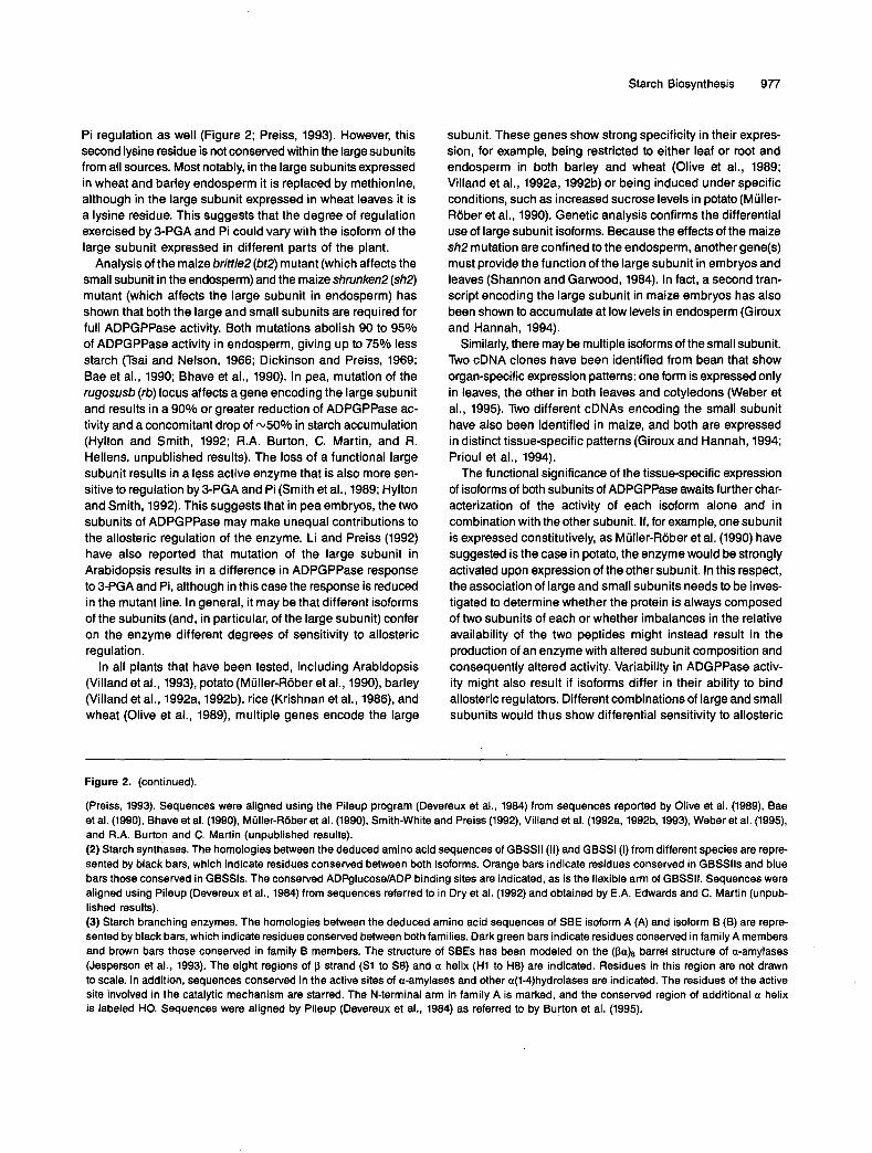
Figure 2. Diagrammatic Representation of the Homologies among Starch Biosynthetic Enzymes.
(1) Subunits of ADPGPPase. The homologies between the deduced amino acid sequences of the large (L) and small (S) subunits from differentspecies are represented by black bars, which indicate residues conserved between both subunits. Red bars indicate residues conserved amonglarge subunits, and pale green bars indicate residues conserved among the small subunits. The sites showing homology with ATP and glucose-1-pnosphate (G1P) binding sites in glycogen synthase from E. coli (Preiss, 1993) are indicated, as is the sequence (SI) around the conserved lysine(starred) in the small subunit that binds the allosteric regulator analog pyridoxal phosphate. This is thought to represent a binding site for 3-PGA.The equivalent site (SI) in the large subunit is also indicated, along with a second site (Sll) that may be involved in both 3-PGA and Pi regulation
Starch Biosynthesis 977
Pi regulation as well (Figure 2; Preiss, 1993). However, this second lysine residue is not conserved within the large subunits from all sources. Most notably, in the large subunits expressed in wheat and barley endosperm it is replaced by methionine, although in the large subunit expressed in wheat leaves it is a lysine residue. This suggests that the degree of regulation exercised by 3-PGA and Pi could vary with the isoform of the large subunit expressed in different parts of the plant.
Analysis of the maizebritt/e2(bf2) mutant (which affects the small subunit in the endosperm) and the maize shrunken2 (sh2) mutant (which affects the large subunit in endosperm) has shown that both the large and small subunits are required for full ADPGPPase activity. Both mutations abolish 90 to 95% of ADPGPPase activity in endosperm, giving up to 75% less starch (Tsai and Nelson, 1966; Dickinson and Preiss, 1969; Bae et al., 1990; Bhave et al., 1990). In pea, mutation of the rugosusb (rb) locus affects a gene encoding the large subunit and results in a 90% or greater reduction of ADPGPPase ac- tivity and a concomitant drop of ~ 5 0 % in starch accumulation (Hylton and Smith, 1992; R.A. Burton, C. Martin, and R. Hellens, unpublished results). The loss of a functional large subunit results in a less active enzyme that is also more sen- sitive to regulation by 3-PGA and Pi (Smith et al., 1989; Hylton and Smith, 1992). This suggests that in pea embryos, the two subunits of ADPGPPase may make unequal contributions to the allosteric regulation of the enzyme. Li and Preiss (1992) have also reported that mutation of the large subunit in Arabidopsis results in a difference in ADPGPPase response to 3-PGA and Pi, although in this case the response is reduced in the mutant line. In general, it may be that different isoforms of the subunits (and, in particular, of the large subunit) confer on the enzyme different degrees of sensitivity to allosteric regulation.
In all plants that have been tested, including Arabidopsis (Villand et al., 1993), potato (Müller-Rober et al., 1990), barley (Villand et al., 1992a, 1992b), rice (Krishnan et al., 1986), and wheat (Olive et al., 1989), multiple genes encode the large
subunit. These genes show strong specificity in their expres- sion, for example, being restricted to either leaf or root and endosperm in both barley and wheat (Olive et al., 1989; Villand et al., 1992a, 1992b) or being induced under specific conditions, such as increased sucrose levels in potato (Müller- Rober et al., 1990). Genetic analysis confirms the differential use of large subunit isoforms. Because the effects of the maize sh2 mutation are confined to the endosperm, another gene(s) must provide the function of the large subunit in embryos and leaves (Shannon and Garwood, 1984). In fact, a second tran- script encoding the large subunit in maize embryos has also been shown to accumulate at low levels in endosperm (Giroux and Hannah, 1994).
Similarly, there may be multiple isoforms of the small subunit. Two cDNA clones have been identified from bean that show organ-specific expression patterns: one form is expressed only in leaves, the other in both leaves and cotyledons (Weber et al., 1995). Two different cDNAs encoding the small subunit have also been identified in maize, and both are expressed in distinct tissue-specific patterns (Giroux and Hannah, 1994; Prioul et al., 1994).
The functional significance of the tissue-specific expression of isoforms of both subunits of ADPGPPase awaits further char- acterization of the activity of each isoform alone and in combination with the other subunit. If, for example, one subunit is expressed constitutively, as Müller-Rober et al. (1990) have suggested is the case in potato, the enzyme would be strongly activated upon expression of the other subunit. In this respect, the association of large and small subunits needs to be inves- tigated to determine whether the protein is always composed of two subunits of each or whether imbalances in the relative availability of the two peptides might instead result in the production of an enzyme with altered subunit composition and consequently altered activity. Variability in ADGPPase activ- ity might also result if isoforms differ in their ability to bind allosteric regulators. Different combinations of large and small subunits would thus show differential sensitivity to allosteric
Figure 2. (continued).
(Preiss, 1993). Sequences were aligned using the Pileup program (Devereux et al., 1984) from sequences reported by Olive et al. (1989), Bae et al. (1990), Bhave et al. (1990), Miiller-RBber et al. (1990), Smith-White and Preiss (1992), Villand et al. (1992a, 1992b, 1993). Weber et al. (1995), and R.A. Burton and C. Martin (unpublished results). (2) Starch synthases. The homologies between the deduced amino acid sequences of GBSSll (11) and GBSSI (I) from different species are repre- sented by black bars, which indicate residues conserved between both isoforms. Orange bars indicate residues conserved in GBSSlls and blue bars those conserved in GBSSls. The conserved ADF'glucose/ADP binding sites are indicated, as is the flexible arm of GBSSII. Sequences were aligned using Pileup (Devereux et al., 1984) from sequences referred to in Dry et al. (1992) and obtained by E.A. Edwards and C. Martin (unpub- lished results). (3) Starch branching enzymes. The homologies between the deduced amino acid sequences of SBE isoform A (A) and isoform B (6) are repre- sented by black bars, which indicate residues conserved between both families. Dark green bars indicate residues conserved in family A members and brown bars those conserved in family B members. The structure of SBEs has been modeled on the barrel structure of a-amylases (Jesperson et al., 1993). The eight regions of p strand (S1 to SE) and a helix (H1 to H8) are indicated. Residues in this region are not drawn to scale. In addition, sequences conserved in the active sites of a-amylases and other a(1-4)hydrolases are indicated. The residues of the active site involved in the catalytic mechanism are starred. The N-terminal arm in family A is marked, and the conserved region of additional a helix is labeled HO. Sequences were aligned by Pileup (Devereux et al., 1984) as referred to by Burton et al. (1995).
978 The Plant Cell
regulation. Such differences have been reported for the en- zyme isolated from cereal endosperm when compared with that isolated from cereal leaves (Duffus, 1993; Kleczkowski et al., 1993) and for the enzyme from bean cotyledons when com- pared with that from bean leaves (Weber et al., 1995).
In addition to affecting the amount of starch produced, ADPGPPase appears to influence starch composition. This is suggested by the fact that the rb mutation of pea, which af- fects the large subunit of ADPGPPase, not only reduces the total amount of starch produced but also alters its quality by increasing the proportion of amylopectin (Kooistra, 1962). Analysis of other pea mutants that show reduced starch accu- mulation through effects on steps involved in the supply of precursors shows that reductions in activity of these enzymes also elevates amylopectin content, suggesting that the effects on composition are a general result of decreased starch syn- thesis rather than a specific effect of ADPGPPase (T. Wang, personal communication).
Why does a decrease in starch synthesis increase the proportion of amylopectin? One possibility is that SBE is nor- mally limiting for the synthesis of amylopectin, so increased branching is observed when the rate of a(l4)glucan synthe- sis is reduced. If this were the case, then a mutation inhibiting SBE activity should severely affect the production of amylopectin, even in heterozygous condition. Maize endosperm contain- ing two doses of the amylose extender (ae) mutation, which reduces activity of one SBE isoform, and just one dose of the wild-type allele show a slight (2 to 8%) decrease in the appar- ent amylopectin content (Shannon and Garwood, 1984). An alternative, but not mutually exclusive, possibility is that re- duced levels of ADPglucose may have particularly extreme effects inside the starch granule, where availability is depen- dent on diffusion through the crystalline matrix. The reduced ADPglucose availability could have a relatively greater effect on the amylose-specific GBSS, which is located exclusively inside the granule, than on the SSs in the soluble phase, which synthesize amylopectin.
Synthesis of a(1-4)Glucan Chains
Collectively, SSs extend a(l4)glucan chains and synthesize both amylose and amylopectin. However, the amylose that they produce in vivo is somehow protected from the activity of SBEs in the plastid, even though SBEs can use it as a substrate in vitro. The key to understanding the synthesis of these discrete products lies in the discovery that wx mutants of maize, rice, sorghum, and Amaranthus, and the amy/ose free (amf) mu- tant of potato, make no amylose and lack the activity of an exclusively GBSS (GBSSI; Sprague et al., 1943; Nelson and Rines, 1962; Tsai, 1974; Okuno and Sakaguchi, 1982; Sano, 1984; Hseih, 1988; Jacobsen et al., 1989) that has also been frequently referred to as the “waxy” protein. This -60-kD pro- tein is made in relatively large amounts and is found exclusively bound to the starch granule. It has a low specific activity in standard assays (Nelson et al., 1978; Macdonald and Preiss, 1985)-so low that in some species it has been impossible
to detect activity in vitro (Smith, 1990; Denyer et al., 1995). How- ever, datafrom mutants on the effect of the loss of this isoform support the view that it is responsible for the synthesis of amy- lose. It may be that the standard assays used to measure SS activity do not accurately reflect the ability of this protein to synthesize a(l4)glucan chains in vivo because it is specifi- cally localized within the starch granule, where the crystalline matrix of glucan chains may provide a specialized environ- ment that promotes the activity of this isoform.
There is some evidence from the equivalent amylose-free mutant of the alga Chlamydomonas that GBSSI may also be responsible for the synthesis of a significant proportion of amylopectin, because the mutant shows deficiencies in a dis- tinct amylopectin fraction as well as lacking amylose (Delrue et al., 1992). In higher plants, mutations that eliminate GBSSI activity do not reduce starch content, implying that the ADP- glucose not used to make amylose is used by other SSs to make amylopectin. If GBSSI contributes to amylopectin bio- synthesis, this suggests that its ability to make amylose is not an intrinsic property of the protein but rather a function of its location in the starch granule. Perhaps it makes amylose when it is relatively deep within the granule and amylopectin when it is closer to the outer edge of the granule, where its products are accessible to the SBEs, which are active predominantly in the soluble phase. This idea is supported by the observa- tion that in starch granules of potato lines in which GBSSI activity is reduced by antisense suppression, amylose is re- stricted to a central core region (Kuipers et al., 1994). This could imply that amylose production requires a starch granule struc- ture in which the GBSSI is “buried.” If so, this might explain in part why amylose production appears to lag behind that of amylopectin during development of storage organs (Shannon and Garwood, 1984).
Other SS isoforms are located in the soluble phase of the amyloplast, although it is probably more useful to thinkof these isoforms in terms of the families to which they belong rather than in terms of their classic biochemical separation into s o b ble and granule-bound forms, because some are clearly located in both phases. One SS from pea embryos is located both in the soluble phase and on the granule and has been called GBSSII, although it is believed to be the major isoform active in the soluble phase in embryos (Smith, 1990; Denyer and Smith, 1992; Dry et al., 1992). Potato tubers contain a homo- log of this isoform (Dryet al., 1992), which isfound in the soluble phase and is also granule bound; there are probable homo- logs in maize and wheat (Mu et al., 1994; Denyer et al., 1995). GBSSll has a much higher specific activity than GBSSI when measured in vitro (Smith, 1990; E.A. Edwards, C. Martin, and E. Murray, unpublished results). However, members of other isoform families are certainly active in different parts of the plant and in different species, and the relative contribution of each isoform to a(l4)glucan synthesis may vary (Denyer et al., 1995).
GBSSll from pea is structurally similar to GBSSI from maize and potato and to glycogen synthase from bacteria (Dry et al., 1992). However, it contains at its N terminus an extra domain not present in the-other two enzymes. This domain accounts
Starch Biosynthesis 979
for the greater size of this SS isoform, which is some 17 kD larger than pea GBSSI. The domain is rich in serine and there- fore predicted to be highly flexible. Three consecutive proline residues near the C terminus of the extension may specify a turn in the folded protein (Figure 2). These residues are also present in the homologous protein from potato. We have termed this domain the “flexible arm” of GBSSII. Full-length GBSSll expressed in E. coli has the same specific activity as the puri- fied plant enzyme, and versions lacking the arm are also fully active, indicating that the arm does not participate in the cata- lytic mechanism of the enzyme. Antisera specific to pea GBSSll cross-react with the E. coli-produced proteins with or without the arm, and antisera specific to GBSSI do not cross-react with either of these proteins, indicating that the structurally similar regions of GBSSI and GBSSll that lie C terminal to the arm (in the case of GBSSII) are antigenically distinct (E.A. Edwards, J. Marshall, A.M. Smith, and C. Martin, unpublished results). This antigenic specificity is maintained toward the homologous proteins from other species-the pea GBSSll antibody rec- ognizes potato GBSSll but not potato GBSSI, for example- implying that there may be significant structural differences between GBSSI and GBSSll isoforms beyond the presence or absence of the N-terminal arm. These differences are likely to determine differences in the specific activities of the enzymes (low for GBSSI, high for GBSSII), whereas the N-terminal do- main of GBSSll is more likely to affect noncatalytic properties of the enzyme, such as the potential for physical association with SBEs or its degree of partitioning between soluble and granule-bound phases of the amyloplast.
The only other SS for which there is detailed structural in- formation is a soluble SS from rice endosperm. Sequencing of cDNA clones (Baba et al., 1993) indicates that this protein contains a C-terminal region with strong homology with E. coli glycogen synthase and other plant SSs. At its N terminus is a proline-rich region that ends in three consecutive proline residues. The amino acids surrounding this Pro-Pro-Pro motif show some homology with the equivalent region at the end of the arm of GBSSll from pea and potato (Dry et al., 1992). This region in the rice-soluble SS has been interpreted as the transit peptide of the protein (Baba et al., 1993), but it does not con- form to typical transit peptides in terms of size, sequence composition, or hydrophobicity, and it is more likely to repre- sent part of a longer N-terminal extension within SS that has certain features in common with GBSSll from pea and potato. The assignment of the end of the mature protein of the rice- soluble SS to a position C-terminal to the potential arm was based on N-terminal sequencing of 57-kD polypeptides pre- pared from partially purified preparations of rice-soluble SS. These may have been derived from a larger form of SS; in- deed, similar potential low molecular weight derivatives of GBSSll have been observed in SS preparations from pea em- bryos (Denyer and Smith, 1992). These maintain SS activity and therefore probably involve loss of parts of the N-terminal arm that may be particularly sensitive to proteases. Such prod- ucts probably also arise in vivo and may explain reports of a high-specific activity, low-molecular weight GBSS from pea (Sivak et al., 1993). Loss of the arm could result in changes
in the physical properties of the soluble SS, affecting its parti- tioning between the soluble and granule-bound phases.
In summary, SS isoforms have a conserved C-terminal re- gion of 4 0 kD that is similar in sequence to glycogen synthase from bacteria (Figure 2). The plant isoforms that are active in the soluble phase and that have been characterized at the mo- lecular leve1 probably also have N-terminal domains of high predicted flexibility that affect their noncatalytic properties. It is possible that these domains are subject to regulated pro- teolysis in vivo that affects their localization or activity. GBSSI has distinct catalytic properties (that is, its low specific activity measured in vitro), suggesting that it has specific structural features associated with its role in amylose synthesis. Any one plant species probably has severa1 SSs that are active in the soluble phase (Macdonald and Preiss, 1985; Denyer et al., 1995). The extent of their participation in starch synthesis may vary from one species to another and between different parts of the plant. These isoforms may also be bound to the starch granule, perhaps as a result of entrapment as the granule grows (Denyer et ai., 1993; Mu et ai., 1994). Their activity within the granule may be limited by the highly crystalline conditions of the granule or by the availability of ADPglucose, whose sup- ply may be severely limiting within the granule.
The relative contributions of different SS isoforms to starch biosynthesis also vary in amount and with time during stor- age organ development. GBSSI is the major protein bound to starch granules, and it is clear that it is present in large amounts relative to other isoforms (such as GBSSll of pea; Smith, 1990). However, the quantitative contribution of each isoform to a(1-4)glucan chain production is difficult to assess because of differences in the specific activity of the isoforms and a lack of understanding of the extent to which these differ- ences are present in vivo.
There are also differences in the expression of the genes encoding the SS isoforms during storage organ development. Because SSs may become trapped within the granule, where their actual activity is unknown, this is difficult to interpret in terms of relative amounts of each protein functioning in starch biosynthesis. However, GBSSI is maximally expressed later in pea embryo development than is GBSSll (Dry et al., 1992). There is some evidence for a similar differential expression in maize, rice, and potato (Nelson et al., 1978; Dry et al., 1992; Nakamura%nd Yuki, 1992). The significance of staggered ex- pression of SS isoforms in developing storage organs probably resides in the long-term formation of starch granules. GBSSll synthesis early in organ development would give early amylopectin production. Amylose would increase later due to GBSSI expression, and the relative balance between amylose and amylopectin synthesis would continue to change during development. Perhaps some of the order of the starch gran- ule results from the staggering of the phases of synthesis.
Production of a(1-6) Branches in Amylopectin
SBE activity is also a function of multiple isoforms. Molecular analysis has identified genes belonging to two SBE families

980 The Plant Cell
in maize, rice, and pea(Boyer and Preiss, 1978a, 1978b; Smith, 1988; Smyth, 1988; Fisher et al., 1993; Mizuno et al., 1993; Stinard et al., 1993; Burton et al., 1995), although it is possi- ble that there are other types of SBE in addition to these (Nakamura and Yamanouchi, 1992). The proteins of the two families are structurally related and are similar to glycogen- branching enzymes from bacteria. Members of family A (which comprises SBEll from maize, SBEI from pea, and SBElll from rice) contain an extra N-terminal domain that is lacking in mem- bers of family B (SBEI from maize, SBE from potato, SBEl from rice, SBEfrom cassava, and SBEll from pea) (Babaet al., 1991; Salehuzzaman et al., 1992; Fisher et al., 1993; Mizuno et al., 1993; Poulsen and Kreiberg, 1993; Burton et al., 1995). This domain is predicted tobe flexible, and like the N-terminal arm of GBSSll from pea and potato, it ends in two or three consec- utive proline residues (Dry et al., 1992; Burton et al., 1995). The functional significance of this extra domain is as yet unknown, although both SBE isoforms may be located within the starch granule as well as in the soluble phase, as is the case in the pea embryo (Denyer et al., 1993), and this domain might af- fect the relative partitioning of the enzymes between these two phases. The immunolocalization of the B isoform in potato in- dicates that it is localized at the interface between the stroma and the starch granule (Kram et al., 1993).
In addition, and perhaps more significantly, the catalytic prop- erties of the isoforms differ (Smith, 1988; Guan and Preiss, 1993; Takeda et al., 1993). Family A isoforms have a lower af- finity for amylose than family B mernbers and preferentially use shorter glucan chains during branch formation. The re- sult is that family A members probably make amylopectin with shorter branch lengths than family B isoforms. The most likely cause for this catalytic difference is a conserved structural distinction between the two isoform types. The structure of branching enzymes can be modeled on the structure of a-amylases, which has been solved by x-ray crystallography (Jesperson et al., 1993). Members of the a-amylase super- family, which hydrolyze a(l-4)-linked glucan chains, consist of a central (Da)* barrel structure involved in hydrolysis. The SBEs conform to this structure and show conservation of amino acid residues predicted to be involved in hydrolysis, especially in the loops connecting each central P-strand with the follow- ing a-helix (Figure 2; Jesperson et al., 1993; Burton ft al., 1995). The loops between P-strand 7 and a-helix 7 and between 0-strand 8 and a-helix 8 have been implicated in determining the length of branches in comparison with the sequences of glycogen branching enzymes and SBEs. The loop between 0-strand 8 and a-helix 8 is very similar among all family A iso- forms and distinct from that in family B isoforms in length and sequence composition, supporting the idea that this loop is involved in the determination of branch length (Burton et al., 1995).
The two SBE isoforms also show distinct developmental differences in the time of their expression. The A isoform is active early in pea embryo development, whereas the activity of the B isoform rises later (Smith, 1988; Burton et al., 1995). This may result in differences in the amylopectin synthesized
early and late in granule formation, because a change from predominantly shorter branch lengths to predominantly longer ones has been detected during pea embryo development (Bufiton et al., 1995). In other species, the developmental regu- lation of the SBEs is less significant (Mizuno et al., 1993). This may reflect a less distinct temporal separation of SBE isoform expression in cereal endosperm or stronger developmental gradients within the developing endosperm that may blur tem- poral differences.
The significance of the contribution of the family A isoform to starch biosynthesis is best appraised through the analysis of mutants; both the rugosus (r) mutant of pea and ae mutants of maize and rice are defective in this activity (Mendel, 1865; Kooistra, 1962; Shannon and Garwood, 1984; Bhattacharyya et al., 1990; Mizuno et al., 1993). Total starch content is re- duced by up to 20% in ae mutant endosperm and up to 50% in r mutant embryos, and the amylopectin content falls from ~ 7 0 to -30%. The remaining starch consists of amylose and an intermediate material that has longer branch lengths than normal amylopectin and a lower molecular weight than amy- lose. Loss of isoform A activity thus limits the synthesis of starch in a way that cannot be compensated for by isoform B. The inhibition of starch synthesis probably results from a decreas- ing availability of nonreducing ends in the glucan polymers, which effectively substrate limits SS activity (Edwards et al., 1988). The effect of the loss of isoform A also indicates that it may be responsible for up to 60% of the amylopectin syn- thesized, although amylopectin synthesis may also be limited indirectly through substrate-limited SS activity in mutant lines. The similarity of the effects of the ae and r mutations suggests that isoform A plays a similar role in starch synthesis in en- dosperms and embryos.
There are no known mutants in isoform B activity. This may be of significance in itself. Mutations in starch synthesis are normally identified by reduced starch synthesis during seed formation, which, through the associated increases in sucrose and osmotic potential of the developing embryos, causes a wrinkling or puckering of the dry seed. The fact that no iso- form B mutants have been identified suggests that inactivation of this isoform does not limit starch biosynthesis sufficiently to give rise to this phenotype (Müller-Rober and Kossmann, 1994; Burton et al., 1995). Indeed, antisense experiments in- hibiting isoform B activity in potato (Kossmann et al., 1991; Müller-Rober and Kossmann, 1994) to undetectable levels resulted in no significant modification in starch synthesis or, indeed, in the amylose-to-amylopectin ratio (Müller-Rober and Kossmann, 1994). Although interpretation of the results is com- plicated by failure, so far, to identify an isoform A counterpart in potato, the results suggest that isoform B plays a quantita- tively minor role in amylopectin biosynthesis in storage organs. It may serve to make amylopectin with somewhat longer dis- tances between the branch points later in granule formation, a qualitative contribution that would be difficult to detect.
Loss of isoform A activity also has a profound effect on starch granule morphology. Granules in ae and r mutants are small and irregular or deeply fissured (Shannon and Garwood, 1984).
Starch Biosynthesis 981
This indicates that the activity of isoform A is essential for the normal regular organization of the starch granule. However, it is not yet clear whether it is the special type of amylopectin synthesized by isoform A that is essential for regular granule formation or rather the relatively early production of amylopectin during granule formation.
An additional enzyme, starch debranching enzyme, may in- fluente the type of glucan produced. The sugary (sul) mutants of maize, rice, and sorghum have reduced starch levels but contain a highly branched, soluble a(l4)/a(+6)-linked glucan polymer, phytoglycogen (Shannon and G a m d , 1984; Kaushik and Khush, 1991). In sul endosperm, starch granules are formed early in development but are subsequently degraded and replaced by phytoglycogen. sul mutants are deficient in the activity of debranching enzyme, an a(1-6)glucosidase (Pan and Nelson, 1984). This supports the hypothesis that the final structure of amylopectin might be determined by a balance between the activities of debranching and branching enzymes. Perhaps debranching enzyme is responsible for reducing the branching in glucan polymers at some stages of storage or- gan development such that in its absence, the more highly branched phytoglycogen is produced. Clearly, further research in this area is needed. To understand the contribution of de- branching enzyme to starch synthesis, it is necessary to ascertain whether the sul locus encodes the structural gene for debranching enzyme and the extent to which its effects on other aspects of starch biosynthesis are direct or indirect.
becomes wrinkled (Kooistra, 1962). Furthermore, the increase in cell size caused by the increased osmotic potential may re- sult in higher lipid content through increased production of cellular membrane (Coxon and Davies, 1982; Bettey and Smith,
The influence of starch synthesis on storage protein accumu- lation may also operate through changes in osmotic potential, which may influence the production of particular storage pro- teins (Turner et al., 1990). It may also involve the coupling of amino acid to sucrose unloading from the phloem supplying the developing storage organs, because if sucrose accumu- lates in the embryos, unloading from the phloem is likely to be reduced. The resulting limitations in amino acid supply could then limit storage protein synthesis (Bhattacharyya et al., 1993). Whatever the primary cause, limitations on starch biosynthe- sis may affect both the transcription and translation of specific storage proteins (Turner et al., 1990; Giroux et al., 1994). Inhi- bition of starch synthesis through antisense inhibition of ADPGPPase activity in potato results in inhibition of patatin production as well and, in addition, affects the overall process of tuber formation, resulting in significantly more tubers per plant (Müller-Rober et al., 1992). These mutants highlight the existence of interactions among the different biosynthetic path- ways in storage organ development and in the development of storage organs themselves.
1990).
It is also important to determine whether debranching activity is significant in the development of starch granules in other
CONCWS~ONS
species or whether its activity is generally limited to periods of starch mobilization. Any finding of such a contribution of starch debranching enzyme must also be reconciled with the claim that the properties of SBE isoforms can themselves ex- plain the structure of amylopectin (Takeda et al., 1993).
THE INTERACTION OF STARCH SYNTHESIS WlTH OTHER BIOSYNTHETIC PATHWAYS IN STORAGE ORGAN DEVELOPMENT
Many of the mutations whose primary effect is on starch bio- synthesis have pleiotropic effects on other aspects of storage organ development. For example, the r mutation of pea con- fers a wrinkled phenotype on the seed and results in the production of relatively more lipid and less legumin storage protein (summarized in Bhattacharyya et al., 1990). Similarly, starch biosynthetic mutants of maize and barley show reduced production of one class of storage protein, the prolamins. Some of these effects may arise from the increase in sucrose con- tent that results from reduced starch biosynthesis and the consequent increases in osmotic potential (Smith and Denyer, 1992). For example, increases in sucrose cause increased wa- ter uptake during development, which stretches the testa or pericarp. However, at maturity, the mutant seed undergo rela- tively greater water loss and the overstretched testa or pericarp
A speculative model for the interaction of the starch biosyn- thetic enzymes is presented in Figure 3. ADPGPPase (1) in the soluble phase of the amyloplast supplies ADPglucose to SSs (2); those SSs in the soluble phase and to the edge of the granule extend a(l4)-linked glucans that are then acces- sible to the SBEs (3) near the edge of the granule, thus forming amylopectin. These a(l-6)-branched polymers can then be extended further by SSs. Somewhat protected from the activ- ity of SBEs is amylose, perhaps because the enzyme that makes it, GBSSI, is active deeper within the granule.
This static model cannot explain the growth rings of starch granules, which appear to represent periodic changes in the degree of crystallinity within the starch granule. Nor can it ex- plain the details of the compositional differences across the granule. There is clearly an enormous amount left to under- stand about the synthesis of starch granules. Central to testing, refining, and developing the model will be the combining of molecular techniques with those of biochemistry and genetics. The enzymes can be produced in E. coli, and their catalytic and physical properties can be studied in greater de- tail. Transgenic plants offer enormous potential for dissecting out the individual contributions of each isoform (Müller-Rober and Kossmann, 1994). In the future, it may be possible to test the model by reconstructing starch biosynthesis in surrogate hosts, such as E. coli. Through developing an understanding of how starch is synthesized, the potential for producing nove1

982 The Plant Cell
Figure 3. Speculative Model for the Role of Isoforms in Starch Synthesis within the Starch Granule.
A diagrammatic representation of an amylopectin molecule (black lines) is overlaid with spots representing various isoforms of starch biosyntheticenzymes. Spots labeled 1, ADPGPPase (green spots, small subunit; red spots, large subunit). This enzyme is thought to be active in the solublephase. Spots labeled 2, SS (blue spots, GBSSI; orange spots, soluble GBSSII; orange spots with yellow ring, GBSSII, which is active in thesoluble phase and is also found bound to the granule, where its relative activity in vivo is unknown [the yellow ring indicates the N-terminal arm,which can be cleaved without loss of activity]; red spots, other starch synthases that are predominantly soluble but also may be bound to thegranule). GBSSI is active within the granule and makes amylose, shown as blue lines, and possibly some amylopectin as well. The other SSsshown synthesize amylopectin. Spots labeled 3, SBE (green spots with yellow ring, isoform A [yellow ring indicates N-terminal arm]; brown spots,isoform B). Both isoforms are active in the soluble phase and are also found bound to the granule. These isoforms make qualitatively differentcontributions to amylopectin synthesis.
starches through genetic modification (Shewmaker and Stalker,1992; Smith and Martin, 1993; Muller-Rober and Kossmann,1994) may be fully realized.
At present, starch is extracted commercially from a limitednumber of sources (predominantly maize and potato). Geneticengineering may allow the modification of maize and potatostarch toward the quality of starches from other plant sources.In addition, the engineering of starch biosynthesis may leadto modification of starch granule shape, amylose-to-amylopectinratio, amylopectin chain length, crystallinity, gelling properties,phosphorylation, and lipid content—all of which will alter sig-nificantly the technological properties of starch for use in bothfood and nonfood industries.
REFERENCES
Baba, T, Kimura, K., Mlzuno, K., Etoh, H., Ishlda, Y., Shida, O.,and Aral, Y. (1991). Sequence conservation of the catalytic regionsof amylolytic enzymes in maize branching enzyme-l. Biochem. Bio-phys. Res. Commun. 181, 87-94.
Baba, T., Nishihara, M., Mizuno, K., Kawasaki, T., Shimada, H.,Kobayashi, E., Ohnishi, S., Tanaka, K.-l., and Aral, Y. (1993). Iden-tification, cDNA cloning, and gene expression of soluble starchsynthase in rice (Oryza saf/Va L.) immature seeds. Plant Physiol.103, 565-573.
Bae, J.M., Giroux, M., and Hannah, L.CO. (1990). Cloning and charac-terisation of the brittle-2 gene of maize. Maydica 35, 317-322.
Beck, E. (1985). The degradation of transitory starch granules in chlo-roplasts. In Regulation of Carbon Partitioning in Photosynthetic
Tissues, R.L. Heath and J. Preiss, eds (Baltimore, MD: WaverlyPress), pp. 27-44.
Bettey, M., and Smith, A.M. (1990). Nature of the effect of the r locuson the lipid content of the embryos of peas (Pisum sativum L.). Planta180, 420-428.
Bhattacharyya, M.K., Smith, A.M., Ellis, T.H.N., Medley, C., andMartin, C. (1990). The wrinkle-seeded character of peas describedby Mendel is caused by a transposon-like insertion in a gene en-coding starch branching enzyme. Cell 60, 115-122.
Bhattacharyya, M.K., Martin, C., and Smith, A.M. (1993). The im-portance of starch biosynthesis in the wrinkled shape character ofpeas studied by Mendel. Plant Mol. Biol. 22, 525-531.
Bhave, M.R., Lawrence, S., Barton, C., and Hannah, L.C. (1990).Identification and molecular characterization of shrunken-2 cDNAclones of maize. Plant Cell 2, 581-588.
Blennow, A., and Johansson, G. (1991). Isolation of a Q-enzyme withMr 103000 from potato tubers. Phytochemistry 30, 437-444.
Boyer, C.D., and Preiss, J. (1978a). Multiple forms of a1—4 a-D-glucan6-glucosyl transferase from developing Zea mays L. kernels. Car-bohydr. Res. 61, 321-334.
Boyer, C.D., and Preiss, J. (1978b). Multiple forms of starch branch-ing enzyme of maize: Evidence for independent genetic control.Biochem. Biophys. Res. Commun. 80, 169-175.
Burton, R.A., Bewley, J.D., Smith, A.M., Bhattacharyya, M.K., Tatge,H., Ring, S., Bull, V., Hamilton, W.D.P., and Martin, C. (1995).Starch branching enzymes belonging to distinct enzyme familiesare differentially expressed during pea embryo development. PlantJ. 7, 3-15.
Buttrose, M.S. (1962). The influence of environment on the shell struc-ture of starch granules. J. Cell Biol. 14, 159-167.

Starch Biosynthesis 983
Copeland, L., and Prelss, J. (1981). Purification of spinach leaf ADP- glucose pyrophosphorylase. Plant Physiol. 68, 996-1001.
Coxon, D.T., and Davies, D.R. (1982). The effect of r a and rb loci on the lipid content of the seed of fisum sativum. Theor. Appl. Genet. 64, 47-50.
Delmer, D.P., and Amor, Y. (1995). Cellulose biosynthesis. Plant Cell 7, 967-1000.
Delrue, B., Fontalne, T., Revtler, F., Deeq, A., Wieruszeski, J.-M., van den Koornhuyse, N., Maddeleln, M.-L., Fournet, B., and Ball, S. (1992). Waxy Chlamydomonas reinhardtii: Monocellular alga1 mu- tants defective in amylose biosynthesis and granule-bound starch synthase activity accumulate a structurally modified amylopectin. J. Bacteriol. 174, 3612-3620.
Denyer, K., and Smith, A.M. (1992). The purification and characteri- sation of the two forms of soluble starch synthase from developing pea embryos. Planta 186, 609-617.
Denyer, K., Sidebottom, C., Hylton, C.M., and Smith, A.M. (1993). Soluble isoforms of starch synthase and starch branching enzyme also occur within starch granules in developing pea embryos. Plant
Denyer, K., Hylton, C.M., Jenner, CF., andSmlth,A.M. (1995). Iden- tification of multiple isoforms of soluble and granule-bound starch synthase in developing wheat endosperm. Planta 196, 256-265.
Detherage, W.L., MacMasters, M.M., and Rlst, C.E. (1955). A par- tia1 survey of amylose content in starch from domestic and foreign varieties of corn, wheat, and sorghum and from some other starch- bearing plants. Trans. Am. Assoc. Cereal Chem. 13, 31-42.
Devereux, J., Haeberll, P., and Smlthles, O. (1984). A comprehen- sive set of sequence analysis programs for the VAX. Nucleic Acids Res. 12, 387-395.
Dlcklnson, D.B., and Prelss, J. (1969). Presence of ADPglucose pyrophosphorylase in shrunken-2 and brittle-2 mutants of maize en- ,dosperm. Plant Physiol. 44, 1058-1062.
Dry, I., Smith, A., Edwards, A., Bhattacharyya, M., Dunn, P., and Martln, C (1992). Characterisation of cDNAs encoding two isoforms of granule-bound starch synthase which show differential expres- sion in developing storage organs of pea and potato. Plant J. 2, 193-202.
Duftus, C.M. (1993). Starch synthesis and deposition in developing cereal endosperms. In Seed Storage Compounds, P.R. Shewry and K. Stobart, eds (Oxford: Oxford University Press), pp. 191-219.
Edwards, J., Green, J.H., and ap Rees, T. (1988). Activity of branch- ing enzyme as a cardinal feature of the r, locus in Pisum sativum. Phytochemistry 27, 1615-1620.
Flsher, D.K., Boyer, C.D., and Hannah, L.C. (1993). Starch branch- ing enzyme II from maize endosperm. Plant Physiol. 102,1045-1046.
French, D. (1984). Organization of starch granules. In Starch: Chem- istry and Technology, R.L. Whistler, J.N. BeMiller, and E.F. Paschall, eds (Orlando, FL: Academic Press), pp. 183-237.
Glroux, M.J., and Hannah, L.C. (1994). ADPglucose pyrophosphory- lase in shrunken2 and brittle-2 mutants of maize. MOI. Gen. Genet. 243, 400-408.
Glroux, M.J., Boyer, C., Felx, G., and Hannah, L.C. (1994). Coordi- nated transcriptional regulation of storage product genes in the maize endosperm. Plant Physiol. 106, 713-722.
Guan, H.-P., and Prelss, J. (1993). Differentiation of the properties of the branching isozymes from maize (Zee mays). Plant Physiol.
J. 4, 191-198.
102, 1269-1273.
Hauxwell, A. J., Corke, F.N.K., Hedley, C.L., and Wang, T.L. (1990). Storage protein gene expression is localized to regions lacking mi- totic activity in developing pea embryos. An analysis of seed development in fisum setivum, XIV. Development 110, 283-289.
Henggedronls, R., and Flsher, D. (1992). ldentification and molecu- lar analysis of glgS, a nove1 growth-phase-regulated and rpo- dependent gene involved in glycogen synthesis in Escherichia coli. MOI. Microbiol. 6, 1877-1886.
Hlll, L.M., and Smlth, A.M. (1991). Evidence that glucose-&phosphate is imported as the substrate for starch synthesis by the plastids of developing pea embryos. Planta 185, 91-96.
Hselh, J.-S. (1988). Genetic studies on the wx gene of sorghum (Sor- ghum bicolor (L.) Moench). 1. Examination of the protein product of the waxy locus. Bot. Bull. Acad. Sinica 29, 293-299.
Hylton, C., and Smlth, A.M. (1992). The rb mutation of peas causes structural and regulatory changes in ADP glucose pyrophosphory- lase from developing embryos. Plant Physiol. 99, 1626-1634.
Ingleslas, A.A., Kaketuda, O., and Prelss, J. (1991). Regulatory and structural properties of the cyanobacterial ADPglucose pyrophos- phorylase. Plant Physiol. 97, 1187-1195.
Ingleslas, A.A., Barry, G.F., Meyer, C., Blocksberg, L., Nakata, P.A., Green, T., Laughlln, M.J., Oklta, T.W., Klshore, G.M., and Prelss, J. (1993). Expression of the potato tuber ADPglucose pyrophosphory- lase in Escherichia coli. J. Biol. Chem. 268, 1081-1086.
Jacobsen, E., Hovenkamp-Hermellnk, J.M.H., Krlgsheld, H.T., Nljdam, H., PlJnacker, L.P., Wltholt, B., and Feenstra, W.J. (1989). Phenotypic and genotypic characterisation of an amylose-free starch mutant of the potato. Euphytica 44, 43-48.
Jespereon, H.M., MacGregor, E.A., Hevrlsaat, B., Slerlce, M.R., and Svensson, B. (1993). Starch and glycogen debranching and branch- ing enzymes: Prediction of structural features of the catalytic barrel domain and evolutionary relationship to other amylolytic en- zymes. J. Protein chem. 12, 791-805.
Kacser, H., and Burns, J.A. (1973). Control of flux. Symp. SOC. Exp. Biol. 27, 65-107.
Kalnuma, K. (1988). The structure and chemistry of the starch gran- ule. In The Biochemistry of Plants, Vol. 14, Carbohydrates, J. Preiss, ed (San Diego: Academic Press), pp. 141-180.
Kaushlk, R.P., and Khush, G.S. (1991). Genetic analysis of endosperm mutants in rice (Oryze sative L.). Theor. Appl. Genet. 83, 146-152.
Kleukowskl, L.A., Vllland, P., Lilthl, E., Olsen, O.A., and Plelss, J. (1993). lnsensitivity of barley endosperm ADPglucose pyrophos- phorylase to 3-phosphoglycerate and orthophosphate regulation. Plant Physiol. 101, 179-186.
Koolstra, E. (1962). On the differences between smooth and three types of wrinkled peas. Euphytica 11, 357-373.
Kossmann, J., Vlsser, R.G.F., MUller-ROber, B., Wlllmltzer, L., and Sonnewald, U. (1991). Cloning and expression analysis of a potato cDNA that encodes branching enzyme: Evidence for co-expression of starch biosynthetic genes. MOI. Gen. Genet. 230, 39-44.
Kram, A.M., Oostergetel, G.T., and van Bruggen, E.F.J. (1993). Lo- calization of branching enzyme in potato tuber cells with the use of immunoelectron microscopy. Plant Physiol. 101, 237-243.
Krlshnan, H.B., Reeves, CD., and OkRa, T.W. (1986). ADPglucose pyrophosphorylase is encoded by different mRNA transcripts in leaf and endosperm of cereals. Plant Physiol. 81, 642-645.
984 The Plant Cell
Kuipers, A.G.J., Jacobsen, E., and Vlsser, R.G.F. (1994). Formation and deposition of amylose in the potato tuber starch granule are affected by the reduction of granule-bound starch synthase gene expression. Plant Cell 6, 43-52.
Li, L., and Preiss, J. (1992). Characterization of ADP-glucose pyrophosphorylase from a starch-deficient mutant of Arabidopsis tbaliana (L). Carbohydr. Res. 227, 227-239.
Lin, T.P., Caspar, T., Somenrllle, C., and Preiss, J. (1988). A starch- deficient mutant of Arabidopsis tbaliana with low ADPglucose pyrophosphorylase activity lacks one of the two subunits of the en- zyme. Plant Physiol. 88, 1175-1181.
Macdonald, F.D., and Preiss, J. (1985). Partia1 purification and char- acterization of granule-bound starch synthases from normal and waxy maize. Plant Physiol. 781, 849-852.
Mendel, 0 . (1865). Versuche Über Pflanzen-Hybriden. Verh. Nat. Forsch. Ver. Brünn 4, 3-47.
Mizuno, K., Kawasaki, T., Shimada, H., Satoh, H., Kobayashl, E., Okumura, S., Arai, Y., and Baba, T. (1993). Alteration of the struc- tural properties of starch components by the lack of an isoform of starch branching enzyme in rice seeds. J. Biol. Chem. 268,
Morell, M., Bloom, M., and Preiss, J. (1988). Affinity labeling of allosteric activator site(s) of spinach leaf ADP-glucose pyrophosphor- ylase. J. Biol. Chem. 263, 633-637.
Moreno, S., Cardini, E.C., and Tandecarz, J.S. (1986). a-Glucan synthesis on a protein primer, uridine diphosphoglucoseprotein transglycolase 1. Separation from starch synthase and phosphory- lase and a study of its properties. Eur. J. Biochem. 157, 539-545.
Mu, C., Harn, C., Ko, Y.T., Slngletary, G.W., Keellng, P.L., and Wasserman, B.P. (1994). Association of a 76 kDa polypeptide with soluble starch synthase activity in maize (cvB73) endosperm. Plant
MÜller-Rtiber, E., and Kossmann, J. (1994). Approaches to influence starch quantity and starch quality in transgenic plants. Plant Cell Environ. 17,. 601-612.
MÜller-Rober, B., Kossmann, J., Hannah, L.C., Wlllmitzer, L., and Sonnewald, U. (1990). Only one of two different ADPglucose pyrophosphorylase genes from potato responds strongly to elevated levels of sucrose. MOI. Gen. Genet. 224, 136-146.
MÜller-Rober, B., Sonnewald, U., and Wlllmitzer, L. (1992). Inhibi- tion of the ADPglucose pyrophosphorylase in transgenic potatoes leads to sugar-storing tubers and influences tuber formation and expression of tuberstorage protein genes. EMBO J. 11,1229-1238.
Nakamura, Y., and Yamanouchi, H. (1992). Nucleotide sequence of a cDNA encoding starch branching enzyme, or Q enzyme I, from rice endosperm. Plant Physiol. 99, 809-819.
Nakamura, Y., and Yuki, K. (1992). Changes in enzyme activities as- sociated with carbohydrate metabolism during the development of rice endosperm. Plant Sci. 82, 18-20.
Nelson, O.E., and Rines, H.W. (1962). The enzyme deficiency in the waxy mutant of maize. Biochem. Biophys. Res. Commun. 9,297-300.
Nelson, O.E., Chourey, P.S., and Chang, M.T. (1978). Nucleoside diphosphate sugar-starch glucosyl transferase activity of wx starch granules. Plant Physiol. 62, 383-386.
Neuhaus, H.E., and Stitt, M. (1990). Control analysis of photosyn- thate partitioning: lmpact of reduced activity of ADPglucose pyrophosphorylase or plastid phosphoglucomutase on fluxes to starch and sucrose in Arabidopsis. Planta 182, 445-454.
19084-19091.
J. 6, 151-159.
Ngernprasirtsiri, J., Takabe, T., and Akazawa, T. (1989). Im- munochemical analysis shows an ATPlADP translocator is associated with the inner envelope membranes of amyloplasts from Acerpseu- doplantanus L. Plant Physiol. 89, 1024-1027.
Okita, T.W. (1992). Is there an alternative pathway for starch synthe- sis? Plant Physiol. 100, 560-564.
Okuno, K., and Sakaguchi, S. (1982). lnheritance of starch charac- teristics in perisperm of Amaranthus bypocondriacus. J. Hered. 73, 467
Olive, M.R., Ellis, R.J., and Schuch, W.W. (1989). lsolation and nucleotide sequences of cDNA clones encoding ADPglucose pyro- phosphorylase polypeptides from wheat leaf and endosperm. Plant MOI. Biol. 12, 525-538.
Pan, D., and Nelson, O.E. (1984). A debranching enzyme deficiency in endosperms of the sugary-1 mutants of maize. Plant Physiol. 74,
Perez-Grau, L., and Goldberg, R.B. (1989). Soybean seed protein genes are regulated spatially during embryogenesis. Plant Cell 1,
Plaxton, W.C., and Preiss, J. (1987). Purification and properties of nonproteolytic degraded ADPglucose pyrophosphorylase from maize endosperm. Plant Physiol. 83, 105-112.
Poulsen, P., and Kfeiberg, J.D. (1993). Starch branching enzyme cDNA from Solanum tuberosum. Plant Physiol. 102, 1053-1054.
Preiss, J. (1991). Biology and molecular biology of starch synthesis and its regulation. In Oxford Surveys of Plant Mofecular and Cell Biology, Vol. 7, B.J. Miflin, ed (Oxford: Oxford University Press), pp.
Preiss, J. (1993). Biosynthesis of starch: ADPglucose pyrophosphory- lase, the regulatory enzyme of starch synthesis-structure-function relationships. Denpan Kogako 40, 117-131.
Preiss, J., and Romero, T. (1989). Physiology, biochemistry and genetics of bacterial glycogen synthesis. Adv. Microbiol. Phys. 36,
Preiss, J., Ball, K., Smith-White, B., Inglesias, A., Kakefuda, G., and Li, L. (1991). Starch biosynthesis and its regulation. Biochem. SOC. Trans. 19, 539-547.
Prioul, J.-L., Jeannette, E., Reyss, A., Glegory, N., Giroux, M., Hannah, L.C., and Causse, M. (1994). Expression of ADPglucose pyrophosphorylase in maize (Zea mays L), grain and source leaf during grain filling. Plant Physiol. 104, 179-187.
Salehuzzaman, S.N.I.M., Jacobsen, E., and Vlsser, R.G.F. (1992). Cloning, partia1 sequencing and expression of a cDNA coding for branching enzyme in Cassava. Plant MOI. Biol. 20, 809-819.
Sano, Y. (1984). Differential regulation of waxy gene expression in rice endosperm. Theor. Appl. Genet. 68, 467-473.
Schünemann, D., Borchert, S., Flügge, U.4, and Heldt, A.W. (1993). ADPlATP translocator from pea root plastids. Comparison with trans- locators from spinach chloroplasts and pea leaf mitochondria. Plant Physiol. 103, 131-137.
Shannon, J.C., and Garwood, D.L. (1984). Genetics and physiology of starch development. In Starch: Chemistry and Technology, R.L. Whistler, J.N. BeMiller, and E.F. Paschall, eds (Orlando, FL: Aca- demic Press), pp. 25-86.
Shewmaker, C.K., and Stalker, D.M. (1992). Modifying starch biosyn- thesis with transgenes in potatoes. Plant Physiol. 100, 1083-1086.
324-328.
1095-1109.
59-1 14.
183-238.
Starch Biosynthesis 985
Sivak, M.N., Wagner, M., and Preiss, J. (1993). Biochemical evidence for the role of waxyprotein from pea (Pisum safivum L.) a$ agranule- bound starch synthase. Plant Physiol. 103, 1355-1359.
Smith, A.M. (1988). Major differences in isoforms of starch branching enzyme in embryos of round and wrinkled seeded peas (Pisum safi- vum L.). Planta 175, 270-279.
Smith, A.M. (1990). Evidence that the waxy protein of pea(Pisum safi- vum) is not the major starch-granule-bound starch synthase. Planta
Smith, A.M., and Denyer, K. (1992). Starch synthesis in developing pea embryos. New Phytol. 122, 21-33.
Smith, A.M., and Martin, C. (1993). Starch biosynthesis and the poten- tia1 for its manipulation. In Biosynthesis and Manipulation of Plant Products, Plant Biotechnology Series, Vol. 3, D. Grierson, ed (Glas- gow: Blackie Academic and Professional Publishers), pp. 1-54.
Smith, A.M., Bettey, M., and Bedford, I.D. (1989). Evidence that the rb locus alters the starch content of developing pea embryos through an effect on ADPglucose pyrophosphorylase. Plant Physiol. 89,
Smith-White, B.J., and Preiss, J. (1992). Comparison of proteins of ADPglucose pyrophosphorylase from diverse sources. J. MOI. Evol. 34, 449-464.
Smyth, D.A. (1988). Some properties of starch branching enzyme from Indica rice endosperm (Oryza sativa L.). Plant Sci. 57, 1-8.
Sprague, Q.F., Brimhill, B., and Nixon, R.M. (1943). Some effects of the waxy gene in corn on properties of the endosperm starch. J. Am. SOC. Agron. 35, 817-822.
Stark, D.M., Timmerman, K.P., Barry, G.I., Preiss, J., and Kishore, G.M. (1992). Regulation of the amount of starch in tissues by ADP- glucose pyrophosphorylase. Science 258, 287-292.
Steup, M., Robenek, H., and Melkonian, M. (1983). In vifro degrada- tion of starch granules isolated from chloroplasts. Planta 158,
Stinard, P.S., Robertson, D.S., and Schnable, P.S. (1993). Genetic isolation, cloning, and analysis of a Mufafor-induced dominant anti- morph of the maize amylose extenderl locus. Plant Cell5, 1555-1566.
182, 599-604.
1279-1284.
428-436.
Takeda, Y., and Hizukuri, S. (1982). Location of phosphate groups in potato amylopectin. Carbohydr. Res. 102, 312-327.
Takeda, Y., Guan, H.-P., and Prelss, J. (1993). Branching of amylose by the branching isoenzymes of maize endosperm. Carbohydr. Res.
Tsai, C.Y. (1974). The function of the waxy locus in starch synthesis in maize endosperms. Biochem. Genet. 11,83-96.
Tsal, C.Y., and Nelson, O.E. (1966). Starch-deficient maize mutant lacking adenosine diphosphate glucose pyrophosphorylase activ- ity. Science 151, 341-343.
Turner, S.R., Barrett, D.H.P., and Casey, R. (1990). The effect of differ- ent alleles at the r locus on the synthesis of seed storage proteins in Pisum safivum. Plant MOI. Biol. 14, 793-803.
240, 253-263.
Tyson, R.H., and ap Rees, T. (1988). Starch synthesis by isolated amyloplasts from wheat endosperm. Planta 175, 33-38.
Villand, P., Aalen, R., Olsen, 0.-A., LÜthi, E., Uinneberg, A., and Kleczkowski, L.A. (1992a). PCR amplification and sequence of cDNA clones for the small and large subunits of ADPglucose pyrophosphorylase from barley tissues. Plant MOI. Biol. 19,381-389.
Villand, P., Olsen, O.A., Kilian, A., and Kleukowski, L.A. (1992b). ADPglucose pyrophosphorylase large subunit cDNA from barley endosperm. Plant Physiol. 100, 1617-1618.
Villand, P., Olsen, O.A., and Kleakowski, L.A. (1993). Molecular characterization of multiple cDNA clones for ADPglucose pyrophosphorylase from Arabidopsis thaliana. Plant. MOI. Biol. 23, 1279-1284.
Weber, H., Heim, U., Borisjuk, L., and Wobus, U. (1995). Cell-type specific, coordinate expression of two ADPglucose pyrophosphory- lase genes in relation to starch biosynthesis during seed development in Vicia faba L. Planta 195, 352-361.
Weiner, H., Stltt, M., and Heldt, H.W. (1987). Subcellular compart- mentation of pyrophosphate and alkaline pyrophosphatase in leaves. Biochim. Biophys. Acta 893, 13-21.
DOI 10.1105/tpc.7.7.971 1995;7;971-985Plant Cell
C Martin and A M SmithStarch biosynthesis.
This information is current as of August 24, 2018
Permissions X
https://www.copyright.com/ccc/openurl.do?sid=pd_hw1532298X&issn=1532298X&WT.mc_id=pd_hw1532298
eTOCs http://www.plantcell.org/cgi/alerts/ctmain
Sign up for eTOCs at:
CiteTrack Alerts http://www.plantcell.org/cgi/alerts/ctmain
Sign up for CiteTrack Alerts at:
Subscription Information http://www.aspb.org/publications/subscriptions.cfm
is available at:Plant Physiology and The Plant CellSubscription Information for
ADVANCING THE SCIENCE OF PLANT BIOLOGY © American Society of Plant Biologists


![Amyloplast-Localized SUBSTANDARD STARCH GRAIN4 Protein ... · Amyloplast-Localized SUBSTANDARD STARCH GRAIN4 Protein Influences the Size of Starch Grains in Rice Endosperm1[W] Ryo](https://static.fdocuments.net/doc/165x107/5e0d7b5872e93a29d062d359/amyloplast-localized-substandard-starch-grain4-protein-amyloplast-localized.jpg)