Stanniocalcin 2 expression is associated with a favourable ...
10
Stanniocalcin 2 expression is associated with a favourable outcome in male breast cancer Coulson-Gilmer, C., Humphries, M. P., Sundara Rajan, S., Droop, A., Jackson, S., Condon, A., Cserni, G., Jordan, L. B., Jones, L. J., Kanthan, R., Di Benedetto, A., Mottolese, M., Provenzano, E., Kulka, J., Shaaban, A. M., Hanby, A. M., & Speirs, V. (2018). Stanniocalcin 2 expression is associated with a favourable outcome in male breast cancer. The Journal of Pathology: Clinical Research, 4(4), 241-249. https://doi.org/10.1002/cjp2.106 Published in: The Journal of Pathology: Clinical Research Document Version: Publisher's PDF, also known as Version of record Queen's University Belfast - Research Portal: Link to publication record in Queen's University Belfast Research Portal Publisher rights Copyright 2018 the authors. This is an open access Creative Commons Attribution-NonCommercial License (https://creativecommons.org/licenses/by-nc/4.0/), which permits use, distribution and reproduction for non-commercial purposes, provided the author and source are cited. General rights Copyright for the publications made accessible via the Queen's University Belfast Research Portal is retained by the author(s) and / or other copyright owners and it is a condition of accessing these publications that users recognise and abide by the legal requirements associated with these rights. Take down policy The Research Portal is Queen's institutional repository that provides access to Queen's research output. Every effort has been made to ensure that content in the Research Portal does not infringe any person's rights, or applicable UK laws. If you discover content in the Research Portal that you believe breaches copyright or violates any law, please contact [email protected]. Download date:16. Mar. 2022
Transcript of Stanniocalcin 2 expression is associated with a favourable ...

Stanniocalcin 2 expression is associated with a favourable outcome inmale breast cancer
Coulson-Gilmer, C., Humphries, M. P., Sundara Rajan, S., Droop, A., Jackson, S., Condon, A., Cserni, G.,Jordan, L. B., Jones, L. J., Kanthan, R., Di Benedetto, A., Mottolese, M., Provenzano, E., Kulka, J., Shaaban, A.M., Hanby, A. M., & Speirs, V. (2018). Stanniocalcin 2 expression is associated with a favourable outcome inmale breast cancer. The Journal of Pathology: Clinical Research, 4(4), 241-249. https://doi.org/10.1002/cjp2.106
Published in:The Journal of Pathology: Clinical Research
Document Version:Publisher's PDF, also known as Version of record
Queen's University Belfast - Research Portal:Link to publication record in Queen's University Belfast Research Portal
Publisher rightsCopyright 2018 the authors.This is an open access Creative Commons Attribution-NonCommercial License (https://creativecommons.org/licenses/by-nc/4.0/), whichpermits use, distribution and reproduction for non-commercial purposes, provided the author and source are cited.
General rightsCopyright for the publications made accessible via the Queen's University Belfast Research Portal is retained by the author(s) and / or othercopyright owners and it is a condition of accessing these publications that users recognise and abide by the legal requirements associatedwith these rights.
Take down policyThe Research Portal is Queen's institutional repository that provides access to Queen's research output. Every effort has been made toensure that content in the Research Portal does not infringe any person's rights, or applicable UK laws. If you discover content in theResearch Portal that you believe breaches copyright or violates any law, please contact [email protected].
Download date:16. Mar. 2022
The Journal of Pathology: Clinical ResearchJ Pathol Clin Res October 2018; 4: 241–249Published online in Wiley Online Library(wileyonlinelibrary.com). DOI: 10.1002/cjp2.106
ORIGINAL ARTICLE
Stanniocalcin 2 expression is associated with a favourableoutcome in male breast cancerCamilla Coulson-Gilmer1, Matthew P Humphries2, Sreekumar Sundara Rajan1, Alastair Droop3, Sharon Jackson1,Alexandra Condon1, Gabor Cserni4, Lee B Jordan5, Louise J Jones6, Rani Kanthan7, Anna Di Benedetto8,Marcella Mottolese9, Elena Provenzano9, Janina Kulka10, Abeer M Shaaban11, Andrew M Hanby1 andValerie Speirs1*
1Leeds Institute of Cancer and Pathology, University of Leeds, Leeds, UK2Centre for Cancer Research and Cell Biology, Queen’s University, Belfast, UK3MRC Medical Bioinformatics Centre, University of Leeds, Leeds, UK4Department of Pathology, Bács-Kiskun County Teaching Hospital, Kecskemét, Hungary5University of Dundee/NHS Tayside, Dundee, UK6Barts Cancer Institute, London, UK7Department of Pathology and Laboratory Medicine, University of Saskatchewan, Saskatoon, Canada8Department of Pathology, Regina Elena National Cancer Institute, Rome, Italy9Department of Histopathology, Addenbrooke’s Hospital, Cambridge, UK102nd Department of Pathology, Semmelweis University, Budapest, Hungary11Department of Cellular Pathology, Queen Elizabeth Hospital Birmingham and University of Birmingham, Birmingham, UK
*Correspondence: Valerie Speirs, Leeds Institute of Cancer and Pathology, University of Leeds, Leeds LS9 7TF, UK.E-mail: [email protected]
AbstractBreast cancer can occur in either gender; however, it is rare in men, accounting for <1% of diagnosed cases. In a pre-vious transcriptomic screen of male breast cancer (MBC) and female breast cancer (FBC) occurrences, we observedthat Stanniocalcin 2 (STC2) was overexpressed in the former. The aim of this study was to confirm the expression ofSTC2 in MBC and to investigate whether this had an impact on patient prognosis. Following an earlier transcriptomicscreen, STC2 gene expression was confirmed by RT-qPCR in matched MBC and FBC samples as well as in tumour-associated fibroblasts derived from each gender. Subsequently, STC2 protein expression was examined immunohisto-chemically in tissue microarrays containing 477 MBC cases. Cumulative survival probabilities were calculated usingthe Kaplan–Meier method and multivariate survival analysis was performed using the Cox hazard model. Gender-specific STC2 gene expression showed a 5.6-fold upregulation of STC2 transcripts in MBC, also supported by datadeposited in Oncomine™. STC2 protein expression was a positive prognostic factor for disease-free survival (DFS;Log-rank; total p = 0.035, HR = 0.49; tumour cells p = 0.017, HR = 0.44; stroma p = 0.030, HR = 0.48) but had nosignificant impact on overall survival (Log-rank; total p = 0.23, HR = 0.71; tumour cells p = 0.069, HR = 0.59;stroma p = 0.650, HR = 0.87). Importantly, multivariate analysis adjusted for patient age at diagnosis, node staging,tumour size, ER, and PR status revealed that total STC2 expression as well as expression in tumour cells was an inde-pendent prognostic factor for DFS (Cox regression; p = 0.018, HR = 0.983; p = 0.015, HR = 0.984, respectively). Inconclusion, STC2 expression is abundant in MBC where it is an independent prognostic factor for DFS.
Keywords: male breast cancer; stanniocalcin 2; immunohistochemistry; survival
Received 27 March 2018; Revised 30 May 2018; Accepted 25 June 2018
No conflicts of interest were declared.
Introduction
Breast cancer (BC) is rare in men, accounting for <1%of diagnosed cases. Treatment is informed by clinical
trials conducted in women, however, recent literaturesuggests that, while similar histologically, there aredifferences in genomic profiles between genders,which may be exploited therapeutically [1–3].
© 2018 The Authors. The Journal of Pathology: Clinical Research published by The PathologicalSociety of Great Britain and Ireland and John Wiley & Sons Ltd.
J Pathol Clin Res October 2018; 4: 241–249
This is an open access article under the terms of the Creative Commons Attribution-NonCommercial License, which permits use, distribution andreproduction in any medium, provided the original work is properly cited and is not used for commercial purposes.

In our efforts to define biological differences inmale breast cancer (MBC) and female breast cancer(FBC), we have previously conducted gene expressionanalysis in matched MBC and FBC [3]. We observedthat Stanniocalcin 2 (STC2) was frequently overex-pressed in MBC with indications that this geneshowed the greatest fold change between genders.STC2 was identified in 1998, cloned from a humanosteosarcoma cDNA library and is related to a secretedglycoprotein found in bony fish, where it plays a rolein calcium and phosphate homeostasis [4]. The STC2gene encodes a 302 amino acid protein, which shares30–39% homology with its sister molecule STC1[4–6]. This 56 kDa secreted glycoprotein forms homo-dimers, and has putative roles in cell survival, dor-mancy, and metastasis. It has been suggested tofunction in an autocrine/paracrine manner [5–10].STC2 is expressed in many mammalian tissues,
including kidney, pancreas, intestine, and liver [8,11].In FBC, STC2 is overexpressed compared to normalhuman breast tissue [12]. STC2 is oestrogen respon-sive, is frequently co-expressed with ER [13,14] and ispreferentially expressed in breast tumours of luminalphenotype [15]. It is overexpressed in other cancers,including lung [16], ovarian [17] as well as in colorec-tal and gastric cancer in which it is thought to play arole in cancer metastasis and progression [9,10]. How-ever, in FBC, STC2 expression appears to be a favour-able prognostic factor, associated with extendeddisease-free and overall survival [15,18,19].As STC2 has not been examined in the context of
MBC, the aim of this study was to validate our initialmicroarray findings, then investigate the expression ofSTC2 on clinical outcome in a large cohort of MBCsby immunohistochemistry (IHC).
Materials and methods
Ethical approval and patient materialLeeds (East) Research Ethics Committee (06/Q1205/156; 15/YH/0025) granted ethical approval. Initialtranscriptomics comparing genders used cases matchedfor age, size, nodal, and survival status, as describedpreviously [3]. An additional three male and threefemale age-matched ER+, PR+, HER− ductal carcino-mas (fresh-frozen) were used to confirm STC2 geneexpression. This was also performed on cultured fibro-blasts derived from a further four male and threefemale samples of the same phenotype, prepared aspreviously described [20].
Gender comparison of STC2 gene expressionGene expression data for male and female BCs wasobtained using the Almac Breast Cancer DSA™ plat-form as described previously [3]. Microarray dataare available on ArrayExpress (www.ebi.ac.uk/arrayexpress) with accession number E-MTAB-4040.The Oncomine™ platform was used for further datamining. Transcriptomics data were confirmed usingqRT-PCR, with reagents from Invitrogen unless other-wise stated. RNA was extracted from fresh-frozen breasttumours and cultured fibroblasts (RNeasy kit, QiagenCat #74106, Manchester, UK) according to manufac-turer’s instructions. Prior to cDNA synthesis, genomicDNA was removed using the TURBO DNA-free™ kit(#AM1907). Following 90 s centrifugation at 8000 × g,the supernatant was transferred to a fresh Eppendorf.Levels and quality of RNA were assessed using Nano-drop. RNA was then reverse transcribed: 1 μl Randomhexamers (50 μM, Invitrogen #N8080127, Paisley, UK),1 μl of 10 mM dNTP stock (#D7295, Sigma-Aldrich,Poole, UK) were added and incubated for 5 min at65 �C, then placed on ice for 2 min. Remaining reagentswere from SuperScript Reverse Transcriptase kit(Invitrogen #18064014) unless otherwise specified. Persample, 4 μl 5× first strand buffer, 2 μl 0.1 M dithiothrei-tol and 1 μl RNase out (Invitrogen #10777019) wereadded and samples incubated for 5 min at room tempera-ture, then for 2 min at 42 �C. Superscript II enzyme(1 μl) was added to each sample, then samples wereheated at 42 �C for 50 min, followed by a 15 min incu-bation at 70 �C. Samples were placed on ice for 2 min,and cDNA concentration measured using Nanodrop.For RT-qPCR, each well contained 90 ng cDNA,
10 μl TaqMan (Universal PCR) MasterMix (II), 1 μlprimer (TaqMan, ×20 Thermo Fisher Scientific, Lough-borough, UK #4331182; STC1 (Hs00174970_m1),STC2 (Hs01063215_m1), RPLP0 (Hs99999902_m1))in a 20 μl reaction volume. cDNA was replaced withdH2O in negative controls.Reactions were heated to 50 �C for 2 min then 90
�C for 10 min followed by 40 cycles of 95 �C for15 s, 60 �C for 1 min using a QS5 PCR machine. Allreactions were performed in triplicate. The meanvalues for the replicates for each sample were calcu-lated and expressed as cycle threshold. Gene expres-sion levels of STC2 were expressed as 2−ΔΔCt, inwhich ΔΔCt was normalised to the Ct value of RPLP0(loading control) and to a calibrator sample when theassay ran across more than one plate.
ImmunohistochemistryLevels of STC2 were examined by IHC in 477 MBCsrepresented on tissue microarrays as described
242 Coulson-Gilmer et al
© 2018 The Authors. The Journal of Pathology: Clinical Research published by The PathologicalSociety of Great Britain and Ireland and John Wiley & Sons Ltd.
J Pathol Clin ResOctober 2018; 4: 241–249

previously [3]. REMARK criteria were employed [21]and patient characteristics are shown in Table 1. Asthe cases covered several tissue microarrays (TMAs),slides were batch stained for consistency. Slides wereplaced on a heat block for 20 min and then placed into1× access revelation solution (Menarini, HighWycombe, UK), which was then heated to 125 �C for2 min in a pressure cooker. Slides were transferred for1 min to 90 �C automation wash buffer before beingplaced under running water for 1 min. Slides weretransferred to TBS-T, then endogenous peroxidaseactivity was quenched by adding 2 drops of peroxidaseblock (Novocastra, Newcastle, UK, RE7101-CE) for20 min. Slides were placed into TBS-Tween (0.1%)for 5 min. Sections were blocked with 1:10 Caseinsolution (Vector Laboratories® #SP-5020, Peterborough,UK) in antibody diluent (Thermo Fisher Scientific#003218) to block non-specific staining, then incubatedovernight at 4 �C with STC2 antibody (manufacturer:Atlas antibodies, supplier: Cambridge Bioscience,Cambridge, UK, HPA045372) solution 1:400 in anti-body diluent (isotype controls were diluted to the same
final concentration). Antibody specificity was confirmedby the manufacturer by Western blot, IHC, and immuno-fluorescence, validated by the Human Protein Atlas(http://www.proteinatlas.org) and has been used success-fully in other published works [22]. We extended this byoptimising the concentration using a multi-tissue blockcontaining positive control tissue (human intestine andliver), and a matched isotype control was used to deter-mine antibody specificity. TMAs were batch stainedalongside the multi-tissue block as well as each TMAincluding its own positive control tissue (human intestineand liver). Slides were then washed three times in TBS-T (5 min each). Novocastra kit (Leica Biosystems,#RE7230-CE) was used for secondary staining accordingto manufacturer guidelines. Following incubation withDAB chromogen, slides were rinsed in 1× PBS (5 min)followed by running tap water (1 min). Slides were thencounterstained with Mayer’s haematoxylin, blued withScotts tap water, then dehydrated and mounted with per-manent aqueous medium DPX (Sigma-Aldrich). TMAswere digitised (×10 magnification, Leica-Aperio AT2ScanScope scanner; Leica Biosystems, Newcastle, UK).Each TMA core was viewed and scored using
QuPath software [23]. In brief, TMAs were identifiedusing the TMA dearrayer tool, and the TMA mapimported. Tissue was detected using the ‘simple tissuedetection’ tool, so that any whitespace was excludedfrom the analysis. Any confounding objects such astissue folds were removed manually at this stage. Cellswere detected using the ‘cell detection’ tool. Polygonswere drawn around a total of 7500 cells across 6 sepa-rate TMAs, setting cell class as tumour or stroma.These ‘training objects’ were then used to create adetection classifier, which recognises a variety of cel-lular features to designate regions as tumour or stroma.The cells were then classified as + [>0.1], ++ [>0.25],+++ [>0.5], or negative [<0.1] (intensity cut-off pointsshown in square brackets). The detection classifier wasrun on all TMAs. STC2 expression was assessed quan-titatively using the H-score [13,22]. The H-score takesinto account both staining intensity and percentage ofcells stained, giving a range of 0–300 using the fol-lowing formula: 1 × (% cells +) + 2 × (% cells ++) + 3 × (% cells +++). Overall scores were averagedfrom duplicate or triplicate cores, which represented acase and a minimum of 200 tumour cells wereevaluated.
Statistical analysisUnpaired two tailed t-tests were used for STC2 expres-sion analysis. Receiver operating characteristic (ROC)curves [24] were generated for tumour and stroma
Table 1. Clinicopathological characteristics for the IHC cohortCharacteristics
Mean age (range) 66 (30–97)Mean follow-up, years (range) 3.9 (0.08–24.5)Mean tumour size mm (range) 21.2 (1–86)
Number (%)HistologyInvasive 419 (88)DCIS 7 (1)Mixed 15 (3)Unknown 36 (8)
Grade1 50 (10)2 193 (41)3 147 (31)Unknown 87 (18)
Lymph node statusPositive 134 (28)Negative 147 (31)Unknown 196 (41)
ERαPositive 404 (85)Negative 30 (6)Unknown 43 (9)
PRPositive 352 (74)Negative 74 (15)Unknown 51 (11)
HER2Positive 6* (1)Negative 291 (61)Unknown 180 (38)
DCIS, ductal carcinoma in situ.*Confirmed by FISH/CISH.
243Stanniocalcin 2 in male breast cancer
© 2018 The Authors. The Journal of Pathology: Clinical Research published by The PathologicalSociety of Great Britain and Ireland and John Wiley & Sons Ltd.
J Pathol Clin ResOctober 2018; 4: 241–249
cells using disease-free survival (DFS; from initialdiagnosis to the diagnosis of local or distant recur-rence), and used to determine clinically relevant cut-off points for STC2 H-scores. Univariate analysis wasthen performed: the STC2 H-score data were dichoto-mised using the identified STC2 cut-off points andassociations with both DFS and overall survival (OS;from initial diagnosis to death) were analysed by Log-rank test. Multivariate analysis was also performedusing the Cox proportional hazards regression model.Clinicopathological variables included in multivariateanalysis were age at diagnosis, node staging, tumoursize, ER, and PR status. Patients were censored at thelast date they were known to be alive.
Results
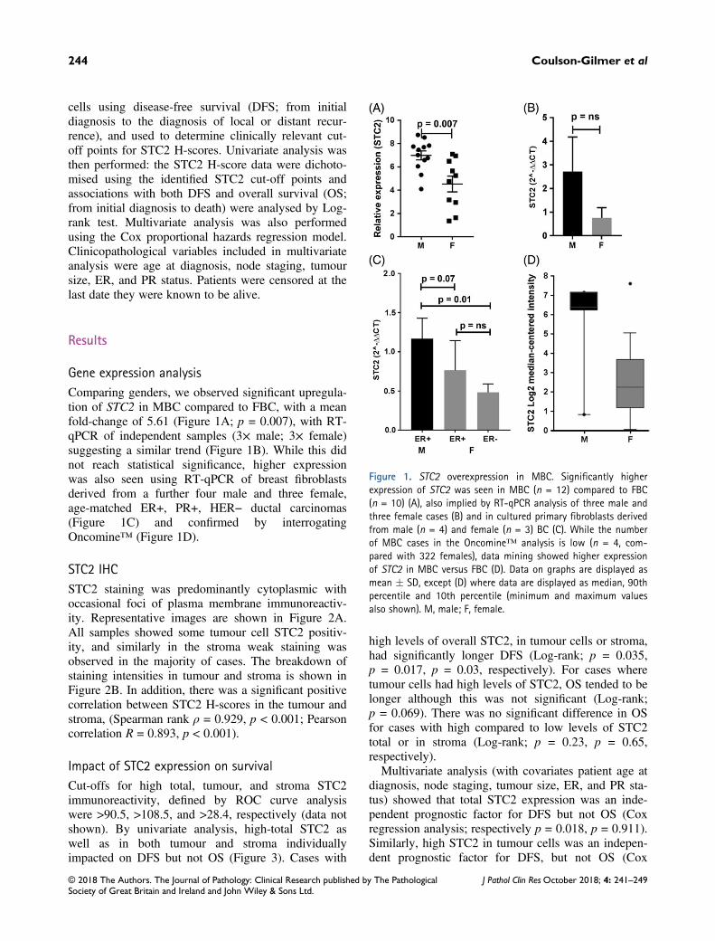
Gene expression analysisComparing genders, we observed significant upregula-tion of STC2 in MBC compared to FBC, with a meanfold-change of 5.61 (Figure 1A; p = 0.007), with RT-qPCR of independent samples (3× male; 3× female)suggesting a similar trend (Figure 1B). While this didnot reach statistical significance, higher expressionwas also seen using RT-qPCR of breast fibroblastsderived from a further four male and three female,age-matched ER+, PR+, HER− ductal carcinomas(Figure 1C) and confirmed by interrogatingOncomine™ (Figure 1D).
STC2 IHCSTC2 staining was predominantly cytoplasmic withoccasional foci of plasma membrane immunoreactiv-ity. Representative images are shown in Figure 2A.All samples showed some tumour cell STC2 positiv-ity, and similarly in the stroma weak staining wasobserved in the majority of cases. The breakdown ofstaining intensities in tumour and stroma is shown inFigure 2B. In addition, there was a significant positivecorrelation between STC2 H-scores in the tumour andstroma, (Spearman rank ρ = 0.929, p < 0.001; Pearsoncorrelation R = 0.893, p < 0.001).
Impact of STC2 expression on survivalCut-offs for high total, tumour, and stroma STC2immunoreactivity, defined by ROC curve analysiswere >90.5, >108.5, and >28.4, respectively (data notshown). By univariate analysis, high-total STC2 aswell as in both tumour and stroma individuallyimpacted on DFS but not OS (Figure 3). Cases with
high levels of overall STC2, in tumour cells or stroma,had significantly longer DFS (Log-rank; p = 0.035,p = 0.017, p = 0.03, respectively). For cases wheretumour cells had high levels of STC2, OS tended to belonger although this was not significant (Log-rank;p = 0.069). There was no significant difference in OSfor cases with high compared to low levels of STC2total or in stroma (Log-rank; p = 0.23, p = 0.65,respectively).Multivariate analysis (with covariates patient age at
diagnosis, node staging, tumour size, ER, and PR sta-tus) showed that total STC2 expression was an inde-pendent prognostic factor for DFS but not OS (Coxregression analysis; respectively p = 0.018, p = 0.911).Similarly, high STC2 in tumour cells was an indepen-dent prognostic factor for DFS, but not OS (Cox
Figure 1. STC2 overexpression in MBC. Significantly higherexpression of STC2 was seen in MBC (n = 12) compared to FBC(n = 10) (A), also implied by RT-qPCR analysis of three male andthree female cases (B) and in cultured primary fibroblasts derivedfrom male (n = 4) and female (n = 3) BC (C). While the numberof MBC cases in the Oncomine™ analysis is low (n = 4, com-pared with 322 females), data mining showed higher expressionof STC2 in MBC versus FBC (D). Data on graphs are displayed asmean � SD, except (D) where data are displayed as median, 90thpercentile and 10th percentile (minimum and maximum valuesalso shown). M, male; F, female.
244 Coulson-Gilmer et al
© 2018 The Authors. The Journal of Pathology: Clinical Research published by The PathologicalSociety of Great Britain and Ireland and John Wiley & Sons Ltd.
J Pathol Clin ResOctober 2018; 4: 241–249

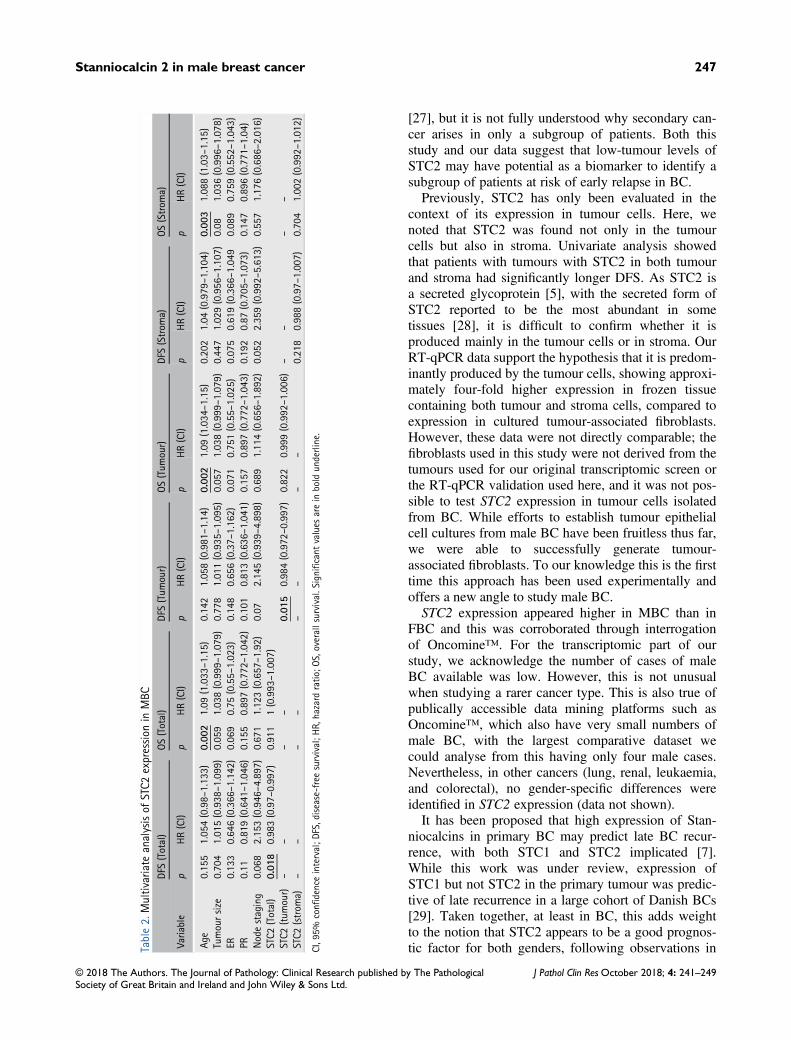
regression analysis; respectively, p = 0.015,p = 0.822). Patients with tumours containing stromawith high STC2 tended to have longer DFS, however,this was not significant (Cox regression analysis;p = 0.218). Nor was there any relationship betweenstroma STC2 levels and OS (Cox regression analysis;p = 0.65). Data are summarised in Table 2, with sig-nificant values in bold underline.
Discussion
A number of studies are beginning to show that STC2expression is a favourable prognostic factor in BC;however, it has not been studied previously in the con-text of MBC. With growing recognition that male andfemale BC may not be identical, there is increasinginterest in elucidating the biology of MBC, to assist indefining indicators of survival. The key findings in thisstudy were elevated expression of STC2 RNA in maleversus female BC and that both total STC2 proteinand its expression in tumour cells was an independentpredictor of patient survival in MBC.Using cell line models, it has been suggested that the
association between STC2 expression and favourableoutcome may be a result of its ability to repress inva-sive behaviour [25]. Hou et al [25] found enhancedmigration, motility, and expression of the transcriptionfactors Slug and Twist in BC cell lines where STC2was silenced, which following radiation were also moreanti-apoptotic compared to non-silenced control cells.Similarly, Raulic et al [5] noted a reduction in cellmotility when BC cell lines were stably transfectedwith STC2, as well as decreased cell viability afterserum withdrawal and reduced proliferation. This find-ing may be unique to BC as, in other cancers, includingneuroblastoma [26], lung [16], ovarian [17], and gastriccancer [9], STC2 expression has been reported to pro-mote metastasis and is thought to be a poor prognosticfactor. These seemingly opposing roles of STC2 againindicate its ability to mediate its effects through differ-ent signaling pathways dependent on the cellular con-text, possibly through dysregulation of calcium andphosphate dependent signaling [25].In a study of 72 paired primary and metastatic BCs
[7], STC2 expression was significantly higher in pri-mary tumours that showed late relapse, leading theauthors to suggest that STC2 may be involved intumour dormancy. This is of particular interest in BC,a disease known for its tendency to recur many yearsafter a patient has been in remission. Formation of dis-tant metastases is believed to be an early event in BC
Figure 2. Representative images of STC2 staining in MBC TMAs.The top panel shows examples of the various staining intensitiesin individual TMA cores, with higher magnification areas shownin the yellow outlined inserts. Black outlined inserts indicate fociof plasma membrane staining. The graph below shows the % ofTMA samples which were categorised into each ‘intensity’ group(where the majority of tumour cells had at least the given inten-sity). +, weak, ++, medium staining, +++, strong staining. Imagesscanned at x20 objective magnification.
245Stanniocalcin 2 in male breast cancer
© 2018 The Authors. The Journal of Pathology: Clinical Research published by The PathologicalSociety of Great Britain and Ireland and John Wiley & Sons Ltd.
J Pathol Clin ResOctober 2018; 4: 241–249

Figure 3. Kaplan–Meier survival curves showing impact of STC2 staining H-score in tumour and stroma on patient prognosis. High STC2H-scores in tumour cells (A), stroma (C) and total (E) were associated with longer DFS (p = 0.017, p = 0.03, p = 0.035, respectively), buthad no significant impact on OS for tumour (B), stroma (D) or total (F) (p = 0.069, p = 0.65, p = 0.23). Grey line, high STC2 H-score;black line, low STC2 H-score, Log-rank test. Cases were dichotomised by STC2 H-score: H-score cut-off point was 108.5 for tumour cells(DFS n = 28 low, n = 23 high; OS n = 76 low, n = 73 high); 28.4 for stroma (DFS n = 16 low, n = 35 high; OS n = 49 low, n = 100 high)and 90.5 for total staining (DFS n = 28 low, n = 23 high; OS n = 77 low, n = 72 high). HR, hazard ratio, followed by confidence intervalsshown in brackets.
246 Coulson-Gilmer et al
© 2018 The Authors. The Journal of Pathology: Clinical Research published by The PathologicalSociety of Great Britain and Ireland and John Wiley & Sons Ltd.
J Pathol Clin ResOctober 2018; 4: 241–249
[27], but it is not fully understood why secondary can-cer arises in only a subgroup of patients. Both thisstudy and our data suggest that low-tumour levels ofSTC2 may have potential as a biomarker to identify asubgroup of patients at risk of early relapse in BC.Previously, STC2 has only been evaluated in the
context of its expression in tumour cells. Here, wenoted that STC2 was found not only in the tumourcells but also in stroma. Univariate analysis showedthat patients with tumours with STC2 in both tumourand stroma had significantly longer DFS. As STC2 isa secreted glycoprotein [5], with the secreted form ofSTC2 reported to be the most abundant in sometissues [28], it is difficult to confirm whether it isproduced mainly in the tumour cells or in stroma. OurRT-qPCR data support the hypothesis that it is predom-inantly produced by the tumour cells, showing approxi-mately four-fold higher expression in frozen tissuecontaining both tumour and stroma cells, compared toexpression in cultured tumour-associated fibroblasts.However, these data were not directly comparable; thefibroblasts used in this study were not derived from thetumours used for our original transcriptomic screen orthe RT-qPCR validation used here, and it was not pos-sible to test STC2 expression in tumour cells isolatedfrom BC. While efforts to establish tumour epithelialcell cultures from male BC have been fruitless thus far,we were able to successfully generate tumour-associated fibroblasts. To our knowledge this is the firsttime this approach has been used experimentally andoffers a new angle to study male BC.STC2 expression appeared higher in MBC than in
FBC and this was corroborated through interrogationof Oncomine™. For the transcriptomic part of ourstudy, we acknowledge the number of cases of maleBC available was low. However, this is not unusualwhen studying a rarer cancer type. This is also true ofpublically accessible data mining platforms such asOncomine™, which also have very small numbers ofmale BC, with the largest comparative dataset wecould analyse from this having only four male cases.Nevertheless, in other cancers (lung, renal, leukaemia,and colorectal), no gender-specific differences wereidentified in STC2 expression (data not shown).It has been proposed that high expression of Stan-
niocalcins in primary BC may predict late BC recur-rence, with both STC1 and STC2 implicated [7].While this work was under review, expression ofSTC1 but not STC2 in the primary tumour was predic-tive of late recurrence in a large cohort of Danish BCs[29]. Taken together, at least in BC, this adds weightto the notion that STC2 appears to be a good prognos-tic factor for both genders, following observations inTa
ble2.Multivariate
analysisof
STC2
expression
inMBC
DFS(Total)
OS(Total)
DFS(Tum
our)
OS(Tum
our)
DFS(Strom
a)OS(Strom
a)
Varia
ble
pHR(CI)
pHR(CI)
pHR(CI)
pHR(CI)
pHR(CI)
pHR(CI)
Age
0.155
1.054(0.98–1.133)
0.00
21.09
(1.033–1.15)
0.142
1.058(0.981–1.14)
0.00
21.09
(1.034–1.15)
0.202
1.04
(0.979–1.104)
0.00
31.088(1.03–1.15)
Tumoursize
0.704
1.015(0.938–1.099)
0.059
1.038(0.999–1.079)
0.778
1.011(0.935–1.095)
0.057
1.038(0.999–1.079)
0.447
1.029(0.956–1.107)
0.08
1.036(0.996–1.078)
ER0.133
0.646(0.366–1.142)
0.069
0.75
(0.55–1.023)
0.148
0.656(0.37–1.162)
0.071
0.751(0.55–1.025)
0.075
0.619(0.366–1.049
0.089
0.759(0.552–1.043)
PR0.11
0.819(0.641–1.046)
0.155
0.897(0.772–1.042)
0.101
0.813(0.636–1.041)
0.157
0.897(0.772–1.043)
0.192
0.87
(0.705–1.073)
0.147
0.896(0.771–1.04)
Nodestaging
0.068
2.153(0.946–4.897)
0.671
1.123(0.657–1.92)
0.07
2.145(0.939–4.898)
0.689
1.114(0.656–1.892)
0.052
2.359(0.992–5.613)
0.557
1.176(0.686–2.016)
STC2
(Total)
0.01
80.983(0.97–0.997)
0.911
1(0.993–1.007)
STC2
(tum
our)
––
––
0.01
50.984(0.972–0.997)
0.822
0.999(0.992–1.006)
––
––
STC2
(strom
a)–
––
––
––
–0.218
0.988(0.97–1.007)
0.704
1.002(0.992–1.012)
CI,95%
confi
denceinterval;D
FS,disease-freesurvival;H
R,hazard
ratio
;OS,overallsurvival.Significant
values
arein
bold
underline.
247Stanniocalcin 2 in male breast cancer
© 2018 The Authors. The Journal of Pathology: Clinical Research published by The PathologicalSociety of Great Britain and Ireland and John Wiley & Sons Ltd.
J Pathol Clin ResOctober 2018; 4: 241–249
FBC, where elevated STC2 expression was associatedwith longer OS and DFS [15,18,19,30]. However, inour study, there was a reduction in its significance onmultivariate compared to univariate analysis. Thismight be explained by the fact that we were unable toobtain complete clinicopathological data from somecenters that contributed cases for our TMAs; as someof this was necessary for multivariate analysis, a noteof caution is warranted.It has been additionally reported that STC2 is asso-
ciated with ER+ FBC [13], supported by our findingsthat fibroblasts from ER+ MBC, or FBC expressedhigher levels of STC2 compared to those from ER−breast tumours. As exemplified in the two largestreported studies on MBC, which examined thousandsof patients, ER expression is very common in MBC[3,31], hence it is not surprising to see the sameassociation.In summary, while overexpressed in male compared
to female BC, STC2 appears to be a good prognosticfactor, irrespective of gender.
Acknowledgements
This study was supported by Yorkshire CancerResearch (grant L378) and Breast Cancer Now(TBLEE2017). We are grateful to the Breast CancerNow Tissue Bank and the Male Breast Cancer Consor-tium who kindly provided cases for this work.
Author contributions statement
VS conceived experiments. CCG, MPH, AC, and SSRcarried out experiments. AD, VS, and CCG carried outdata analysis. SSR and SJ provided clinical data. JLJ,GC, LBJ, RK, ADB, MM, EP, JK, AMS, and AMHprovided patient material. CCG and VS wrote themanuscript. All authors read and approved the finalmanuscript.
References
1. Johansson I, Nilsson C, Berglund P, et al. High-resolution genomic
profiling of male breast cancer reveals differences hidden behind
the similarities with female breast cancer. Breast Cancer Res Treat
2011; 129: 747–760.2. Johansson I, Nilsson C, Berglund P, et al. Gene expression profil-
ing of primary male breast cancers reveals two unique subgroups
and identifies N-acetyltransferase-1 (NAT1) as a novel prognostic
biomarker. Breast Cancer Res 2012; 14: R31.
3. Humphries MP, Sundara Rajan S, Droop A, et al. A case-matched
gender comparison Transcriptomic screen identifies eIF4E and
eIF5 as potential prognostic markers in male breast cancer. Clin
Cancer Res 2017; 23: 2575–2583.4. Ishibashi K, Miyamoto K, Taketani Y, et al. Molecular cloning of
a second human stanniocalcin homologue (STC2). Biochem
Biophys Res Commun 1998; 250: 252–258.5. Raulic S, Ramos-Valdes Y, DiMattia GE. Stanniocalcin 2 expres-
sion is regulated by hormone signalling and negatively affects
breast cancer cell viability in vitro. J Endocrinol 2008; 197:517–529.
6. Roch GJ, Sherwood NM. Stanniocalcin has deep evolutionary
roots in eukaryotes. Genome Biol Evol 2011; 3: 284–294.7. Joensuu K, Heikkila P, Andersson LC. Tumor dormancy: elevated
expression of stanniocalcins in late relapsing breast cancer. Cancer
Lett 2008; 265: 76–83.8. Yeung BH, Law AY, Wong CK. Evolution and roles of stanniocal-
cin. Mol Cell Endocrinol 2012; 349: 272–280.9. Arigami T, Uenosono Y, Ishigami S, et al. Clinical significance of
stanniocalcin 2 expression as a predictor of tumour progression in
gastric cancer. Oncol Rep 2013; 30: 2838–2844.10. Chen B, Zeng X, He Y, et al. STC2 promotes the
epithelial-mesenchymal transition of colorectal cancer cells through
AKT-ERK signaling pathways. Oncotarget 2016; 7: 71400–71416.11. Shin J, Sohn YC. cDNA cloning of Japanese flounder stanniocalcin
2 and its mRNA expression in a variety of tissues. Comp Biochem
Physiol A Mol Integr Physiol 2009; 153: 24–29.12. Zubor P, Hatok J, Moricova P, et al. Gene expression profiling of
histologically normal breast tissue in females with human epider-
mal growth factor receptor 2positive breast cancer. Mol Med Rep
2015; 11: 1421–1427.13. Bouras T, Southey MC, Chang AC, et al. Stanniocalcin 2 is an
estrogen-responsive gene coexpressed with the estrogen receptor in
human breast cancer. Cancer Res 2002; 62: 1289–1295.14. McBryan J, Howlin J, Kenny PA, et al. ERalpha-CITED1
co-regulated genes expressed during pubertal mammary gland
development: implications for breast cancer prognosis. Oncogene
2007; 26: 6406–6419.15. Esseghir S, Kennedy A, Seedhar P, et al. Identification of NTN4,
TRA1, and STC2 as prognostic markers in breast cancer in a
screen for signal sequence encoding proteins. Clin Cancer Res
2007; 13: 3164–3173.16. Na SS, Aldonza MB, Sung HJ, et al. Stanniocalcin-2 (STC2): a
potential lung cancer biomarker promotes lung cancer
metastasis and progression. Biochim Biophys Acta 2015; 1854:
668–676.
17. Wu J, Lai M, Shao C, et al. STC2 overexpression mediated by
HMGA2 is a biomarker for aggressiveness of high-grade serous
ovarian cancer. Oncol Rep 2015; 34: 1494–1502.18. Parris TZ, Kovacs A, Aziz L, et al. Additive effect of the AZGP1,
PIP, S100A8 and UBE2C molecular biomarkers improves outcome
prediction in breast carcinoma. Int J Cancer 2014; 134:1617–1629.
19. Todd JR, Ryall KA, Vyse S, et al. Systematic analysis of tumour
cell-extracellular matrix adhesion identifies independent prognostic
factors in breast cancer. Oncotarget 2016; 7: 62939–62953.
248 Coulson-Gilmer et al
© 2018 The Authors. The Journal of Pathology: Clinical Research published by The PathologicalSociety of Great Britain and Ireland and John Wiley & Sons Ltd.
J Pathol Clin ResOctober 2018; 4: 241–249

20. Speirs V, Green AR, Walton DS, et al. Short-term primary culture
of epithelial cells derived from human breast tumours. Br J Cancer
1998; 78: 1421–1429.21. McShane LM, Altman DG, Sauerbrei W, et al. REporting recom-
mendations for tumour MARKer prognostic studies (REMARK).
Br J Cancer 2005; 93: 387–391.22. Jansen MP, Sas L, Sieuwerts AM, et al. Decreased expression of
ABAT and STC2 hallmarks ER-positive inflammatory breast can-
cer and endocrine therapy resistance in advanced disease. Mol
Oncol 2015; 9: 1218–1233.23. Bankhead P, Loughrey MB, Fernandez JA, et al. QuPath: open
source software for digital pathology image analysis. Sci Rep 2017;
7: 16878.24. Budczies J, Klauschen F, Sinn BV, et al. Cutoff finder: a compre-
hensive and straightforward web application enabling rapid bio-
marker cutoff optimization. PLoS One 2012; 7: e51862.25. Hou J, Wang Z, Xu H, et al. Stanniocalicin 2 suppresses breast
cancer cell migration and invasion via the PKC/claudin-1-mediated
signaling. PloS One 2015; 10: e0122179.
26. Volland S, Kugler W, Schweigerer L, et al. Stanniocalcin 2 pro-
motes invasion and is associated with metastatic stages in neuro-
blastoma. Int J Cancer 2009; 125: 2049–2057.27. Husemann Y, Geigl JB, Schubert F, et al. Systemic spread is an
early step in breast cancer. Cancer Cell 2008; 13: 58–68.28. Jellinek DA, Chang AC, Larsen MR, et al. Stanniocalcin 1 and
2 are secreted as phosphoproteins from human fibrosarcoma cells.
Biochem J 2000; 350: 453–461.29. Brantley KD, Kjaersgaard A, Cronin-Fenton D, et al. Stanniocalcin
expression as a predictor of late breast cancer recurrence. Cancer
Epidemiol Biomarkers Prev 2018; 27: 653–659.30. Gyorffy B, Lanczky A, Eklund AC, et al. An online survival anal-
ysis tool to rapidly assess the effect of 22,277 genes on breast can-
cer prognosis using microarray data of 1,809 patients. Breast
Cancer Res Treat 2010; 123: 725–731.31. Cardoso F, Bartlett JMS, Slaets L, et al. Characterization of male
breast cancer: results of the EORTC 10085/TBCRC/BIG/NABCG
International Male Breast Cancer Program. Ann Oncol 2018; 29:405–417.
249Stanniocalcin 2 in male breast cancer
© 2018 The Authors. The Journal of Pathology: Clinical Research published by The PathologicalSociety of Great Britain and Ireland and John Wiley & Sons Ltd.
J Pathol Clin ResOctober 2018; 4: 241–249