European Anesthesia, Respiratory and Sleep Management Device Market
STANDARDS FOR ADULT RESPIRATORY AND SLEEP SERVICES IN NEW ZEALAND
A document produced for the Thoracic Society of Australia and New Zealand (New Zealand Branch)
Initial Report 1989, revised 1996, 2002 and 2004
1
Summary and Recommendations
Summary of the current situation
The importance of a well organised, efficient and accessible Respiratory Service in the New Zealand Health Care system scarcely needs emphasising. Respiratory disease is a major health problem in New Zealand. In fact, respiratory disorders are the most common reason for primary health care consultations and are responsible for 35�50 percent of all medical admissions. With the increasing prevalence of respiratory conditions such as asthma, Chronic Obstructive Pulmonary Disease (COPD), TB, Obstructive Sleep Apnoea (OSA) and pulmonary malignancy, the costs, in human and economic terms, are large and continue to escalate. Four respiratory disorders (Lower Respiratory Tract Infections (LRTIs), COPD, TB and lung cancer) are amongst the 10 leading causes of disease burden in the world1 and four respiratory disorders (COPD, LRTIs, asthma, and lung cancer) are amongst the 10 leading causes of disease burden in New Zealand.2 Asthma and COPD are the highest-ranking causes of years lost to disability (YLD) in males in New Zealand and rank third and seventh respectively for females.3 Respiratory disease has now overtaken coronary heart disease and cancer as the most common cause of mortality.4 Respiratory illness is the most common cause of: long term illness among children, ED utilisation and general practice visits, as well as hospital admissions and therefore costs the health system more than any other medical disorder. Further, clinicians are now recognising the importance of other conditions, such as a range of sleeping disorders, bronchiolitis and interstitial lung disorders. Over the past 30 years, the burden of respiratory illness has increased substantially and will continue to do so, with greater consequent demands on primary and secondary health care services. The World Health Organization (WHO) has defined respiratory disorders as one of the key areas requiring special attention in the 21st century and advised that new models of care should be considered. The burden of respiratory illness can be reduced and, in many instances, prevented. Avoiding or stopping smoking,5 vaccination, better early childhood respiratory care, improved occupational surveillance, better access to specialist care and investigations and screening, all contribute to reduction and/or prevention of respiratory illness. Earlier diagnosis using spirometry in the community,6 together with subsequent introduction of effective therapy at an earlier time, could also be expected to reduce the burden of respiratory disease over time. The current structure of the New Zealand health care system mitigates against early diagnosis. The majority of respiratory physicians work in public hospitals and are more frequently referred patients with advanced disease.
i
It is important to recognise that financial barriers to primary health care, including excessive co-payments on drug therapy, have an adverse effect on respiratory disorders. The timing of intervention during acute exacerbations of respiratory conditions is of critical importance to successful management. Financial barriers are an important reason behind New Zealand�s high admission rates for asthma, COPD, bronchiectasis and pneumonia. The current Primary Health Care Strategy, which has begun to address some of these issues, including co-payments on prescriptions should therefore contribute to reductions in both morbidity and mortality for patients with a range of respiratory disorders. With the development of 21 District Health Boards (DHBs), we believe there is an urgent need to develop an infrastructure to advise and support the management of respiratory disorders in each of the districts. A template for comprehensive regional respiratory services has been developed and is described in this report, using staff that move across traditional health care boundaries and facilitate continuity of care between the community and hospital. However, because expert respiratory opinion is not readily available in some DHBs and there are substantial variations in the practice of respiratory medicine in New Zealand, we perceive a need for both regional and national overview. We therefore propose the infrastructure defined in Figure 1 and which is similar to that envisaged under the Cancer Control Strategy (New Zealand) http://www.moh.govt.nz/moh.nsf/0/3D7504AD140C7EF0CC256D88000E5A16/$File/CancerControlStrategy.pdf Because most respiratory disorders can be managed in the community if the appropriate infrastructure exists, developing a system for improved management of respiratory disorders may offer a model that can readily be adopted by other specialities. As a professional body we would like to offer our services and those of our members to the Ministry of Health (MoH) and DHBs to support what we hope will become an exciting period of change in health care delivery. In anticipation of this, TSANZ have developed this proposal in collaboration with RACGP, RACP, ANZSRS and specialist nurses, radiologists and physiotherapists affiliated with the TSANZ.
Recommendations
! The Ministry of Health (Ministry of Health) needs to recognise respiratory disorders as a major health problem in New Zealand and create an infrastructure to provide oversight and direction of management of respiratory diseases in New Zealand, centrally co-ordinate activities, and support the development of the initiatives described in this document.
! There is an urgent need for health education and greater self-management of respiratory disease. Increased educational services, particularly at primary care level, are strongly recommended and incentives for these and for more comprehensive management of acutely unwell respiratory patients need to be put in place.7,8,9,10
! Access to respiratory physicians and to new technology (sleep laboratories, high-resolution Computed Tomography (CT) scans, and detailed lung function testing) needs to be improved to identify disease at an earlier stage.
ii
! General practice facilities for respiratory diagnosis and management need to be better supported and made more efficient with greater financial incentive for managing acute respiratory illness in the community.
! Secondary and tertiary care facilities must be of a more uniform standard. At the same time, first referrals to outpatient clinics should be increased and reimbursed more appropriately, and long term follow up patients should be better monitored and returned in greater number to their general practitioner. Prioritisation criteria for admission and outpatient care have already been developed and implemented by the Ministry of Health in association with the TSANZ (Appendix III).
! Information on waiting times to outpatient clinic attendance are not currently collected and reported on. Collection and evaluation of waiting times is necessary to determine if the prioritisation criteria are being followed.
! Case management of high risk (eg, frequent hospitalisation, ICU admissions) needs further study and support.
! Greater provision of day patient facilities could reduce inpatient caseloads provided staffing and facilities are adequate. It is recommended there be at least 1.0 FTE specialist respiratory physician per 75�100,000 population11,12,13,14,15 in New Zealand, together with adequate resident medical, nursing and allied health staff and other support personnel.
! Access and impact indicators for respiratory disorders are not well defined; outcomes need to be assessed and compared nationally and internationally.
! A respiratory adviser should be appointed to each of the DHBs with an opportunity for regular regional meetings (centred on Auckland, Hamilton, Wellington, Christchurch and Dunedin) and a representative from each of the regions should sit on a National Executive Committee. The National Executive Committee would also receive representation from the TSANZ, primary, secondary and tertiary care communities, nursing, allied health, lay societies and Maori and Pacific communities and would report both to the Ministry of Health and to DHBs.
! An infrastructure to advise and support the management of respiratory disorders in each DHB needs to be developed. We recommend this is actioned as follows: ! perform an assessment of the burden of lung disease in New Zealand ! collect and collate information by DHB to assess whether any deficiencies in
practice presently exist ! update paediatric respiratory service, quality of care and primary care
components of the Standards Recommendations ! credential DHBs to ensure basic respiratory services are available and of
reasonable standard (Appendix XIV) ! work in close association with the Ministry of Health and Public Health
Departments to develop evidence based strategies to reduce the prevalence of respiratory disease.
! disease specific management systems could then be proposed using a common but flexible template to develop integrated models of care.
iii
Acknowledgements
The RNZCGP, ANZSRS, RNZSP and RACP have all made important contributions to this document: Standards for New Zealand Respiratory Services 1996 Committee: Dr J Garrett (Chairman), Dr C Drennan, Dr A Watson, Dr C Wong, and Prof TV O�Donnell. The Committee would like to gratefully acknowledge the contributions made by: RNZCGPs, Dr N Karalus, Dr A Harrison, A Prof J Kolbe, Dr K Whyte, Dr T Christmas, Dr A Veale, Prof H Rea, Dr I Asher, Dr A Wells and Dr M Wilsher. Standards for New Zealand Respiratory Services 2002: A Prof J Garrett (Chairman), A Prof R Taylor (President). We would like to gratefully acknowledge contributions by: A Prof J Kolbe, A Prof M Wilsher, Dr A Neil, Dr D Milne, Mr P Alison, Ms C Chalmers, Prof I Town, Ms M Swanney, Mrs P Young, and Dr Peter Jansen.
iv
About this Publication
The New Zealand Branch of the TSANZ has produced this document, �Standards for Adult Respiratory and Sleep Services in New Zealand� to present its views on the best configuration for Respiratory Services for the future against the current backdrop of increasing service demands and continuing financial constraint. The report encompasses the philosophy of the WHO in acknowledging the need for health policy formulation, regulation and assessment of performance by collection and analysis of outcome generated data. The aim is to help develop an efficient respiratory health system which is of good quality, responsive to the population�s needs and which is resourced with a fair and equitable distribution of money. This report appeared initially in 1989 and was substantially rewritten in 1996. It was revised in 2002 and again in 2004 to reflect changes in health care and in respiratory medicine in New Zealand over the past five years. The latest revision no longer incorporates comprehensive sections on primary care or paediatric respiratory disorders. These would be a priority if a national committee were formulated.
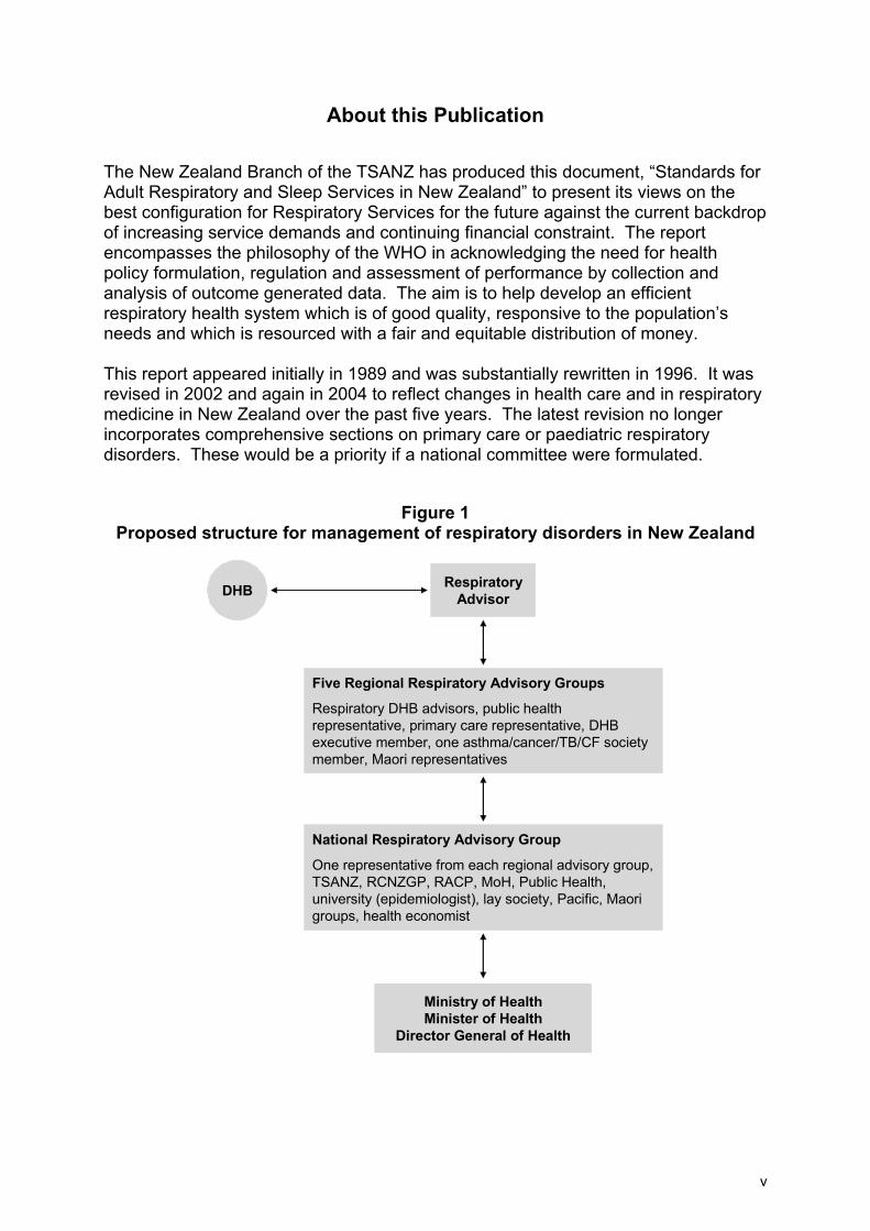
Figure 1 Proposed structure for management of respiratory disorders in New Zealand
DHB Respiratory Advisor
Five Regional Respiratory Advisory Groups
Respiratory DHB advisors, public health representative, primary care representative, DHB executive member, one asthma/cancer/TB/CF society member, Maori representatives
National Respiratory Advisory Group
One representative from each regional advisory group, TSANZ, RCNZGP, RACP, MoH, Public Health, university (epidemiologist), lay society, Pacific, Maori groups, health economist
Ministry of HealthMinister of Health
Director General of Health
v
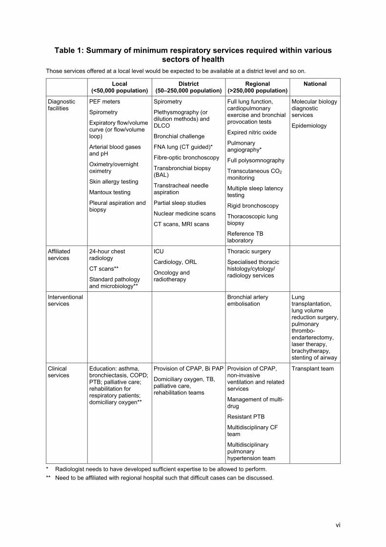
Table 1: Summary of minimum respiratory services required within various sectors of health
Those services offered at a local level would be expected to be available at a district level and so on.
Local (<50,000 population)
District (50�250,000 population)
Regional (>250,000 population)
National
Diagnostic facilities
PEF meters
Spirometry
Expiratory flow/volume curve (or flow/volume loop)
Arterial blood gases and pH
Oximetry/overnight oximetry
Skin allergy testing
Mantoux testing
Pleural aspiration and biopsy
Spirometry
Plethysmography (or dilution methods) and DLCO
Bronchial challenge
FNA lung (CT guided)*
Fibre-optic bronchoscopy
Transbronchial biopsy (BAL)
Transtracheal needle aspiration
Partial sleep studies
Nuclear medicine scans
CT scans, MRI scans
Full lung function, cardiopulmonary exercise and bronchial provocation tests
Expired nitric oxide
Pulmonary angiography*
Full polysomnography
Transcutaneous CO2 monitoring
Multiple sleep latency testing
Rigid bronchoscopy
Thoracoscopic lung biopsy
Reference TB laboratory
Molecular biology diagnostic services
Epidemiology
Affiliated services
24-hour chest radiology
CT scans**
Standard pathology and microbiology**
ICU
Cardiology, ORL
Oncology and radiotherapy
Thoracic surgery
Specialised thoracic histology/cytology/ radiology services
Interventional services
Bronchial artery embolisation
Lung transplantation, lung volume reduction surgery, pulmonary thrombo-endarterectomy, laser therapy, brachytherapy, stenting of airway
Clinical services
Education: asthma, bronchiectasis, COPD; PTB; palliative care; rehabilitation for respiratory patients; domiciliary oxygen**
Provision of CPAP, Bi PAP
Domiciliary oxygen, TB, palliative care, rehabilitation teams
Provision of CPAP, non-invasive ventilation and related services
Management of multi-drug
Resistant PTB
Multidisciplinary CF team
Multidisciplinary pulmonary hypertension team
Transplant team
* Radiologist needs to have developed sufficient expertise to be allowed to perform. ** Need to be affiliated with regional hospital such that difficult cases can be discussed.
vi
Contents
Glossary .................................................................................................................................. ix
1 Introduction ...................................................................................................................12 1.1 Purpose and use of guidelines ............................................................................12 1.2 The scope of respiratory services........................................................................12 1.3 The burden of respiratory disease .......................................................................13 1.4 Maori and Pacific health ......................................................................................15 1.5 Service history .....................................................................................................15 1.6 Future directions for respiratory services.............................................................16 1.7 Predictions ...........................................................................................................18 1.8 New technologies ................................................................................................19
2 Guidelines for Development of Respiratory Services....................................................22 2.1 Prevention of disease ..........................................................................................22 2.2 Maori and Pacific health ......................................................................................22 2.3 Culturally acceptable care ...................................................................................23 2.4 Ethnicity data .......................................................................................................24 2.5 Primary and community care ...............................................................................24 2.6 Health education and self-management of respiratory disorders ........................25 2.7 Secondary and tertiary health care......................................................................25 2.8 Quality assurance and peer review .....................................................................26
3 Respiratory Services in General Practice .....................................................................28 3.1 Education.............................................................................................................28 3.2 Referral to secondary and tertiary care ...............................................................28 3.3 Quality control......................................................................................................29 3.4 Planning services.................................................................................................29 3.5 Integrated electronic information systems ...........................................................29
4 Specialist Respiratory Services.....................................................................................30 4.1 Respiratory services at a national level ...............................................................30 4.2 Respiratory services at a regional level ...............................................................30 4.3 Respiratory services at a district level (between 50,000 - 250,000) ................ 37354.4 Respiratory Services at a rural level (below 50,000) 37
5 Support Facilities for Inpatient Respiratory Services at a Regional Level.....................39
6 Requirements for Outpatient Respiratory Services at a Regional Level .......................42
7 Minimum Requirement for Staffing at a Regional and District Level.............................43 7.1 Senior consultant staff .........................................................................................43 7.2 Resident medical staff .........................................................................................44 7.3 Nursing staff.........................................................................................................44 7.4 Respiratory nurse practitioner..............................................................................45 7.5 Respiratory physiotherapists ...............................................................................45 7.6 Secretarial, clerical and administrative staff ........................................................45
vii
7.7 Respiratory physiology scientists / technologists / technicians............................46 7.8 Staff definitions,....................................................................................................46 7.9 Recommendations...............................................................................................46
8 Respiratory Services at a Subspecialty Level ...............................................................48 8.1 Tuberculosis ........................................................................................................48 8.2 Cystic fibrosis.......................................................................................................50 8.3 Bronchiectasis .....................................................................................................52 8.4 Occupational lung disease...................................................................................56 8.5 Sleep disordered breathing service (adults) ........................................................57 8.6 Lung cancer .........................................................................................................60 8.7 Interstitial lung diseases ......................................................................................63 8.8 Pulmonary vascular disorders .............................................................................64 8.9 Asthma.................................................................................................................65 8.10 Chronic obstructive pulmonary disease (COPD), ................................................68
Integration of Medical Services with Community Groups.......................................................72
Health Service Data Requirements ........................................................................................73
Research and Education Requirements.................................................................................74
Appendices Appendix I: National hospital admissions for respiratory diseases 1970�98,
and projections for 2005 ......................................................................75 Appendix II: Measurement of performance and outcome indicators .......................76 Appendix III: Respiratory medicine national referral guidelines................................84 Appendix IV: TSANZ/RACP standards for training in respiratory medicine
requirements for physician training, adult medicine, 2001 ..................85 Appendix V: Respiratory standards/training in other specialty areas.......................97 Appendix VI: Bronchoscopy services......................................................................105 Appendix VII: Laser, stenting and brachytherapy ....................................................106 Appendix VIII: Respiratory function assessment ......................................................107 Appendix IX: Lung transplantation ..........................................................................109 Appendix X: Lung volume reduction surgery .........................................................111 Appendix XI: Service specification � home oxygen therapy services .....................118 Appendix XII: Sleep related breathing disorders, a position paper of the New
Zealand branch of the Thoracic Society of Australia and New Zealand..............................................................................................119
Appendix XIII: Chronic disease management: recommendations on asthma services for District Health Boards ....................................................133
Appendix XIV: Accreditation of Specialist Services...................................................140
References ...........................................................................................................................174
viii
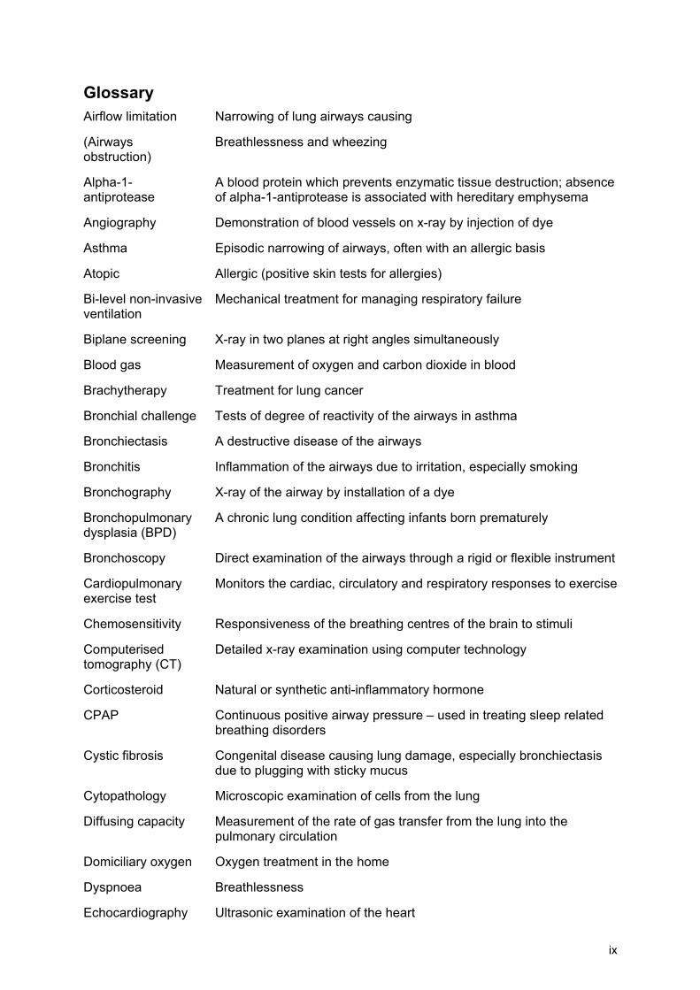
Glossary Airflow limitation Narrowing of lung airways causing
(Airways obstruction)
Breathlessness and wheezing
Alpha-1-antiprotease
A blood protein which prevents enzymatic tissue destruction; absence of alpha-1-antiprotease is associated with hereditary emphysema
Angiography Demonstration of blood vessels on x-ray by injection of dye
Asthma Episodic narrowing of airways, often with an allergic basis
Atopic Allergic (positive skin tests for allergies)
Bi-level non-invasive ventilation
Mechanical treatment for managing respiratory failure
Biplane screening X-ray in two planes at right angles simultaneously
Blood gas Measurement of oxygen and carbon dioxide in blood
Brachytherapy Treatment for lung cancer
Bronchial challenge Tests of degree of reactivity of the airways in asthma
Bronchiectasis A destructive disease of the airways
Bronchitis Inflammation of the airways due to irritation, especially smoking
Bronchography X-ray of the airway by installation of a dye
Bronchopulmonary dysplasia (BPD)
A chronic lung condition affecting infants born prematurely
Bronchoscopy Direct examination of the airways through a rigid or flexible instrument
Cardiopulmonary exercise test
Monitors the cardiac, circulatory and respiratory responses to exercise
Chemosensitivity Responsiveness of the breathing centres of the brain to stimuli
Computerised tomography (CT)
Detailed x-ray examination using computer technology
Corticosteroid Natural or synthetic anti-inflammatory hormone
CPAP Continuous positive airway pressure � used in treating sleep related breathing disorders
Cystic fibrosis Congenital disease causing lung damage, especially bronchiectasis due to plugging with sticky mucus
Cytopathology Microscopic examination of cells from the lung
Diffusing capacity Measurement of the rate of gas transfer from the lung into the pulmonary circulation
Domiciliary oxygen Oxygen treatment in the home
Dyspnoea Breathlessness
Echocardiography Ultrasonic examination of the heart
ix
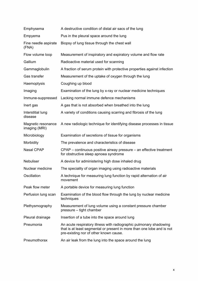
Emphysema A destructive condition of distal air sacs of the lung
Empyema Pus in the pleural space around the lung
Fine needle aspirate (FNA)
Biopsy of lung tissue through the chest wall
Flow volume loop Measurement of inspiratory and expiratory volume and flow rate
Gallium Radioactive material used for scanning
Gammaglobulin A fraction of serum protein with protective properties against infection
Gas transfer Measurement of the uptake of oxygen through the lung
Haemoptysis Coughing up blood
Imaging Examination of the lung by x-ray or nuclear medicine techniques
Immune-suppressed Lacking normal immune defence mechanisms
Inert gas A gas that is not absorbed when breathed into the lung
Interstitial lung disease
A variety of conditions causing scarring and fibrosis of the lung
Magnetic resonance imaging (MRI)
A new radiologic technique for identifying disease processes in tissue
Microbiology Examination of secretions of tissue for organisms
Morbidity The prevalence and characteristics of disease
Nasal CPAP CPAP � continuous positive airway pressure � an effective treatment for obstructive sleep apnoea syndrome
Nebuliser A device for administering high dose inhaled drug
Nuclear medicine The speciality of organ imaging using radioactive materials
Oscillation A technique for measuring lung function by rapid alternation of air movement
Peak flow meter A portable device for measuring lung function
Perfusion lung scan Examination of the blood flow through the lung by nuclear medicine techniques
Plethysmography Measurement of lung volume using a constant pressure chamber pressure � tight chamber
Pleural drainage Insertion of a tube into the space around lung
Pneumonia An acute respiratory illness with radiographic pulmonary shadowing that is at least segmental or present in more than one lobe and is not pre-existing nor of other known cause.
Pneumothorax An air leak from the lung into the space around the lung
x
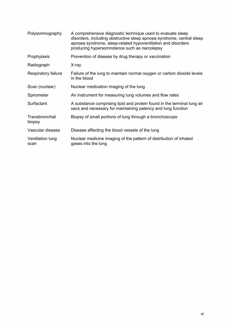
Polysomnography A comprehensive diagnostic technique used to evaluate sleep disorders, including obstructive sleep apnoea syndrome, central sleep apnoea syndrome, sleep-related hypoventilation and disorders producing hypersomnolence such as narcolepsy
Prophylaxis Prevention of disease by drug therapy or vaccination
Radiograph X-ray
Respiratory failure Failure of the lung to maintain normal oxygen or carbon dioxide levels in the blood
Scan (nuclear) Nuclear medication imaging of the lung
Spirometer An instrument for measuring lung volumes and flow rates
Surfactant A substance comprising lipid and protein found in the terminal lung air sacs and necessary for maintaining patency and lung function
Transbronchial biopsy
Biopsy of small portions of lung through a bronchoscope
Vascular disease Disease affecting the blood vessels of the lung
Ventilation lung scan
Nuclear medicine imaging of the pattern of distribution of inhaled gases into the lung
xi
1 Introduction
1.1 Purpose and use of guidelines
These strategic guidelines are primarily written for professionals planning, developing and managing Respiratory Services in the Ministry of Health and DHBs. If accepted, these guidelines would underpin the activities of a National Respiratory Advisory Committee and of regional committees. The guidelines look to include the full scope of services from prevention of respiratory illness, to promotion of primary and community health care, to hospital based services and ambulatory care. Their purpose is to facilitate the development of an efficient, accessible and equitable Respiratory Service in all regions of New Zealand. They are �service� rather than �disease management� guidelines, and do not attempt to provide detailed protocols for patient care other than by way of occasional examples (pertaining to transplantation, lung volume reduction surgery, sleep related breathing disorders and oxygen therapy). They indicate the minimum services, manpower, equipment and level of training anticipated at the different levels of our health care system. The New Zealand health system has entered a new era with the formation of DHBs, which have responsibility for community and hospital-based health care, thus allowing greater integration of primary and specialised health services. It is expected that DHBs will meet the standards suggested in these guidelines for essential Respiratory Services within the next five years. However, it is important to recognise the requirement to satisfy present needs, whilst also planning for anticipated needs. These guidelines will require review within five years of publication, because of changing health systems and health needs. The reviewing panel should include general practitioners, secondary health care providers, members of the TSANZ, representatives of Maori and Pacific people, lay societies and the DHBs and Ministry of Health.
1.2 The scope of respiratory services
Respiratory services provide community- and hospital-based facilities for the prevention, detection, assessment, investigation and management of diseases of the respiratory system. The service should include an educational and counselling role. Conditions that fall within the jurisdiction of respiratory services include: ! asthma and COPD ! malignant intrathoracic diseases (lung cancer, mesothelioma, thymoma etc) ! sleep related breathing disorders and daytime hypersomnolence ! pulmonary infections, particularly pneumonia and tuberculosis ! interstitial lung disorders ! bronchiolitis, bronchiectasis, cystic fibrosis ! bronchopulmonary dysplasia ! occupational lung diseases
12
! pulmonary vascular disorders ! chest trauma ! respiratory failure due to neuromuscular disorders, chest wall deformity, obesity
and COPD.
1.3 The burden of respiratory disease
Respiratory disease is a major contributor to morbidity and mortality in New Zealand and worldwide.
1. Respiratory illness is the most common reason for consultations in general practice and accounts for 35�50 percent of medical admissions to hospital. Respiratory disorders make up nine percent of New Zealand Burden of Disease estimates, ranking behind cardiovascular disease, cancer (15 percent of which are due to lung cancers) and mental disorders.13 However, this estimate does not include all respiratory disorders (eg, lung cancer, sleep related breathing disorders) which would account for another four to five percent. The diversity of respiratory disease is often not appreciated by those who work outside of the specialty, so it is important to highlight that respiratory disorders, when taken together, kill more people than cancer or cardiovascular disease.15
2. Asthma is a serious cause of morbidity in children and young people, the single most frequent reason for hospitalisation in childhood,16,17 and the most common cause of ED attendance in New Zealand.18 Asthma is also increasing in prevalence.19 During two different time periods, New Zealand had the highest asthma mortality rates in the world.20,21 Currently, New Zealand has the highest admission rate in the world, and asthma remains a significant cause of respiratory morbidity (loss of time from school, absenteeism from work, suboptimal performance, etc). Direct and indirect costs associated with asthma in New Zealand have been estimated at around $825 million annually.19 Asthma ranks third in New Zealand for specific causes of years lost to disability (highest in males)14 and eighth for both males and females in New Zealand Burden of Disease estimates.13
3. COPD is the fourth most common cause of hospitalisation in male adults (and eighth among females), and is the third most common cause of death. The prevalence of COPD (emphysema/chronic bronchitis) has been estimated to be around 834/100,000 (of any age) but the COPD prevalence in the New Zealand population aged over 45 years is estimated to be around 40,000.22 Direct costs of COPD management in New Zealand have not been accurately defined but are estimated at between $120�234 million per year and indirect costs are usually at least as high. COPD ranks second for males and fifth for females in New Zealand Burden of Disease estimates.13
4. Lung cancer is the most common cause of cancer death in men and more recently women. The direct costs associated with lung cancer management in New Zealand range from $18 to $29 million and will increase substantially if international guidelines on the use of chemotherapy in non small cell lung cancer are endorsed.22 Lung cancer ranks fifth in New Zealand males in New Zealand Burden of Disease estimates and 12th in females, but is rising rapidly and should rank ahead of breast cancer, (currently ranked fourth), within 10 years.14
13
5. Pneumonia and lower respiratory tract infections are a major cause of death in middle-aged and elderly persons and the most common cause of hospital admission. Respiratory tract infections rank third for males in New Zealand Burden of Disease estimates.18
6. Serious sleep related breathing disorders occur in 4 percent of middle-aged adult males and one to 2 percent of females, and may contribute to upwards of 22 percent of serious road traffic crashes (RTCs).23 To date, insufficient data is available to estimate the costs (direct and indirect) associated with these conditions and so no burden of disease estimates have been formulated (though a New Zealand study has recently been commissioned). However, given the likely prevalence of the disorder and its association with a number of common conditions (RTCs, hypertension, cardiac failure, CVA), the associated costs are likely to be high.
7. In 1984 the WHO declared pulmonary tuberculosis a worldwide emergency. Together with an increasing incidence in Western countries, the development of multi-drug resistant disease TB will provide a major medical challenge for the future. TB presently ranks sixth in the world in global burden of disease estimates.17
Respiratory illness is a particular problem among Maori and Pacific people of all ages, with considerable premature deaths attributable to bronchiectasis.24 A substantial proportion of the total burden of respiratory illness � specifically that related to cigarette smoking,25 occupational health26 and lack of immunisation27,28 � is theoretically preventable. DHBs should ensure that services are available to effectively meet the needs of people with the above disorders, and should support all measures, including prevention and adequate access to primary health care, that help avoid such diseases and reduce their impact in the community. Respiratory services need to be easily accessible to all individuals. Organisational and financial barriers to health care have a more immediate impact on patients with respiratory disease than those with most other medical conditions, and remain the major and most preventable component of escalating admission rates for a variety of respiratory disorders.29 Financial barriers to primary care have an exaggerated effect on respiratory disorders partly because respiratory illness is more common and more severe in lower socioeconomic groups (as well as in Maori and Pacific populations). Respiratory disorders are also inclined to worsen acutely, often demanding the need for after hours care and further costs to the patient. The Primary Health Care Strategy has, as a key focus, the reduction of financial barriers to health care, which may go some way towards improving this situation. Early diagnosis and treatment of the majority of respiratory illnesses could be expected to reduce morbidity and mortality. Particular emphasis should be placed on the needs of �at risk� groups in the community, such as children of smoking parents,30 children of atopic parents, immigrants at high risk of tuberculosis,31,32,33 smokers34 and overweight snorers. A well-organised multidisciplinary approach will be required to identify individual needs and to implement effective management plans.
14
1.4 Maori and Pacific health
Maori have a unique place in New Zealand society, being an indigenous minority with special needs due to a significant disparity in their health status compared to the whole population. The Treaty of Waitangi and Crown objectives for the health of Maori provide a framework for planning and actions to address these disparities. Maori comprise about 15 percent of the population currently, but like the Pacific population have a greater rate of growth and a significantly lower health status compared to the remainder of the New Zealand population.35 The burden of chronic disease on Maori and Pacific populations has been well described but is only explained in part by poverty. For example, Marwick et al36 in their analysis of the 1996/97 New Zealand Health Survey concluded, with regard to primary care services, that there are other barriers for Maori besides income and all the identified variables. Furthermore, it is apparent that Maori with chronic disease do not achieve the same health outcomes, even when attending general practice, as often as non-Maori. Having a health care provider who is empathetic and communicates well with37 the patient has consistently been shown to achieve patient satisfaction and raise the acceptability of treatment.38,39 This is equally true for Maori.40 However, the lack of cultural agreement38,41 between Maori patients and many non-Maori health providers suggests that a key factor in improving access to care,36 adherence to treatment42 and outcomes would be to develop the cultural competence of health care providers. Consistent and accurate collection of patient ethnicity data from Maori, Pacific and other disadvantaged communities is a priority.
1.5 Service history
The great majority of respiratory illness has been and continues to be managed at the primary health care level. Hospital-based respiratory medical and surgical services were originally concerned with treatment and prevention of tuberculosis, severe COPD, severe asthma and lung cancer. As the prevalence of tuberculosis declined in New Zealand, Respiratory Services became increasingly involved in detection, diagnosis and management of diseases of airways obstruction (asthma, COPD, bronchiectasis, cystic fibrosis, bronchiolitis), lung infections (including pneumonia) and malignancies.
15
However, tuberculosis is again on the increase, and certain conditions, such as bronchiectasis, OSA and asthma, are clearly more prevalent than initially recognised. Diagnostic and management strategies for pneumonia have advanced, particularly in the last decade.43,44 New technology has led to recognition, diagnosis, and thus treatment of new conditions such as sleep related breathing disorders, opportunistic infection and a variety of interstitial lung disorders. Technologic advances in other areas of medicine and new diseases (eg, HIV)45 and new therapies have led to new challenges, eg drug induced lung disease, pulmonary infection in the immune compromised host, and pulmonary manifestations of organ rejection in transplantation (bone marrow and lung). Improved management and understanding of certain paediatric conditions, including cystic fibrosis, BPD, and conditions such as bronchiectasis, has meant improved survival and led to transfer of patients to adult services for care. Development of facilities and expertise in support services has also occurred for aspects of respiratory disease such as respiratory physiology, chest radiology, pulmonary allergy and immunology, DNA based diagnosis and molecular epidemiology, cytopathology, bronchoscopy and bronchoscopic techniques, thoracic surgery and non-invasive ventilation. This newer technology has contributed to improved management of patients in secondary and tertiary health care institutes. Non-invasive ventilation, for example, has enabled closer relationships between respiratory physicians and intensive care units. This technology has also helped with the investigation of immuno-compromised patients, enabling closer ties between patients and physicians managing lymphoproliferative disorders, AIDS and transplantation services. The development of recombinant DNA based therapies, such as r DN�ase46 and alpha 1 antitrypsin replacement,47 as well as monoclonal antibody therapies targeting cancer cells or IgE will add to the need for highly specialised respiratory services.
1.6 Future directions for respiratory services
There are strong indications of a pending rise in the need for respiratory services. Many respiratory illnesses are increasing in prevalence or severity. These include asthma, COPD, bronchiectasis, pulmonary malignancy, interstitial lung disorders, occupational lung disease, pulmonary tuberculosis and pulmonary problems in the immuno-suppressed. The prevalence of some diseases are influenced by environmental factors, especially cigarette smoking,5,25 and it is likely that smoking-related lung diseases will not peak for another 10 years. Further, passive smoking exposure increases childhood admission rates in several disease categories and has many other negative health effects.30 The increased longevity of children with cystic fibrosis means that most children born now will survive to adulthood, which in turn will require increasing use of specialist adult respiratory services.46,48,49 In addition, improved neonatal care will result in larger numbers of children and hence adults with bronchopulmonary dysplasia (BPD) due to prematurity and its complications.
16
The increasing diagnostic capabilities within respiratory medicine, including radiology (pulmonary angiography, CT scanning, bronchial arteriography) and other modalities of organ imaging (magnetic resonance imaging (MRI), isotope scanning), physiology, sleep study, bronchoscopy, immunology and investigation of infection in immuno-compromised hosts (for transplant patients, leukaemia and lymphoma, and HIV for example) have increased specialist referrals for assessment. The effect of the ageing population has also increased the prevalence of respiratory illness, and raised the numbers of patients admitted to hospital with pneumonia and COPD. Thus, not only have the numbers of respiratory patients increased, but also the increased complexity of disease and associated co-morbidity will place increasing demands on respiratory services. Appendix I illustrates changes in hospital admissions for various types of respiratory illness across all age groups at five yearly intervals from 1970 to 1998. (These data need to be interpreted in the context of markedly reduced admissions (ie, heightened threshold) for certain conditions (eg, TB and lung cancer) and the markedly reduced duration of admission for other conditions (eg, asthma, COPD). Admissions for tuberculosis are again increasing, predominantly because of immigration from developing countries where this disease is epidemic. There are increasing numbers of patients with multi-drug resistant TB. There has also been a substantial increase (350 percent) in admissions for diseases of airways obstruction (asthma, COPD) and a doubling in the number of admissions with lung malignancy over the past 20 years. The numbers of admissions due to pulmonary infection gradually reduced up to 1985, but have increased again mainly because of increasing rates of antibiotic resistance. (Pneumococcal infections were found to be resistant to penicillins in 9 percent of cases in 1996, compared with 0 percent in 1980.) Overall, during a 23-year period, there has been a 250 percent increase in admissions for all respiratory causes. Admissions in 2005 are predicted to increase by 16 percent, with the greatest increase occurring in patients with COPD. These figures make for sober reading. Respiratory admissions are increasing at a disproportionate rate to other medical conditions. Winter admissions due to respiratory disorders paralyse hospitals, with respiratory patients displacing elective surgical patients, causing cancellation of surgical lists, and lengthening surgical waiting lists. New, more integrated approaches to respiratory health care delivery are clearly needed if we are to reverse this trend.
17
1.7 Predictions
On the basis of recent trends and current practices, it is predicted that: ! the incidence of tuberculosis will gradually increase even with more adequate
screening of immigrants than exists presently. Management will remain predominantly an outpatient/community service, but patients who are aged or infirm, are recent immigrants, or have co-existent HIV infection will need initial inpatient therapy. Those who have drug resistant TB, who are infectious or who are likely to be non-compliant with therapy will need in-patient treatment for at least a month and directly observed outpatient therapy for at least six months.33,50,51 Well co-ordinated approaches to care, characterised by close working relationships between Public Health and hospital based services, are therefore required. These need to be underpinned by up-to-date national guidelines on TB management http://www.moh.govt.nz/moh.nsf/0/4760DF3580A6F5B5CC256C86006ED394/$File/ TBControlGuidelines03.pdf
! there will continue to be a steady increase in the prevalence of diseases associated with smoking, specifically COPD and lung cancer. These will require greater community and hospital-based facilities for diagnosis and management. Smoking-related pulmonary disease is predicted to peak around the year 2010. Integration of primary and secondary care services, with use of hospital outreach programmes and community services, will be an essential requirement. Early discharge at either the ED or ward level, with management by a hospital outreach team (case management), or the provision of funding to the primary care sector to support follow-up or achieve more comprehensive intervention in the community during acute exacerbations, has been shown to be effective in the Counties Manukau DHB.91 Assisted ventilation given to selected patients with COPD, who were admitted in respiratory failure, has been shown to be effective and to reduce ICU admissions and mortality rates
! the prevalence of asthma appears to be gradually increasing. A 30�50 percent reduction in hospital admissions could be achieved20,29,52,53,54,55,56 if: ! community and outpatient management programmes are upgraded and
implemented more effectively ! inhaled steroids and long acting inhaled beta agonists are more appropriately
prescribed ! patient self-management education is made more available ! factors that impair the acquisition of knowledge and the development of
appropriate self-management behaviour are addressed, and ! financial barriers to primary health care are reduced
! there will be a steady increase in pulmonary infections in the immune suppressed, which will place considerable demands on time and resources. Community acquired infections requiring hospitalisation, particularly pneumonia, are also increasing and may reflect (along with asthma admissions) the ageing population and poor access to primary health care by people living in disadvantaged communities.29,43,57,58 Strategies to manage patients with less severe community-acquired pneumonia (perhaps, grades one through three in severity) in the community could be evaluated
18
! as a larger pool of patients with cystic fibrosis and bronchiectasis with moderately severe or severe disease evolve, there will be a further increase in admission rates for support during infective exacerbations. Such patients are more likely to develop infection resistant to commonly employed antibiotics, requiring expensive antibiotic regimens and, on occasions, assisted ventilation. These patients could often be managed in the community, if home IV programmes were in place, along with visits from the hospital outreach team24,48
! sleep related breathing disorders are common59 and the screening and appropriate management of people in New Zealand with this range of illnesses is poorly co-ordinated and inadequately funded. No budget has been agreed to nationally, for either studying the condition or providing therapy (nasal CPAP, BiPAP). We fall far below accepted international standards of care for managing this disorder, which has important public health implications due to its known association with fatal RTCs and occupational injuries. In addition, OSA is at least twice as common in Maori and Pacific people60
! non-invasive ventilation for muscular dystrophy, musculoskeletal disorders, and other causes of respiratory failure, need to be introduced and appropriate funding ensured. Assisted ventilation for ICU patients coming off mechanical ventilation, and ventilatory support for COPD exacerbations (for which there are five randomised controlled trials demonstrating benefit)61,62,63,64 will place added demands on hospital services, but will lead to earlier discharge of patients with these disorders as well as patients with COPD, OSA and bronchiectasis.
The major impact of these changes in prevalence and severity will initially be at the primary health care level, where provision of services in respiratory diagnosis and management will need to be increased, made more sophisticated and also more accessible. DHBs will need to develop strategies to ensure that the increased respiratory workload in both primary and hospital-based services is met. However, development of strategies to use existing services more intelligently may lead to a need to increase resources at a secondary and tertiary care level in the future. This need should be reduced over time if the benefits from better quality secondary and tertiary health care evolve into the community.
1.8 New technologies
DHBs will need to consider the relative priorities and cost/effectiveness of the following services, and will need to give consideration as to how these should be developed within their own regions to ensure adequate access and quality.
1. Domiciliary oxygen therapy is increasingly used for children with bronchopulmonary dysplasia and cystic fibrosis, and adults with interstitial lung disorders, COPD, bronchiectasis, pulmonary hypertension and terminal malignant disease.65,66 A review of the domiciliary oxygen service in Auckland showed it was often used too late in the course of COPD treatment.67 Targeting the use of oxygen in patients with less severe disease may not only lead to greater improvement in survival, but to greater improvements in quality of life and reduced need for hospitalisation. International guidelines on domiciliary oxygen,68 if fully adopted in New Zealand, will have important resource implications (for example, in Auckland the rate of prescription was 24/100,000 compared with 52/100,000 in Australia, 60/100,000 in Canada and 240/100,000
19
in the USA). Currently, portable oxygen therapy, the most efficacious form of oxygen therapy, is unfunded and the amount spent on oxygen in New Zealand is 50 percent of that spent in Australia and England and five percent of the spend in the USA. We are unable to apply international guidelines on Oxygen therapy because of this lack of funding (see also Appendix XI).
2. Cancer treatments such as chemotherapy are currently standard treatment for small cell lung cancers. Laser therapy, airway stenting and brachytherapy69 should be used for palliation in a greater number of selected cancer patients. Chemotherapy for non small cell lung cancer is being used increasingly internationally, and presently consumes more than 50 percent of the total chemotherapy budget in the USA. However, it is not routinely available in New Zealand. Guidelines for the use of chemotherapy in non small cell lung cancer patients have been developed in Australia70,71 and have been endorsed by the TSANZ. Since chemotherapy for non small cell lung cancer is more cost effective than mammography in women over the age of 55 or chemotherapy for breast cancer (two presently funded initiatives),72 this issue needs to be urgently addressed in New Zealand as we are unable to implement these guidelines.
3. Replacement therapies such as alpha 1 antiprotease (for adults with hereditary emphysema),47 and gamma-globulin (for children and adults with immune deficiencies) can now be administered to individuals with these life-threatening deficiencies.
4. Prophylaxis and treatment of opportunistic infections related to immune deficiency will increase, particularly if the incidence of AIDS rises along with the projected increase in patients with organ transplants (heart, bone marrow, lung, kidney and liver).
5. Organ imaging technologies, specifically computerised tomography (CT) scanning, (an existing service) and nuclear magnetic resonance imaging (MRI) will enhance the quality of diagnostic services but at moderate cost. High resolution CT scanning of the lung and CTPA have only been developed in the last 10 years and have revolutionised the way patients with a wide spectrum of respiratory disorders are evaluated. However, CT scanners will need to be upgraded and respiratory radiologists trained at regional centres to ensure scans are accurately reported. PET scanning is a newer form of scanner, which is particularly useful in evaluating the chest wall and mediastinum, and will have a small but important part to play in the investigation of lung cancer and chest wall and mediastinal disorders.
6. Organ transplantation (see Appendix IX) including single lung, bilateral sequential lung and heart-lung transplantation is appropriate for a selected group of patients.73 A national lung transplant service has been established at Auckland City Hospital. This programme is limited by a lack of donor organs, which allows only 10 patients to undergo lung transplantation annually, but could be revolutionised by advances in xenografting. The present standardised lung transplant rate is 50 percent that of Australia. Rejection is noted more commonly in lung transplantation than in other solid organ transplant programmes and more expensive immunosuppressants will need to be quickly accommodated into the programme, as has already occurred for liver transplantation in New Zealand.
20
7. Sleep related breathing disorders are common and variously reported as affecting between 1 and 4 percent of the population.59 Whilst criteria about who should be prescribed nasal CPAP therapy still need to be accurately defined, there is clear symptomatic and probable survival benefit with this treatment in those with moderate and severe disease. Treatment is not uniformly available in New Zealand and there is no funding for investigation of paediatric patients. However, the ARHTAC Report on the effectiveness of nasal CPAP, which was commissioned, by the HFA and the Ministry of Health (see Appendix XII) concluded that nasal CPAP is an effective treatment for OSA. The Report also defined specific patient groups who should receive therapy.
8. Non-invasive ventilation74 is becoming increasingly utilised and has led to a closer working relationship with Intensive Care Units, with shared care/step-down care of selected patients with primary respiratory failure. Level two data now exists showing survival benefit with use of nocturnal ventilatory support or non-invasive ventilation therapy in patients with a wide variety of neuromuscular disorders.75,76 Such treatment is not funded in New Zealand.
9. Pulmonary hypertension is an increasingly recognised condition, which can complicate a number of disorders. Pulmonary endarterectomy77 can be offered to a small subset of patients with central thrombus complicating pulmonary emboli that has not resolved on anticoagulant therapy. This procedure can be performed at Auckland City Hospital. In patients with primary pulmonary hypertension unable to be controlled on calcium antagonists, prostacyclines have a proven role in management, but only limited funding exists for this care in New Zealand.
10. Lung volume reduction surgery (Appendix X) is routinely available in most Western countries to a highly selected group of patients with emphysema. It is not routinely available in New Zealand, though 12 cases have been performed at Auckland City Hospital with no post- operative deaths and 30 percent improvement (on average) in lung function and exercise capacity. Data has been forwarded to the Australasian database.78 The rate of LVRS in Australia is five per million per year and in New Zealand one per million per year.
11. Nebulised antibiotics have an established role in the management of cystic fibrosis and evidence suggests benefit in bronchiectasis.79 They are available in New Zealand for use in cystic fibrosis patients only. Preservative free tobramycin is not listed on the pharmaceutical schedule and therefore not available free to most patients.
New drug therapies are about to be made available. IFN gamma is likely to be confirmed as the only pharmacologic agent benefiting CFA (alone or associated with connective tissue disorders).80 Other therapies include anti IgE therapy for asthma,81 monoclonal antibody therapy for acute severe pneumonia and long-acting inhaled anticholinergic therapy in COPD.82
21
2 Guidelines for Development of Respiratory Services
2.1 Prevention of disease
To help detect early disease and prevent disease occurrence, attention should be directed to: ! the primary prevention of lung disease of pre-term infants ! genetic counselling in cystic fibrosis families ! universal newborn screening ! screening and replacement therapy for alpha-1 antiprotease deficiency ! prevention or cessation of smoking ! prevention of occupational lung disease ! surveillance of contacts of patients with tuberculosis. Preventive strategies should also include: ! community health education programmes ! avoidance of work related pulmonary injury ! vaccination programmes, especially for those at highest risk ! screening of children with frequent lower respiratory tract infections in infancy ! earlier referral to respiratory specialists of patients with chronic respiratory
symptoms not easily classifiable or which do not respond satisfactorily to available therapies, especially when certain specific conditions are suspected.
It is expected that the National Respiratory Committee would work closely with the Ministry of Health, and Regional Public Health Units (through DHBs) and District Health Boards New Zealand (DHBNZ) to develop effective, evidence based strategies to help prevent respiratory illness.
2.2 Maori and Pacific health
Maori and Pacific people have a considerably younger age structure compared with the whole New Zealand population. Certain respiratory conditions (pneumonia, OSA, tuberculosis, and bronchiectasis) are both more common and more severe in Maori and Pacific people, contributing to premature mortality.57 In addition, Maori and Pacific people have higher hospital admission rates for COPD, asthma, bronchiectasis, acute lower respiratory infections, tuberculosis and sleep related breathing disorders. Maori and Pacific people also utilise services differently from the European/Pakeha population (for example, there is greater use of the emergency department by Pacific people in particular)42 and they have additional barriers to primary care that are not explained by deprivation.36
22
In contrast to the reduction in smoking prevalence seen in European/Pakeha populations over the last decade, there has been no decrease in smoking prevalence in Maori or Pacific populations.83,84 Together these data confirm the greater burden of illness suffered by Maori and Pacific people due to respiratory illnesses compared to the European/Pakeha population. In order to address this significant disparity, attention must be focused on providing care and preventive services that are culturally acceptable.
2.3 Culturally acceptable care
Patients are more satisfied with care provided by services that are a part of, or in tune with, their culture.37,41 When there is agreement between the different cultural beliefs, and there is greater understanding between the provider and the patient, access is improved and adherence to treatment is enhanced.38 For example, Maori and Pacific patients are much less likely to question treatment plans than Pakeha. In large part this is because Pakeha health professionals are seen to have a position of authority, and should not be questioned, as that would be disrespectful. Clinicians therefore need to check on the understanding of Maori and Pacific patients in different ways, such as through indirect questioning, the use of family members, and by using Maori or Pacific health workers. While initiatives to increase the numbers of Maori and Pacific providers are underway,85 there remains a need to increase the cultural competency of all providers in order to improve access and health outcomes for Maori and Pacific people.35,36,39,40,42,59 Culturally acceptable care59 begins with community involvement. This may require Maori and Pacific Island and other ethnic groups to become included at all stages of service development, including: ! staff training ! policy and resource materials development ! complaints processes ! assessments of patient satisfaction ! relationships with Maori and Pacific providers ! evaluations and planning for service improvements. Assistance with these matters can also be sought from the Maori and Pacific community, Maori and Pacific health professional groups, qualified Maori and Pacific consultants, Maori and Pacific patient advocacy groups, Maori and Pacific staff of hospitals, DHBs, the Ministry of Health, Te Kete Hauora and others. Appropriate recompense and support for these groups should be considered.
23
2.4 Ethnicity data
All of these activities should be supported by reliable ethnicity information. Services must ensure that the self-identified ethnicity (including all iwi and hapu that are relevant to the individual) is included in the patient information management systems as well as any patient records used by provider staff. This data should be collected in an approved and consistent manner, so that individual patients can be offered culturally acceptable and safe care and so that their care outcomes can be appropriately evaluated. Collecting and reporting demographic, epidemiological and clinical outcome data broken down by ethnicity is the first step to making improvements to services for Maori, Pacific and other disadvantaged groups.
2.5 Primary and community care
The majority of respiratory problems should be managed in the community. These include most respiratory infections and most cases of asthma, bronchiectasis, and COPD.86 Continuing care should also be available in the community for patients with lung cancer or sleep related breathing disorders. Such primary and community health care needs to involve general practitioners and practice nurses, physiotherapists, nurses from health development units and voluntary agencies, supported and resourced if necessary by the base hospital facilities. DHBs now have the opportunity to direct their attention to the community, and provide planning for integrated services by, for example, working with general practitioners, local Asthma Societies and the Asthma and Respiratory Foundation of New Zealand, TSANZ, Cancer Society, local Hospices, Cystic Fibrosis Association, Sleep Association, Tuberculosis and Chest Diseases Association, Health Promotion Services and Occupational Safety and Health Unit, as well as local iwi and Pacific communities. The integration of community, professional and hospital-based services should reduce the current pressure on hospital-based services, including the use of EDs for management of acute asthma, COPD and acute respiratory infection, and reduce morbidity from respiratory diseases in the community. We recommend that health providers involved in community care be directly involved in the planning and management of these services. Exciting possibilities exist for respiratory specialists to be made more available to the community, for example through links with PHOs and use of super clinics.87 Furthermore, the potential for disease-specific funding could be explored. However, these initiatives should be closely monitored, and the DHB may need to consider apportioning money to evaluate health outcomes as a result of these endeavours so that they can be improved over time.
24
2.6 Health education and self-management of respiratory disorders
There is a growing recognition of the need to provide patients with greater information and hence deliver them greater responsibility for preservation of their health and for management of illness. The educational material should be produced in multiple languages and be culturally appropriate. The development of asthma services in several centres over the last 10 years is one example of the envisaged education services. These services should be available through multiple referral routes � such as general practice, patient self-referral, or hospitals. They will need to be developed and funded to the appropriate referral level. Adequate resources will also allow: ! home visits by respiratory nurse practitioners ! group sessions in individual practices ! community centres ! employment groups and groups like this ! training of other patients and their families ! education of colleagues and other health professionals ! liaison with practice nurses and general practitioners and ! provision of advice and assistance with treatments (eg, use of inhalation devices,
monitoring of lung function, and use of prescribed management plans). In addition, educators and specialist respiratory nurses must have roles in the development of public awareness programmes, and co-ordination with other lay and support groups, Maori health workers, asthma societies, etc. Under new legislative arrangements, respiratory nurse practitioners should be in a position to case manage and co-ordinate care (within their defined scope of practice eg, COPD), whilst maintaining close liaison with general practitioners or physicians/paediatricians. The GP and physician should maintain primary responsibility for the management programmes and for the prescription and use of treatments, but in certain instances repeat prescriptions could be carried out by nurse practitioners. Respiratory nurse practitioners should also consult other nursing services, such as district nursing services in asthma, public health nurses in TB and Cancer Society and hospice nurses in lung cancer.
2.7 Secondary and tertiary health care
The extent of respiratory hospital services will vary according to the size of the region. In general, in-patient beds should be available for investigation and management of acute and chronic adult respiratory illnesses. ED services and intensive care should be available in each region. Extensive support services, including organ imaging, physiology, pathology and associated medical, and surgical disciplines (including cardiology, thoracic surgery, otorhinolaryngology, immunology, oncology and radiotherapy, sleep clinics and polysomnography) should be available in larger centres (see Table 1).
25
Hospital outpatient departments and private specialists should provide clinics for adult respiratory diseases, while day patient facilities could provide an increasingly used alternative to inpatient stay for specialised investigations and procedures (for example, administration of chemotherapy, immunoglobulin transfusions, early management of COPD exacerbations and investigation of respiratory disorders requiring multiple investigations such as lung function, percutaneous needle biopsy, CT scanning and bronchoscopy). Presently, there is too much emphasis on the use of expensive acute secondary health care services, particularly for the management of respiratory disorders. In our opinion, the reduction of hospital admissions will not occur simply by diverting funds from secondary to primary services. It is our view that this could be achieved through: ! better integration of primary and secondary services ! improved communication (electronic) between primary and secondary services ! improved tracking of patients discharged from hospital, with improved follow-up in
outpatient clinics ! definition of at-risk patients for multidisciplinary respiratory outreach team follow-
up, and subsequent transfer to community based groups ! increased availability of specialist advice ! implementation of evidence based guidelines in primary/secondary care sectors ! agreed to criteria as to when and how to discharge patients from outpatient clinic
follow-up. Respiratory nurse practitioners and electronic information systems can both contribute to continuity of care between the primary, secondary and tertiary sectors.
2.8 Quality assurance and peer review
Quality needs to be assessed by clinical audit, credentialing, clinical pathways, guidelines, clinical indicators, quality feedback processes and measurement of clinical outcomes (including morbidity, mortality and quality of life measures).88 These assessments should be monitored nationally with the assistance of an epidemiologist or specialist in community medicine. Health providers responsible for patient care at all levels should maintain accurate, full and confidential records which should be available to authorised personal for the purposes of audit, assessment of outcomes, study of trends in prevalence, referral patterns, and standards of care. A quality assurance programme incorporating peer review should be maintained by the DHB. DHBs should also make data available for within-region, inter-regional and international comparisons with respect to access to health care, standards of health care and effect of preventative measures. Examples of how the limited data currently available for lung cancer and asthma can be used are given in Appendix II.
26

The following will require more specific evaluation: ! rates of attendance for asthma at EDs ! patterns of admission for asthma and COPD ! the length of time from GP referral to surgery/radiotherapy for lung cancer ! the proportion of patients referred for surgery, radiotherapy or chemotherapy ! the proportion of patients staged with N2 disease post surgery and ! five year survival figures.
27
3 Respiratory Services in General Practice
The general practitioner is, in most instances, the primary caregiver for patients with respiratory disorders, providing assessment, treatment, referral, education and ongoing surveillance. Equipment available to PHOs should include spirometers and pulse oximeters (both of which require calibration and regular maintenance), mobile nebulising units (both venturi and ultrasonic, and each of a suitable standard, with regular maintenance checks), oxygen for nebulisation and appropriate education material. General practitioner services should be of the highest standard possible, and be available 24 hours a day for acute assessment and treatment of all respiratory problems, or there should be an appropriate back-up service available to patients over 24 hours. We recommend that strategies be developed to reduce financial barriers to primary health care for selective patients with chronic respiratory disorders.
3.1 Education
The general practitioner and the practice nurse should undertake basic and continuing education. Education should: ! be flexible enough to fit all disorders and age groups ! be socially and culturally appropriate ! occur over time as patient requirement varies. In communities with high incidences of morbidity/mortality from respiratory disorders the concept of a community based education centre or of a multidisciplinary and possibly multicultural respiratory health team may need to be explored. This approach may require communication and co-ordination with lay societies such as the Cancer Society, Asthma Society, Cystic Fibrosis Society and others.
3.2 Referral to secondary and tertiary care
The services described in the secondary and tertiary guidelines, especially the consultative services, should be readily available to patients under the care of general practitioners. Guidelines for appropriate referral and for the information to be supplied to patients need to be developed. There is also a need to further develop the national access and priority assessment criteria prepared by the TSANZ and HFA (see Appendix III). Communication between services must be rapid and effectual, and further investigation of modern secure electronic systems (email) is needed. Specialist advice and services such as respiratory physiotherapy/rehabilitation, and lung function laboratories should be made more available and receive more secure funding, as should specialist outpatient care. We believe that hospital admission rates will decline with improved funding and access to primary and ambulatory care.
28
3.3 Quality control
Maintenance of professional standards of general practitioner services should be under the auspices of the Royal New Zealand College of General Practitioners (RNZCGP), which should set standards for medical care in the community. As part of an ongoing quality assurance programme, the RNZCGP should assess the requirements of a general practitioner who is caring for patients with respiratory diseases and establish guidelines for these, in conjunction with the DHBs, PHOS, the Thoracic Society of Australia and New Zealand, the Cochrane Collaboration and the New Zealand Ministry of Health Guidelines Committee. International guidelines for management of asthma,88,89,90,91 COPD,92,93 community acquired pneumonia,44 lung cancer,94 oxygen therapy65 and cystic fibrosis95,96 and the investigation of suspected sleep breathing disorder have been developed and should be adopted for use in New Zealand, with any necessary adaptations. Evaluations should be undertaken to ensure they are implemented at primary, secondary and tertiary healthcare levels. Currently, there is no mandate for any of the DHBs to implement guidelines, whether developed in New Zealand or Australasia (ie, developed by TSANZ). National evidence-based guidelines for the management of interstitial lung disorders, sleep related breathing disorders and bronchiectasis still need to be developed in New Zealand. These need to be along the same lines as those for asthma, COPD and tuberculosis.33
3.4 Planning services
General practitioners, working at the divide between health and disease and caring for people over time, clearly see the need for health services planning to be based on primary care. Secondary and tertiary services should be complementary to primary care, and act in a supportive, educational and backup role. The general practitioner should also work with patients in the same way, to help with independence, encourage self-reliance and avoid over medication.
3.5 Integrated electronic information systems
Improved electronic information systems are urgently required to: ! help develop an integrated health work force ! improve communication between hospital and community based health care
providers ! improve communication between the five regional respiratory centres and
secondary health care providers.
29
4 Specialist Respiratory Services
4.1 Respiratory services at a national level
In view of the relatively small size of New Zealand�s population, certain low volume and highly technical procedures should only be developed at one centre. This could be reviewed annually so that, if volumes did increase, some procedures could be developed in regional centres. Presently, lung transplantation, lung volume reduction surgery, and pulmonary thromboendarterectomy are available only at Auckland City Hospital. This unit also has two bronchoscopists trained in laser therapy and stenting of airways. Strong consideration should be given to the development of a brachytherapy unit to supplement the programme currently being offered. Regional TB reference laboratories and diagnostic laboratories offering molecular biology techniques97 should be co-ordinated to develop new techniques and make these increasingly available internationally, and also to monitor the quality of the techniques already developed. For example, quality assurance programmes surrounding respiratory histopathology and cytology are of paramount importance, as is the issue of quality assurance in relation to thoracic radiology (see Appendix V).
4.2 Respiratory services at a regional level
Regional respiratory units should provide a high standard for the majority of services in respiratory medicine, and should take patient referrals from neighbouring DHBs where highly specialised staff and facilities are not available. These regional respiratory units should be training centres in respiratory medicine (see Appendices IV and V), and should provide resource and educational facilities to adjacent DHBs. They should also provide regional oversight to management of conditions such as lung cancer, TB, interstitial lung disorders, sleep related breathing disorders, pulmonary vascular diseases and cystic fibrosis. More comprehensive training in adult respiratory medicine is currently available at seven respiratory units in New Zealand, which have been approved by the Royal Australasian College of Physicians. These units are in Auckland, Hamilton, Wellington, Christchurch, Dunedin, Palmerston North and Hastings. The TSANZ executive in association with the New Zealand and Australian Medical Councils are in the process of determining what minimal resources are required for departments of respiratory medicine to be accredited to continue training in postgraduate respiratory medicine. It is likely, as a result, that the number of training sites will be reduced in New Zealand. With the exception of Dunedin, Palmerston North and Hastings, these units serve regions with populations in excess of 250,000 people. It is recommended that five centres serve as the regional resource centres for New Zealand (viz Auckland, Hamilton, Wellington, Christchurch and Dunedin). The regional units will set and maintain standards and act as centres of excellence for the practice of respiratory medicine.98,99,100 How this is achieved will depend on the geography of the region,
30
the socio-demographic characteristics of the populations served and existing clinical services. For example, Wellington, although servicing a large geographic area, has two other hospitals within its region, which can provide good quality tertiary services (Palmerston North and Hastings Hospitals). So Wellington will require a different organisation than Auckland. This is because Auckland has a more specialised tertiary centre located centrally at Auckland City Hospital and two other major hospitals (Middlemore, Waitemata), which serve populations greater than 350,000 and which also provide a number of tertiary services. Furthermore, Auckland has only one geographically remote hospital (Northland). It is envisaged that the regional committees will offer most of the respiratory services to their region. This will require a number of changes in practice, or an extension of what is already available. These changes might include outreach clinics performed away from the regional tertiary hospital by a multidisciplinary team. The regional committees may support respiratory physicians who have sub specialised in certain areas (for example, sleep or transplantation) to be employed by more than one DHB. The first step is to credential each hospital�s ability to provide reasonable standards of care for patients with respiratory illness (see Appendix XIV). This will involve assessment by a committee appointed by the TSANZ (or National Respiratory Committee), Ministry of Health and with possible Medical Council representation. The aim of such a process is to evaluate the respiratory physician�s training with respect to the 10 major areas of respiratory disease they might be expected to manage, which are underpinned by this document. An assessment will also be made as to whether physicians have appropriate support in areas such as histopathology, radiology, physiology and equipment to care for the needs of the population they serve. An assessment will also be needed to ensure appropriate procedure manuals and guidelines are in place and that appropriate quality assurance programmes are available. The assessments should be designed to complement the present directive of the Ministry of Health.101 These assessments have been successfully performed at Middlemore, Auckland City, Waikato and Christchurch hospitals. This will ensure that all patients with respiratory disorders have available to them the full range of respiratory services and are overseen by physicians with the appropriate mix of skills and training. This process will also ensure that physicians have the appropriate range of resources and other health personnel to support them in their practice. It will also influence workforce development, continuing medical education (CME) for respiratory physicians, nurses and allied health professionals, and the way respiratory health care is managed regionally and nationally. Planning indicators DHBs will plan their services including community and hospital care on a regional basis, drawing on local estimates of disease prevalence, to ensure that: ! primary services are commensurate with the actual or anticipated need ! detection, diagnosis, investigation, assessment and management of respiratory
system disorders are co-ordinated
31
! referral systems are established in a formalised manner to ensure that specific respiratory conditions needing specialist care are referred to the most appropriate treatment facility, and that appropriately educated and trained staff are available at these regional centres and are available for consultation to the community and to smaller hospitals98
! integrated care systems are developed to better support a wide variety of respiratory disorders
! morbidity and mortality rates are equal to or better than those achieved in comparable services in highly developed countries88
! both the referring agent and the patient are fully satisfied with the service offered and received
! planning acknowledges the Treaty of Waitangi. Access indicators For most respiratory diseases, incidence rates and outcomes should be similar for all races, socioeconomic groups and for urban and rural dwellers. However, it is acknowledged that even in health care systems where there are no financial barriers to primary health care during acute illness (as opposed to New Zealand), respiratory diseases such as asthma, bronchiectasis, pneumonia, COPD, OSA, TB and lung cancer are either more prevalent or more severe in working class neighbourhoods (as well as in ethnic minority groups). Primary care must be available 24 hours a day for acute problems, with secondary and tertiary consultations available immediately by telephone, and emergency hospital admission facilities available with transfer provided if needed. Patients referred by primary care physicians for urgent specialist assessment of management of their serious illness (such as lung cancer) should be seen within two weeks of referral, either by a rural or urban domicile. For life-threatening illnesses, such as respiratory failure, massive haemoptysis or severe asthma, adult specialist services, together with essential backup services, must be immediately available 24 hours daily for consultation and (if necessary) referral and admission (see Appendix III). Patients with suspected active infectious tuberculosis or pneumonia or empyema should be seen within 24 hours. For urgent cases � for example, assessment/ consultation for difficult asthma, COPD, interstitial lung disease, diagnosis of occupational lung disease, suspected severe OSA, unexplained dyspnoea or chest pain � consultation within one month is desirable. Delay of over six weeks in seeing such patients should prompt an examination of service load, staffing levels, and priorities within the services (see National Access Criteria for First Assessment and National Clinical Priority Assessment Criteria developed in 2000 by TSANZ/Ministry of Health Appendix III). Consideration should also be given to accessibility of primary care and outpatient services outside usual working hours to cater for employed persons requiring regular reviews. Outpatient clinics conducted by respiratory specialists from large regional centres should be available in smaller centres on a regular (1�3 monthly) basis.
32
Marae-based respiratory programmes, hospital in the home and refugee programmes need exploration. As an example of a simple access indicator that could be readily monitored, the time from initial presentation with lung cancer to the start of treatment could be compared in city and rural areas, and also against international standards.102 Impact indicators With appropriate emphasis on preventive measures and greater patient self-management, primary health care attendance and, hospital admissions should gradually decrease for conditions such as asthma, COPD and bronchiectasis. These conditions are amenable to good control by self-management, and as a result of greater use of outpatient and ancillary services such as rehabilitation and education. Effective use of such facilities should reduce the number of follow-up visits, because greater reliance on good primary care, when integrated into a total respiratory service, will result in decreasing morbidity and mortality. There are likely to be increased outpatient visits for diagnosis, assessment and management of COPD over the next 15 years and continued stable mortality or only a slight decrease. Outcome indicators Outcomes should be regularly assessed and compared with other regions nationally and internationally (for example, see Appendix II). Expected outcomes from a better quality respiratory service will include: ! a reduction in prevalence and severity of preventable tuberculosis103 (if
immigration screening and contact tracing of friends, workmates and relatives of infectious cases is properly performed)
! reduced morbidity from COPD ! increased survival of patients with lung cancer and COPD ! reduced morbidity and mortality from asthma ! reduced prevalence of smoking ! decreased exposure to pollutants and occupational sensitisers reducing the
prevalence of occupational asthma ! reduced fatal RTCs, work related accidents, from earlier and accurate diagnosis of
sleep related breathing disorders. Influenza vaccination also reduces the risk of infective exacerbation of COPD by about 50 percent and would therefore be expected to reduce hospitalisation.28 For these reasons, a national and regional data-monitoring programme needs to be established using the National Health Statistics Centre as a core repository. The collection of certain data would be made compulsory. This should include, for example, ED asthma /COPD attendances (re-attendances), route of admission to hospital (GP versus self referral versus outpatient), time from referral to first appointment, to diagnosis, to first treatment (chemotherapy/surgery/radiotherapy) for lung cancer patients etc.
33
Pharmac continues to make decisions on behalf of the Government as to which registered medications receive government subsidy. The recently re-created respiratory sub-committee of the Pharmacology and Therapeutics Advisory Committee (PTAC) should provide advice on rationalising existing drug therapies and the appropriate application of new drug therapies. For example, inhaled steroids confer benefit to only 10�15 percent of the COPD population, yet are prescribed to as many as 60 percent. No collaborative effort has been undertaken to attempt to reduce prescribing rates. Further, the decision, in the 1990s,to substantially restrict access to inhaled long acting beta agonists (LABAs) in the management of asthma, together with the publication of non evidence based criteria for their use led to over-prescribing of inhaled steroids at a dose well above that considered cost effective. Currently, combined inhaled steroids and LABAs are utilised in a way that is more influenced by the pharmaceutical industry than by Pharmac, MedSafe or PTAC. Non CFC inhaled steroid MDIs (eg, QVAR) have been available since 1998 with an efficacy 2.5 times that of CFC MDIs, using the same formulation and with an improved safety profile. These have not been subsidised in New Zealand. QVAR inhalers make up 40 percent of sales in Australia and the UK where they are considered an important advance in asthma care. Despite their known adverse effect on the environment, CFC inhalers are only just being phased out in New Zealand despite non-CFC alternatives being available since 1998. A National Respiratory Committee would envisage a closer working relationship with Pharmac, and with the respiratory subcommittee of PTAC. Electronic capture of prescribing information on a National Database through the use of NHI numbers linked to each prescription would make doctors more accountable for their prescribing habits and would allow post marketing surveillance and pharmacoepidemiology studies to be performed. These could be expected to impact positively on the pharmaceutical budget and on quality of care. A health intranet would also allow better communication between health professionals and improved self- and patient-directed education. Recommended structures of regional respiratory services The comprehensive services outlined later in this document should be provided and organised by the five regional respiratory committees. To enable this to happen, strategic plans will need to be developed along with guidelines for management of referral strategies. The population served by each will range from around 300,000 to 1.5 million. These regional respiratory services would be the responsibility of the respiratory advisors for each DHB in each region.104 The advisors would report back to the funding and planning arm of their DHBs as well as to the provider arm, and the National Respiratory Committee, who in turn would advise the Ministry of Health, and report directly to the Director General of Health, the Minister of Health and DHBNZ (Figure 1). The regional respiratory services would be organised to accommodate regional variations in population, geography, ethnic mix etc.
34
Respiratory services at a district level (between 50,000�250,000 population) Voluntary organisations such as local asthma societies, the Asthma and Respiratory Foundation of New Zealand, the Cystic Fibrosis Society and the New Zealand TB Foundation could be involved in public education and health promotion and support for patients with respiratory disease. At a district level, specialist respiratory nurses may be employed by two different agencies � for example, PHO and Asthma Society or hospital � and thus be capable of combining their roles. All district hospitals provide care for patients with acute respiratory illness but the level of care varies. However, all hospitals serving populations of >50,000 should have an intensive care unit and ventilatory and non-invasive ventilatory support (eg, BiPAP therapy). General physicians usually provide medical care. For populations of 50�150,000, one or two of the physicians employed need to have a special interest in respiratory disease. For populations of between 150�250,000, ideally 2�3 specialist respiratory physicians should be employed. Thoracic surgical services are generally not available or warranted at district level. However, some general surgeons have training in thoracic surgery and should be allowed to continue to deliver this service as long as the number of thoracic surgical cases is sufficient to maintain expertise and the case-mix and standards meet RACS criteria (see Appendix V). Access indicators DHBs should ensure that provision is made for detection, diagnosis and management of disorders of the respiratory system, and that referral systems are established to ensure that, when necessary, specific respiratory conditions are referred to the most appropriate regional treatment facilities. Delays in diagnosis and treatment, or access to screening, should be no greater or less than for patients living close to a regional centre. The district hospital may operate a retrieval service to treat and stabilise patients in smaller (local) units before transfer to the district level facility. For certain conditions such as sleep related breathing disorders, troublesome bronchiectasis or asthma,105,106,107 cystic fibrosis,48,49 and certain interstitial lung disorders, an occasional review by a regional centre or a visiting respiratory physician and respiratory nurse specialist from a regional centre should be mandatory. There is documented evidence of survival benefit from review of cystic fibrosis patients in tertiary centres48,49 and the Calman recommendations and British Thoracic Society108 argue strongly for regional care co-ordination of lung cancer patients. This allows respiratory physicians working out of centralised tertiary units to maintain expertise in subspecialty areas, which in turn lifts the level of care offered in that subspecialty, both regionally and nationally. In this way, patients with these conditions have access to care which should be of world standard quality.
35
Patients accessing the regional service would have access to the multidisciplinary team working out of the regional centre � for example, a cystic fibrosis nurse, an oncology nurse, an oxygen therapy nurse, a respiratory physiotherapist, a thoracic radiologist/histopathologist and a physiologist). Members of the multidisciplinary team may also travel to geographically isolated places from time to time. Respiratory nurse practitioners should be trained through regional centres for deployment in geographically remote areas to allow more sophisticated and better quality of care to be delivered to such regions. Impact and outcome indicators The same impact and outcome indicators that are used for a regional service are appropriate, with the data generated from these services being incorporated into both the regional and national database. Inpatient services Admissions to the district hospital may include all types of respiratory illness for which the patient can be adequately treated with the medical/nursing staff available, and for which the outcome is as good at this level as at a regional level. Where possible, a specialist physician with an interest in respiratory disorders should manage more complicated respiratory patients. The beds in which these patients are nursed should be equipped with piped oxygen and suction, with suitable monitoring equipment available. The services available at district level will depend on staff and facilities, and may include some or most of those provided at the regional level. Transbronchial biopsies should not be undertaken at this level unless the bronchoscopist performs at least 20 per year,109 and has oxygen and ECG monitoring and resuscitation equipment. The bronchoscopist must also have formal links with reference pathology services for review of cytology and histology. The TSANZ (and American Thoracic Society)109,110,111 recommends bronchoscopists perform at least 50 bronchoscopies per year to maintain expertise (see Appendix VI). Fine needle lung biopsies of nodules less than 2 cm in size should only be undertaken with biplanar or CT screening by appropriately trained persons with adequate resuscitation equipment and expertise (see Appendix V). Difficult biopsies, or in situations where patients are at particular risk of complications from biopsy, should be undertaken in a regional centre. Patients with respiratory disorders requiring specialised investigation may require transfer to a regional centre with a respiratory service (for example, polysomnography, pulmonary angiography, cardio-pulmonary testing, level II intensive care unit facilities). Consultation should be immediately available through the regional service to allow transfer of these patients when necessary.
36
Special facilities A radiology service providing chest radiographs must be available on a 24-hour basis, and other radiological investigations should be available on site or at the regional centre (see Appendix V). The district hospital will need to have an intensive care unit, facilities to perform partial sleep studies, a basic respiratory laboratory (capable of performing spirometry and DLCO), CT scanner and ancillary medical and paramedical services (Table 1) as in a regional hospital. The exception will be thoracic surgery (apart from straightforward cases undertaken by a surgeon with approved thoracic surgical training) (see Appendix V). The general laboratory service should be comprehensive, but samples may be referred to a regional hospital laboratory for some specialised tests. Outpatient facilities Resident or visiting respiratory specialists will provide outpatient clinics, although some patients will require referral to a regional outpatient clinic for more detailed investigations or assessments. Staffing Hospitals at a district level should endeavour to appoint at least one physician with specialist training in respiratory medicine, whose responsibilities may also include general medicine. The proportion of physician time given to respiratory services should be equivalent to the recommended level of 1 FTE respiratory specialist per 75�100,000 people, after allowing for the level of service extended to the district hospital from the regional centre. The remainder of the staff required for services at this level can be calculated pro rata using the recommendations for regional centres.
4.3 Respiratory services at a rural level (below 50,000)
General practitioners and practice nurses provide primary health care. Voluntary organisations, particularly the Asthma Society, are also involved in education and health promotion and support for patients with respiratory disease. Most smaller hospitals provide care for patients with acute respiratory illness but the level of care varies widely. General practitioners often provide medical care, some of who have developed a special interest in respiratory disease. Access indicators Areas with populations under 50,000, which are not within a reasonable travelling time from a district service, will need to make special provisions for secondary health care. DHBs should ensure that provision is made for detection, diagnosis and initial management of respiratory system disorders and that referral systems are established to ensure specific respiratory conditions are directed to the most appropriate treatment facilities. The local hospital needs to establish an appropriate arrangement with the district and regional hospital to facilitate prompt and safe transfer of ill patients. The regional/base hospital may also need to operate a retrieval service to treat and stabilise patients prior to transport. Such a system will ensure that patients can be established on ventilatory or non-invasive ventilatory
37
support prior to transfer. Delays in diagnosis and treatment, or access to screening, should be not substantially greater than those occurring at a regional or district level. Inpatient services Admission to the local hospital should be limited to cases of relatively minor illness with clear-cut diagnosis, where the patient can be adequately treated with the medical/nursing staff available. Patients with respiratory disorders requiring investigation should be referred to a district or regional hospital service where there is ready access to a respiratory service. Consultation should be formalised with a regional service to allow transfer of these patients when necessary. At a local level, general practitioners may be the only medical staff available to look after these patients, but where possible a specialist physician with an interest in respiratory disorders should take over patient management. The beds in which these patients are nursed should be equipped with piped oxygen and suction, and there need to be adequate monitoring facilities. The services available at local level may include some or all of these provided at the district level, depending on the size. There should be adequate facilities for the resuscitation and stabilisation of critically ill patients prior to transfer to district or regional services. Special facilities A radiology service providing chest radiographs must be available on a 24-hour basis. The general laboratory service may not always be available at a local level but where possible should provide basic haematology and biochemistry including blood gas analysis. Outpatient facilities Visiting specialists will provide outpatient clinics, or patients will be referred to a district or regional clinic. Staffing For a population of 50,000, there will not generally be a resident specialist respiratory physician, but specialist services will be provided from the appropriate district or regional centre.
38
5 Support Facilities for Inpatient Respiratory Services at a Regional Level
1. Intensive care with facilities for artificial ventilation, in which the adult or paediatric respiratory physician can retain clinical involvement with his or her patients. The intensive care unit should be in the same hospital as the ordinary inpatient facilities, and should be co-ordinated with respiratory services. �Assisted ventilation� (see guidelines) allows for step-down treatment in appropriate facilities overseen by respiratory physicians.
2. Radiology services which, in addition to standard radiology, provide biplane screening, ultrasound, computerised tomography, bronchial arteriography and embolisation, and pulmonary angiography. CT scanning, bronchial arteriography and pulmonary angiography, along with routine radiology, must be available 24 hours daily, with a radiologist available at all times. An adequately trained radiologist(s) with a respiratory interest is essential (see Appendix V). Bedside radiology facilities are essential. A nuclear medicine department will provide ventilation and perfusion lung scans, gallium scans and �gated� ventriculograms, supervised by a physician experienced in interpretation of nuclear imaging.
3. A respiratory physiology laboratory with facilities for spirometry,112 including bronchodilator response testing, measurement of lung volumes by body plethysmography, helium dilution or nitrogen washout techniques, flow volume loops, measurement of gas transfer, cardiopulmonary exercise testing, bronchial challenge testing pharmacological (methacholine or histamine) and physical (exercise and/ or hyper-tonic saline and/or hyperventilation), respiratory muscle strength, blood gas and acid base sampling and analysis, co-oximetry, monitoring of oxygen saturation with pulse oximetry, pulmonary shunt estimation, polysomnography during sleep, and chemo-sensitivity studies.113 All results not requested by a respiratory physician should include an interpretation in the report.
4. A sleep laboratory run by an accredited thoracic/sleep physician. The laboratory should adhere to the standards outlined in the Accreditation of Sleep Disorders Services document prepared by the TSANZ and Australasian Sleep Association (ASA) 2000 (see Appendix XII).
5. General laboratory services, including cytopathology, histopathology, immunohaematology, biochemistry and microbiology, with 24-hour availability of blood gas analysis and other essential biochemistry and haematology, drug assays and microbiology. Access to a national reference TB laboratory97 and molecular biology diagnostic laboratory is essential. At least one pathologist should have a special interest in pulmonary diseases and have access to electron microscopy. Paediatric facilities will need to include microsampling and microanalysis of blood specimens.
39
6. Bronchoscopy facilities with several flexible fibreoptic bronchoscopes as well as facilities for rigid bronchoscopy must be available 24 hours daily. A full range of sizes of both rigid and fibreoptic bronchoscopes from 3.0 mm upwards should be included with accessories. The minimum team for performing fibreoptic bronchoscopy is a respiratory physician, and two trained assistants.109 Bronchoscopy theatres should have x-ray screening facilities available if required. Laser and endobronchial stenting equipment should be available nationally since there is inadequate demand to make this a regional service. This service presently exists at Auckland City Hospital. A teaching extension or preferably a videoscope is necessary for training. Paediatric bronchoscopies should only be carried out where there are surgeons trained in paediatric thoracic surgery.
7. Access to oncology and radiotherapy services. There should be ready access to oncologists and radiotherapists for consultation and, if desired, referral of patients with pulmonary malignancies. Terminal care for such patients may be a joint responsibility. Regular liaison is preferable � for example, through weekly meetings � combined with the thoracic surgical service, radiology and oncology and radiotherapy services.
8. Patient education facilities. One or more health educators trained in respiratory medicine and particularly asthma, cystic fibrosis, bronchiectasis, tuberculosis, COPD and rehabilitation need to be based in the respiratory unit, with responsibilities for inpatients and outpatients. They will also act as resource persons to smaller units.
9. Liaison with other clinical departments, including ICU, thoracic surgery, cardiology, clinical immunology/allergy, ORL and liaison psychiatry and anaesthesia including pain clinic, should be readily available. Respiratory medical procedures have become more sophisticated (for example, stenting, laser therapy, brachial artery embolisation) and have increased the need to have immediate thoracic surgical back-up. Thoracic surgery intervention needs to be critically timed to increase the likelihood of success and reduce post-operative morbidity (for example, through surgical management of empyema and pneumothorax).
10. Special services (a) Palliative care teams to help in the management of patients with end stage
respiratory disease should be available. The team should be made up of a palliative care nurse, physiotherapist, social worker and respiratory physician, and have strong liaison with the oncologists, radiotherapists, pain clinic and hospices in view of the large number of patients with terminal lung cancer managed by respiratory services.
(b) Oxygen service. Each region should have a centralised oxygen service to ensure oxygen concentrators and portable oxygen are delivered to patients satisfying the international criteria for long term oxygen therapy, (LTOT) as defined by the TSANZ92. Guidelines are more likely to be accurately implemented and follow-up maintained if a respiratory physician oversees the service.67 Ideally, the service should maintain close professional contacts with the rehabilitation service and with community
40
support services, and maintain a database of all patients in the DHB on LTOT (Appendix XI).
(c) Cystic fibrosis team. Each region should have a centralised cystic fibrosis team made up of a respiratory physician, respiratory paediatrician, dietician, cystic fibrosis nurse, physiotherapist, clinical psychologist, social worker and psychiatrist. Such resourcing acknowledges the specialised requirements of successfully managing cystic fibrosis.46,95,96
(d) Rehabilitation services for chronically ill patients with respiratory conditions, particularly bronchopulmonary dysplasia, COPD, severe asthma, cystic fibrosis and bronchiectasis, rehabilitation services should be available.114 Physiotherapists, occupational therapists, dieticians, social workers and psychologists should develop a multidisciplinary approach to rehabilitation with close liaison with physicians through the outpatient department.
11. Other services. To maintain good levels of care the following services are essential: (a) appropriate levels of clerical/secretarial support (b) information services with appropriate computer facilities (c) translation service, to accommodate the increasing population of
immigrants. This service should also look to help develop educational material that is culturally appropriate and available in a variety of languages.
41
6 Requirements for Outpatient Respiratory Services at a Regional Level
1. The respiratory clinic should be identifiable and separate from general medical outpatient clinics, and must have adequate clinic space, nursing staff, appropriately trained secretarial and clerical staff and a satisfactory appointments system. Medical records and x-rays, whether maintained as a separate file or part of the hospital records, must always be readily available, and the clerical/secretarial system set up so that relevant information is rapidly communicated back to the referring practitioner, preferably within 48 hours of consultation.
2. Standard outpatient equipment should be available, with the addition of spirometry, peak flow monitoring, pulse oximetry, blood gas analysis, nebulisers, electrocardiography, and facilities for allergy and tuberculin skin testing.
3. There should be good access to plain chest radiology and CT scanning. This should be either immediately available or within a six-week waiting time, depending on clinical circumstances.
4. There should be ready access to a fully equipped respiratory physiology laboratory.
5. Routine outpatient cyto-pathology services should be readily accessible (for example, diagnostic FNA).
6. There should be ready access to non-invasive cardiological tests, echocardiography, and nuclear medicine investigations.
7. Space and resources for patient and family education should be provided.
8. The opportunity to fully investigate patients with interstitial lung disorders, opportunistic infection, PTB, and lung cancer, as day patients should be explored. (This would require readily available microbiology, CT scanning, percutaneous needle biopsy, cytology, bronchoscopy and respiratory physiology.) With good planning, this could be performed in a day stay facility with subsequent reduction in hospital admissions and follow-up outpatient attendances.
42
7 Minimum Requirement for Staffing at a Regional and District Level
7.1 Senior consultant staff
In 1986, the British Thoracic Society99 reviewed respiratory specialist staffing in Great Britain. Including full-time (FTE) consultants, senior registrars, research fellows and lecturers, and associate specialists, senior registrars, research fellows and lecturers, and associate specialists of hospital practitioner grade or clinical assistant, Great Britain had 0.85 respiratory specialists FTE per 100,000 population. This figure is greater than that currently in New Zealand (0.8/100.000), yet the British Thoracic Society and the Royal College of Physicians15 regarded staffing in Britain as inadequate. In Australia the number of respiratory physicians varies depending on the state (from 1.5/100,000 in NSW to 0.85/100,000 in Victoria) with all states apart from Victoria having >1.0/100,000 respiratory physicians.16 Given the severity of respiratory illness, particularly asthma, COPD, bronchiectasis and OSA in New Zealand, it is recommended that DHBs move to a minimum respiratory physician staffing of not less than one FTE per 75�100,000 people as a matter of urgency. In determining consultant levels, consideration needs to be given to ensuring adequate time for patient investigations, inpatient and outpatient service needs, administrative duties, audit, research, teaching and continuing education, together with provision for time off-call and coverage while absent. An appropriately trained and credentialed respiratory sleep physician (minimum one FTE per region) should be available for all regional services. In addition, respiratory physicians with appropriate experience in the diagnosis and management of sleep disorders should be available to provide a clinical service to support the needs of the local population. The Royal College of Physicians in the United Kingdom have suggested that, given current demand, one consultant physician is required for between 50 and 100,000 population in each of the following specialities: cardiology; diabetes and endocrinology; gastroenterology; nephrology; neurology; respiratory medicine; and rheumatology.15 A review of New Zealand�s clinical workforce suggests that, of these specialities, only neurology (1/106,230) and respiratory medicine (1/120,000) (Table 1)15 fall below the guidelines suggested by the Royal College of Physicians, (which recommends1/50�100,000 people not 1/75�100,000). The ratio we propose for New Zealand can thus be seen as reasonable.
43
7.2 Resident medical staff
Two registrars and two house physicians will be required per 24�36 bed ward unit. This acknowledges that registrars have other responsibilities such as providing outpatient and day patient care. Additional staff will be required for outpatient and day patient facilities. Consideration must be given to training needs, particularly for registrars. Medical registrars need to fulfil training requirements to satisfy the Royal Australasian College of Physicians147 and TSANZ (see Appendix IV). Respiratory registrars must undertake training in bronchoscopy, lung function testing, sleep study interpretation, and so on, and complete research projects to satisfy the requirements of the TSANZ. Planning needs to be undertaken to ensure adequately trained Respiratory Physicians are available to maintain and advance standards in respiratory medicine in the future. Presently seven centres in New Zealand (Auckland, Hamilton, Palmerston North, Hastings, Wellington, Christchurch and Dunedin) are recognised by the TSANZ for training, although Auckland is the only centre that presently offers comprehensive training in all aspects of respiratory medicine. Hamilton, Wellington, Christchurch and Dunedin�s services need to be improved to provide more comprehensive training, as well as services to the regions they serve, and to comply with the TSANZ standards for respiratory training that are currently under review.
7.3 Nursing staff
Nurses should have had specific training in the care of patients with respiratory illness, including intensive care nursing.105 A registered nurse should be responsible and accountable for the care of inpatients in a respiratory unit 24 hours a day. The number of nurses and their qualifications will be determined by the workload at any one time. If separate intensive care facilities are not readily available, there will be a need for trained registered nurses to undertake individualised patient care as part of a step-down unit from the ICU. There is a need to develop national and regional respiratory nurse training programmes to improve standards amongst respiratory nurses and specialist skills. Such a programme would include training in: ! cystic fibrosis ! asthma ! bronchiectasis ! COPD ! TB ! sleep related breathing disorders ! assisted ventilation ! intensive care and palliative care for respiratory patients with a terminal illness
(particularly lung cancer).
44
Nurses with these skills can also immensely benefit the outpatient clinic, where their role would extend into education of patients and families. In smaller centres, there may be a need to combine the roles of a general or community nurse with that of a specialist respiratory nurse.
7.4 Respiratory nurse practitioner
Nurse practitioners with wide experience in respiratory medicine and proven communication skills can increase the awareness, knowledge, practical skills and confidence of their patients and their families. This approach should assist in reducing morbidity/mortality associated with respiratory illness. The respiratory nurse practitioner, in consultation with the medical team, should help implement a consistent team approach along with dieticians, other nurses (including public health nurses, district nurses, staff nurses and practice nurses), occupational therapists, pharmacists, physiotherapists, rehabilitation officers, social workers, teachers and psychologists. Their role could also expand to help develop education programmes for other health professionals. They must maintain their own knowledge by reading, attending and contributing to seminars and conferences in respiratory medicine. These nurse practitioners should be accredited as their role and number increases. It is recommended that one FTE respiratory nurse practitioner per 50�100,000 people be employed, and that consideration be given to employing nurses with specific responsibilities, for example TB, asthma, cystic fibrosis, and palliative care nurses. To a large extent this will depend on the volumes of patients with these disorders being managed by any given service.
7.5 Respiratory physiotherapists
Respiratory physiotherapists will have had specific training in respiratory care procedures and specialised knowledge and skill in managing breathlessness, decreased exercise tolerance, sputum removal techniques, and breathing pattern disorders. They will have an important role in managing hyperventilation syndrome and will make up part of the multidisciplinary team in COPD, cystic fibrosis and bronchiectasis. They have an increasing role in patient rehabilitation and education. It is recommended that one FTE respiratory physiotherapist per 50�100,000 population be employed.
7.6 Secretarial, clerical and administrative staff
There should be enough staff to meet the needs of each unit and its Clinical Director. Taking into account the move towards computerised patient and other record information, these people will need computer skills for entry and retrieval of information for clinical and statistical purposes. At least one staff person should be permanently attached to the respiratory unit in smaller units serving less than 250,000 people. One quality assurance person should be employed for each regional respiratory unit. They in turn should report to the regional committee and should also be available to smaller centres within the region.
45
7.7 Respiratory physiology scientists / technologists / technicians
Appropriately trained laboratory staff100 will help raise the quality of physiological services. Laboratory staff perform lung function assessments and are responsible for the calibration, maintenance and quality control of equipment. Staff should meet standards for accreditation as outlined by the TSANZ.
7.8 Staff definitions115,116
Technician or technologist. Technical staff perform a limited range of respiratory function tests and associated duties under the supervision of senior laboratory staff. Scientist. Scientific staff are involved in all aspects of respiratory function assessment including patient testing, equipment maintenance, developing/evaluating new methods and quality assurance. Head scientist. In addition to their role as a scientist, the head scientist is involved in staff supervision and training and has immediate responsibility for the operation of the service. The head scientist may have significant involvement in research and teaching outside the routine operation of the laboratory.
7.9 Recommendations
The following qualifications are recommended for scientific and/or technical staff employed in the assessment of respiratory function in New Zealand.
Minimum entry qualifications
Formal training in respiratory science
CRFS credential Laboratory experience
Technician/ technologist
Diploma or technical certificate
Recommended Recommended
Respiratory scientist
Degree Postgraduate training recommended
Recommended
Head scientist Degree Postgraduate training strongly recommended
Strongly recommended
3�5 years
Technicians or technologists should have at least two years tertiary training in biological or physical science. Scientists and the head scientist should possess at least a Bachelor of Science. It is recommended that scientific and or technical staff without substantial practical or theoretical experience in respiratory function measurement, particularly staff commencing their career, obtain formal, relevant professional training. This training should preferably be through a tertiary course of study, such as Charles Stuart University Postgraduate Certificate, or the Postgraduate Diploma in Respiratory Science from Auckland Technical Institute.
46
It is recommended that scientific and or technical staff acquire the certified respiratory function scientist credential, based on examination by the ANZSRS. This credential is strongly recommended for head scientists because of their training responsibilities. The head scientist should have a minimum of three years experience in the measurement of respiratory function at the time of appointment to the position. More experience may be appropriate depending on the nature of previous experience, range of tests performed in the laboratory and any other associated duties. In all situations, head scientists must demonstrate experience in solving equipment problems, analysing test protocols and the implementation of rigorous quality assurance programme. Head scientists may require greater experience and tertiary qualifications (eg, MSc, PhD) if teaching and research, outside of routine operation of the laboratory, forms a significant part of their duties. Continuing professional education is essential for all scientific and or technical staff. This may be through involvement in professional associations, attendance and or presentations at meetings, attendance of post-graduate courses. Funds should be available in the budget for education and attendance at annual scientific meetings.
47
8 Respiratory Services at a Subspecialty Level
8.1 Tuberculosis
Current situation and future directions The new case notification rate of tuberculosis (TB) reached its lowest point in 1988, and since then there has been a small but persistent increase. The halt in decline in the incidence of TB, and rise since the mid-late 1980s has occurred in America, Britain, Canada and Australia, as well as New Zealand. The increased rate of TB in under-developed countries and projected figures for TB infection, disease and death after the year 2000 led the WHO to declare TB a global emergency in 1994. Not only is the increasing incidence a major concern, but the incidence of multi-drug resistant TB is also increasing. Increased financial resources and disciplined adherence to guidelines are needed if these two aspects of TB are to be overcome both in New Zealand and overseas. There are five districts in New Zealand where the incidence of tuberculosis (TB) is substantially greater than the national average. The main factors responsible for this are: ! migration and visitation from overseas ! the higher than average incidence of tuberculosis amongst Maori ! poverty and subsequent housing and nutritional deficiencies. The highest incidence of tuberculosis in New Zealand occurs in non-New Zealand born, followed by Maori and the elderly. Although HIV is an important risk factor for development of tuberculosis, to date HIV has not had a significant impact on tuberculosis notification rates in New Zealand. Pre-existing resistance to anti-tuberculous drugs is an important consideration in non-New Zealand born individuals. The development of drug resistance in this country is likely to occur if treatment is incorrectly prescribed or taken, or if immigrants with multi-drug resistant TB are not carefully screened at the borders and then either treated comprehensively or refused entry. In order to improve the quality of patient care and reduce the national tuberculosis rates, we need to focus on unifying the services provided by hospitals and community health services. A cohesive team approach is required, not only for individual cases, but also in joint planning, auditing and the review of systems such as communication needed to carry out a cohesive tuberculosis policy. Medical and nursing staff involved with managing tuberculosis in hospitals and in the community health services throughout New Zealand need to hold regular regional meetings. Close liaison between these groups is an expected activity in regional tuberculosis control, and should be monitored by DHBs to ensure quality of care is maintained and encouraged.
48
Revised Guidelines for Tuberculosis Control in New Zealand are now available http://www.moh.govt.nz/moh.nsf/0/4760DF3580A6F5B5CC256C86006ED394/$File/ TBControlGuidelines03.pdf33 and have been updated. These should provide the basis for any tuberculosis service. Guidelines for referral to secondary and tertiary health care Initiation of anti-tuberculous treatment and subsequent supervision of management is a function of secondary and tertiary healthcare services. There are a number of factors that necessitate this approach: 1. Multiple TB drug regimens exist and selection of a correct regimen requires
experience and training. 2 There is a risk of encouraging drug resistance if an incorrect treatment is
prescribed, or if patient non-compliance occurs. 3. Close monitoring and education is required in order to ensure compliance. 4. There is a relatively high incidence of drug side effects, which require
management by an experienced clinician so that optimal and effective treatment is continued.
Referral to a tertiary healthcare centre is required for: 1. cases with extensive disease 2. cases with multi-drug resistant tuberculosis (or where multi-drug resistance is
suspected). Clinicians with only occasional experience of this condition should not manage cases of tuberculosis. A comprehensive management term involving experienced nursing staff is an important component of successful clinical tuberculosis management. National / regional / local practice 1. Population 50,000: straightforward cases will be managed under the care of a
respiratory physician. 2. 50�250,000 population: as per a). 3. National tuberculosis centres. Cases with extensive disease and or multi drug
resistant tuberculosis should be referred to a regional tertiary care institution for isolation and supervision by a tuberculosis specialist. Highly infectious cases must be referred to a centre with facilities for isolation in a negative pressure environment. National tuberculosis centres should be associated with national TB reference laboratories and should also be responsible for the provision of the second and third-line antituberculous drugs, which are otherwise unavailable to hospital and other pharmacies.
Quality assurance measures ! Local compared with national incidence of tuberculosis. ! Percentage of confirmed (namely TB culture positive) cases � local compared with
national.
49
! Percentage of cases successfully treated. These should be subdivided as to cases where treatment was self-administered and those where directly observed treatment (DOT) was taken.
! Time between onset of symptoms and (1) diagnosis of TB, (2) notification, (3) initiation of treatment.
! Compliance with visits and adherence to the treatment regimen. ! Contact surveillance statistics: the number of contacts successfully investigated;
the percentage needing chemoprophylaxis; the percentage needing treatment. ! Rates for drug resistant organisms. ! Laboratory standards: This subject is dealt with in the NZ National Tuberculosis
Guidelines 1996.33 Briefly, technical aspects of performing smear and culture tests on specimens need to be followed, and safety standards upheld. Laboratories performing smear tests should be able to report smears within 24 hours for urgent cases. Laboratories, which do not use liquid culture, should send specimens to a reference laboratory immediately when tuberculosis is suspected, particularly if there is a risk of drug resistance.
! DHBs should require an annual report to be prepared by Medical Officers of Health, laboratories and the principal clinician involved with tuberculosis, dealing with the above measures.
! Regional budgets: The budget for managing TB should either be held by the tertiary referral centre, the Medical Officer of Health involved with managing TB, or shared between the two. This will ensure standards of care are maintained and appropriate budgets defined to meet the needs of the joint (hospital and community) services managing a condition with increasing incidence. Since an important cause of the increase in PTB in New Zealand is as a consequence of immigration, specific efforts need to be undertaken to screen our borders more effectively. Screening of immigrants is considered suboptimal by the TSANZ. Therefore, specific information needs to be collected on all TB patients not born in New Zealand. The Immigration Department should also store x-rays and medical records on successful applicants from countries where the incidence of TB is > 30/100,000/year.
8.2 Cystic fibrosis
Background Cystic fibrosis (CF) is the most common lethal genetic disorder in New Zealand, occurring in one in 3200 non-Polynesian births and currently affecting approximately 300 children and young adults in this country. Although a multi-system disease, affecting the respiratory tract, pancreas, hepato-biliary system and tubular gut, most CF morbidity and mortality in adults is due to cardio-respiratory complications. The survival rate of patients with CF has risen dramatically in the last two decades and in Victoria 80 percent of new patients will reach the age of twenty years and 60 percent will survive to the age of thirty. (Recent evidence suggests that New Zealand survival figures are very similar to those of Victoria). Median survival in some North American centres is 20�25 years in
50
females and 30�35 years in males. This improvement in survival is due most likely to the improvement in overall care supplied by the multidisciplinary team approach. A number of potentially important advances in the area of cystic fibrosis have occurred in recent years. The most important of these has been the discovery of the CF gene and elucidation of the structure and function of the gene product (CFTR � cystic fibrosis transmembrane regulator). Large scale multi-centre trials have proven the efficacy of certain treatments. ! High dose ibuprofen slows decline in lung function and maintains weight when
used long-term, in comparison with placebo. ! DN�ase (nebulised) has been shown to decrease the rate of decline in FEV1 and
to decrease �infective� exacerbations in comparison to placebo. Other treatments showing promise include α1 anti-protease supplementation, nebulised amiloride and nebulised hypertonic saline. Lung transplantation is a viable option for a proportion of CF patients. In the future, there is the definite prospect of gene replacement therapy. Clinical trials are currently taking place. Criteria for the management of paediatric CF patients in New Zealand have been developed46 and the TSANZ has published guidelines on management.117 The outcome for patients with CF is better if they are managed by multidisciplinary teams in a secondary or tertiary institution, �The need for cystic fibrosis centres is virtually indisputable�.27,28 These centres should manage a minimum of 10 CF patients. Centres with < 10 patients should establish links with �regional� centres and patients should be reviewed by these centres at least six monthly. The multidisciplinary team should consist of: ! paediatricians/physician(s) experienced in the management of patients with CF ! CF charge nurse co-ordinator ! respiratory physiotherapist ! dietician ! social worker ! liaison psychiatrist/clinical psychologist. CF regional centres also require access to services such as: ! porta cath insertion ! gastrostomy tube insertion ! gastroenterology/diabetes/growth and nutrition/rheumatology/ORL consultation ! domiciliary oxygen ! provision of nebulisers ! provision of special medications ! thoracic surgery ! full lung function testing facilities
51
! microbiology laboratory ! drug monitoring facilities. A close relationship with a lung transplant unit should be established, with agreed guidelines as to who and when to refer (these exist at Auckland City Hospital). Inpatient requirements include: ! suitably trained nursing staff ! separate room accommodation ! precautions to prevent or minimise transmissions of infection ! physiotherapy or exercise facilities ! kitchen and recreation facilities ! ongoing involvement of the CF team, during admission. Quality assurance measurements 1. Data on individual patients is contributed annually to the national database. 2. Individual centre databases. 3. Although CF is currently an inevitably fatal condition, outcomes such as:
! median survival ! lung function ! growth parameters, could be compared with those obtained in overseas centres.
4. Comparison of management with international guidelines.96
8.3 Bronchiectasis
Background Bronchiectasis remains a major source of mortality and morbidity in New Zealand, with admission rates of 16/100,000 and mortality rates of 1.5/100,000 in 1991.24 Mortality rates now exceed that of asthma, with no decline between 1986 and 1993. Mortality per unit population is substantially higher in Maori (16/100,000) and Pacific Islanders (21/100,000) than in Europeans (< 1/100,000) in those > 50 years and is paralleled by a higher rate of hospital admissions. Despite the high prevalence of the disease, there is no overall consensus as to how best to maximise the utility of community and hospital resources in investigation and management. An improvement in short-term morbidity is an obvious and worthwhile aim, but does not, in isolation, meet the needs of patients with bronchiectasis. Mortality and morbidity rates may be reduced if there were a systematic approach in place, which aimed at preventing progression of disease. Prevention of disease progression is partially or wholly attainable in most patients, but requires early diagnosis and optimum management. Since the disorder often declares itself for the first time in childhood, then a combined approach to management utilising paediatric and adult respiratory physicians is essential. The introduction of antibiotics in the 1950s resulted in a major reduction in mortality; there is little doubt that the improved use of
52
antibiotics, inhaled corticosteroids, physiotherapy and rehabilitation would bring about a further reduction in morbidity and mortality. The first step in management is to make a confident diagnosis of bronchiectasis, using thin section CT scanning, which has now supplanted bronchography and should be performed in all patients with unexplained chronic purulent sputum production. Accurate diagnosis requires not only CT technology but also, crucially, radiological expertise in the interpretation of CT abnormalities. However, CT technology is new and expertise in New Zealand is uneven and lacking in some regions. Moreover, the grey area between normal and abnormal is a matter of perspective and can only be acquired with experience, even by the most skilled radiologists. The next decade should see the dissemination of CT expertise to secondary centres. In the meantime, regional centres with a large CT experience have a central role to play in diagnosis. The widespread development of digital telemedicine facilities should allow CT images to be transmitted for review by experienced practitioners. Once the diagnosis of bronchiectasis has been secured, the best site of initial investigation and management depends upon disease severity. Mild, non-progressive disease with minimal morbidity can be reasonably managed by collaboration between smaller centres (population < 100,000) and community services. By contrast, moderate to severe disease may be best managed initially in regional centres. Mild disease, which is progressive, despite the prompt use of antibiotics for infective exacerbations, should be referred regionally, before irreversible deterioration has occurred. Optimal management should include: ! the identification of underlying causes of bronchiectasis (including exclusion of
immunodeficiency states and cystic fibrosis variant) ! the exclusion and treatment of concurrent rhinosinusitis and gastro-oesophageal
reflux ! physiotherapist guided training in pulmonary clearance techniques with rigorous
follow-up ! the development of an individualised management strategy (incorporating the best
use of antibiotics and anti-inflammatory treatments) ! the consideration of surgery in selected cases. The management of bronchiectasis requires a cohesive approach, incorporating physician diagnostic and management skills, the definitive staging of disease severity (to allow subsequent assessment of progression), physiotherapeutic expertise, ready access to ORL services and, ideally, the input of a bronchiectasis nurse practitioner. The major argument for the regional management of bronchiectasis is that such an approach allows a concentration of resources and expertise to a larger group of patients. However, patients with mild disease should be monitored at regular intervals with spirometry and if there is a loss of lung function of > 50 mls per year over three years, then referral to a regional centre for assessment should be mandatory.
53
Bronchiectasis is seen most commonly in lower socioeconomic groups and it is essential that expertise in management by general practitioners and general physicians be increased during the next decade. For this to be effective, bronchiectasis needs to be given a higher profile in district hospitals, with the designation of physicians and physiotherapists who have a particular interest in the disease. GPs and community services need to be more proactive in following patients and, as with other respiratory disorders, there is a strong case for spirometry to be routinely available in general practice. Ultimately, bronchiectasis should be managed at subregional centres, with close community liaison. However, this approach is at least a decade away; the present recommendations for the best use of resources are geared to the early 21st century. Guidelines for referral to secondary and sub-regional/district services All patients suspected of having bronchiectasis (chronic productive cough, frequent chest infections) should be referred initially to regional or sub-regional or district units. The reasons for this recommendation are: ! CT scanning is required for diagnosis ! definitive staging of disease severity and monitoring for progressive disease
should include detailed lung function tests ! initial evaluation to define specific causes or complications requires a stereotyped
list of investigations, including immunoglobulin levels, sweat testing and cystic fibrosis genotyping and aspergillus serology in selected cases, along with the exclusion or treatment of rhinosinusitis, atypical TB and gastro-oesophageal reflux
! specialist physiotherapist instruction and follow-up ! specialist medical expertise and experience is required for complex management
issues ! access to immunology, infectious diseases and thoracic surgical services. National / regional / local practice 1. Population 50,000. Mild disease should be managed initially by a physician
with access to a CT scanner and subsequently by community services (with stated indications for re-referral). It is essential that progression of disease is monitored by spirometry and chest x-ray and possibly CT scanning in selected cases and progressive disease be referred to a regional centre. Follow-up of moderate and severe disease will be undertaken by a respiratory physician, with regular physiotherapeutic evaluation. There should be a low threshold for re-referral to a regional centre if disease is progressive (eg, > 50 ml decline in FEV1 per year over three years) or morbidity unacceptable despite treatment.
2. Population 50-250,000. In general, exactly as for a). The best division in management and follow-up between a) and b) will depend upon the severity of disease, socioeconomic constraints, and the availability of medical and physiotherapeutic expertise in (a). Ideally, most routine follow-up should be undertaken in (b) but if local facilities are unequal to this aim, patients should be offered regular review in larger units.
54
3. Population > 250,000. Regional centres remain a vital resource that should be available to all patients with advanced or progressive bronchiectasis. In time, much of the radiologic, microbiologic, medical, physiotherapeutic and rehabilitation expertise will be distributed to smaller centres, but even in the distant future, there will still be an important role filled by regional centres in managing difficult cases or in those where there is disease progression. Independent of the need for specialist supervision, the majority of patients with bronchiectasis will be managed in the community by GPs. Recognising that both Maori and Pacific Island populations have a higher prevalence of bronchiectasis and more severe disease than Europeans, a case may exist for respiratory physicians and respiratory physiotherapists to work in close association with Maori and Pacific Island community health centres.
Quality assurance measures There are difficulties in instituting short-term quality assurance (QA) measures. The prevention of progression of disease is essential, but by definition, this aim will be successfully met if disease severity is unaltered. A reduction in disease severity is attainable in some but not all patients. Thus, the overall success of definitive management in preventing decline can only be evaluated over the course of decades. Short-term QA measures must therefore be directed towards reductions in admission and mortality rates, improved quality of life and patient compliance (to outpatient visits and medical management) and to preventing further decline in lung function.
55

8.4 Occupational lung disease
Numbers As in other Western nations118 it is difficult to be sure of the true incidence of occupational lung disease in New Zealand. However, the National Asbestos and Occupational Asthma Medical Advisory Panels report the following incidence for the period 1992�94:
Occupational asthma (79 validated by panel) 291 Asbestos pleural disease 229 Asbestosis 81 Mesothelioma 69 Lung cancer 38 COPD 14
The incidence of asbestos related pleuro-pulmonary disease is not expected to diminish until after 2010 because of the lag between exposure and manifestation of disease.119,120 Guidelines for referral (1) All patients suspected of having occupational lung disease including
asymptomatic impairment considered due to environmental exposures. (2) All patients considered to have asbestos related lung disease independent of
whether there is functional impairment.120 (3) All patients thought to have occupational asthma.121 National vs regional vs local Occupational lung disease should be managed at sub regional level only if there are specialist occupational and respiratory services. Because of the need for detailed lung function testing, interpretation and CT scanning, referral to a regional centre may be necessary. Rare disorders, for example hard metal lung disease and cadmium induced emphysema, may require regional referral, with access to a specialised laboratory with pathology and/or biochemistry laboratory facilities. In addition, whenever lung biopsy or medical thoracoscopy is required, referral to a regional centre is indicated. The majority of patients referred who are thought to have an occupational lung disease should also be evaluated by an occupational physician who should maintain a close liaison with respiratory physicians. Whatever the pattern of referral, accurate notification to OSH should be mandatory so that trends in disease incidence can be monitored. (This is not currently happening and there is anecdotal evidence that asbestos related lung disorders are only being reported to OSH in 25% of instances.) Further, current ACC policies mitigate against fair remuneration for patients with occupational lung disorders where there is a long latency period between exposure and manifestation of disease.
56
Quality assurance measures 1. Accurate disease incidences need to be recorded. This should be co-ordinated
by OSH and should be mandatory.
2. Morbidity and mortality statistics should be monitored by OSH and trends notified to occupational and respiratory physicians.
3. Respiratory physicians with an interest in occupational lung disease should have continuing education in this area. Close liaison with an occupational physician and occupational health nurses is essential.
4. Radiologists reporting x-rays and CT films on patients with occupational lung disease should have particular expertise in this area. Inter-observer variability in reporting the extent of interstitial disease should be determined. Until this is achieved ideally a thoracic radiologist at a regional centre should check all plain radiographs or CT scans.
8.5 Sleep disordered breathing service (adults)
Background Sleep relating breathing disorders are common. Of these, obstructive sleep apnoea (OSA) is the most widespread. Although it is difficult to determine prevalence accurately, 2�4 percent of the adult population in New Zealand will suffer from OSA.122 Between 0.5 and 1.0 percent have moderate to severe OSA, and are at risk of excessive daytime sleepiness and severe end-organ damage (respiratory failure, cardiomyopathy, hypertension, ischaemic heart disease and cardiovascular disease). The confounding factor in diagnosis is that the symptoms of OSA, snoring and sleepiness, are common in the general population. This disease carries a major socioeconomic and health burden linked to the cardiovascular morbidity, including hypertension. OSA patients have a high incidence of road traffic crashes (RTCs),122,123 occupationally related accidents, under-performance at work and unemployment and divorce rates. From data collected at Auckland City Hospital, OSA appears both more prevalent and more severe in Maori and Pacific Island populations.80 Early intervention in this disease has been shown to dramatically decrease mortality, and to a lesser extent morbidity, with considerable savings to the health service achieved through avoiding the need to treat resultant end-organ failure.124 Diagnosis Patients under investigation of OSA or excessive daytime somnolence have excessive daytime sleepiness, witnessed apnoeas or snoring and frequently a combination of all three. Since these symptoms are not specific, clinical diagnosis is not possible without detailed respiratory monitoring during sleep. Polysomnography remains the gold standard for the diagnosis of sleep related breathing disorders and other conditions that can produce excessive daytime somnolence such as periodic limb movements of sleep and narcolepsy. In an effort to improve cost-effectiveness and the long waiting lists for study which exist in New Zealand, patients with a high pre-test clinical probability of OSA can receive split polysomnography, where nasal
57
CPAP treatment is initiated during the second half of the study on confirmation of OSA. There is also increasing evidence that polysomnography can be used in the home environment. Simple overnight oximetry is the least sophisticated monitoring available but, whilst capable of confirming the diagnosis in most patients with severe OSA, it lacks sensitivity and to a lesser extent specificity. Updated guidelines regarding the provision of polysomnography in a hospital, and home environment, will need to be provided. There is evidence that some patients can be effectively evaluated utilising a comprehensive respiratory sleep study. Ensuring adequacy of treatment with continuous positive airway pressure (CPAP) is difficult. Nonetheless, simple oximetry may have a limited role at either end of the spectrum: that is, in severe disease to confirm diagnosis, or to exclude the diagnosis in loud snorers with no other features of OSA (that is low pre-test probability where normal oximetry would exclude significant OSA if the patient slept well). Limited respiratory monitoring systems are now available at reasonable cost, which measure oxygen saturation, airflow, and in most cases chest wall and abdominal movements. Whilst many of these devices have not been adequately validated, they are the most practical, cost effective system currently available, and should be acquired in hospitals servicing populations between 100,000 and 250,000 if a more comprehensive service is not geographically available. They are capable of confirming the diagnosis and monitoring success of therapy in the majority (70�80 percent) of patients. They are relatively simple to use and the results easy to interpret in most cases, though they should only be used under the supervision of a trained respiratory or sleep physician who is able to recognise the limitations of the equipment. Studies should be carried out in a medical ward (preferably in a dedicated, quiet sound-proofed room). Further evaluation of treatment strategies based on home diagnosis and treatment is required before it can be recommended. Interaction with ORL services for upper airway assessment and when required, surgery, is mandatory. In a significant proportion of patients (20 percent), a diagnosis will not be able to be made, or the diagnosis of OSA cannot be satisfactorily excluded. These patients, along with those where there is difficulty in establishing effective therapy, should be referred to a regional sleep laboratory with full polysomnography.123 Ideally the district and regional service should develop protocols for diagnosis, treatment and referral for polysomnography to ensure equity of access to a regional specialist service when required. Regional sleep laboratory service The regional sleep laboratory should be staffed by a minimum of one FTE sleep physician, with dedicated support staff and a full polysomnography facility. It should have close links with ORL and with psychology or psychiatric and neurology services. It should act as a tertiary referral centre for the district services and have a major role in education and resource support of the district services. Regional sleep laboratories should serve a population of greater than 0.5 million to ensure efficient utilisation of resources, and should be funded accordingly. The quality of the regional sleep laboratories and of its personnel should comply with the ASA/TSANZ
58
paper �Accreditation of Sleep Laboratories� (see Appendix XIV), and all should be accredited. Support staff At a district and regional level it is essential to have clearly identified support staff (respiratory nurse or physiotherapist or physiology technician) trained in mask fitting, machine maintenance etc. These personnel are crucial to achieving patient confidence and compliance. Quality assurance Each regional centre should be responsible for standards of sleep disordered breathing services, and should report annually on the number of patients tested in the region and the proportion conforming to guidelines for nasal CPAP therapy. As a result, each DHB within a region can be assessed to ensure that sleep services are being applied evenly across the region and rates of testing can be compared with international rates. To allow more specific information to be recorded, specific guidelines for diagnosis and definition of severity must be agreed upon. This will allow patient subgroups to be treated with CPAP or splints to be defined. Each district hospital will therefore be asked to maintain a database which will record information on patients who have been studied in relationship to follow-up, outcome, compliance with therapy, and so on. Presently, sleep related breathing disorders have not been appropriately recognised by the HFA, and subsequently the Ministry of Health and DHBs, and only patients with severe disease are allowed access to CPAP therapy. Due to the restricted facilities available, waiting lists are rapidly increasing. For these reasons, access criteria to the sleep laboratory need to be defined and patients categorised on an urgent, semi-urgent or routine basis. Prioritisation criteria are outlined below. Patient numbers should be recorded in each category and, if waiting times for the various categories cannot be maintained, this should be reported to the Ministry of Health and Regional Respiratory Service Committee.
59
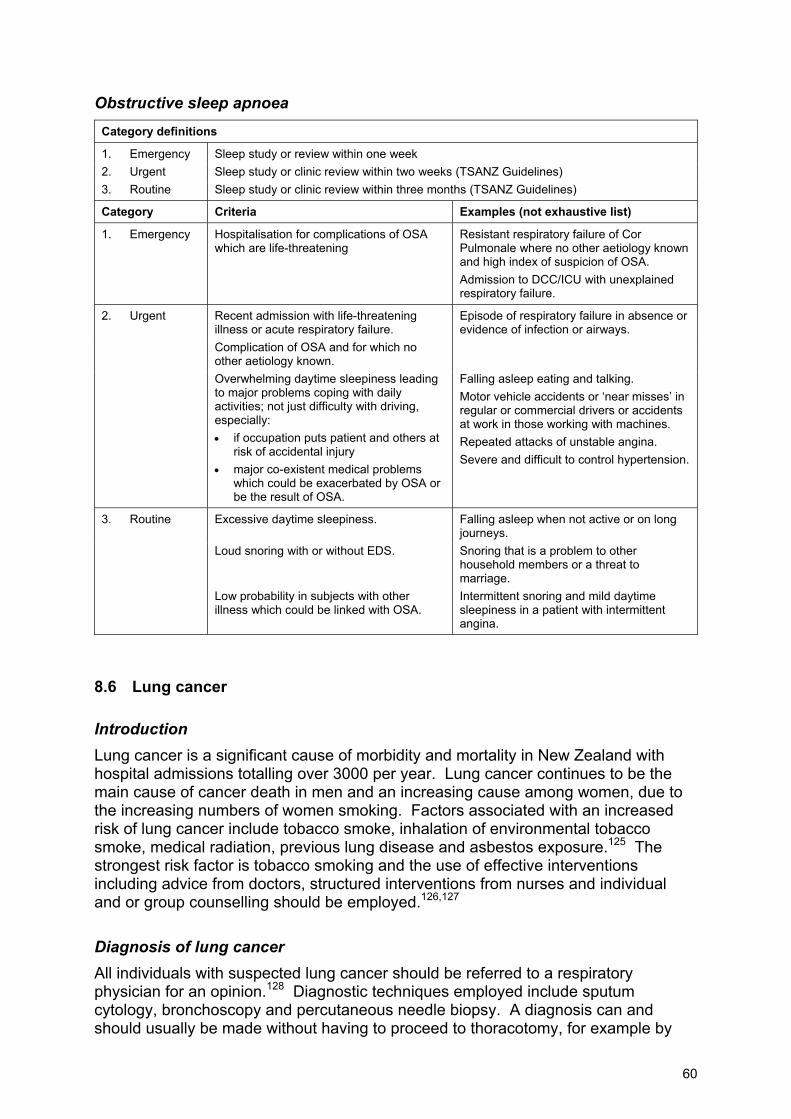
Obstructive sleep apnoea Category definitions
1. Emergency Sleep study or review within one week 2. Urgent Sleep study or clinic review within two weeks (TSANZ Guidelines) 3. Routine Sleep study or clinic review within three months (TSANZ Guidelines)
Category Criteria Examples (not exhaustive list)
1. Emergency Hospitalisation for complications of OSA which are life-threatening
Resistant respiratory failure of Cor Pulmonale where no other aetiology known and high index of suspicion of OSA. Admission to DCC/ICU with unexplained respiratory failure.
2. Urgent Recent admission with life-threatening illness or acute respiratory failure.
Episode of respiratory failure in absence or evidence of infection or airways.
Complication of OSA and for which no other aetiology known.
Overwhelming daytime sleepiness leading to major problems coping with daily activities; not just difficulty with driving, especially: • if occupation puts patient and others at
risk of accidental injury • major co-existent medical problems
which could be exacerbated by OSA or be the result of OSA.
Falling asleep eating and talking. Motor vehicle accidents or �near misses� in regular or commercial drivers or accidents at work in those working with machines. Repeated attacks of unstable angina. Severe and difficult to control hypertension.
3. Routine Excessive daytime sleepiness. Falling asleep when not active or on long journeys.
Loud snoring with or without EDS. Snoring that is a problem to other household members or a threat to marriage.
Low probability in subjects with other illness which could be linked with OSA.
Intermittent snoring and mild daytime sleepiness in a patient with intermittent angina.
8.6 Lung cancer
Introduction Lung cancer is a significant cause of morbidity and mortality in New Zealand with hospital admissions totalling over 3000 per year. Lung cancer continues to be the main cause of cancer death in men and an increasing cause among women, due to the increasing numbers of women smoking. Factors associated with an increased risk of lung cancer include tobacco smoke, inhalation of environmental tobacco smoke, medical radiation, previous lung disease and asbestos exposure.125 The strongest risk factor is tobacco smoking and the use of effective interventions including advice from doctors, structured interventions from nurses and individual and or group counselling should be employed.126,127 Diagnosis of lung cancer All individuals with suspected lung cancer should be referred to a respiratory physician for an opinion.128 Diagnostic techniques employed include sputum cytology, bronchoscopy and percutaneous needle biopsy. A diagnosis can and should usually be made without having to proceed to thoracotomy, for example by
60
bronchoscopy or FNA. Pre-operative staging of non small cell lung cancer may require a number of procedures: plain chest radiograph, CT (to more accurately stage and to assess whether mediastinal nodes are enlarged), and trans-tracheal aspiration via bronchofibroscope or rigid bronchoscope, mediastinoscopy or mediastinotomy if mediastinal lymph nodes are > 1 centimetre in size. Treatment of non-small cell lung cancer Surgery Patients are considered potentially operable if, on history and examination, the patient is fit and deemed to have adequate respiratory reserve to withstand surgery and lung resection, and there is no evidence of local (T4), regional (N2�3) or metastatic disease (M1). Prognosis based on the TNM classification (T = size of tumour, N = nodal station and M = metastases) shows that early stage one lung cancer T1�2 No Mo has a 65�90 percent five-year survival which declines to 20�65 percent as staging increases.129 All surgical resections should be performed with mediastinal node sampling to more accurately stage patients. Radical mediastinal lymph node resection confers no benefit.130 Chemotherapy Recent studies suggest that perioperative chemotherapy should be considered in locally invasive cases (Stage IIIA),131 which may render these previously untreatable tumours resectable. Improved survival can be achieved with this combined treatment. However, further studies are required before induction chemotherapy becomes standard practice. For unresectable lung cancer (Stage IIIB and IV), treatment has been mainly palliative with radiotherapy, however a number of recent studies have shown that modern platinum based chemotherapeutic regimens alone or combined with radiotherapy should be considered in patients with good performance status. These treatments have achieved response rates of 30�40 percent and modest survival advantage.132 Currently chemotherapy for advanced carcinomas of the breast is considered standard despite only modest improvement in survival. Therefore, chemotherapy for advanced lung cancer also needs consideration, particularly since it has been shown to be more cost effective than chemotherapy for disseminated breast cancer. Recent studies have shown that palliative chemotherapy can not only improve symptom control, but also reduce days spent in hospital, and thus cost. Radiotherapy Radical radiotherapy may be helpful in small tumours where the patient�s lung function or co-existing medical problems do not allow resection. This yields variable results of between 6 and 42 percent five-year survival rates for stage one disease. However, survival of patients with surgical resection is superior to radical radiotherapy. Palliative external beam radiotherapy presently remains the mainstay of treatment for extensive non small cell lung cancer. Brachytherapy appears equally effective at controlling symptoms and has the benefit of being administered in one day compared with five to eight days for external beam radiotherapy. Such therapy exists in Hamilton only. Results are at least as good as external beam irradiation and less expensive.133
61
Small cell lung cancer Small cell lung cancer is the second major group of lung cancers accounting for 15�25 percent of all lung tumours. Early tumour dissemination is the rule and surgery even in limited stage disease has not been shown to increase survival. Small cell lung cancer is chemosensitive and prognosis is determined by the extent of tumour at diagnosis. Limited stage small cell lung cancer (confined to one hemithorax), treated with combined chemotherapy has been shown to have a response rate of 80�90 percent and complete response rate of up to 50 percent with a median survival of 16�20 months compared with 8�12 weeks if untreated. Small numbers of patients with limited disease live beyond two years (5�10 percent) following chemotherapy.134,135 In advanced disease, prognosis is worse with no long-term survivors following chemotherapy. However, chemotherapy does have a role in symptom control and improves median survival from 6 to12 weeks. The addition of thoracic radiotherapy to a chemotherapeutic regimen for limited stage small cell carcinoma has also been shown to improve survival by approximately 5 percent and should be considered part of standard treatment for limited stage small cell carcinoma, especially in patients who have had a complete response. In addition, prophylactic cranial radiotherapy has been shown by meta-analysis to reduce the incidence of cerebral metastases and improve survival by a further 5 percent.136 Summary Our recommendation to DHBs in terms of resourcing, investigation and management of lung cancer is that every patient considered to have a lung cancer should be referred to a respiratory physician for an opinion. All necessary investigations required to make a therapeutic decision should have been completed within six weeks of referral, regardless of where patients live. Acknowledging that New Zealand has variable and relatively limited access to chemotherapy (and therefore cannot conform with international evidence based guidelines), the development of New Zealand Guidelines for lung cancer management should be an urgent priority. These guidelines should be developed with input from the Ministry of Health, NZ Guidelines Group, TSANZ and the New Zealand Society of Radiotherapists and Oncologists, along with representation from Palliative Care Physicians and Cancer Societies. Given the variable access to both radiotherapy (unacceptably long waiting times in some regions) and chemotherapy and the lack of a national database on surgical outcomes (post operative mortality rates and five year survival figures), each region should develop strategies to ensure care is optimised and well co-ordinated and collect data for national and international comparisons. The data should be collected in such a manner that comparisons as reported in Appendix II can be made. Fundamental to these requirements are: ! good access to a respiratory physician trained in bronchoscopy to obtain biopsy
material for diagnosis, and to assess whether the tumour is potentially resectable ! radiological support with radiologists skilled in the techniques of fine needle
aspiration and biopsy, thoracic CT scanning ! exercise testing and ventilation/perfusion scanning for pre-operative assessment
in those patients perceived to have borderline lung function
62
! a surgeon trained in thoracic surgery, who should have the ability to perform thoracotomy, mediastinal dissection, mediastinoscopy and mediastinotomy. More recently there has been a trend to perform thorascopic surgery and each region needs to give consideration to encouraging training in this technique. Quality assurance markers should be maintained by all Thoracic Surgical Units and by all surgeons undertaking thoracic surgery and which must be performed to accepted standards and using ATS Guidelines (see Appendix V)
! associated with the Thoracic Surgical Unit there should be an intensive care unit able to manage post-thoracotomy patients where appropriate
! each region should have a radiotherapy unit and treatment should not be delayed for > 2 weeks, whether it be curative or palliative. There should be a national brachytherapy unit using an HDR Selectron and a national bronchoscopy theatre with facilities for endobronchial laser and stent placement
! small cell lung cancer should be managed either by an oncologist or a respiratory physician with a particular interest in oncology (preferably patients should be co-managed)
! there is an urgent need to address new developments in lung cancer and which have important resource implications, that is: ! the use of chemotherapy in non small cell lung cancer (regionally) ! the use of brachytherapy as opposed to external beam radiotherapy in the
palliation of respiratory symptoms in non small cell lung cancer (national unit).
8.7 Interstitial lung diseases
Current situation and future directions There are over 200 interstitial lung diseases (ILDs) comprising about 15 percent of respiratory practice.137 They encompass a very wide spectrum of pathologies, presentations and outcomes. There is concern that ILDs may be poorly recognised and it is considered that respiratory physicians are the only group with appropriate training and skills to deal with the complexity of the diagnosis and management of these conditions. There is, however, evidence of wide variation in the management of ILDs amongst respiratory physicians.138,139 For that reason, the British Thoracic Society has formulated recommendations on management beginning with the statement that patients with ILD or suspected ILD should be under the direct or joint care of a respiratory physician.140 The investigation of ILD has been simplified enormously with the development of High-Resolution Computerised Tomography (HRCT).141 Within respiratory departments there is growing dependence on high quality reporting (refer to the radiology standards in Appendix V) and less referral to surgeons for open lung biopsy. There are very few therapeutic options for the treatment of ILDs but the future promises novel (and) probably expensive new therapies.142 It is envisaged that these will be offered on a case-by-case basis or in the context of a clinical trial. In view of the above factors it is recommended that all patients with ILD be referred to a secondary centre for evaluation by a respiratory physician and that complex, rare or difficult cases be discussed at, or referred to, a tertiary centre.
63

Guidelines for referral to secondary health care Primary health services or general physicians with no designated respiratory interest should not manage patients with ILD. Thus referral to secondary or tertiary services is mandatory. Guidelines for referral to tertiary health care The following indications for tertiary evaluation can be formulated: ! Initial evaluation. Cases in whom the secondary physician is insecure about the
diagnosis or management or where complex diagnostic procedures are required for example open lung biopsy.
! Monitoring disease progression where complex lung function or superior HRCT reporting is required.
! When the patient is receiving a novel or experimental drug for the treatment of ILD.
! Where facilities exist for multidisciplinary care of complex patients for example rheumatology and respiratory in the case of ILD associated with connective tissue disorder.
! Referral for consideration of lung transplantation. National/regional practice 1. Population 50,000: Straightforward cases and routine follow-up to be managed
locally by a respiratory physician or general physician with a respiratory interest. There should be regional access to bronchoscopy (BAL and transbronchial biopsy), cyto-pathology, HRCT and lung function testing (plethysmography and DLCO).
2. Population 50,000�250,000: as for a). 3. National centre: Cases meeting the above tertiary criteria should be referred to
regional or national centres. Those centres should ideally have a clinician(s) with post fellowship training in and an academic interest in ILD, high level of competency in HRCT interpretation access to respiratory subspecialty cyto-pathology expertise and a fully equipped lung function laboratory (exercise, DLCO, compliance and body plethysmography) and thoracic surgical services.
8.8 Pulmonary vascular disorders
Venous thromboembolism remains a significant cause of morbidity.143 Approximately 10 percent of patients with deep venous thrombosis will develop a pulmonary embolism, 10 percent of them will die. Chronic pulmonary embolism with subsequent pulmonary hypertension is rare (about 0.1 percent of those that suffer embolism). With the exception of superior vena caval obstruction, most commonly due to thoracic malignancy, other pulmonary vascular disorders are rare (primary pulmonary hypertension, veno-occlusive disease, arterio/venous malformations, hepatopulmonary syndrome).
64
National/regional/local practice a) Population 50,000: Peripheral venous thrombosis should be diagnosed and
treated by local physicians. If pulmonary embolus is suspected and imaging facilities are available for diagnosis, management should also be by a local physician. There should be facilities for basic resuscitation, arterial blood gas and measurement of blood coagulation profile.144
b) Population 50,000�250,000: Facilities for the diagnosis of thromboembolism, for example scintigraphy, ultrasonography, +/- angiography should exist. Appropriate laboratory facilities for the diagnosis of inheritable coagulopathy should be available. There should be intensive care facilities for the management of major pulmonary embolus with hemodynamic compromise.
c) Population >250,000: Radiologic facilities should include digital subtraction angiography, spiral CT, intravascular stenting, placement of IVC filters and intravascular occlusion devices. There should also be appropriate facilities for right and left cardiac catheterisation and trans-oesophageal cardiac ultrasound. Thoracic surgery and intensive care should be available.
Criteria for referral to a tertiary centre a) Complex pulmonary vascular disease requiring:
! sophisticated imaging ! vascular stenting, coil or filter placement ! vascular surgery (pulmonary endarterectomy � Auckland City Hospital).
b) Rare pulmonary vascular disorders include: ! pulmonary veno-occlusive disease ! pulmonary AV malformations ! hepatopulmonary syndrome.
Quality assurance measures ! Fully trained staff. ! Regular continuing education and peer review with participation in external
accreditation programmes where appropriate. ! Safe well maintained equipment and facilities.
8.9 Asthma
Background In New Zealand, hospital admissions and deaths from asthma have fallen between 1989 and 1994, although the reasons for this are not entirely clear. From this it might be hoped that the morbidity of the disease in the population is decreasing. However, there is no data to support this; in fact, sales of inhaled B-agonists continue to rise and asthma admissions increased by 35 percent between 1994 and 1996, indicating that morbidity may be increasing. The incidence and prevalence of the disease has continued to rise both locally and internationally though there appears to have been a
65

plateau over the past five years. There is no information on whether current asthma therapy is improving the natural history of the disorder, although the reduction in asthma admissions and mortality, which coincided with increasing use of inhaled steroids in the late 1980s, suggests that inhaled steroids may substantially modify the course of the disorder.20 Consensus guidelines for the diagnosis and management of asthma have been produced by a number of groups and are generally very similar in their recommendations.89,90,91 In New Zealand comprehensive consensus guidelines were developed in 2000 but there has been no mandate for DHBs to introduce these into practice and take-up has consequently been variable.145 Requirements for diagnosis of asthma ! Asthma is a symptomatic disease. The possibility of asthma is frequently first
identified by the patient or patient�s family. Public education about how to recognise the disorder is therefore an essential initial part of any local or national strategy.
! Diagnosis is usually made within the primary care setting by a general practitioner. In addition to the usual skills of history taking and examination, the general practitioner will need ready access to, and understanding of: ! peak expiratory flow meters and recordings ! simple spirometry ! skin prick testing ! routine laboratory blood testing ! chest radiology.
! Cases where the diagnosis is uncertain, where occupational asthma is considered, or where good control is difficult to achieve, should be referred to a respiratory physician. Also: ! more complex respiratory laboratory testing, including static lung volumes, flow
loops, simple exercise challenge testing or bronchial provocation testing ! CT chest scanning (either locally or on referral to another centre), expired nitric
oxide and induced sputum testing.
! Occasional referral to a tertiary centre may be necessary, particularly in cases where asthma or bronchial hyperactivity is associated with other pulmonary conditions such as bronchiolitis. Such a centre will have access as needed to: ! TSANZ Category 3 respiratory laboratory ! open lung/thoracoscopic lung biopsy.
66
Requirements for treatment The patient ! Asthma is a condition that requires active ongoing self-management in order to
achieve a successful outcome. This will require adequate motivation, education and access to medical care for the patient and family.
Primary care ! Appropriately skilled general practitioner. ! Appropriately skilled practice nurse. ! These to include � therapeutics, education, management plans and access for
urgent advice and treatment. ! Emergency services and after hours clinics need trained staff and adequate
equipment, including oxygen, nebulisers, oximetry and spirometry. Secondary care ! Specialist outpatient clinics and general medical inpatient care facilities. ! Specialist asthma educators available for patient, healthcare professional and
community education. Tertiary ! As above. ! Development of treatment guidelines, research, and so on. The community ! Access to local asthma societies and other community education groups. ! The role of schools, sports clubs and other community groups needs to be
considered. ! The image of asthma in the community is likely to significantly affect treatment. The research community ! Improvement in the treatment of asthma needs ongoing investment in all areas of
research that translate through to the assessment and improvement of care of asthma generally.
The need for a national consensus The National Asthma Campaign in Australia90 has been developed in co-operation with a number of groups. New Zealand does not have a similar programme, although there has been co-operation between the Royal College of General Practitioners, the Asthma and Respiratory Foundation of New Zealand, the Royal Australasian College of Physicians, the Thoracic Society of Australia and New Zealand (New Zealand branch) and the government in producing a number of asthma self-management plans.
67
It may be appropriate to develop a local programme building on the experience in Australia and other countries. This could follow on the recent development of New Zealand guidelines for the diagnosis and treatment of asthma, along with the introduction of strategies for prevention, directions for local research, initiatives for education and related topics. The need for a co-ordinated approach to service delivery The recent health reforms have dramatically changed the way in which the groups involved with the treatment of people with asthma can interact with each other. Dialogue is needed between secondary and primary health care providers (through PHOs) and the Royal College of General Practitioners, specialists (through the TSANZ) and the Royal Australasian College of Physicians, and community and national organisations such as the Asthma and Respiratory Foundation of New Zealand. Issues to consider include how the current environment should be applied to produce the best outcomes for the patients with asthma, and to point out areas where improvements are needed.
8.10 Chronic obstructive pulmonary disease (COPD)92,93
COPD is a significant health problem. In New Zealand it is the third most common cause of death (8 percent of male and 4 percent of female deaths). Using New Zealand hospital discharge data from 1999 as a marker of morbidity, it ranks second for males (first for Maori) and sixth for females (fourth for Maori). A cross-sectional study in New Zealand in 1989 estimated that the total health care costs attributable to smoking were $185 million (7 percent of the total State expenditure on health). Of the $128 million spent on hospital costs, $22 million was for COPD admissions. These are likely to be substantially higher in 2004, particularly if one includes the costs associated with COPD when it is defined as a secondary diagnosis and contributes to the length of hospital stay. Depending on the prevalence of COPD a recent review in 2003 estimated that between $120 million and $235 million is spent on the direct costs of health services.22 Due to the ageing population, COPD is making a significant contribution to the rising incidence of acute hospital admissions. It is now the commonest cause of respiratory admission. In spite of the frequency of admission and cost associated with managing COPD, service development has lagged behind other medical conditions. There is an urgent need for the development of a comprehensive vertically integrated service. Routine management should be devolved for the most part to general practitioners, other primary care providers and both general and respiratory physicians. The major role for respiratory physicians is: ! to provide a consultation service regarding diagnosis and management,
particularly of those with severe disability ! lead the development of multidisciplinary management and rehabilitation
programmes ! assist in the development and implementation of standards and best practice
guidelines
68
! assist in audit and quality assurance processes ! develop new treatment modalities, for example volume reduction surgery, portable
oxygen, lung transplantation, and assisted ventilation during acute exacerbations when indicated
! research. The devolution of routine management to primary care will require the development of an integrated service across the primary/secondary health care interface. This will require discussions with general practice groups, PHOs and purchasing health authorities. Models successfully developed in the Counties Manukau DHB and Canterbury DHB could be further explored. Secondary care services In these situations there should be a specialist respiratory physician or a general physician with an interest in respiratory disease who closely associates with the regional respiratory department in the tertiary centre. Services that should be provided include: ! physiological assessment including spirometry, flow volume loops, transfer factor,
arterial blood gas analysis and pulse oximetry ! clinical management including acute exacerbations based on TSANZ guidelines
199592 ! intensive care facilities with the capability of ventilating selected cases. High
dependency units (HDUs) or allocated areas in respiratory wards with appropriate training and guideline development can provide assisted ventilation (biPAP) during an exacerbation
! a service for long term domiciliary oxygen therapy and, in selected cases, ambulatory oxygen therapy
! a limited level three sleep diagnostic and treatment service for COPD cases with associated OSA or nocturnal hypoventilation
! a multidisciplinary education and rehabilitation programme emphasising self-care, action plan, rehabilitation programme, activities of daily living, medication and nebuliser use, nutrition and psychosocial support
! an outreach programme assisting integration with primary health care services ! a structured smoking cessation programme, such as the Smokescreen
programme for the 1990s (University of New South Wales).146 This could be a combined service with general practice and other primary health care providers.
Tertiary care services The five tertiary centres should have well developed secondary care services as outlined above and should facilitate and support the development of secondary care services, such as education, rehabilitation, smoking cessation and oxygen therapy services, within the region.
69
In addition to those secondary services, the regional centre should be able to provide the following: ! additional respiratory function assessments such as lung volume estimations and
exercise testing ! a regional centre for the development of a home ventilation service for selected
patients ! a full sleep assessment and management service ! a regional thoracic surgical service for the operative and thoracoscopic
management of selected cases of pneumothorax associated with COPD ! a physician with responsibility for preliminary assessment and follow up of COPD
cases selected for transplantation or lung volume reduction surgery. National service There should be one centre in the country responsible for: ! lung transplantation, lung volume reduction surgery and complicated surgery for
major bullous disease ! a national centre (Canterbury) has been established for the registration of cases of
alpha 1 antitriptysin deficiency. This centre is endeavouring to co-ordinate a study into replacement therapy through a worldwide multicentre trial, which involves Auckland as the other principal study centre.
Prevention COPD has a long pre-symptomatic phase with symptoms often not developing until the FEV1 reduces to around 50 percent of predicted. COPD is an ideal case for secondary prevention. Spirometry is a simple, reliable and inexpensive test for pre-symptomatic identification of this disease but needs to be introduced carefully to ensure that good quality recordings are achieved. This could be used for screening of smokers to identify the 15 percent or 20 percent who are developing COPD. Those exhibiting a reduction in FEV1 to less than 65 percent of predicted could be subjected to more intensive smoking cessation efforts, with utilisation of newer programmes that, in the current non-smoking social environment, are becoming increasingly successful. Spirometric recordings have also been shown to increase the likelihood of smoke cessation. For those who are unable to quit, close follow up would allow other timely interventions and revisiting of the smoking issue. There needs to be a major service development in the field of COPD prevention. A future prevention programme could include: ! spirometric screening of all smokers over the age of 30, preferably in the general
practice or other community settings, but also during admission to other hospital departments, for example pre-operative assessment
! the provision of intensified smoking cessation services integrated across the primary -secondary care interface
! active discouragement of smoking by older children with asthma or recurrent bronchitis by paediatricians and general practitioners
70
! environmental measures such as reduction in air pollution and occupational exposure to lung irritants
! a screening programme for families of cases identified with alpha 1 antitriptysin deficiency.
Quality assurance The absence of strong evidence based support for much of contemporary COPD management makes it difficult to apply quality assurance measures. Nevertheless, standards and current best practice guidelines should be developed and implemented as part of service development. These should be used as a benchmark to audit the process, outcome and cost effectiveness of care, including improving the quality of life for those who live with this progressive disease.
71

Integration of Medical Services with Community Groups DHBs will be responsible for liaison with general practitioners, practice nurses, health development staff, and lay community groups (eg, Asthma Societies, Cancer Society, Hospice Society, AIDS Foundation, Cystic Fibrosis Association, Tuberculosis and Chest Diseases Association) with regard to provision of primary medical care, preventive programmes, screening services, educational initiatives, continuing medical education and quality control. Other government bodies such as the Occupational Safety and Health Committee and National Advisory Committee on Health and Disability need to have a closer relationship with regional respiratory services development. The board should encourage the work of voluntary organisations, and such groups should, where appropriate, be represented on decision-making committees.
72
Health Service Data Requirements DHBs must ensure that adequate records are kept of patient consultations at all levels, admissions, follow-up and other activities, so that regional data are available for audit and cost analysis and for comparisons of services, outcomes, and other epidemiologic and service-related purposes.
73
Research and Education Requirements DHBs have responsibility to encourage research appropriate to identified local and regional needs including health service needs, and to facilitate research funded by other independent sources, for example Health Research Council, Lottery Health Research, local Medical Research Foundations, and lay organisations. All research must be approved by an appropriately constituted ethics committee, and subject to peer scientific review. DHBs and the Ministry of Health need to urgently review systems to support postgraduate education as well as research. Evolving better quality of care from hospital settings into the community will require upskilling of staff at all levels. Regional centres of expertise should work with local universities or technical institutes to ensure postgraduate training of respiratory physicians147 (see also Appendix XII) updating of general physicians and general practitioners, postgraduate training of respiratory scientists, training of respiratory nurse practitioners,105 respiratory physiotherapists and of public health and practice nurses (see Appendix XIII). These endeavours need to be overseen by the respective DHBs and the regional groups to ensure the moneys are being distributed appropriately and that those entrusted with administering funds are made accountable. National meetings should also be held to review respective DHB�s progress and to prevent duplication of effort.
74
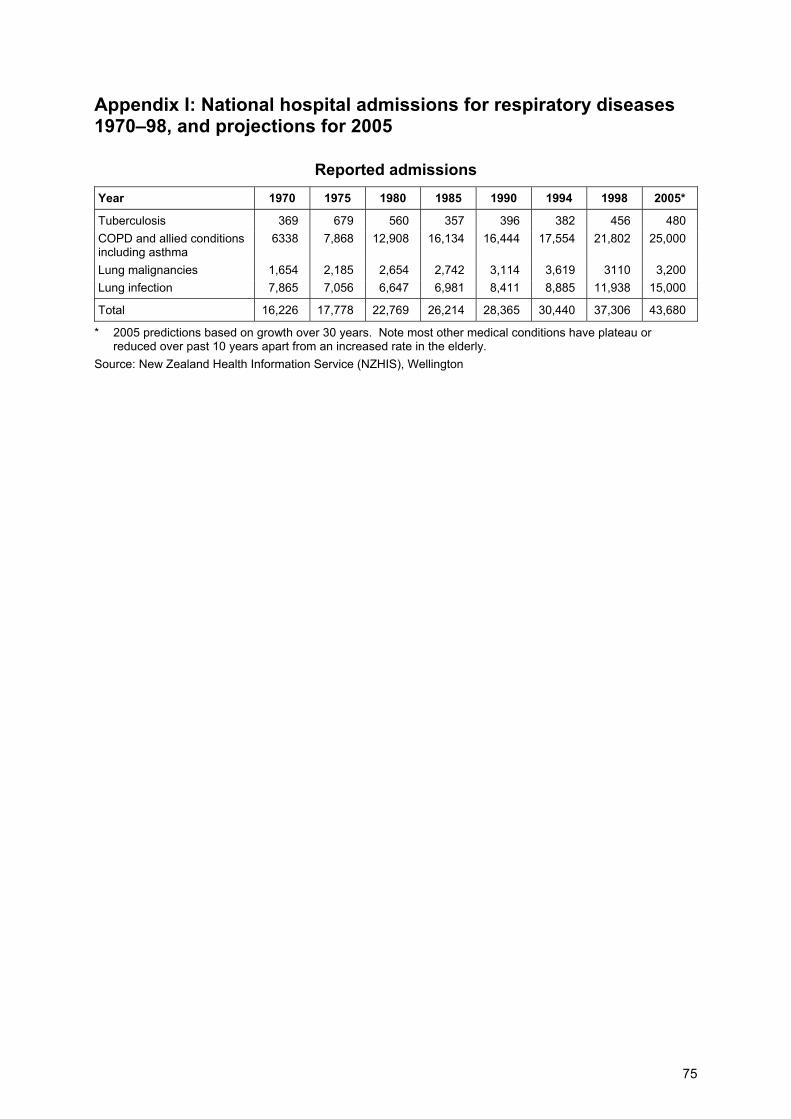
Appendix I: National hospital admissions for respiratory diseases 1970�98, and projections for 2005
Reported admissions Year 1970 1975 1980 1985 1990 1994 1998 2005*
Tuberculosis 369 679 560 357 396 382 456 480 COPD and allied conditions including asthma
6338 7,868 12,908 16,134 16,444 17,554 21,802 25,000
Lung malignancies 1,654 2,185 2,654 2,742 3,114 3,619 3110 3,200 Lung infection 7,865 7,056 6,647 6,981 8,411 8,885 11,938 15,000
Total 16,226 17,778 22,769 26,214 28,365 30,440 37,306 43,680
* 2005 predictions based on growth over 30 years. Note most other medical conditions have plateau or reduced over past 10 years apart from an increased rate in the elderly.
Source: New Zealand Health Information Service (NZHIS), Wellington
75
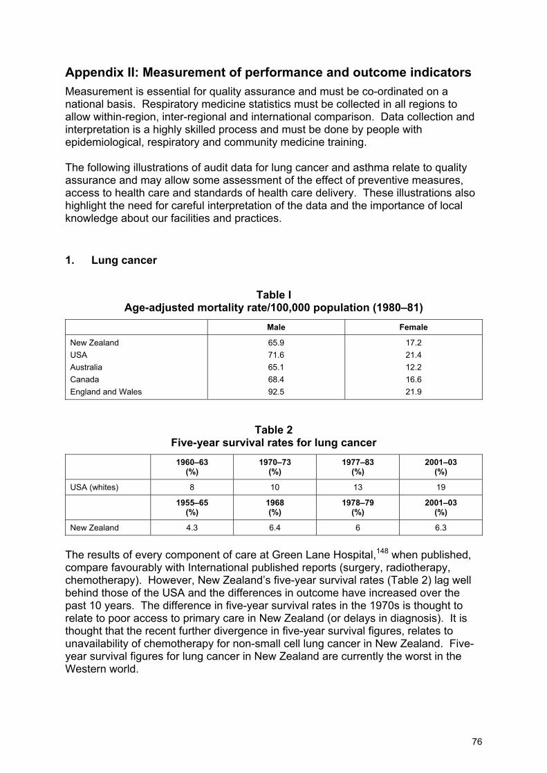
Appendix II: Measurement of performance and outcome indicators Measurement is essential for quality assurance and must be co-ordinated on a national basis. Respiratory medicine statistics must be collected in all regions to allow within-region, inter-regional and international comparison. Data collection and interpretation is a highly skilled process and must be done by people with epidemiological, respiratory and community medicine training. The following illustrations of audit data for lung cancer and asthma relate to quality assurance and may allow some assessment of the effect of preventive measures, access to health care and standards of health care delivery. These illustrations also highlight the need for careful interpretation of the data and the importance of local knowledge about our facilities and practices.
1. Lung cancer
Table I Age-adjusted mortality rate/100,000 population (1980�81)
Male Female
New Zealand 65.9 17.2 USA 71.6 21.4 Australia 65.1 12.2 Canada 68.4 16.6 England and Wales 92.5 21.9
Table 2 Five-year survival rates for lung cancer
1960�63 (%)
1970�73 (%)
1977�83 (%)
2001�03 (%)
USA (whites) 8 10 13 19
1955�65 (%)
1968 (%)
1978�79 (%)
2001�03 (%)
New Zealand 4.3 6.4 6 6.3
The results of every component of care at Green Lane Hospital,148 when published, compare favourably with International published reports (surgery, radiotherapy, chemotherapy). However, New Zealand�s five-year survival rates (Table 2) lag well behind those of the USA and the differences in outcome have increased over the past 10 years. The difference in five-year survival rates in the 1970s is thought to relate to poor access to primary care in New Zealand (or delays in diagnosis). It is thought that the recent further divergence in five-year survival figures, relates to unavailability of chemotherapy for non-small cell lung cancer in New Zealand. Five-year survival figures for lung cancer in New Zealand are currently the worst in the Western world.
76
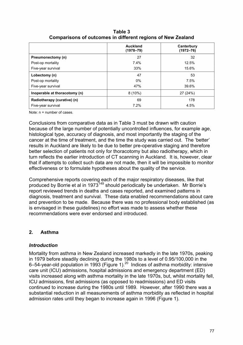
Table 3 Comparisons of outcomes in different regions of New Zealand
Auckland (1978�79)
Canterbury (1972�76)
Pneumonectomy (n) 27 32 Post-op mortality 7.4% 12.5% Five-year survival 33% 15.6%
Lobectomy (n) 47 53 Post-op mortality 0% 7.5% Five-year survival 47% 39.6%
Inoperable at thoracotomy (n) 8 (10%) 27 (24%)
Radiotherapy (curative) (n) 69 178 Five-year survival 7.2% 4.5%
Note: n = number of cases. Conclusions from comparative data as in Table 3 must be drawn with caution because of the large number of potentially uncontrolled influences, for example age, histological type, accuracy of diagnosis, and most importantly the staging of the cancer at the time of treatment, and the time the study was carried out. The �better� results in Auckland are likely to be due to better pre-operative staging and therefore better selection of patients not only for thoracotomy but also radiotherapy, which in turn reflects the earlier introduction of CT scanning in Auckland. It is, however, clear that if attempts to collect such data are not made, then it will be impossible to monitor effectiveness or to formulate hypotheses about the quality of the service. Comprehensive reports covering each of the major respiratory diseases, like that produced by Borrie et al in 1973149 should periodically be undertaken. Mr Borrie�s report reviewed trends in deaths and cases reported, and examined patterns in diagnosis, treatment and survival. These data enabled recommendations about care and prevention to be made. Because there was no professional body established (as is envisaged in these guidelines) no effort was made to assess whether these recommendations were ever endorsed and introduced.
2. Asthma
Introduction Mortality from asthma in New Zealand increased markedly in the late 1970s, peaking in 1979 before steadily declining during the 1980s to a level of 0.95/100,000 in the 6�54-year-old population in 1993 (Figure 1).20 Indices of asthma morbidity: intensive care unit (ICU) admissions, hospital admissions and emergency department (ED) visits increased along with asthma mortality in the late 1970s, but, whilst mortality fell, ICU admissions, first admissions (as opposed to readmissions) and ED visits continued to increase during the 1980s until 1989. However, after 1990 there was a substantial reduction in all measurements of asthma morbidity as reflected in hospital admission rates until they began to increase again in 1996 (Figure 1).
77
Many reasons were cited for the increase in both mortality and admission rates in the late 1970s including: ! an increase in the prevalence or severity of asthma ! changes in the provision of and access to primary health care against a
background of social and economic decline ! direct drug toxicity due either to individual drugs (fenoterol) or a combination of
drugs ! management error by patient, relatives or doctor during the acute attack. Despite an analysis of the wealth of epidemiological data now available to us20 we are still unable to specifically differentiate between the relative contributions each of these factors may have had on either admission or mortality rates. Whilst debate continues about the contribution fenoterol MDIs may have had on the increase in mortality rates, there is agreement that overuse of inhaled B agonists as a class and underuse of inhaled steroids were major contributing factors. Withdrawal of fenoterol (a poorly selective and relatively high dose B agonist) and warnings about the potential dangers of overuse of inhaled B agonists in association with the release of high dose inhaled steroids undoubtedly contributed to the reduction in both mortality and morbidity in the late 1980s. While decreases in asthma mortality suggest that current asthma strategies are effective, it is important to continue to monitor these trends as they may change quickly as happened in the late 1970s. Indeed, hospital admissions have again risen by 35 percent since 1995. Other quality care measurements should include: ! intensive care unit admission rates ! readmission ratios ! emergency department visits and relapses and corticosteroid to ß agonist inhaler
prescription ratios. Mortality rates Whilst death occurs in only a very small proportion of the asthmatic population, most deaths are considered preventable, and many asthmatics who have died had previously declared themselves to be at high risk of experiencing severe attacks. The 1993 mortality rate for 5�54-year-olds was 0.93 per 100,000 and reflects the trend towards falling mortality over the last 10 years. An important goal should be to keep mortality rates below an �acceptable� level and below the current rate as has continued to happen. Because the total number of asthmatics dying within DHBs is relatively small there can be considerable yearly variation (see Table 1). It is therefore suggested that regional mortality rates be evaluated over a period of at least three years (see Table 1), and to include 5�54-year-olds rather than 5-34 year olds since the accuracy of diagnosis in 35�54-year-olds is 90 percent (ie, acceptable) and would allow larger numbers of asthmatics to be incorporated.
78
Admission rates Looked upon as a form of treatment, the decision to admit a patient with asthma to hospital is subject to three factors: ! the organisation of the medical care system ! asthma management ! illness behaviour. Each of these factors may be difficult to change. For example, the distribution of the population around a hospital will influence how that population use the resource. If the population residing in close proximity to the hospital is poor and carries a high number of patients belonging to ethnic minority groups then the use of the emergency department for acute asthma may be high because patients will use the emergency department in preference to the community based medical care. Whilst three separate audits have shown that the quality of care available in New Zealand urban emergency departments is as good or better than equivalent sized overseas centres, New Zealand patients tend to use the resource later in an attack than they would a community-based centre and subsequently are more likely to be admitted. Over the past three years there has been a general downward trend in hospital admission rates within most districts in New Zealand but there are still two-fold differences between DHBs. Readmission ratios This is defined as the number of readmissions divided by total admissions within a calendar year (though readmissions within one or three months of first admission may be a better measure). Although it is a relatively crude method of measuring readmissions (patients admitted in the latter half of the year and who are readmitted in the early stages of the following year would not be included as a readmission), such measurements are easily calculated from information available from the New Zealand Health Information Services or Ministry of Health, and can be compared from year to year and between regions (see Table 2). Asthmatics who require readmission are acknowledged to be a high-risk group and should be targeted for specialist outpatient care. Readmission ratios began to drop in New Zealand in 1986 (see Figure 2), coinciding with the launching of high dose inhaled steroids which were available on specialist only prescription and which may have exaggerated the positive effect of hospital based asthma clinics which were being established on a larger scale in New Zealand at the time. A reduction in readmissions is the aim of any outpatient clinic, (along with rationalisation of therapy, improvement of asthma control and the teaching of self management skills). Therefore, readmission rates are a good indicator of the quality of follow-up care after discharge.
79
Intensive care unit admissions (ICU) Intensive care unit (ICU) admissions are relatively easy to monitor in those areas that have such a resource. Although ICUs may have different criteria for admission, the accuracy of this information may be improved by only including those patients who either require ventilation or who have a pH of less than 7.2 or a PaCO2 of greater than 6.7 kPa (or 45 mmHg) at the time of their admission to hospital. All patients admitted with these criteria should spend at least some time in an ICU. Asthmatics who have required ICU admission are at very high risk of mortality and morbidity and should be targeted for close supervision in asthma clinics. Effective intervention in this group is likely to reduce mortality and also impact on admission rates. In Auckland reductions in ICU admissions paralleled reductions in hospital admissions (see Figure 2). Emergency department (ED) use In the past, information on emergency department (ED) use has been difficult to obtain due to inadequate data collection, but this should improve as EDs incorporate electronic information systems. It would be of use to DHBs to compare ED rates with other centres, since ED rates correlate well with hospital admission and which in turn may explain variations in admission rates between centres. Patients requiring re-attendance at the ED within one week of assessment expressed as a percentage of the total number of asthmatics using the ED is a useful measure of quality of care within the ED and should be < 5 percent. Inhaled preventers/inhaled bronchodilator ratio Information on the quality of asthma care outside of the hospital environment is difficult to define unless cross sectional questionnaire based studies on asthmatics in the community are undertaken. However, data relating to prescription of medications is available. It is generally accepted that adoption of appropriate doses of inhaled steroids and to a lesser extent long acting inhaled B agonists would reduce asthma morbidity rates. The use of regular inhaled steroids instead of reliance on bronchodilator medication is a widely accepted principle in asthma management. A ratio of inhaled corticosteroid prescription rates to inhaled bronchodilator prescription rates for asthma might be a useful measurement of quality of care (see Table 2) though others have found as we did that this was a relatively crude measure of quality of care and in part is as a consequence of our inability to differentiate between COPD and asthma with prescriptions filled (approximately 60 percent of prescriptions (based on IMS (Intercontinental Medical Statistics) figures) are for asthma). Converting all corticosteroid therapy to 100 µg per puff equivalent and all ß agonists to 100 µg equivalent per puff then total sales nationally are roughly equivalent. Based on an estimation of cost then inhaled steroid to inhaled ß agonist ratio should be greater than 2:1. The quality of this information would improve if sales and prescribing information to 6�54-year-old asthmatics were included. However, the quality of pharmaceutical based information would improve substantially if New Zealand developed a national drug register.
80
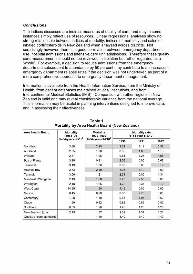
Conclusions The indices discussed are indirect measures of quality of care, and may in some instances simply reflect use of resources. Linear regressional analyses show no strong relationship between indices of mortality, indices of morbidity and sales of inhaled corticosteroids in New Zealand when analysed across districts. Not surprisingly however, there is a good correlation between emergency department use, hospital admissions and intensive care unit admissions. Therefore these quality care measurements should not be reviewed in isolation but rather regarded as a �whole�. For example, a decision to reduce admissions from the emergency department subsequent to attendance by 50 percent may contribute to an increase in emergency department relapse rates if the decision was not undertaken as part of a more comprehensive approach to emergency department management. Information is available from the Health Information Service, from the Ministry of Health, from patient databases maintained at local institutions, and from Intercontinental Medical Statistics (IMS). Comparison with other regions in New Zealand is valid and may reveal considerable variance from the national average. This information may be useful in planning interventions designed to improve care, and in assessing their effectiveness.
Table 1 Mortality by Area Health Board (New Zealand)
Mortality rate 6�54-year-old/103
Area Health Board Mortality 1988�90
6�54-year-old/105
Mortality 1990�1992
6�54-year-old/105 1990 1991 1992
Northland 3.38 2.25 2.24 1.12 3.38 Auckland 2.85 1.28 0.85 1.88 1.13 Waikato 2.67 1.26 0.84 1.26 1.68 Bay of Plenty 3.20 0.91 2.06 0.00 0.68 Tairawhiti 3.19 1.06 0.00 0.00 3.19 Hawkes Bay 2.73 2.40 3.08 4.10 0.00 Taranaki 3.06 1.21 2.33 0.00 1.31 Manawatu/Wanganui 2.13 1.60 2.25 2.55 0.00 Wellington 2.18 1.26 1.72 0.34 1.72 West Coast 14.95 1.50 4.49 0.00 0.00 Nelson 0.45 0.90 0.00 2.70 0.00 Canterbury 1.08 1.40 0.65 1.94 1.62 Otago 1.90 0.82 0.82 0.82 0.82 Southland 0.85 1.28 1.28 1.28 1.28
New Zealand (total) 2.45 1.37 1.32 1.37 1.21 Quality of care standards 1.40 1.40 1.40 1.40
81
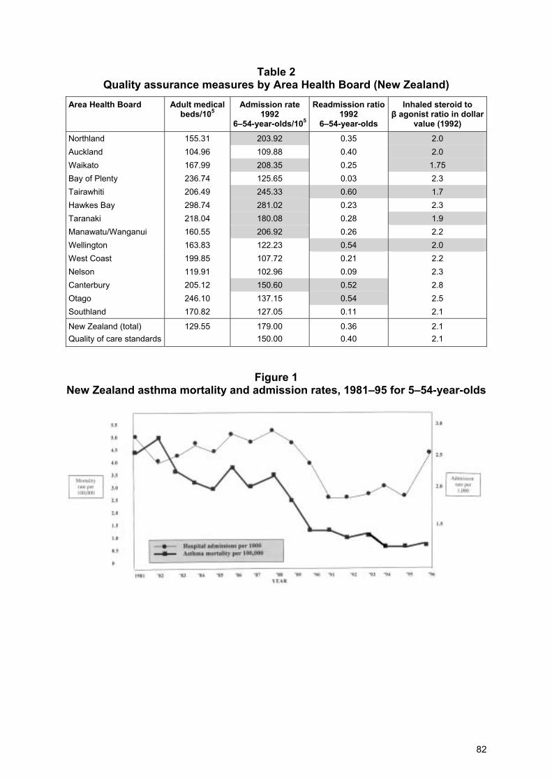
Table 2 Quality assurance measures by Area Health Board (New Zealand)
Area Health Board Adult medical beds/105
Admission rate 1992
6�54-year-olds/105
Readmission ratio1992
6�54-year-olds
Inhaled steroid to β agonist ratio in dollar
value (1992) Northland 155.31 203.92 0.35 2.0 Auckland 104.96 109.88 0.40 2.0 Waikato 167.99 208.35 0.25 1.75 Bay of Plenty 236.74 125.65 0.03 2.3 Tairawhiti 206.49 245.33 0.60 1.7 Hawkes Bay 298.74 281.02 0.23 2.3 Taranaki 218.04 180.08 0.28 1.9 Manawatu/Wanganui 160.55 206.92 0.26 2.2 Wellington 163.83 122.23 0.54 2.0 West Coast 199.85 107.72 0.21 2.2 Nelson 119.91 102.96 0.09 2.3 Canterbury 205.12 150.60 0.52 2.8 Otago 246.10 137.15 0.54 2.5 Southland 170.82 127.05 0.11 2.1
New Zealand (total) 129.55 179.00 0.36 2.1 Quality of care standards 150.00 0.40 2.1
Figure 1 New Zealand asthma mortality and admission rates, 1981�95 for 5�54-year-olds
82
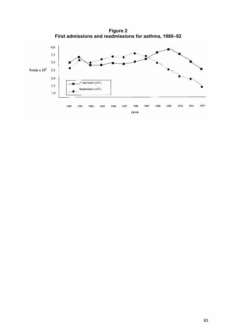
Figure 2 First admissions and readmissions for asthma, 1980�92
83
Appendix III: Respiratory medicine national referral guidelines
Guidelines for specialist referrals: diagnosis-based http://www.electiveservices.govt.nz/respiratory_medicine_referral.html Guidelines for specialist referrals: symptom-based http://www.electiveservices.govt.nz/respiratory_medicine_referral.html National access criteria for first assessment (ACA) http://www.electiveservices.govt.nz/pdfs/respiratory_medicine.pdf National clinical priority assessment criteria (CPAC) http://www.electiveservices.govt.nz/pdfs/respiratory_medicine_CPAC.pdf
84
Appendix IV: TSANZ/RACP standards for training in respiratory medicine requirements for physician training, adult medicine, 2001
Thoracic and Sleep Medicine Supervising Committee
Specialist Advisory Committee in Thoracic and Sleep Medicine (SAC)
Introduction
Thoracic medicine and sleep medicine are intimately related areas of internal medicine. Currently supervision of trainees wishing to gain experience in either thoracic medicine or sleep medicine (or both) is by the specialist advisory committee (SAC) in thoracic and sleep medicine. Sleep medicine is considered an integral part of thoracic medicine and each trainee must complete at least three months of core sleep medicine.
Thoracic medicine
Definition of speciality Thoracic medicine is a subspecialty of internal medicine encompassing diseases of the respiratory system including the upper airway, the lung, the chest wall and the ventilatory control system. Thoracic medicine is a cohesive blend of: clinical knowledge of respiratory diseases; the respiratory sciences of normal and disordered respiratory function; and experience with specialised diagnostic techniques, tests and procedures employed in clinical assessment. General principles of training 1. Training in thoracic respiratory medicine consists of a structured three-year
programme. Advanced training commences with approval of a submitted programme. At least two years� experience in clinical thoracic medicine is necessary. The third year of training may be spent in research or in an approved complementary year in a related discipline. Complementary training will normally not be considered until at least one year of core thoracic medicine training has been completed.
2. Trainees are encouraged to become conversant with all diagnostic procedures available, with the current literature, and with research activities in the respiratory field, but are not expected to become expert in all branches of thoracic medicine and in all techniques.
3. It is desirable that training be undertaken at more than one institution and it is envisaged that this will be necessary for many trainees, to enable them to acquire a sufficient breadth of experience.
85
4. Advanced trainees transferring to the specialist advisory committee in thoracic and sleep medicine may be advised to undertake some post FRACP supervised training in thoracic medicine to complete their three-year training programme.
5. Normally at least one year of advanced training should be undertaken in Australia or New Zealand.
Components of training Core training Clinical thoracic medicine Training should include a wide exposure to all common respiratory disease including lung cancer, tuberculosis and experience in respiratory intensive care. It is expected that all advanced trainees in clinical thoracic medicine will also gain first-hand experience in a respiratory function laboratory and a sleep disorders clinic. This experience will enable trainees to become familiar with the performance and application of commonly used respiratory function tests in the management of patients with respiratory disease. Clinical sleep medicine All thoracic medicine trainees must undertake the equivalent of at least three months� training in sleep medicine, as part of their core training in thoracic medicine. This training should occur in an institution with a sleep laboratory and its associated clinic(s) where the trainee should receive: ! knowledge of basic sleep physiology ! experience in the diagnosis and management of cardiorespiratory sleep disorders ! knowledge of the symptomatology and management of non-respiratory sleep
disorders, particularly those that enter into the differential diagnosis of sleep apnoea (eg, disorders that cause excessive daytime sleepiness).
Trainees should receive experience in the clinical application of polysomnography and basic training in the polysomnographic techniques used to measure and score sleep, and abnormal sleep-related respiratory events. Desirable options It is recommended that some experience with subspecialty and related disciplines is obtained and these include: ! thoracic surgery ! infectious diseases including pulmonary infection in the immuno-compromised ! adult cystic fibrosis ! lung transplantation ! ENT surgery ! clinical allergy and immunology and occupational lung disease.
86

Procedural skills Essential procedural skills These include fibreoptic bronchoscopy and transbronchial lung biopsy, pleural biopsy and tube thoracostomy. As a guide to the numbers required to gain competence, trainees should aim to complete approximately the following numbers of procedures during their training period:
Bronchoscopies 200 Transbronchial lung biopsies 50 Pleural biopsies 20 Tube thoracostomies 20
Desirable (optional) procedural skills These include rigid bronchoscopy, laser bronchoscopy, fine needle aspiration biopsy, transbronchial needle biopsy and intensive care procedures including intubation, central line placement and Swan Ganz catheter insertion. Complementary (elective) training The specialist advisory committee in thoracic and sleep medicine may approve a maximum of one year of complementary (non-core) training which may be spent in related clinical medicine, respiratory research or laboratory work. Clinical complementary training will generally be restricted to related disciplines such as intensive care, infectious diseases, clinical allergy/immunology and cardiology. Any departure from this would require exceptional circumstances for consideration. Complementary training must be approved by the specialist advisory committee before training commences. The requirements for core training, as outlined under core training in the guidelines must be satisfied. Retrospective approval of the complementary training will not normally be granted. Training in research will be strongly encouraged. During the period of complementary training ongoing contact with a respiratory supervisor is required. Projects Trainees are expected to: ! present (or to be principal author of) at least one paper to a meeting of a national
or international society (eg, Thoracic Society of Australia and New Zealand (TSANZ) or RACP)
! prepare an article accepted for publication by a peer reviewed journal. In general, single case reports will not satisfy this criterion.
Other specific requirements Attendance at meetings Trainees are expected to attend and take part in at least one annual scientific meeting of the TSANZ during the three years of training.
87

Sleep medicine Trainees wishing to make sleep medicine an important area of their practice (eg, to manage complex sleep breathing disorders and to report sleep studies) will be expected to undertake at least once year of training in clinical sleep medicine in an approved centre (see Sleep Medicine Guidelines, Levels I and II). Logbook Advanced trainees are required to maintain a logbook which documents clinical procedures including bronchoscopy, transbronchial lung biopsy, tube thoracostomy and pleural biopsy. The information required for each procedure includes the medical record number, the procedure performed and the outcome of the procedure including any complications or unusual features. The logbook should state whether these procedures were supervised or unsupervised. Competence will be assessed by supervisors who will be asked to verify the details of the logbook and the trainee�s experience and competence. Clinical respiratory physiology Those intending to make clinical respiratory physiology an important area of their practice (eg, to specialise in complex respiratory function testing) are expected to spend at least one year of non-core training in a comprehensively equipped and staffed laboratory performing a wide range of respiratory function tests under the supervision of physicians in this area of medicine. Those advanced trainees who wish to practise predominantly or solely in clinical respiratory physiology will be expected to undertake two years of core training in thoracic medicine and two years in clinical respiratory physiology. One year of respiratory physiology training may be devoted to full-time research. Trainees who wish to consider this option are encouraged to discuss it with the Chair of the specialist advisory committee in thoracic and sleep medicine in advance. Research It is strongly recommended that trainees undertake a significant research project during their training in order to understand and apply appropriately research methodologies in laboratory and clinical settings. Trainees should become actively involved in research activities including quality assurance. Experience should be gained in: ! study design ! literature research and review ! writing submissions for grants and Ethics Committee approval ! data collection ! storage and analysis ! computer programme evaluation for results analysis, statistics and graphics. Success in these activities should be demonstrated by publication or presentation of a significant project at a national or international meeting.
88
Training posts Centres at which programmes of advanced training in thoracic medicine are undertaken ideally should have the following: ! a full-time staff thoracic physician or full-time equivalent by visiting thoracic
physicians ! opportunities for the trainee to acquire broad clinical experience in respiratory
diseases as well as relevant aspects of immunology, epidemiology, pathology, microbiology, and pharmacology
! facilities for the performance of fibreoptic bronchoscopy, nuclear medicine studies, thoracic surgery and acute respiratory intensive care, as well as relevant clinics in thoracic oncology and immunology
! a respiratory function laboratory with facilities for spirometry and for measurements of absolute lung volumes, gas transfer, arterial blood gas tensions, pulmonary and respiratory muscle mechanics, and the cardiopulmonary response to exercise
! a facility for the investigation and management of respiratory sleep disorders ! regular clinical respiratory meetings designated for teaching and for consultation
with related disciplines ! library facilities with ready access to all major respiratory journals and texts, and to
literature search facilities such as medicine.
Sleep medicine
Definition of speciality Sleep medicine encompasses sleep breathing disorders, disorders of daytime somnolence, insomnias, parasomnias, disorders of chronobiology, and the neurological and psychiatric disorders affecting sleep. Sleep medicine is a cohesive blend of: ! clinical knowledge of sleep disorders ! knowledge of the basic sciences of normal and disordered sleep processes ! experience with specialised diagnostic techniques, tests and procedures employed
in clinical assessment ! expertise in the management of all clinical sleep disorders. Levels of training In addition to the compulsory three months of sleep medicine that must be undertaken by all thoracic medicine trainees (detailed in the section on thoracic medicine) there are a further two levels of training in sleep medicine that lead to qualification to practise sleep medicine. These are: ! Level I: 12 months core training for thoracic medicine trainees, to enable them to
gain clinical practice in sleep medicine and to report sleep studies (this may include three months� training achieved during the course of a general thoracic core year)
89
! Level II: three years training (two core years plus an approved complementary year) for advanced trainees to enable them to gain clinical practice mainly in sleep medicine, and the opportunity to direct a clinical sleep laboratory.
Advanced training programme in sleep medicine � Level I General principles 1. Completion of Level I advanced training in sleep medicine qualifies thoracic
medicine trainees in sleep medicine (eg, manage sleep apnoea syndromes and more complex sleep-related breathing problems), and for credentialing for reporting sleep studies (as required by the Health Insurance Commission in Australia). Twelve months of training in clinical sleep medicine is necessary.
2. This may be undertaken as an approved complementary year within the three years of advanced training in thoracic medicine, although three months of the 12 months may be undertaken during the two years of core training in clinical thoracic medicine (as the mandatory three months of training in sleep medicine).
3. Alternatively the training may be undertaken as post-FRACP supervised training in sleep medicine.
4. It may be undertaken in New Zealand, Australia or overseas.
5. Advanced trainees from non-thoracic medical disciplines will generally not be eligible to undertake Level I training.
6. Successful completion of Level I training will enable trainees to reach a satisfactory of level of competence to report sleep studies.
Components of training Core training Clinical sleep medicine Quality training should include a wide exposure to all common sleep disorders including sleep breathing disorders, disorders of daytime somnolence, and other non-respiratory sleep disorders. Trainees will be expected to have a detailed practical knowledge of: ! sleep physiology ! the instrumentation, recording, scoring techniques and interpretation of
polysomnographic studies. They will need to obtain detailed experience and skill in the management of sleep breathing disorders (including CPAP and nasal ventilation). They should also be familiar with the diagnosis and management of non-respiratory sleep disorders. It is desirable that training should be undertaken in a multidisciplinary sleep disorders service.
90
Procedural skills Essential procedural skills Full polysomnography, MSLT, MWT, overnight oximetry and transcutaneous CO2 monitoring, CPAP and nasal ventilation. Other specific requirements The trainee�s supervisor(s) will confirm in their written report(s) that a full 12 months of training has been spent in the clinical areas of sleep medicine outlined in the programme of nine months, if three months are undertaken during the two core years of training in thoracic medicine. A sufficient number of procedures must be performed to allow the trainees to develop competence in these procedures. Competence will be assessed on the basis of the written assessment by supervisors, and by interview to assess the trainee�s experience and competence. A logbook of procedures will be required to support the assessment of experience. Training posts See section under Advanced Training Programme in Sleep Medicine. Advanced Training Programme in Sleep Medicine � Level II General principles 1. Level II advanced training in sleep qualifies trainees to practise predominantly
or solely in sleep medicine.
2. Career training in sleep medicine consists of a structured three-year programme with expectations from training different from those of trainees spending one year in sleep medicine as part of their training in respiratory medicine (Level I training). Advanced training commences with the approval of a submitted programme. At least two years� experience in clinical sleep medicine is necessary. The third year of training may be spent in research or an approved complementary year in a related discipline. Approval of complementary training will not be considered until at least one year of core sleep training has been completed.
3. For thoracic medicine trainees who wish to gain dual recognition (ie, Level II sleep plus thoracic medicine), a minimum of four years of advanced training would be required (two years of core training in each discipline). In this case, the clinical sleep medicine component may precede of follow approved thoracic advanced medical training.
4. Advanced trainees from a non-thoracic background (eg, neurology) who wish to gain dual recognition should prospectively seek the advice of the specialist advisory committee in thoracic and sleep medicine as to the suitability of their prior training as approved complementary training in sleep medicine. Retrospective approval of non-relevant complementary training will not normally be granted. For non-thoracic sleep trainees undertaking sleep medicine advanced training, at least one year of their sleep training must involve
91
substantial experience in sleep breathing disorders and their commonly associated respiratory diseases.
5. Trainees are encouraged to become conversant with all diagnostic procedures available, with the current literature and with research activities in the sleep field.
6. It is desirable that training be undertaken at more than one institution, and it is envisaged hat this will be necessary for many trainees, to enable them to acquire a sufficient breadth of exposure and experience.
7. Advanced trainees transferring to the specialist advisory committee in thoracic and sleep medicine for sleep medicine training may be advised to undertake some post-FRACP supervised training in sleep medicine to complete their training programme.
8. Normally at least one year of advanced training should be undertaken in Australia and New Zealand.
9. Successful completion of Level II training will enable trainees to reach a satisfactory level of experience and competence to report sleep studies and direct a sleep laboratory.
Components of training Core training Clinical sleep medicine Quality training should include a wide exposure to all common sleep disorders including sleep breathing disorders, disorders of daytime somnolence, insomnia, parasomnias, disorders of chronobiology and psychiatric disorders affecting sleep. Trainees will be expected to have a detailed practical knowledge of: ! sleep physiology ! the instrumentation, recording, scoring techniques and interpretation for all
varieties of polysomnographic studies. They will need to obtain detailed experience and skill in the management of sleep breathing disorders (including CPAP and nasal ventilation) and other non-respiratory sleep disorders. They should also obtain experience in the diagnosis and management of other respiratory, cardiovascular and neurological disorders affecting sleep. Wherever possible, training should be undertaken in a multidisciplinary sleep disorders service where interactions with other relevant disciplines are possible. At least one year must be spent in a service with a laboratory that has substantial expertise in sleep breathing disorders and their commonly associated respiratory disorders. For non-thoracic trainees, there must be a core component of training in chronic respiratory diseases (eg, chronic airflow limitation, respiratory failure).
92
Desirable options In addition to the core training in the fields of respiratory, cardiovascular and neurological disorders affecting sleep, it is recommended that some experience in other subspecialty fields is obtained and these include: endocrinology, psychiatry, ENT surgery and paediatrics. Procedural skills Essential procedural skills Full polysomnography (including audiovisual recordings), MSLT, MWT, overnight oximetry and transcutaneous CO2 monitoring, CPAP and nasal ventilation. Desirable (optional) procedural skills It is recommended that some experience be gained in one or more of the following procedures: ! actigraphy ! EEG ! respiratory function testing ! oesophageal pH monitoring ! light therapy ! penile tumescence monitoring ! endoscopic nasopharyngoscopy. Complementary (elective) training The specialist advisory committee in thoracic and sleep medicine may approve a maximum of one year of complementary (non-core) training which may be spent in related clinical medicine, sleep research or in laboratory work. Clinical complementary training will generally be restricted to related disciplines such as thoracic medicine, neurology, cardiology and endocrinology. Any departure from this would require exceptional circumstances for consideration. Approval for complementary training must normally be given by the specialist advisory committee before commencement. The requirements for core training as outlined under �Core Training� must be satisfied. Retrospective approval of complementary training will not normally not be accepted, apart from trainees seeking dual specialty recognition (see General Principles). Training in research will be strongly encouraged. Projects or case reports Trainees are expected to: ! present (or to be principal author of) at least one paper to a meeting of a national
or international society, for example the Thoracic Society of Australia and New Zealand (TSANZ) or RACP
! prepare an article accepted for publication by a peer reviewed journal. In general, single case reports will not satisfy this criterion.
93
Other specific requirements 1. Trainees are expected to attend and take part in at least one annual scientific
meeting of the Australasian Sleep Association (or other relevant national or international society) during the three years of training.
2. A sufficient number of procedures must be performed to allow the trainee to develop competence in these procedures. Competence will be assessed on the basis of written assessment by supervisors, and by interview to assess the trainee�s experience and competence. A logbook of procedures will be required to support the assessment of experience.
Research It is strongly recommended that trainees undertake a significant research project during their training in order to understand and apply appropriate research methodologies in laboratory and clinical settings, and to be actively involved in research activities including quality assurance. Experience should be gained in: ! study design ! literature research and review ! writing submissions for grants and ethics committee approval ! data collection, storage and analysis ! computer program evaluation for results analysis, statistics and graphics. The aim it to complete significant project for presentation at a national meeting or for publication. Teaching Supervised experience in teaching for undergraduate, graduate, nursing and lay audience groups is desirable. Regular presentation at hospital activities and participation in peer groups in expected. Training posts Centres at which programmes of advanced training in sleep medicine (Level I or II) are undertaken should have the following: ! the equivalent of a full-time staff sleep physician of full-time equivalent by visiting
sleep physicians ! opportunity for the trainee to acquire a broad clinical experience in sleep disorders
as well as relevant aspects of epidemiology, pathology and pharmacology ! a sleep-disorders laboratory with facilities for full polysomnography, MSLT, CPAP,
and nasal ventilation ! regular clinical sleep meetings designed for teaching and for consultation with
related disciplines ! library facilities with ready access to major sleep journals and texts, and to
literature search facilities such as Medline.
94
Ideally centres should have access to facilities for the performance of fibreoptic endoscopy, neurophysiology studies, respiratory function studies, ENT surgery and acute respiratory intensive care, as well as relevant clinics in psychiatry and psychology. Training posts will generally be evaluated at regular intervals and may be designated as suitable for limited periods of training. Recognition of advanced training programmes in thoracic and sleep medicine There are a number of pathways by which trainees may complete training in thoracic medicine, sleep medicine or both. The confirmation of completion of training from the RACP will reflect the level of training achieved. Combined thoracic and sleep medicine training Two core years of thoracic medicine training and two core years of sleep training are necessary to complete full training in both disciplines. This would likely mean a year of post-FRACP training (after three years completed satisfactorily, eligible to be recommended to fellowship). The letter from the college at the end of the four-year training period will state that the fellow is a fully trained thoracic physician with Level II training in sleep medicine. Thoracic medicine training only The letter from the college would reflect the fact that this trainee had completed training as a thoracic physician, with or without Level I training in sleep medicine. Sleep medicine training only The fellow will be recommended to fellowship by the specialist advisory committee in thoracic and sleep medicine, but not as a thoracic physician. The letter from the college will acknowledge the training in Level II sleep medicine. Special societies Thoracic Society of Australia and New Zealand (TSANZ) Trainees are encouraged to join the Society as associate members and to participate in scientific meetings of the Society at both state and national levels. For further information regarding the requirements for registration with the society, please contact:
TSANZ (New Zealand Branch) Dr Denise Aitken Honorary Secretary TSANZ (New Zealand Branch) C/- Rotorua Hospital Private Bag Rotorua E-mail: [email protected]
95

Australasian Sleep Association (ASA) GPO Box 295 Sydney NSW 1043 Tel: 0500 500 701 Fax: 0500 500 702 E-mail: mailto:[email protected]
Please note: Advice relating to training requirements should be sought from the specialist advisory committee through the college office.
96
Appendix V: Respiratory standards/training in other specialty areas
1. Cardiorespiratory physiotherapy
Definition of speciality Cardiorespiratory physiotherapy is a sub speciality in which a sound knowledge of respiratory physiology and pathology is integrated with physiotherapeutic intervention based on assessment of the cardiorespiratory system and analysis of the effect of change on cardiorespiratory mechanics, gas exchange, peripheral muscle function and exercise tolerance. Interventions address ventilatory dysfunction, impaired airway clearance and limitations to exercise and functional performance. General principles of training 1. The general qualification of BSc (physiotherapy) or BHSc (physiotherapy) from
Otago University or Auckland University of Technology which includes competency in cardiorespiratory physiotherapy.
2. There is no defined pathway to specialisation and currently no post graduate training courses. Specialisation is by experiential learning commencing two years after obtaining initial qualifications.
3. It is most likely that the experience will be gained in a tertiary hospital working with senior physiotherapists and respiratory physicians.
4. Training should include: ! respiratory physiology with application to physiotherapeutic techniques ! interpretation of lung function tests, ECGs, CXRs and HRCT scans ! exercise testing and prescription of exercise for populations with cardiac or
ventilatory impairment ! use and interpretation of findings from auscultation of the chest with
stethoscope ! indications for and application of non-invasive positive pressure ventilation ! appropriate prescription of airway clearance techniques including autogenic
drainage, PEP, flutter device, sputum induction ! assessment of quality of life , impairment and disability ! principles and practice of pulmonary rehabilitation ! assessment and management of disordered breathing ! management of lung transplant and/or lung volume reduction surgery ! manual techniques for thoracic dysfunction ! involvement in research.
Desirable options Progression to MSc/MHSc. Masters level cardiorespiratory practice papers will be available in 2002 at Auckland University of Technology and Otago University.
97
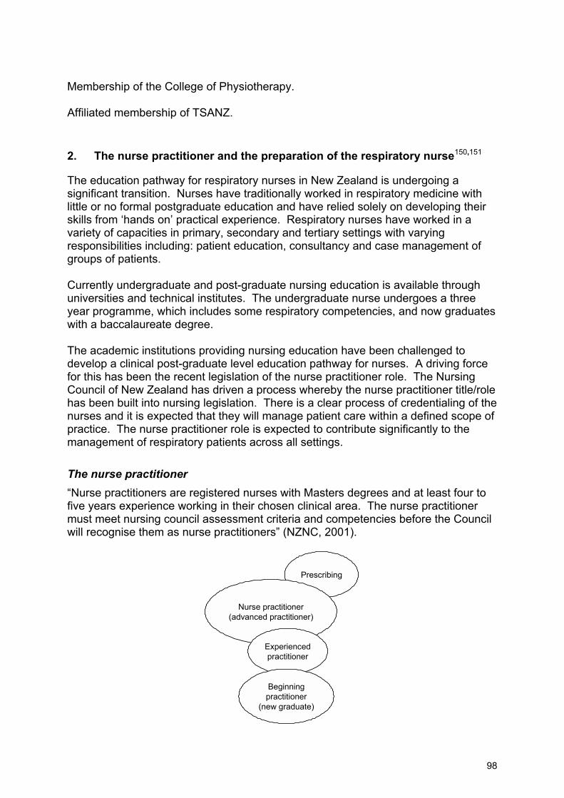
Membership of the College of Physiotherapy. Affiliated membership of TSANZ.
2. The nurse practitioner and the preparation of the respiratory nurse150,151
The education pathway for respiratory nurses in New Zealand is undergoing a significant transition. Nurses have traditionally worked in respiratory medicine with little or no formal postgraduate education and have relied solely on developing their skills from �hands on� practical experience. Respiratory nurses have worked in a variety of capacities in primary, secondary and tertiary settings with varying responsibilities including: patient education, consultancy and case management of groups of patients. Currently undergraduate and post-graduate nursing education is available through universities and technical institutes. The undergraduate nurse undergoes a three year programme, which includes some respiratory competencies, and now graduates with a baccalaureate degree. The academic institutions providing nursing education have been challenged to develop a clinical post-graduate level education pathway for nurses. A driving force for this has been the recent legislation of the nurse practitioner role. The Nursing Council of New Zealand has driven a process whereby the nurse practitioner title/role has been built into nursing legislation. There is a clear process of credentialing of the nurses and it is expected that they will manage patient care within a defined scope of practice. The nurse practitioner role is expected to contribute significantly to the management of respiratory patients across all settings. The nurse practitioner �Nurse practitioners are registered nurses with Masters degrees and at least four to five years experience working in their chosen clinical area. The nurse practitioner must meet nursing council assessment criteria and competencies before the Council will recognise them as nurse practitioners� (NZNC, 2001).
Prescribing
Nurse practitioner (advanced practitioner)
Experienced practitioner
Beginning practitioner
(new graduate)
98
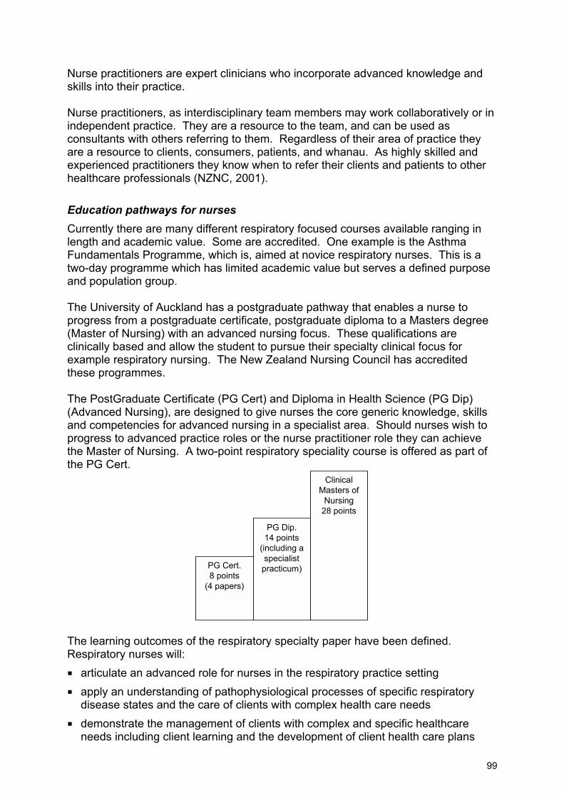
Nurse practitioners are expert clinicians who incorporate advanced knowledge and skills into their practice. Nurse practitioners, as interdisciplinary team members may work collaboratively or in independent practice. They are a resource to the team, and can be used as consultants with others referring to them. Regardless of their area of practice they are a resource to clients, consumers, patients, and whanau. As highly skilled and experienced practitioners they know when to refer their clients and patients to other healthcare professionals (NZNC, 2001). Education pathways for nurses Currently there are many different respiratory focused courses available ranging in length and academic value. Some are accredited. One example is the Asthma Fundamentals Programme, which is, aimed at novice respiratory nurses. This is a two-day programme which has limited academic value but serves a defined purpose and population group. The University of Auckland has a postgraduate pathway that enables a nurse to progress from a postgraduate certificate, postgraduate diploma to a Masters degree (Master of Nursing) with an advanced nursing focus. These qualifications are clinically based and allow the student to pursue their specialty clinical focus for example respiratory nursing. The New Zealand Nursing Council has accredited these programmes. The PostGraduate Certificate (PG Cert) and Diploma in Health Science (PG Dip) (Advanced Nursing), are designed to give nurses the core generic knowledge, skills and competencies for advanced nursing in a specialist area. Should nurses wish to progress to advanced practice roles or the nurse practitioner role they can achieve the Master of Nursing. A two-point respiratory speciality course is offered as part of the PG Cert.
PG Cert.8 points
(4 papers)
PG Dip.14 points
(including a specialist
practicum)
Clinical Masters of
Nursing28 points
The learning outcomes of the respiratory specialty paper have been defined. Respiratory nurses will: ! articulate an advanced role for nurses in the respiratory practice setting ! apply an understanding of pathophysiological processes of specific respiratory
disease states and the care of clients with complex health care needs ! demonstrate the management of clients with complex and specific healthcare
needs including client learning and the development of client health care plans
99
! demonstrate competency in respiratory client assessment skills ! demonstrate competency in respiratory skills or the use and management of
specialty based technology ! analyse the impact of illness and health care experience on the client/family ! evaluate the ethical and legal parameters of current nursing practice. The respiratory section of the New Zealand Nurses Organisation is in the process of defining standardised criteria, which will further guide respiratory education for nurses in New Zealand. It will provide a guide, for nurses seeking further qualifications, as to the level of education being offered, whether it has been accredited and by whom, and how it fits into the overall education framework. Recommendations for this document are: ! that nationally recognised programmes be available for all nurses seeking further
knowledge and skills in respiratory medicine ! the programmes be accredited and offered from education institutions in the North
and South Islands. Respiratory nurses are closely aligned with respiratory physicians and can become affiliated members of the TSANZ. They will be expected to fulfil executive functions on the New Zealand branch of the TSANZ and also on regional and national respiratory advisory boards.
3. Radiology support for respiratory services
Local Imaging services ! CXR, CT, fluoroscopy, ultrasound Radiologist staff ! Radiologist on call 24 hours ! Radiologist to complete CME guidelines recommended by Royal Australian and
New Zealand College of Radiologists (RANZCR) ! Non-vascular intervention to include some biopsy, aspiration or drainage under
CT, U/S or fluoroscopic control to a level determined by the local radiology staff. District Imaging services ! CXR, CT, fluoroscopy, ultrasound ! Non-vascular intervention to include biopsy, aspiration or drainage under CT, U/S
or fluoroscopic control.
100
Radiologist staff In addition to guidelines for local services, there should be at least one radiologist who has support for the clinical respiratory service as a key performance indicator. Regional Imaging services ! CXR, CT, fluoroscopy, ultrasound, angiography, nuclear medicine, MRI. ! Non-vascular intervention to include biopsy, aspiration or drainage under CT, U/S
or fluoroscopic control. The degree of technical difficulty of referred cases may be greater than that of the district services.
! Vascular intervention to include pulmonary angiography, bronchial embolisation, venous stenting and IVC filter placement. Pulmonary arterial embolisation may be offered depending on expertise.
Radiologist staff Radiologists supporting regional respiratory services should be trained at a fellowship level in respiratory radiology. They should be encouraged to be members of TSANZ. Their clinical practice should involve providing the imaging services listed above and in addition, they should support regular regional clinical, pathological, radiological meetings where respiratory and related imaging topics are discussed. These radiologists should be known to district level clinicians and radiologists and they should be available to give written or verbal opinions on cases referred.
4. Proposal on the definition of the structure of general thoracic surgery in New Zealand
I. Definition of general thoracic surgery (GTS) General thoracic surgery encompasses the factual knowledge, technical skill and judgement required to diagnose accurately and to manage surgically diseases of the thorax (chest). The knowledge base includes, but is not limited to, diseases of the chest wall, pleura, lungs, trachea and bronchi, mediastinum, diaphragm and oesophagus. General thoracic surgery requires in-depth knowledge of physiology, diagnostic imaging, organ function testing, semi-invasive and invasive investigation, pre-operative evaluation, post operative care, critical care, trauma, oncology and transplantation. Clinical competence Competency in general thoracic surgery entails the continued appropriate and skilled management of general thoracic surgical problems. This requires an active caseload of diseases of the thorax as detailed above and continued interest in the practice of general thoracic surgery as evidenced by attendance and participation in appropriate speciality meetings and symposia. Involvement in research and education is necessary wherever possible.
101
Thoracic surgeons are specially qualified to manage surgical complications that involve the organ systems detailed above. They are also qualified to assist in the management of pulmonary, pleural, oesophageal, mediastinum and chest wall problems as well as tracheal problems that arise in the course of patient management by allied specialists. Core competencies General thoracic surgery includes all kinds of open or video-assisted surgical procedures in both children and adults. These surgical procedures include: 1. operations involving resection, reconstruction, repair and biopsy of the lung 2. operations involving the chest wall and pleura, including resection and
reconstruction of the chest wall for neoplasms, pleurectomy, decortication, drainage and resection of empyema, thoracoplasty and repair of pectus excavatus and pectus carinatum and other chest wall deformities, as well as the management of traumatic chest wall instability
3. operations involving resection, reconstruction, and repair of the trachea and bronchi for neoplasms, strictures and trauma
4. operations involving resection, reconstruction, and repair of the oesophagus, including laparoscopic or thoracoscopic techniques and endoluminal procedures
5. operations involving resection, reconstruction, and repair of the diaphragm 6. operations involving the mediastinum, including biopsy and resection of
neoplasms, drainage of infections, mediastinal lymphadenectomy, mediastinotomy and mediastinoscopy
7. operations of the pericardium involving resection, reconstruction and drainage 8. endoscopic procedures using both the flexible and rigid scopes and
instrumentation of the tracheobronchial tree and oesophagus 9. operations of biopsy of mediastinal and lymph nodes 10. operations on the thoracic sympathetic nerves 11. operations to correct abnormalities of the thoracic outlet/chest wall 12. operations necessary for airway control including tracheostomy, tracheal
intubation and endoluminal procedures 13. operations for management of pleural space problems, including thoracentesis,
tube thoracostomy, and shunting for pleural effusion and management of pneumothorax
14. operations to provide exposure for thoracic spine surgery 15. all operations incidental to the performance of the above operative procedures 16. critical care management and procedures including placement of central venous
lines, Swan-Ganz catheters, arterial lines, ventilator management, and total enteral and parenteral nutrition management
102
17. operations for traumatic injuries to the chest or to organs within the chest and their sequelae
18. operations on vascular structures related to the management of any pathology treated within the field of GTS.
In major tertiary centres: 19. operations for functional corrections of emphysema 20. operations involving transplantation of one or two lungs or lobes, including all
diagnostic or therapeutic procedures related to the field. II. Structure of a general thoracic surgical unit To ensure quality patient care within the field of GTS and to promote continuous development of the speciality itself, GTS needs to be performed within the logistical and economical framework of specialised units. The structure of these units should be designed to allow: 1. patient care at the level of accepted international standards 2. education and training of surgical trainees in the field of GTS according to
accepted standards 3. continuous clinical and experimental research in the field of GTS. To meet these demands and to become accepted as a unit specialised in GTS a certain organisational background and a number of minimum requirements depending on the individual level of standard or excellence are thought to be necessary. Institutional status GTS units of standard can either be completely free-standing or within the combination with cardiac, vascular, general surgery. The unit should be headed preferentially by an Australasian recognised fellowship. Recommended 1. Structural resources
! A general thoracic surgery unit should have access to ICU. ! General thoracic surgery patients should be on a general thoracic surgery
dedicated surgical ward or ward with general thoracic surgery experience. One wound treatment room should be available on every ward. There should be facilities for outpatient visits at a rate of one room per 750 visits per year. General thoracic surgery units need own in-house facilities for thoracic diagnosis. This includes a laboratory for bronchoscopies and oesophagoscopies, a laboratory for respiratory pathophysiology and in specialised centres one for oesophageal pathophysiology. There must be access to x-ray, CT scan, laboratory medicine and clinical pathology. Preferably access to nuclear medicine and PET scan should be given.
! General thoracic surgery units need qualified physiotherapists. ! General thoracic surgery units should have affiliated endoscopy.
103
2. Staff resources General thoracic surgery units should have a minimum of two surgeons with recognised Australasian Fellowship or equivalent with audit and peer review per 50 major thoracic procedures per year. Conduct ongoing or audit consistent with national standards. 3. Procedural guidelines 1. The number of major thoracic procedures per year should be more than 50 in
standard centres and more than 100 in centres of excellence. 2. Oesophageal resections should be performed only in units with special interest. 3. Lung transplantation should be performed only at Green Lane Hospital. 4. LVRS along with major tracheal resection should only be undertaken at units
with training in the area and whereby post operative mortality rates can be maintained at < 10 percent.
Quality surveillance Quality surveillance has to be performed in every general thoracic surgery unit. There ideally should be documentation of all procedures performed together with a documentation of all major adverse events. Results should be analysed on a regular basis and recorded to all surgeons. There should be a recognised and generally accepted system for risk stratification. Complications should be discussed regularly and a feedback of risk stratified individual results should be given to every surgeon. Procedure specific mortality standards � op mortality/morbidity for lobectomy/ pneumonectomy pleurodesis, oesophageal resection, LVRS, one year survival for lung transplantation should be in the official range given by the European Registry (please specify what the rates are when I reviewed our results for 1983�89 at GLH post op mortality for lobectomy was 1.7 percent and pneumonectomy 3 percent. Presently (n=9) no post op deaths for LVRS. General thoracic surgery units of excellence should provide the possibility for advanced postgraduate education for general thoracic surgery surgeons. Centres of excellence must have access to animal laboratories and to basic science laboratories.
104
Appendix VI: Bronchoscopy services
Guidelines for the: ! training of bronchoscopists109,152 ! maintenance of competency of110 ! bronchoscopists, and ! bronchofibroscopy services, have been produced and recently updated by the Thoracic Society of Australia and New Zealand (TSANZ) (New Zealand branch).109 Additional pertinent documents include: ! TSANZ position paper, Sedation for Bronchoscopy (1995)153 ! TSANZ position paper, AIDS and the Thoracic Physician (1995)45 ! TSANZ position paper, Advanced Training Programme in Respiratory Medicine
(1995)147 ! bronchoscopy services (including transbronchial biopsy and BAL techniques)
should be available at all subregional hospitals (serving a population of > 100,000). Implicit in this is the availability of appropriately trained bronchoscopist and provision of suitable facilities (as outlined earlier). There must be appropriate diagnostic microbiology (as support for BAL in the immuno-compromised host (not just AIDS)).
More complex techniques including removal of difficult foreign bodies, transtracheal (transbronchial) aspiration and rigid bronchoscopy would be available only at the five regional centres. There should be a single national centre for highly specialised techniques such as endobronchial laser, placement of stents, and brachytherapy (Appendix VIII).
Quality assurance
Quality assurance should be an integral part of such a service and relevant measures would include: ! procedural morbidity/complications and mortality ! diagnostic rate (under different but clearly designated circumstances) ! obtaining adequate tissue at transbronchial biopsy ! tolerance and acceptability of the procedure. This should be undertaken in conjunction with quality assurance measures of supporting services, for example: ! radiology ! histopathology ! cytology ! microbiology.
105
Appendix VII: Laser, stenting and brachytherapy
Numbers
Based on local and UK experience approximately 30�40 patients per annum are suitable for laser endobronchial surgery and 5�10 for endobronchial stenting in New Zealand.69 Green Lane Hospital has a Ng Yag laser and operators experienced with its use, and equipment for endobronchial stenting.
Guidelines for re-referral
1. Laser Patients with intrinsic endobronchial tumour (benign or malignant) or hemoptysis with visualised bleeding site or benign tracheal webs. 2. Stenting Patients with benign or malignant airway stenosis or torsion, or severe tracheo-bronchomalacia.
National vs regional
The small numbers justify only one national centre in order to maintain expertise and this has already been established at Green Lane Hospital. Ideally laser and stenting should be performed in a hospital with thoracic and ORL surgical services. In New Zealand this hospital should also be the same as that performing lung transplantation, so that anastomotic stenoses can be treated. A close association with a radiotherapy department preferably with brachytherapy is also essential. Brachytherapy has the potential to be administered in a single treatment and is as effective as more expensive external beam irradiation in palliating an array of respiratory symptoms caused by bronchial carcinomas. This needs to be given a high priority for development in Auckland.
Quality assurance measures
1. Operator training and continuing education is mandatory for all procedures. Currently this can only be performed in other countries.
2. Maintenance of equipment should be regular and the theatre should comply with Australian National Standards for laser safety.154
3. A hospital laser safety committee should exist. A complete record of all procedures be kept, recording all outcomes, including complications.
106
Appendix VIII: Respiratory function assessment Objective functional assessment of the respiratory system is an essential component of Respiratory medicine. Virtually all patients with respiratory symptoms which last for more than a few weeks or which recur should have at least rudimentary evaluation of pulmonary function (this includes children and adults). The indications for pulmonary function tests include: ! assistance in establishing a diagnosis ! objective assessment of severity : baseline from which to measure change or
assessment of disability ! fitness assessment: for compensation, prior to surgery, flying or diving ! determine treatment or treatment response, for example need for pulmonary
rehabilitation, oxygen or response to steroids. The need for pulmonary function assessments is growing and will continue to grow in New Zealand; prevalence of most respiratory diseases are increasing and there is a need for occupational and compensation related assessment. There is a strong feeling that current facilities are not meeting the need and that even where service is provided quality may be suboptimal. With the emphasis on epidemiological and biochemical aspects of respiratory medicine in the 1970s and 1980s many respiratory services have allowed their physiological sections to decline. This trend needs to be reversed.
Recommendations
These are based on a position paper of the TSANZ guidelines for respiratory function laboratories.115 Accreditation of respiratory function assessment services 1. Five regional respiratory centres in New Zealand should have respiratory
laboratories capable of comprehensive pulmonary function testing such as TSANZ. At least category three standard assessment of respiratory function including measurement of static lung volumes (TLC, RV, FRC and VC); maximum expiratory flow rates before and after bronchodilator (maximum expiratory flow volume curves; carbon monoxide gas transfer; and maximum respiratory pressures measured at the mouth; arterial blood gas analysis, pharmacological and non-pharmalogical bronchial provocation tests and cardiopulmonary exercise tests). Laboratory staff should have special training in pulmonary function testing in children. All five centres should seek TSANZ accreditation. Senior staff should be certified with the CRFS credential.
2. At �district� level category two testing should be available that is all of the above except bronchial provocation and exercise testing.
3. At �local� level spirometry, flow volume loop and pulse oximetry should be available. GP practices should have ability to measure peak expiratory flow rate and spirometry, and have access to pulse oximetry.
107
Each regional centre should be responsible for standards of pulmonary function testing and interpretation reporting in their region, that is arrange audit systems for the district and local testing within their region.155 These standards apply to both the laboratory and its director and are defined within the TSANZ guidelines.115 A system for ongoing monitoring of quality of a national basis would be desirable. The medical directors of the regional pulmonary function laboratories (and the senior respiratory scientists) should meet annually. They should establish themselves as a group to report on contentious tests for bodies such as the ACC or for medicolegal purposes.
108
Appendix IX: Lung transplantation Single and bilateral sequential lung transplants are available in New Zealand at Green Lane Hospital. The number of transplants are limited by donor availability. The availability of donor organs in New Zealand is 10 per million per year but as only 15�25 percent are suitable for lung transplantation this translates into only 10�12 transplants/year. As such there can be only one National Centre, to maintain expertise. Success rates should be equivalent to top overseas centres. Although depending to an extent on patient selection, if one year survival should fall to less than 75 percent the programme should be reviewed. To date 54 lung transplants have been performed and the one year survival rate is 85 percent and which is above the international average (78 percent). Heart-lung transplants are only required in patients with poor right ventricular function or irreparable congenital cardiac defects and should not exceed 5 percent of all patients considered for lung transplantation. Such patients should receive their surgery in Australia since the operation technique is different to that of lung transplantation.
Criteria for acceptance for lung transplantation
Indications for lung transplantation73 include patients with end stage lung disease with an anticipated poor prognosis and symptoms that are severe and progressive despite optimal medical treatment. These patients should, except for their lung disease, be healthy individuals with strong family support. They should be able to comply with medical advice and be keen to proceed with transplantation. Patients aged over 50 years or under 15 years will be considered only in exceptional circumstances. Owing to unsatisfactory outcome worldwide, patients over 60 have not been considered for the programme. The following factors are contra-indications to consideration of lung transplantation: ! Systemic diseases (such as lupus etc): Peripheral vascular disease, chronic fungal
lung infections, insulin dependant diabetes are ischaemic heart disease are relative contra-indications. Moderately severe or severe coronary artery disease not amenable to angioplasty or with left ventricular ejection fractions below 45 percent is an absolute contraindication as is diabetes with end organ damage.
! Unresolved pulmonary infection: Infections exhibiting panresistance to antibiotics are an absolute contra-indication, those exhibiting multi-drug resistance a relative contra-indication.
! Renal or hepatic failure: Unless mild and secondary to right heart failure. ! Obesity/severe malnutrition: BMI 20�25 ideal. ! History of severe or continuing mental illness and or alcohol or drug abuse:
Patients should have stopped smoking for at least two years. ! Immobility: Patients must be able to walk 50 metres unassisted, with oxygen as
required.
109
! Continuous steroid therapy: A relative contra-indication if prednisone therapy < 7.5 mg average dose in previous year; > 7.5 mg particularly if associated with side-effects of therapy is a definite contra-indication.
! Malignancy: Evidence of complete remission for five years prior to referral is essential, but remains a relative contra-indication depending on the type of tumour.
Criteria for lung transplantation in patients with cystic fibrosis
Inclusion criteria ! FEV1 < 30 percent predicted. ! Respiratory disease major determinant of current morbidity. ! Absence of absolute exclusion criteria. Absolute exclusion criteria ! Biliary cirrhosis complicated by portal hypertension (as evidenced by Doppler
portal vein flow, oesophageal varices, massive splenomegaly) or previous episode encephalopathy.
! Multi-drug* resistant bacterial infection (eg, Burkholderia cepacia, gladioli or other pseudomonas species resistant to four or more IV antibiotics on two or more occasions including MRSA).
! Aspergilloma. ! Insulin dependent diabetes mellitus with evidence of �end organ� damage (eg,
retinopathy, proteinuria). NB: General contraindications for lung transplantation apply. Relative exclusion criteria ! Severe nutritional impairment, for example BMI less than 17. ! Unstable insulin dependent diabetes mellitus. ! Regular oral corticosteroid therapy greater than 7.5 mg per day for > 6 months in
last two years. ! Coagulopathy. ! Previous pleurodesis or thoracic or mediastinal surgery (unless pleurodesis is
limited and performed through thoracoscope). ! Severe malabsorption unable to be corrected by pancreatic supplements (inability
to maintain cyclosporin levels). ! Current ABPA as evidenced by persisting fungal hyphae in sputa, typical CXR
infiltrates and positive serology. ! Colonisation with Aspergillus species.
110
Appendix X: Lung volume reduction surgery
Introduction
Airflow limitation in COPD is due to varying combinations of airways disease and emphysema. Major pathophysiological consequences of emphysema can be attributed to a loss of elastic recoil, and consist of static and dynamic hyperinflation as well as a preferential obstruction of expiratory airflow due to a loss of traction on the airways and increased work of breathing. The main symptom of patients with very advanced emphysema is dyspnoea which may be contributed to by impaired pulmonary mechanics. The only treatment proven to reduce the rapid decline in FEV1 is smoking cessation, and the sole treatment proven to prolong life is long-term oxygen therapy. Inhaled bronchodilators can ameliorate symptoms and improve quality of life but have little effect on lung function tests. Pulmonary rehabilitation does NOT improve lung function, but can improve exercise performance, quality of life, and reduce morbidity. Lung transplantation and lung volume reduction surgery (LVRS)156 have gained acceptance as palliative procedures for a subgroup of patients with advanced emphysema. The criteria for selection of patients for LVRS are currently being investigated. The goals of LVRS are: ! an improvement of the lung elastic recoil to enhance radial traction on the airways,
thus lowering airway resistance and increasing driving force for maximal expiratory flow
! a reduction in pulmonary hyperinflation, with a more physiological diaphragmatic configuration for generating inspiratory force and reduced work of breathing.
To date 10 patients have proceeded to LVRS at Green Lane Hospital. There have been no post operative deaths and mainly good outcomes in highly selected patients aged less than 70. Whilst overseas data suggest better results are obtained in centres also undertaking transplantation, if centres outside Auckland perform LVRS it should be mandatory that data be forwarded to the Australasian LVRS database.78
Patient evaluation and selection (note recently modified in TSANZ position paper which accompanies this guideline)
Inclusion criteria ! Age < 70 years (less than 80 years if health otherwise excellent). ! Emphysema confirmed on HRCT. ! No evidence of ischaemic heart disease (angiogram needed prior to surgery). ! Dyspnoea at rest, or with minimal activity resulting in severe limitation of daily
activity with impaired quality of life (six-minute walk test 150�350 metres). ! Completed a rehabilitation programme and be shown to comply with programme. ! Severe airflow obstruction (FEV1 < 35 percent predicted).
111
! No significant improvement with bronchodilators (< 15 percent improvement in FEV1 with nebulised bronchodilator), 20 mg prednisone for three weeks.
! Functional aspects of emphysema (ie, RV > 200 percent predicted, TLC > 140 percent predicted, impaired DLCO (but > 30 percent predicted))
! reduced perfusion to major bullae on quantitative V/Q scan. Exclusion criteria ! Smoking within two years of consideration of LVRS. ! �Vanishing lung� on CT (namely independent of bullae, little or no evidence of
normal functioning lung). ! Coronary artery disease > 50 percent diameter reduction of more than one
coronary artery. ! Neoplastic disease with life expectancy of < 2 years. ! Other important medical problems (eg, renal failure, severe steroid-induced
osteoporosis; PH CVA) ! Prednisone > 7.5 mg/day for > 3 months. ! Bronchiectasis (cylindrical as opposed to traction). ! Aspergillosis (aspergilloma). ! Colonisation of airways with multi-drug resistant organisms. ! Prior pleurodesis � unless limited thoracoscopic (relative). ! Cor pulmonale (right ventricular ejection function < 30 percent, determined by
gated nuclear scan, or good quality 2D echo). ! Chronic productive cough of > 1 tablespoon per day. ! dLCO < 30 percent predicted. ! Hypercapnoea > 7.3 kPa (50 mmHg). ! Addiction to alcohol, drugs, psychiatric disturbance, non-compliance with drug
regimens (or rehabilitation) ! BMI < 17 (unresponsive to Pulmocare/Ensure Plus), or BMI > 25.
A position statement of the Thoracic Society of Australia and New Zealand
GI Snell Department of Respiratory Medicine, Alfred Hospital, Melbourne, Victoria
M Peacock Department of Thoracic Surgery, Queen Elizabeth Hospital, Adelaide, South Australia
J Garrett Department of Respiratory Medicine, Green Lane Hospital, Auckland, New Zealand
Published in Internal Medicine Journal 2001; 31:112�115. This document will be reviewed in 2006 or earlier if significant developments occur. http://www.thoracic.org.au/lvrsposition29nov2000.pdf
112
Abstract Lung volume reduction surgery involves the removal of emphysematous lung tissue with the aim of palliating symptoms in selected patients with severe emphysema. This form of surgery is being practised in Australia with favourable short-term outcomes, similar to those reported in the literature. Large multicentre trials are currently underway in North America and the United Kingdom to clarify issues of safety and long-term efficacy. As a result, it is to early to apply an evidence-based approach to this procedure. In the meantime, local audits of practice need to be undertaken to define patient subgroups at higher risk of morbidity and mortality (Intern Med J 2001; 31: 112�115). Keywords: emphysema, lung reduction surgery. Correspondence to:
Dr G I Snell Department of Respiratory Medicine Alfred Hospital Prahran VIC 3181 Email: [email protected]
Overview Lung volume reduction surgery (LVRS) is a general term encompassing a variety of surgical procedures that involve resection of lung tissue with the aim of reducing symptoms in patients with severe emphysema. The first series of patients treated with pulmonary resection for emphysema was reported by Brantigan et al in 1957.157 He postulated that removal of the most diseased lung tissue would increase radial traction on small airways, reversing the collapsibility that had caused airflow obstruction and normalising diaphragm and chest wall respiratory mechanics. The postoperative mortality rate was 18 percent and the patient�s subjective benefits were not confirmed objectively. Consequently, the procedure was not widely accepted. In 1995, following observations made in lung transplant surgery and taking advantage of developments in anaesthesia and postoperative care, Cooper et al revitalised the concept of surgical treatment of non-bullous emphysema utilising a linear stapler reinforced with bovine pericardial strips to avoid excessive air leaks.158 Vigorous exclusion criteria were applied and a peri-operative rehabilitation programme was included to optimise fitness. His first 20 patients had a surprisingly large improvement in forced expiratory volume in (FEV1) of 82 percent and no mortality. Other centres have subsequently published their results using a variety of surgical techniques.159 These studies confirm the improvement in FEV1 and patient quality of life (QOL) noted by Cooper et al.158
113
Although Cooper advised caution and suggested LVRS be restricted to specialised centres initially, early success led to a rapid rise in the number of operations performed at multiple sites across the USA. It has since become clear that LVRS can be complicated by serious morbidity and mortality.159 A review of Medicare (USA) billing in 1995 noted a mortality rate of 26 percent.160 As a result, Medicare (USA) funding was withdrawn for LVRS in 1996 and a large National Heart, Lung and Blood Institute multicentre randomised trial was proposed to compare LVRS with best practice.160,161 This seven-year trial has now commenced162 and other large national trials are underway in Canada163 and the United Kingdom. Three studies, including two short-term small randomised studies, have been recently reported comparing LVRS and medical therapy.164,165,166 Although not uniform in their analyses or results, these three studies tend to favour improvements in measures of pulmonary function and QOL in the LVRS group. By the end of 2000, LVRS will have been performed on over 400 patients in more than 15 Australian centres from all mainland states.167,168,169 A National Lung Volume Reduction Surgery Database has been established, in conjunction with the Royal Australasian College of Surgeons� Australian Safety and Efficacy Register of New lnterventional Procedures � Surgical (ASERNIP-S), and analysis will help to define selection criteria, outcomes and resource allocation.169,170 To avoid a repeat of the experience in the USA, the Australian medical profession needs to review continually local results, follow closely the evolving literature and, if necessary, set minimal Australian standards of medical care and technical excellence. This position statement, and the recent ASERNIP-S LVRS Review,170,171 reflect a commitment to that process. Mechanisms of improvement in respiratory symptoms with LVRS An enhanced understanding of the pathophysiological mechanisms altered through LVRS has much to tell us about lung diseases in general. Recent studies tend to confirm Brantigan�s hypotheses.159 Sciurba et al have confirmed an improvement in elastic recoil following LVRS172 and others have shown an improvement in respiratory muscle performances.159,161 The procedure may also act by improving ventilation/perfusion matching, decreasing the effects of dynamic hyperinflation on the venous circulation or even by improving cardiac output.161,172 Variations in LVRS technique Generally, LVRS programmes advocate a pulmonary rehabilitation programme preoperatively. Purported benefits include the optimisation of physical condition and the opportunity for patient education. Although the limited evidence available suggests that LVRS is complementary to pulmonary rehabilitation alone,158,173 patients may derive sufficient benefit from the pulmonary rehabilitation to forgo or defer surgery.
114
Different surgical approaches to LVRS have been used. There is general agreement that maximal benefits obtained by operating on both lungs simultaneously using some form of reinforced excision-stapling technique.159,161 Initial experience revealed that the unilateral operation afforded half the improvement, with the same risk of early mortality and an increased risk of late mortality,161 but recent studies have found no difference in survival outcomes between the procedures.174 Reinforcement material is expensive and its value in reducing air leaks has still to be determined. There seems to be no significant difference in clinical outcomes using midline sternotomy, bilateral thoracotomy or thoracoscopic techniques.161,171 Operator experience with any given approach appears to be the most important factor. The limited Australian experience so far reveals that most units are performing bilateral LVRS using a stapling technique.169 Open and thoracoscopic168 techniques are practised equally.169 Expected outcomes of LVRS Based on results reported in peer-reviewed journals, abstracts and presentations at international meetings, the procedure appears efficacious for some, but not all, patients with advanced chronic obstructive pulmonary disease due to emphysema.159,161,167 Perioperative mortality should vary between five and 15 percent, depending on case selection and centre expertise. Causes of morbidity include respiratory failure, sepsis, persistent intercostal catheter drainage, late pneumothorax, atrial dysrhythmias and myocardial infarction.157�168 In a recent large series, mortality approached 30 percent at three years post-procedure.174 Two to six months following bilateral LVRS, an improvement in FEV, and the six-minute walk test of approximately 50 percent can be shown.159 Patient-reported dyspnoea, exercise tolerance and QOL improve similarly. The peak improvement in FEV1, is noted after six months, with a variable decline back to baseline over the next two to four years.175,176,177 A small proportion of patients fail to improve lung function significantly.176,177 Selection and assessment of LVRS candidates Candidate selection will determine clinical and functional outcomes. Although trials have not been performed to compare different selection criteria, the literature allows some broad generalisations (see Table 1).158,159,171 Certain preoperative variables, including a six-minute walk of less than 200 m, PCO2 greater than 55 mmHg, gas transfer factor (DLCO) less than 30 percent predicted165 or pulmonary arterial hypertension, have been shown to portend a higher perioperative risk of morbidity and mortality. Advanced age (beyond 75 years), left ventricular impairment or significant coronary artery disease are also likely to define patients at higher risk.174,178
115
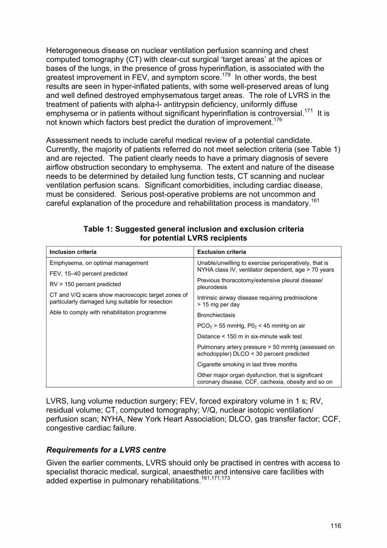
Heterogeneous disease on nuclear ventilation perfusion scanning and chest computed tomography (CT) with clear-cut surgical �target areas� at the apices or bases of the lungs, in the presence of gross hyperinflation, is associated with the greatest improvement in FEV, and symptom score.179 In other words, the best results are seen in hyper-inflated patients, with some well-preserved areas of lung and well defined destroyed emphysematous target areas. The role of LVRS in the treatment of patients with alpha-l- antitrypsin deficiency, uniformly diffuse emphysema or in patients without significant hyperinflation is controversial.171 It is not known which factors best predict the duration of improvement.176 Assessment needs to include careful medical review of a potential candidate. Currently, the majority of patients referred do not meet selection criteria (see Table 1) and are rejected. The patient clearly needs to have a primary diagnosis of severe airflow obstruction secondary to emphysema. The extent and nature of the disease needs to be determined by detailed lung function tests, CT scanning and nuclear ventilation perfusion scans. Significant comorbidities, including cardiac disease, must be considered. Serious post-operative problems are not uncommon and careful explanation of the procedure and rehabilitation process is mandatory.161
Table 1: Suggested general inclusion and exclusion criteria for potential LVRS recipients
Inclusion criteria Exclusion criteria
Emphysema, on optimal management
FEV, 15�40 percent predicted
RV > 150 percent predicted
CT and V/Q scans show macroscopic target zones of particularly damaged lung suitable for resection
Able to comply with rehabilitation programme
Unable/unwilling to exercise perioperatively, that is NYHA class IV, ventilator dependent, age > 70 years
Previous thoracotomy/extensive pleural disease/ pleurodesis
Intrinsic airway disease requiring prednisolone > 15 mg per day
Bronchiectasis
PCO2 > 55 mmHg, P02 < 45 mmHg on air
Distance < 150 m in six-minute walk test
Pulmonary artery pressure > 50 mmHg (assessed on echodoppler) DLCO < 30 percent predicted
Cigarette smoking in last three months
Other major organ dysfunction, that is significant coronary disease, CCF, cachexia, obesity and so on
LVRS, lung volume reduction surgery; FEV, forced expiratory volume in 1 s; RV, residual volume; CT, computed tomography; V/Q, nuclear isotopic ventilation/ perfusion scan; NYHA, New York Heart Association; DLCO, gas transfer factor; CCF, congestive cardiac failure. Requirements for a LVRS centre Given the earlier comments, LVRS should only be practised in centres with access to specialist thoracic medical, surgical, anaesthetic and intensive care facilities with added expertise in pulmonary rehabilitations.161,171,173
116
Conclusion Lung volume reduction surgery is still to establish its exact role in the management of severe emphysema and an evidence-based approach cannot be applied at this time. The recent ASERNIP-S review of LVRS has concluded that LVRS is an acceptable short-term treatment in highly selected cases, but longer follow up is needed to assess long-term outcomes. A Cochrane review on the subject preferred to await the results of the large randomised controlled trials in the USA, Canada and England before drawing any strong conclusions.162,163,180 While there are still many questions to be answered, the most important will be to define the sub-group who gain the greatest physiological improvement for the longest duration.171,177,180 Audits of practice need to be undertaken to define patient subgroups at higher risk of morbidity and mortality. The National LVRS Database provides a mechanism to help answer some of these questions and aids in dissemination of information on Australian and New Zealand clinical practice back to the profession.169�171 Open discussion and the opportunity to measure the experience carefully are both critical to the development of this new technology.
117

Appendix XI: Service specification � home oxygen therapy services
http://www2.moh.govt.nz/QuickPlace/nsfl/PageLibraryCC256B4A00721041.nsf/h_E5674C2E19B044ADCC256B4A0072264F/F6DADB2693B02EFFCC256E93000EF827/?OpenDocument
118
Appendix XII: Sleep related breathing disorders, a position paper of the New Zealand branch of the Thoracic Society of Australia and New Zealand Prepared by a working party of the TSANZ New Zealand branch: Dr Alister M Neill, Dr D Robin Taylor, Dr KF Whyte.
Overview
Aims ! To ensure the adequate provision of a quality service for the diagnosis and
management of sleep related disorders in adults to the population of New Zealand. ! To ensure equal access to these services. ! To ensure services reach a recognised professional standard. ! Sleep related disorders in children differ in their presentation and management
from those in adults, and paediatric sleep disorders are not part of the remit of this document. A position paper on paediatric sleep disorders is in preparation by the Respiratory Committee of the Paediatric Society of New Zealand.
Obstructive sleep apnoea Prevalence ! Minimum estimates are 4 percent of adult male population based on US
population. ! Minimum estimates are 2 percent of adult female population. ! Evidence that Micronesian and Polynesian races at higher risk and present with
more severe disease and more associated comorbidity. Impact ! Proven 5�10 times increase in risk of road traffic accidents. ! Proven independent risk factor for systemic hypertension. ! High probability it is an independent risk factor for cardiovascular and
cerebrovascular disease. ! Proven impact on quality of life across many domains including employment and
increased divorce rates. Intervention ! Effective treatment available for majority of sufferers from moderate to severe
disease.
119
Other causes of excessive daytime sleepiness (EDS) Prevalence ! Periodic leg movement disorder (PLMD), prevalence unknown, probably 0.5�1.0
percent of adult populations. ! Narcolepsy, prevalence in non-New Zealand populations 0.025�0.1 percent of
adults but diagnosis difficult and prevalence is likely to be higher. ! Other rarer disorders include: post traumatic head injury, post cerebrovascular
events and idiopathic hyper-somnolence. Impact ! EDS is associated with increased risk of accidents (work and road traffic). ! EDS is associated with increased risk of unemployment, failing to achieve
occupational skills in line with 10 and potential. ! EDS is associated with increased incidence of divorce and mental illness. Intervention ! In symptomatic PLMD effective pharmacological therapy is available. ! In narcolepsy a combination of patient education, lifestyle changes, nap strategies,
coping skills and judicious use of pharmacological agents can improve QOL, social and economic performance.
Nocturnal hypo-ventilation and respiratory failure Prevalence Wide range of causes and prevalence widely variable, these conditions are all rare, with the exception of chest disease such as chronic bronchitis emphysema. Other conditions include: ! chest wall deformities (eg, kyphoscoliosis) ! neuromuscular disease (wide range from acute degenerative diseases, eg, motor
neurone disease, through slowly progressive muscle disease (eg, Duchenne Muscular Dystrophy), to stable chronic conditions (eg, post-polio syndrome)).
! respiratory conditions which place a major load on the respiratory (breathing) muscle pump including morbid obesity.
Impact Sleep fragmentation with daytime sleepiness and reduced functioning and QOL includes: ! low oxygen levels at night leading to right heart failure ! polycythaemia (thickened blood) with increased risk of strokes and heart attacks
due to the low nocturnal oxygen levels ! morning headaches from carbon dioxide retention ! increased numbers of chest infections due to shallow breathing and poor drainage
of lung secretions
120
! increased hospital admissions with chest infections; heart failure; cardiovascular problems
! premature death. Intervention Nasal support ventilation at night restores sleep quality, improves oxygenation, reduces carbon dioxide levels and cardiac function. It reduces hospital admissions, incidence of respiratory infection and in some diseases it prolongs life expectancy by averting death from respiratory failure. It does not alter the natural progression of the underlying neuromuscular disease. The treatment is well tolerated by the vast majority but demands expertise in patient assessment, initiating therapy and monitoring the adequacy of therapy. Insomnia (disorders of the initiation and maintenance of sleep (DIMS)) Prevalence Affects 9�15 percent of the adult population. Impact Association with major impact on individuals quality of life (across most domains) and with psychiatric illness. Intervention Will remain principally a disorder dealt with by primary care providers, however there is a clear need for provision of support to primary care teams by sleep services in terms of: ! upskilling providers in non-pharmacological interventions ! appropriate use of psychological and sleep therapists ! ongoing education and support ! specialist referral service for diagnostic problems and difficult management
problems, for example co-exists with other sleep disorders. Adequate provision of service Current deficiencies ! Limited access to services throughout the country. ! Limited and variable extents of funding for OSA in terms of provision of diagnostic
sleep studies (varies from 1:1000 to 1:5000), funding of CPAP machines and organisation of services.
! No provision for the on-going care of the increasing number of patients on nasal CPAP therapy.
! No funded service for the investigation and treatment of nocturnal hypoventilation. ! Patchy funding of excessive daytime somnolence. ! No funded service for insomnia in New Zealand.
121
! In absence of dedicated funding and clearly defined contractual criteria, development of high quality services capable of reaching a recognised professional standard is impossible.
Proposed solutions ! Establishment of a national quality service offering both comprehensive and
portable diagnostic sleep studies appropriate to each locality�s and the country�s need.
! National funding of sleep clinic slots at a rate of 1:850 of population and provision of diagnostic sleep studies at a level of 1:1000 of the population with funding of an appropriate number of CPAP machines.
! Funding of the ongoing costs of treatment patients established on long-term CPAP therapy.
! Educational and support services for primary carers involved in the care of patients with insomnia with a focus to limit the widespread inappropriate use of pharmacological agents (hypnotics). This would include some investment in a network of sleep therapists and psychologists with training in this area.
! Adopt TSANZ standards for the diagnosis and treatment of sleep breathing disorders.
Background
Aims ! To ensure the adequate provision of a quality service for the diagnosis and
management o sleep-related disorders to the adult population of New Zealand. ! To ensure equal access to these services. ! To ensure services reach a recognised professional standard. What is sleep medicine? The study of sleep related disorders is less than 40 years old, and the recognition of illness and morbidity of these disorders is much more recent. The vast majority of sleep disorders fall into two categories, although there is some overlap between them. 1. Disorders leading to excessive daytime sleepiness (EDS):
! Obstructive sleep apnoea. ! Periodic leg movement disorder. ! Narcolepsy. ! Central sleep apnoea, often secondary to cardiac or cerebrovascular
disease. ! Idiopathic hyper-somnolence. ! Sleep hygiene problems. ! Disruption of the sleep-wake rhythm and cycles.
122
2. Disorders of initiation and maintenance of sleep include: ! primary insomnia ! secondary insomnia.
Excessive daytime sleepiness (EDS) is a major clinical complaint. Apart from its subjective impact, it leads to loss of productivity and increased accident rates. For some of the conditions listed above, as well as EDS there are associated direct effects on personal health, both physical and mental.181,182 Insomnia is a common problem for which there are no secondary or tertiary level services provided. In the majority of cases, insomnia can be managed in the primary care setting. There is a need to educate general practitioners regarding the different forms of insomnia and non-pharmacological methods of management. These strategies are particularly successful in short to medium duration insomnia. The field of sleep medicine has developed in response to the recognition of the importance of sleep disorders, such as obstructive sleep apnoea syndrome, and the development of highly effective therapy. The recognition of the different causes of EDS requires experienced practitioners who are able to appropriately assess and determine the likely need of sophisticated technique. Sleep and respiratory failure An ancillary area that involves sleep medicine is the treatment of respiratory failure. During sleep respiration is shallower, especially in dream sleep (REM sleep). In patients with limited respiratory reserve, because of neuromuscular diseases (eg, muscular dystrophy, post-polio syndrome); chest wall deformity (eg, kyphoscoliosis) or advanced lung disease, then their respiratory insufficiency manifests first in sleep leading to sleep fragmentation, with the inevitable daytime sleepiness and other effects of sleep deprivation, followed by carbondioxide retention and hypoxia, finally culminating in cardiac and respiratory failure. Sleep practitioners, many with a background in respiratory medicine, are in a unique situation in having the skills to assess these individuals and initiate non-invasive nocturnal ventilatory support, if indicated, when their condition deteriorates sufficiently to justify the introduction of such therapy.183 The results of non-invasive ventilation studies indicated impressive improvements in respiratory failure, reduced readmission rates and illness events combined with improved quality of life.184,185,186,187 In addition in the acute setting nasal ventilation reduces hospital stay, costs and intubation rates.188 Excessive daytime sleepiness: a significant problem? Excessive daytime sleepiness (EDS) is now widely recognised as a major clinical problem in our community. A US congress report has recently documented its extent and its cost to society.181 The report contained a �wake-up call� to society. EDS is often linked to socioeconomic factors such as shift work, limited time in bed because of work patterns, or social pressures. Continuous process and transport industries have increasingly identified EDS as an important problem, often driven by their insurers on safe practices to minimise accident rates.182
123
In many cases EDS is due to a primary sleep disorder with associated sleep disruption and poor quality sleep. The fact that EDS has only been recognised in the last 25 years and that only in this decade have reliable epidemiological data emerged regarding prevalence and long term consequences, has meant that this very difficult �nettle� has not yet been adequately grasped by health providers. Data on prevalence, consequences and scope for successful treatment of EDS will soon be available within New Zealand. These will provide the imperative for providing a nationwide sleep service that ensures adequate investigation for all New Zealanders. Current evidence suggests that failure to investigate and treat sleep disorders has major costs for one society as a whole ranging from direct costs of accidents to the often hidden costs in terms of reduced productivity of affected individuals leaving aside the socioeconomic costs to the affected individuals and their families. The costs to the health services of leaving these individuals untreated is not well documented but it is known that there are costs such as the cost of treating hypertension and other resulting health problems.189 Obstructive sleep apnoea: just doze v snores? Obstructive sleep apnoea (OSA) is an important sleep related breathing disorder and a major cause of EDS. Its relatively high prevalence compared to other causes highlights the issue of sleep disorders for health providers. Recurrent obstructive apnoeas lead to both increased cardiac work, and physiological arousals during sleep with associated stimulation of the sympathetic nervous system. Sleep disruption also occurs, resulting in EDS. Depending on a number of factors including the frequency and duration of apnoeas, and individual patient�s resistance to sleep disruption, EDS may be very significant, particularly when external stimulation is not intense, for example when driving or continuously operating machinery. There is now very convincing evidence that patients with OSA have a greatly increased risk of road traffic accidents (odds ratios in most studies indicate a five to 10-fold increase in risk.190,191,192 There is also evidence that when treated, at least with continuous positive airway pressure (CPAP), the risk of road traffic accidents is reduced.193 Other effects on physical health are becoming increasingly apparent. Two large prospective epidemiological studies (The Wisconsin Sleep Cohort Study and The Sleep Heart Health Study) have provided evidence that OSA is an independent risk factor for systemic hypertension, similar in magnitude to that of cigarette smoking.194,195 These data indicate that even mild OSA may lead to hypertension. The role of OSA as a risk factor for cardiovascular and cerebrovascular disease is both theoretical and based on objective evidence. Firstly, OSA results in increased left ventricular work and stimulation of the sympathetic nervous system during apnoeas.196 Secondly OSA is an independent risk factor for hypertension, which in turn is a known risk factor for cardiovascular and cerebrovascular events. There is also evidence that patients with pre-existing cardiac and respiratory disease are at increased risk of morbidity if they have co-existent OSA.197 These observations have prompted the National Heart Lung Blood Institute to commence an epidemiological study designed to provide even stronger evidence. This study is known as the �sleep heart health study�.198
124
Is OSA common? The most robust epidemiological data from the Wisconsin Sleep Cohort Study indicate that 4 percent of males and 2 percent of females have clinically significant OSA (that is more than 15 apnoeas per hour and EDS).199 No comparable data are yet available for New Zealand. However a study in Auckland revealed increased incidence of sleep apnoea in Maori and Pacific Island communities.200 A further study from Otago has shown that the cranio-facial structure of Polynesian skulls puts these races at increased risk of OSA.201 Now that definitions of obesity have been standardised worldwide using WHO obesity taskforce criteria, there is no evidence of a significant difference in obesity rates between new Zealand and the USA.202 Given that obesity is a marker for OSA risk, it is likely that the Wisconsin data apply in New Zealand. Thus, using the widely accepted model that not everyone with a disease will seek investigation and treatment, then the need for treatment for OSA will be of the order of 0.5�1.5 percent of the middle aged male population and 0.25�0.75 percent of the middle aged female population. Current estimates from the USA suggest that at least 82 percent of male sufferers and 93 percent of female sufferers remain undiagnosed.203 Is effective therapy available? For many patients with OSA syndrome, conservative treatment which includes sleeping in a lateral position,204 alcohol reduction, smoking cessation and weight reduction may be successful. The weight loss is rarely sustained with evidence from long-term studies indicating that only 10 percent of patient who lose weight will maintain the lower weight in the long term. Nasal continuous positive airway pressure (CPAP) treatment is established as first line therapy for obstructive sleep apnoea syndrome. Nasal CPAP consists of a nasal mask and machine that provides position airway pressure to the upper airway during sleep and effectively �splints� open the upper airway. The New Zealand and Australian Health Ministries commissioned a report into the efficacy and cost utility of nasal CPAP under the auspices of the Australasian Health Research and Technology Advisory Committee (AHRTAC). Their report concluded that CPAP was a proven and effective therapy in patients with moderate to severe OSA, and at reasonable cost.205 This ground-breaking report recommended categories of patients who should be offered therapy. These were based on disease severity and the likelihood of clinical benefit, as well as benefit to society. The criteria outlined in the AHRTAC report have been adopted by most sleep medicine specialists in New Zealand as a guideline for management, whilst accepting that a small percentage of patients with gross EDS but apparently mild OSA will be offered therapy based on clinical judgement. For milder sleep apnoea, there is a place for both mandibular advancement splints and other similar devices. Their success is based on repositioning the mandible and the tongue, thus helping to maintain a patent airway.206 Upper airway surgery may be successful in selected cases, and there is a rapidly increasing range of surgical approaches. Previously used upper airway surgical techniques have been applied with inconsistent results, these include uvulopalatopharyngoplasty, laser pharyngoplasty and palatal somnoplasty, and their role is unproven and hence cannot be recommended in OSA. These techniques should generally be regarded as treatment for snoring only as they have a very inconsistent effect in the
125
management of obstructive sleep apnoea but do have a role in mild sleep apnoea where alternative therapies are not tolerated or available.207,208 Newer surgical approaches involving the maxilla and mandible show promise and are likely to be a viable treatment option in selected younger patients with obstructive sleep apnoea syndrome.209
Other causes of excessive daytime sleepiness (EDS)
Periodic limb movement Estimates of the prevalence of clinically significant periodic limb movement disorder vary widely. Figures range from 2�16 percent of patients referred for investigation of EDS. Although periodic limb movements may be seen with no evidence of sleep disruption and no daytime sleepiness, some patients may have severe daytime sleepiness as a result of gross sleep disruption, and will respond dramatically to treatment, the therapeutic approaches include benzodiazepines, for example clonazepam and the anti-parkinsonian drugs (carbidopa and bromocriptine).210 Narcolepsy Narcolepsy is an inherited sleep disorder characterised by excessive daytime sleepiness; paroxysmal episodes of daytime muscle weakness (cataplexy) sleep paralysis and vivid life-like dreams. The disease onset is often during adolescence or young adult life.211 The prevalence of this condition in New Zealand is unknown, estimates in other populations have varied between 0.1�0.025 percent. Diagnosis requires careful clinical assessment, an overnight sleep study and a multiple sleep latency test. During multiple sleep latency tests, patients undergo a series of daytime naps at two hourly intervals. Sleep latency and the onset of rapid eye movement sleep are recorded. Treatment includes nap strategies to reduce daytime sleepiness, tricyclic antidepressants to suppress associated cataplexy, and the use of stimulant medications, for example Methylphenidate, dexamphetamine, to relieve daytime sleepiness. It is important that therapy is initiated and monitored through recognised sleep clinics to ensure correct diagnosis and management.212 Access to a skilled clinician and appropriate sleep clinic based investigation will decrease the likelihood of drug abuse. The investigation of OSA and other causes for hyper-somnolence: why a separate service? The investigation of EDS involves the appropriate use of an investigative technique designed to examine sleep. Even in the most common disorder, obstructive sleep apnoea symptoms and examination features alone are not sufficiently reliable to allow a clinical diagnosis.213 A sleep study is essential in order that the correct diagnosis is established, to allow an assessment of severity and to ensure rational treatment planning.
126
A sleep study involves the physiological evaluation of patients during sleep. A range of technical sophistication is now available. Fully polysomnography, in which brainwave and breathing pattern variables are all simultaneously recorded is the only investigation which has been convincingly shown to offer reliable results. It remains the gold standard. The use of partial sleep study devices, which mostly record a combination of respiratory signals, has increased but the reliability of many of these devices has not been adequately assessed to determine definitively their exact role in clinical diagnosis.214 Current devices have no role in identifying alternative causes of EDS in patients who do not turn out to have significant obstructive sleep apnoea syndrome. Oximetry alone has been shown to completely miss approximately one third of OSA cases and thus even its role as a screening tool is controversial. Full polysomnography remains an essential tool.215,216 It may be carried out in dedicated sleep laboratories or in the home, as increasingly reliable portable systems to allow adequate data capture in the home are developed.217 A significant number of patients will not be able to be studied at home for a variety of factors, ranging from the presence of serious associated heart or lung disease to home circumstances such as overcrowding to geographical reasons. Therefore, dedicated sleep laboratories will continue to be required in the foreseeable future and it is recommended that these laboratories are in the best position to determine the appropriate use of home-based studies in any given individual patient. Depending on the clinical circumstances and particular expertise of the sleep laboratory staff, nasal CPAP can be initiated during an attended single night polysomnography study (a split sleep study), or during a further overnight study or using a CPAP machine with a micro-processor driven algorithm that adjusts the pressure in response to each apnoea, so called �smart� CPAP. These studies are essential for determining the most appropriate pressure for long term therapy using a standard CPAP machine. It is important to establish patients on CPAP using a very careful and proactive approach during the first few nights and weeks of treatment, and easy access to support in the first weeks of therapy for trouble shooting problems enhances compliance rates which have been reported to vary from 45�90 percent depending on the degree of support offered.218 Various strategies are available and can be tailored to the individual patient�s clinical problem to achieve an appropriate clinical outcome. Insomnia and other disorders of sleep initiation and maintenance The disorders are common, and large proportions of patients with chronic insomnia have psychological or psychiatric disease.219 Formal sleep study (polysomnography) is rarely needed as the history is usually highly suggestive and can be confirmed by getting the patient to perform a simple sleep diary. Many of the important initial clinical steps in the diagnosis and management of insomnia should be undertaken by general practice. The absence of a dedicated service for such patients and a therapist with sleep expertise makes this area difficult for GPs who often resort to the use of hypnotics, which is often not appropriate or cost effective.220,221 There is a role for sleep clinics to provide the GPs with further education, advice and support, but it is neither cost effective nor practical for the sleep clinic to take over the care of these individuals who would swamp the services. Patients with strong psychological or psychiatric features should be appropriately referred to those services. A group of
127
patients who have sleep phase disorders may be identified and referred to sleep clinics for further advice and management in order to help shift the circadian rhythm. Nocturnal hypoventilation leading to respiratory failure During normal sleep both ventilation and the bodies metabolic rate decrease. The net effect is that the amount of oxygen carried in blood to the tissues continues to meet the body�s requirement. However, in a range of disease states where there is either intrinsic lung disease or limited respiratory muscle function, this fall in ventilation in sleep leads to respiratory failure with resultant carbondioxide and hypoxia (low blood levels). This can have the following effects on the patient: ! sleep fragmentation and daytime sleepiness ! morning headaches from CO2 retention ! right heart failure from recurrent and prolonged nocturnal hypoxia ! polycythaemia from recurrent and prolonged nocturnal hypoxia ! increased numbers of respiratory tract infections from inadequate depth of
breathing leading to stiffer lungs and increased risk of infection ! marked impaired quality of life. These patients are frequently referred to sleep services for investigation of their daytime sleepiness or because it is recognised that they have nocturnal hypoventilation and require assessment for initiation of nocturnal ventilatory support using nasal mask non-invasive ventilation. There are a wide range of conditions that can lead to nocturnal respiratory failure but these can be grouped into the following categories: Deformities of the chest wall Any condition leading to major deformity of the chest wall will inevitably lead to a loss of respiratory reserve and the potential for nocturnal hypoventilation. These conditions range from congenital kyphoscoliosis to iatrogenic chest wall deformities from thoracoplasty procedures used in the 1950s and 1960s to treat pulmonary tuberculosis. Neuromuscular disease involving respiratory muscles This encompasses a wide range of both primary muscle disease and neurological diseases that affect respiratory muscles. These can broadly be divided into two categories:
1. Rapidly progressive diseases such as motor neurone disease where respiratory muscle involvement may be a relatively late development and leads to a combination of problems of both hypo-ventilation and difficulty in maintaining adequate clearance of respiratory secretions due to inadequate cough and so on. These patients may have major problems in using nasal ventilation at night due to weakness of arm muscles and/or involvement of throat muscles (bulbar
128
palsy) but in a proportion of such patients nasal nocturnal ventilation may have a role to play in alleviating distressing symptoms such as dyspnoea in the latter part of the course of these diseases and should be seen as part of their palliative care.
2. Slowly progressive neuromuscular diseases for example Duchenne�s Muscular Dystrophy or post-polio syndrome. In these conditions there tends to be relatively early involvement of respiratory muscles in a patient who has otherwise good health and quality of life. Often in the case of the hereditary muscle diseases when the individual is still attending educational establishments or working. The onset of symptoms from hypo-ventilation are often insidious but progressive with increasing daytime sleepiness, repeated chest infections and eventually cardiac and respiratory failure. Nocturnal nasal ventilatory support often leads to a significant improvement in quality of life, fewer hospital admissions and prolonged life expectancy.184,185,186,187
Respiratory failure secondary to intrinsic lung disease Chronic nasal support ventilation in patient with lung disease has principally been seen as having potential value in chronic obstructive lung disease and airway disease associated with suppuration and lung destruction, bronchiectasis and cystic fibrosis. In chronic obstructive lung disease respiratory muscle pump failure leading to CO2 retention is a central part of the disease process in many patients, but there are also other profound disturbances of both respiratory mechanics, ventilation/perfusion matching and gas exchange in these patients to a variable extent. As a rule, their hypoxia and hypercapnia (CO2 retention) are greater at night but the contribution of the nocturnal deterioration in blood gases to outcomes is unclear. Preliminary research suggests that a proportion of these patients would benefit from chronic domiciliary nasal support ventilation at night but this awaits further research study.222 Nonetheless in the interim there will be occasional patients who following a period of acute nasal ventilatory support in hospital during an acute exacerbation of their obstructive lung disease will require ongoing support to allow discharge from hospital. In bronchiectasis and cystic fibrosis, respiratory failure is inevitable in the later stages of the disease and nasal support ventilation can alleviate symptoms as part of the palliative care of such patients or as a bridge to pulmonary transplantation. There is no evidence of a significant effect on survival, however this has not been systematically evaluated. At this stage we recommend that this treatment be trialled in selected patient on symptomatic grounds. Those patients with a large volume of respiratory secretions or major nasal and sinus disease it may neither be appropriate nor well tolerated. Obesity-related hypoventilation Respiratory and cardiac failure in the morbidly obese is, unfortunately increasingly common in New Zealand as we seen an explosion in obesity in our community. There is a very high prevalence in Maori and Pacific Island people.223 The causes of the respiratory and cardiac failure observed so frequently in these patient is complex and often mutifactorial.
129
As the vast majority are snorers and in respiratory failure they are referred to sleep clinics as cases of obstructive sleep apnoea, in many cases there is indeed a degree of obstructive sleep apnoea ranging from mild to very severe OSA, however in a proportion (possibly a third) there is no evidence of significant upper airway obstruction. Morbid obesity itself alters the mechanics of the lung and the efficiency of the lung as a gas exchanger leading to a dramatically increased load on the respiratory muscles. Upper airway obstruction, whether total as in obstructive sleep apnoea, or partial as in upper airway resistance syndrome, will further load the respiratory muscle pump. Thus these patients are primed to develop nocturnal respiratory failure and eventually, as chemoreceptors down regulate, chronic hypercapnic respiratory failure. Cardiac failure from a combination of hypoxia, obesity related cardiomyopathy and other factors that may be present such as diabetes or hypertension is common in these patients. In some of these patients CPAP will treat any coexistant sleep apnoea and, by increasing lung volume, will alter lung mechanics sufficient to improve the respiratory failure. Others will require nocturnal ventilatory support to improve the situation. Outcome studies are not available in this condition, but a number of such patients have been treated in New Zealand with nocturnal support ventilation simply to stabilise their condition and allow discharge from hospital with many returning to good levels of premorbid function. High cervical spinal injuries Cervical spinal cord injuries above C4/C5 level leads to loss of some respiratory muscle function and very high injuries leads to total ventilatory dependency. These patients care involves a number of specialist services including sleep services in assessing the degree of respiratory insufficiency and the degree of respiratory support, if any, that is required. What is an adequate provision of service? With increasing recognition of sleep disorders and the availability of highly effective therapy, sleep disorder clinics around New Zealand have received increasing numbers of referrals and experienced difficulty in managing their waiting lists. Funders have responded by providing a relatively fixed level of service provision and not acknowledged the important impact of sleep breathing disorders on health, the new nature of the service and the expected increase in referrals that accompany any recently identified disease and hence failed to provide an adequate level of funding. To identify important and treatable clinical populations with sleep disorders, and reduce unnecessary referrals, several strategies are required. Firstly, the education of general practitioners about the whole gamut of sleep disorders and the treatments available will allow them to make informed decisions regarding which patients to refer. Secondly, increasing the level of funding and service provision for sleep disorders focussing particularly on the sleep related breathing disorders, for which there is good evidence for treatment benefit. It is clear that the current level of funding is inadequate given the exponential growth in clinic waiting list times. A pragmatic approach is to limit studies in patients with symptoms suggestive of sleep apnoea and to patients with significant daytime sleepiness. Such an approach is based on
130
the premise, only partially supported by the literature, that patients with OSA and EDS form the overwhelming majority of patients who will benefit from and comply with CPAP therapy. Another group in whom a lower threshold for conducting sleep studies is appropriate is patients with established cardiovascular disease in whom milder degrees of OSA associated with mild daytime sleepiness who may still potentially benefit from therapy in terms of decreased cardiovascular morbidity. A degree of clinical freedom in determining the potential benefits of investigation is required. Within a sleep disorders service, it has to be recognised that waiting list management in order to maximise effective use of scarce resource will be required. This is recognised by all clinicians working in this field. A reasonable level of service provision would be to fully fund the evaluation of one OSA patient per thousand of the population per year in the region stated. The sleep disorders clinic would aim to see one patient per 900 population in order to provide an adequate throughput to the diagnostic service. The ethnic makeup and the demographics of the population may require to be taken into account in different regions. Specific data are not available regarding this aspect. Funding would need to cover the assessment, investigation, cost of CPAP machine or alternative therapy, follow-up, ongoing costs of CPAP maintenance and eventual replacement costs of CPAP machines (current life expectancy approximately eight years). Current inequity of access and funding At present there is a very limited and unequal provision of sleep disorders services within New Zealand and none can meet the above proposed provision of service. In addition, there are marked variations in the funding of CPAP machines and in the contracts regarding the type of clinical problems that services are being asked to investigate, ranging from �severe obstructive sleep apnoea� through �excessive daytime sleepiness� to �provision of appropriate sleep services�. The present approach is very much �ad hoc�. There is no country wide approach to the funding of sleep disorders consultations, diagnostic sleep studies, the provision of CPAP machines or more sophisticated bi-level machines used to treat respiratory failure. Similarly, there is variation in the type and quality of sleep investigation services. This is being addressed to some extent by the Thoracic Society of Australia and New Zealand who are encouraging sleep laboratories to achieve TSANZ accreditation and practising sleep physicians to attain TSANZ sleep training credentialing. The success of treatment depends on an active CPAP monitoring programme with adequate patient access to maintenance and trouble-shooting services. The provision of such a service has to include provision for the increasing workload imposed on a service by the increasing number of such patients under the care of the service year by year. Such provision has to include systems for CPAP machine maintenance and the funding of a CPAP machine replacement programme (machine life span is six to eight years). Currently the need to fund replacement machines is eroding into the budget providing machines to newly diagnosed patient and this will become an escalating problem in future years.
131
Would a booking service allow control of demand? Booking systems are based on objective and validated instruments for assessing severity of disease and thus prioritising access to treatment. In the investigation of sleep disorders, particularly in the investigation of excessive daytime somnolence, the rate limiting factor is access to clinical assessment and objective diagnostic investigation by a sleep study (likely cost $750�$800). Thereafter treatment is relatively cheap, maximum average annual cost of $300 (cost of CPAP machine (minimum life expectancy six years) $1100/6 = $190 per year plus $50�$75 annual maintenance cost plus the provision of a �trouble shooting service� at $50 per year on average leads to an annual treatment cost of $300 maximum). Patient self assessment of subjective daytime sleepiness is poor compared to objective measurement (multiple sleep latency test). Patients concerns about driving risks, employment risks and so on. often bias them to under report the extent of their daytime sleepiness. Use of questionnaires for prioritising new referrals is not convincingly validated,224 nor is their use in predicting final diagnosis.225 In Green Lane Hospital in Auckland a booking system based on a questionnaire was attempted but the current evidence strongly suggests that a booking system is not a feasible or defensible option in this condition. This is because the booking system is for a diagnostic test designed to identify one of a series of conditions that are notoriously difficult to distinguish without sleep studies. Towards an acceptable sustainable level of service There is overwhelming need to increase and rationalise service for sleep related disorders within New Zealand. Equitable access to diagnostic services as well as to treatments requires to be established. Achieving adequate standards for both clinical assessment and investigation is an essential part of providing an adequate service. With the recognition of the importance of these disorders, many hospitals have developed a sleep investigation service that includes polysomnography. There remains significant fragmentation and variation regarding funding. A planned service is essential for the long term. Solving these problems clearly requires decision making at a national level. Within this service strategy a crucial decision will be the mixture of services available in different regions but a provision for key tertiary level sleep laboratories will be required for the specific use of polysomnography to allow the identification of complex sleep disorders, the provision of ventilatory support services using bi-level ventilation and the diagnosis and management of conditions such as narcolepsy-cataplexy syndrome.
132
Appendix XIII: Chronic disease management: recommendations on asthma services for District Health Boards This statement has been prepared by the Asthma Working Group, a Ministry of Health Advisory Group. It has been endorsed by the Executive of the Thoracic Society of Australia and New Zealand (New Zealand branch).
Key recommendations for DHBs to incorporate into their strategic plans
1. All patients should be enrolled with a Primary Health Organisation providing continuity of care in an integrated framework along with community and hospital based services.
2. Financial barriers to primary care and co-payments on asthma medications should be eliminated or substantially reduced.
3. Iwi and Pacific people�s involvement should be sought to ensure culturally appropriate services including medical care, education and whanau support are provided.
4. Access to education in the community must be improved so that patients with asthma have access to education from a suitably trained provider.
5. Education needs to focus on self-management skills, the appropriate use of inhaled corticosteroids and long-acting beta agonists (LABAs) and should be based within the primary care setting.
6. High risk patients (eg, oral corticosteroid dependant, previous life threatening attack, two or more admissions in past year) should be followed-up wherever possible in a multi-disciplinary Asthma Clinic until their control has stabilised.
7. All patients with persisting symptoms or severe episodic attacks should be offered a written self-management plan.
8. The New Zealand Asthma Guidelines for Adults should be promoted to primary and secondary care organisations in each region. Guidelines for children should also be developed.
9. Planning for asthma services should be informed by analysis of health outcomes and which should be collected and analysed annually with comparisons between regions, DHBs as well as nationally (and internationally).
10. A regional respiratory advisory group with representation from all stakeholders should be established to provide ongoing advice to the DHBs concerning asthma management.
Background
Asthma is the most common chronic condition affecting the health of young people in New Zealand. Data from recent research indicate that 15 percent of adults and up to 27 percent of children have symptoms suggestive of asthma.226 Asthma is a very high cost disease in terms of both direct and indirect health care costs. These costs have been conservatively estimated at $349 million per annum in New Zealand227 and asthma ranks sixth in New Zealand Burden of Disease estimates.228 For Maori
133
and Pacific peoples the burden of asthma is higher with admission rates which are two to three times higher than Europeans. The cause appears to be mainly socioeconomic although health care utilisation practices and socio-cultural issues are also undoubtedly important.229,230,231 In 1999 the then Health Funding Authority developed a chronic disease initiative and set aside $10 million annually for three years to provide for additional services in diabetes, asthma and cardiovascular disease. Some $2.3 million was spent on new asthma services in the 2000�01 financial year, mainly on new primary care projects for Maori and Pacific peoples. While diabetes was subsequently identified as a health priority in the 2001 Health Strategy, for unexplained reasons asthma was not. This was difficult to understand given the burden of asthma in the community. Diabetes, respiratory disorders including asthma and COPD, along with mental health have been listed by the WHO as global health issues requiring specific strategies in the 21st century. The Asthma Working Group, nested within the New Zealand Guidelines Group (NZGG) structure, has achieved a number of important goals since it was established. These include: 1. a stocktake of asthma education resources/providers (available from The
Asthma and Respiratory Foundation of New Zealand) 2. a discussion paper on barriers to care in the primary sector (available on the
New Zealand Guidelines Group website www.nzgg.org.nz) 3. a desktop computer template for use in primary care to improve the quality and
content of asthma review visits (in final development available on request) 4. a project on clinical indicators which will define the asthma management
outcomes that should be prioritised for action/recording by the Ministry of Health (in final draft)
5. the development of a Guideline for the management of adult asthma in primary care (http://www.nzgg.org.nz/guidelines/0003/Full_text_Guideline.pdf). A guideline for the management of childhood asthma is also in development.
Purpose of this document
The purpose of this document is to outline the nature of asthma services that are to be expected in New Zealand in order to: ! provide appropriate access to care in the correct cultural framework ! ensure that providers meet agreed standards and are working in an effective well-
organised service ! promote integration of services ! allow effective patient/health professional partnerships to evolve where the prime ! goal is acquisition by patients (and their families) of appropriate self management
skills ! provide cost-effective care.
134
1. Primary care services
Primary health organisations (PHOs) should provide a co-ordinated community based service with ready access for people with asthma to medical, nursing and educational services. General practitioners should establish and maintain a register of asthma patients in the practice and ensure that all patients with significant asthma on inhaled corticosteroid treatment are seen at least once a year for review. The annual review appointment should include a review of the following: ! accuracy of the diagnosis ! level of asthma control ! short acting bronchodilator dose and usage ! inhaled corticosteroid dose ! inhaler techniques ! self-management knowledge. Nurses and the use of desktop computer-based review packages will support this process. The review should also include a measure of airflow obstruction such a spirometry or peak expiratory flow rate. All PHOs should provide appropriate education for their patients and all patients requiring inhaled corticosteroid therapy and or who have required after hours attendance at hospital or GP clinic should have a written self management plan. Smoking status should be documented for all asthma patients and smoking cessation advice offered for all smokers with specific targeted to those who are well motivated to stop smoking.232 DHBs should review the primary care based education projects funded in 2001 to ensure that the desired outcomes have been achieved before deciding on further funding. The major changes in asthma management which require implementation are listed below. 1. Development and implementation of the adult asthma management guidelines. 2, Implementation of asthma self-management packages (to patients with
persistent symptomatic asthma or severe episodic asthma). 3. Development of an extended role for clinical nurse specialists in asthma
management. 4. Collaboration and integration across the interface of primary and secondary
care. Work conducted by the AWG has established that the major barrier to good quality asthma care in New Zealand is financial and DHBs/Ministry of Health should explore methods of eliminating or reducing co-payments on medication and on GP visits.
135
2. Secondary care services
Where respiratory physicians are employed they should provide oversight of the asthma service and they and their team should provide timely support of general physicians in their hospital and in their region. Where there is no respiratory physician appointed, one of the general physicians should be nominated to oversee the asthma programme and develop strategies in line with regional and national developments with maintenance of regular contact with the regional respiratory physician.233 Within hospitals, professionals should ensure that guidelines on asthma management are available in the emergency department234 and all medical wards, that self management plans are available where appropriate for patients on discharge, and that high-risk patients are followed up in an outpatient clinic (preferably an asthma clinic). Each DHB should record basic statistics for the Ministry of Health. These should include: ! hospital admission and readmission (3 to 12 months) rates ! route of admission (GP, ED) ! emergency department attendance and re-attendance ! outpatient attendances and ICU admission rates broken down by age, sex, and
race ! appropriate statistics from primary care using the clinical indicators developed by
the AWG. The main regional public hospitals (Auckland Healthcare, Waitemata Health, South Auckland Health, Waikato, Wellington, Christchurch and Dunedin) should provide specialist services that include specialist medical staff, education programmes, and multidisciplinary teams with appropriate cultural representation as a regional resource for other providers with an appropriate funding provision. Paediatric services should be provided in a paediatric facility where possible and be led by a paediatrician and other appropriate stakeholders. DHB clinical advisors should refer to the Paediatric Tertiary Services Review document for detailed recommendations.
3. Regional co-ordination and integration
Asthma services should be adequately coordinated between primary and secondary providers to avoid fragmentation of care. There should be adequate communication between primary and secondary care providers underpinned by electronic information systems, especially for those with more severe asthma. A regional advisory committee for asthma (preferably in association with other respiratory disorders) should be established by DHBs to ensure that there is a forum for providers to communicate about the services they provide, and to avoid duplication of services.
136
Members of the liaison committee should include: ! respiratory physician ! paediatrician ! primary care physician ! practice nurse ! pharmacist ! Asthma Society representative ! health planner (DHB representative) ! Maori health worker ! respiratory nurse specialist. In regions where there is a high density of Maori and/or Pacific peoples, DHBs should be encouraged to contract to iwi and/or Pacific Island services who can provide culturally appropriate, effective, integrated services rather than relying solely on mainstream services. Such providers should be under the same scrutiny, including regular reporting of statistics, as exist for primary and secondary healthcare services and should be capable of achieving the same health outcomes.
4. Iwi and Pacific providers
Asthma services for Maori and Pacific peoples are sub-optimal in many areas of New Zealand. Many of these patients suffer social and economic deprivation which is an important risk factor for asthma morbidity and mortality. Independent of ethnicity, asthmatics residing in poor compared with affluent socioeconomic areas (using the New Zealand Deprivation Index Scale), are 2.3 times more likely to be admitted, 3.8 times more likely to require an intensive care init admission and 5.7 times more likely to die.231 Resources should be directed towards accredited iwi and Pacific providers to ensure that culturally appropriate care is readily accessible if there is a deficiency of Maori and Pacific health care workers in mainstream multidisciplinary clinics. Education for community health workers need to be developed within their own communities with support from accredited training agencies or their local respiratory service. It is essential that close communication be maintained between hospital asthma management teams and iwi and Pacific providers, through the regional advisory board.
5. Education services
The Asthma Working Group has identified in an audit of education services that there is a very large gap in the services provided in many regions of New Zealand, particularly in rural and remote areas. Training needs to be provided for community based nurses, pharmacists and health workers to provide greater one to one education for patients to improve upon training in self management skills. This training must be delivered in a culturally appropriate way to those working with Iwi and Pacific providers or other ethnic minorities.
137
Access to certified training programmes which fulfil NZQA standards needs to be sustained though directed funding initiatives to accredited providers of education programmes. Such programmes should be overseen by a national committee of professionals expert in education and asthma management to ensure they are of a high standard. Professionals who attend such courses should be re-certified regularly to ensure that their skills are maintained and that community sensitivities are at an acceptable standard. Nurse practitioners in primary and secondary role will have a central role in providing education for patients with asthma. Asthma education has been shown to be of greatest benefit when provided in association with an asthma clinic and with utilisation of self-management plans.235,236 It is likely only to be of use if the strategies are realistic, relevant to individual patients and reinforced over time and thus conducted as part of usual health care delivery, that is research supports greatest benefit when provided in the context of a team approach rather than by an educator working in isolation.
6. Guideline development and dissemination
The Asthma Working Group is fully committed, in conjunction with the New Zealand Guidelines Group, to the development of guidelines for the management of asthma in both adults and children in primary care. These evidence-based guidelines which will be launched in May 2002 should be adopted, disseminated, and implemented by primary providers. PHOs should be required to report on dissemination and implementation strategies.
7. Appropriate use of pharmaceuticals
The AWG will work with PHARMAC, the Thoracic Society of Australia and New Zealand (TSANZ) and the Royal New Zealand College of General Practitioners (RNZCGP) to promote effective best practice in asthma management. Initiatives already underway include adoption of appropriate self-management plans, which encourage back titration of inhaled corticosteroid medications to the lowest effective dose and the appropriate use of inhaled long acting beta agonists. The AWG strongly supports the need for a prescription drug plan along the lines established in Saskatchewan with the use of unique NHI number on prescriptions to allow electronically linked data collection.237
8. Community organisations
There are a large number of community organisations and groups that contribute to the care of people with asthma. Examples include the Asthma and Respiratory Foundation of New Zealand and their affiliated societies and Asthma New Zealand. These groups should be supported and encouraged to form partnerships with primary and secondary providers along with iwi and Pacific providers. Education services provide by such groups should be delivered by staff who have received appropriate training and are recertified regularly.
138
9. Future initiatives
9.1 Nurse-run clinics The Asthma Working Group (AWG) encourages the role of practice nurses and specialist nurses in the management of patients with asthma. Nurses can, with appropriate training, contribute in a major way to long-term asthma care including education, training in self-management skills, review of medications and assessment of adequacy of treatment. The AWG recommends that such clinics should be conducted as part of a larger primary or secondary care team with a doctor available nearby, whilst the outcome of a full scientific evaluation of the place of nurse run clinics is undertaken. 9.2 Nurse prescribing The AWG recommends that pilot programmes be funded to investigate the role of Nurse Practitioners prescribing for carefully selected patients with asthma. This would include provision of reliever medication and adjustment of the dose of inhaled corticosteroid medication, including device changes for patients already established on these medications. Only nurse practitioners with appropriate training and ongoing accreditation programmes should be allowed to prescribe within a collaborative team model. 9.3 Annual review visit The AWG believes that primary care teams of doctors and nurses should conduct an annual review using recommended criteria developed as a desk-top computer proforma. The clinician conducting these reviews would seek input from other team members where necessary. It is recommended that wherever possible such reviews be conducted in situations where there is a doctor present. Pharmacists have an acknowledged place in the multidisciplinary approach to asthma management. They are qualified to review medication use including device technique and self-management skills and provide basic education where needed. Their input must be communicated to the primary health care team. The AWG recommends that the Ministry of Health and DHBs investigate the cost-effectiveness of providing a subsidy for annual review visits to ensure better attendance and to encourage appropriate titration of medication, which could accrue significant cost savings on pharmaceuticals. Professor Ian Town Chairman The Asthma Working Group February 2002
139
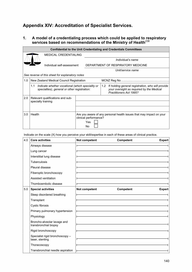
Appendix XIV: Accreditation of Specialist Services.
1. A model of a credentialing process which could be applied to respiratory services based on recommendations of the Ministry of Health238
Confidential to the Unit Credentialing and Credentials Committees
MEDICAL CREDENTIALING
Individual�s name
DEPARTMENT OF RESPIRATORY MEDICINE Individual self-assessment
Unit/service name See reverse of this sheet for explanatory notes
New Zealand Medical Council Registration MCNZ Reg No ...........................................................1.0
1.1 Indicate whether vocational (which speciality or specialties), general or other registration:
1.2 If holding general registration, who will provide your oversight as required by the Medical Practitioners Act 1995?
2.0 Relevant qualifications and sub-specialty training
3.0 Health Are you aware of any personal health issues that may impact on your clinical performance?
Yes No
Indicate on the scale (X) how you perceive your skill/expertise in each of these areas of clinical practice.
Core activities Not competent Competent Expert
Airways disease
Lung cancer
Interstitial lung disease
Tuberculosis
Pleural disease
Fiberoptic bronchoscopy
Assisted ventilation
4.0
Thomboembolic disease
Special activities Not competent Competent Expert
Sleep disordered breathing
Transplant
Cystic fibrosis
Primary pulmonary hypertension
Physiology
Broncho-alveolar lavage and transbronchial biopsy
Rigid bronchoscopy
Specialist rigid bronchoscopy � laser, stenting
Thoracoscopy
5.0
Transbronchial needle aspiration
140
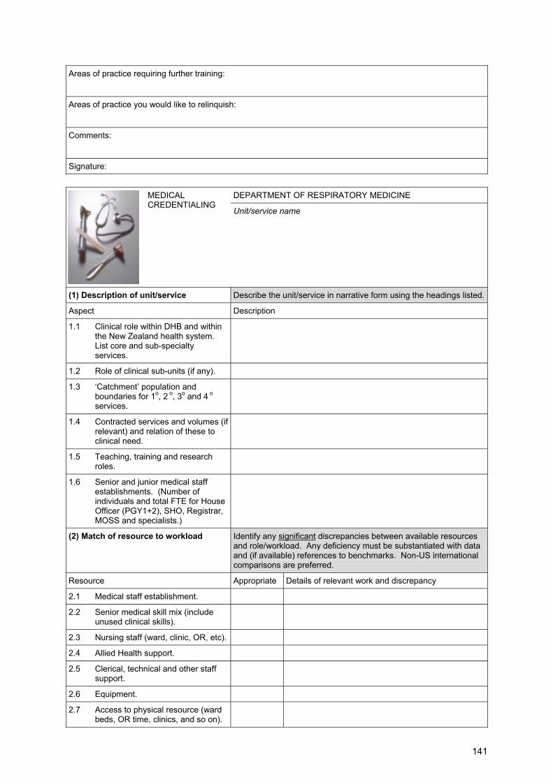
Areas of practice requiring further training:
Areas of practice you would like to relinquish:
Comments:
Signature:
DEPARTMENT OF RESPIRATORY MEDICINE
MEDICAL CREDENTIALING
Unit/service name
(1) Description of unit/service Describe the unit/service in narrative form using the headings listed.
Aspect Description
1.1 Clinical role within DHB and within the New Zealand health system. List core and sub-specialty services.
1.2 Role of clinical sub-units (if any).
1.3 �Catchment� population and boundaries for 1o, 2 o, 3o and 4 o services.
1.4 Contracted services and volumes (if relevant) and relation of these to clinical need.
1.5 Teaching, training and research roles.
1.6 Senior and junior medical staff establishments. (Number of individuals and total FTE for House Officer (PGY1+2), SHO, Registrar, MOSS and specialists.)
(2) Match of resource to workload Identify any significant discrepancies between available resources and role/workload. Any deficiency must be substantiated with data and (if available) references to benchmarks. Non-US international comparisons are preferred.
Resource Appropriate Details of relevant work and discrepancy
2.1 Medical staff establishment.
2.2 Senior medical skill mix (include unused clinical skills).
2.3 Nursing staff (ward, clinic, OR, etc).
2.4 Allied Health support.
2.5 Clerical, technical and other staff support.
2.6 Equipment.
2.7 Access to physical resource (ward beds, OR time, clinics, and so on).
141
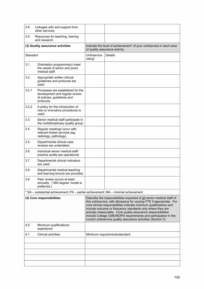
2.8 Linkages with and support from other services.
2.9 Resources for teaching, training and research.
(3) Quality assurance activities Indicate the level of achievement* of your unit/service in each area of quality assurance activity.
Standard Unit/service rating*
Details
3.1 Orientation programme(s) meet the needs of senior and junior medical staff.
3.2 Appropriate written clinical guidelines and protocols are used.
3.2.1 Processes are established for the development and regular review of policies, guidelines and protocols.
3.2.2 A policy for the introduction of new or innovative procedures is used.
3.3 Senior medical staff participate in the multidisciplinary quality group.
3.4 Regular meetings occur with relevant linked services (eg, radiology, pathology).
3.5 Departmental clinical case reviews are undertaken.
3.6 Individual senior medical staff practice audits are operational.
3.7 Departmental clinical indicators are used.
3.8 Departmental medical teaching and learning forums are provided.
3.9 Peer review occurs at least annually. (�360 degree� model is preferred.)
* SA � substantial achievement; PA � partial achievement; MA � minimal achievement
(4) Core responsibilities Describe the responsibilities expected of all senior medical staff of this unit/service, with allowance for varying FTE if appropriate. For core clinical responsibilities indicate minimum qualifications and include outcome or frequency standards only where they are actually measurable. Core quality assurance responsibilities include College CME/MOPS requirements and participation in the current unit/service quality assurance activities (Section 3)
4.0 Minimum qualifications/ experience
4.1 Clinical activities Minimum requirements/standard
142
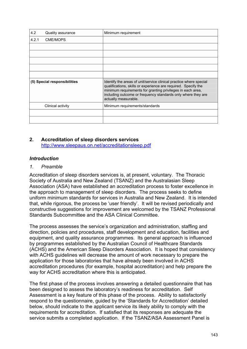
4.2 Quality assurance Minimum requirement
4.2.1 CME/MOPS
(5) Special responsibilities Identify the areas of unit/service clinical practice where special qualifications, skills or experience are required. Specify the minimum requirements for granting privileges in each area, including outcome or frequency standards only where they are actually measurable.
Clinical activity Minimum requirements/standards
2. Accreditation of sleep disorders services http://www.sleepaus.on.net/accreditationsleep.pdf
Introduction 1. Preamble Accreditation of sleep disorders services is, at present, voluntary. The Thoracic Society of Australia and New Zealand (TSANZ) and the Australasian Sleep Association (ASA) have established an accreditation process to foster excellence in the approach to management of sleep disorders. The process seeks to define uniform minimum standards for services in Australia and New Zealand. It is intended that, while rigorous, the process be �user friendly�. It will be revised periodically and constructive suggestions for improvement are welcomed by the TSANZ Professional Standards Subcommittee and the ASA Clinical Committee. The process assesses the service�s organization and administration, staffing and direction, policies and procedures, staff development and education, facilities and equipment, and quality assurance programmes. Its general approach is influenced by programmes established by the Australian Council of Healthcare Standards (ACHS) and the American Sleep Disorders Association. It is hoped that consistency with ACHS guidelines will decrease the amount of work necessary to prepare the application for those laboratories that have already been involved in ACHS accreditation procedures (for example, hospital accreditation) and help prepare the way for ACHS accreditation where this is anticipated. The first phase of the process involves answering a detailed questionnaire that has been designed to assess the laboratory�s readiness for accreditation. Self Assessment is a key feature of this phase of the process. Ability to satisfactorily respond to the questionnaire, guided by the �Standards for Accreditation� detailed below, should indicate to the applicant service its likely ability to comply with the requirements for accreditation. If satisfied that its responses are adequate the service submits a completed application. If the TSANZ/ASA Assessment Panel is
143
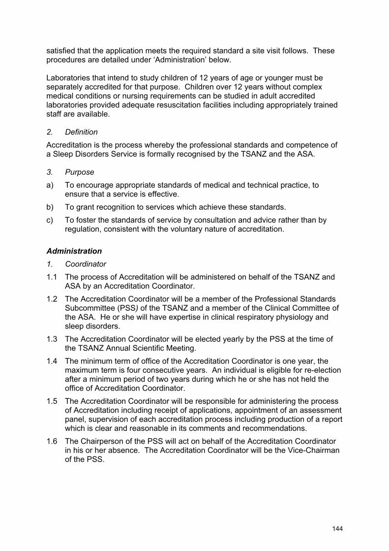
satisfied that the application meets the required standard a site visit follows. These procedures are detailed under �Administration� below. Laboratories that intend to study children of 12 years of age or younger must be separately accredited for that purpose. Children over 12 years without complex medical conditions or nursing requirements can be studied in adult accredited laboratories provided adequate resuscitation facilities including appropriately trained staff are available. 2. Definition Accreditation is the process whereby the professional standards and competence of a Sleep Disorders Service is formally recognised by the TSANZ and the ASA. 3. Purpose a) To encourage appropriate standards of medical and technical practice, to
ensure that a service is effective. b) To grant recognition to services which achieve these standards. c) To foster the standards of service by consultation and advice rather than by
regulation, consistent with the voluntary nature of accreditation. Administration 1. Coordinator 1.1 The process of Accreditation will be administered on behalf of the TSANZ and
ASA by an Accreditation Coordinator. 1.2 The Accreditation Coordinator will be a member of the Professional Standards
Subcommittee (PSS) of the TSANZ and a member of the Clinical Committee of the ASA. He or she will have expertise in clinical respiratory physiology and sleep disorders.
1.3 The Accreditation Coordinator will be elected yearly by the PSS at the time of the TSANZ Annual Scientific Meeting.
1.4 The minimum term of office of the Accreditation Coordinator is one year, the maximum term is four consecutive years. An individual is eligible for re-election after a minimum period of two years during which he or she has not held the office of Accreditation Coordinator.
1.5 The Accreditation Coordinator will be responsible for administering the process of Accreditation including receipt of applications, appointment of an assessment panel, supervision of each accreditation process including production of a report which is clear and reasonable in its comments and recommendations.
1.6 The Chairperson of the PSS will act on behalf of the Accreditation Coordinator in his or her absence. The Accreditation Coordinator will be the Vice-Chairman of the PSS.
144

2. Process 2.1 Applications for accreditation will be received by the Executive Secretary of the
TSANZ.
2.2 The Executive Secretary will respond to all applications by providing applicant laboratories with Accreditation guidelines and the application forms which seek information regarding the laboratory and investigations/measurements that it performs (�the accreditation package�). These forms include questions designed to indicate the laboratory�s readiness for accreditation. Self assessment is a key feature of this phase of the process. Once satisfied it can respond to the questions adequately the laboratory completes the forms and returns them to the Executive Secretary, along with an �initial assessment� fee which covers the cost of initial assessment of submitted material. Copies of all correspondence will be sent to the Accreditation Coordinator. A further fee (the �site visit� fee) will be charged if the application is found to be acceptable and a site visit is arranged (see below). These fees, which are set to recover costs, will be determined by the TSANZ and revised from time to time. The current fee schedule is obtainable from the TSANZ office.
2.3 On receipt of the �initial assessment� fee and application forms an Assessment Panel will be appointed. The assessors will be recognised experts in the diagnosis and management of sleep disorders and/or the technical aspects of their assessment. The assessment panel will have three members, at least one of whom will be from a city other than the one in which the service undergoing accreditation is located. The Chairperson will be a member of both the TSANZ and the ASA and will be appointed jointly by the Accreditation Coordinator of the TSANZ and the Chairperson of the Clinical Committee of the ASA. The other two members of the panel will be nominees of the ASA and TSANZ respectively, one of whom need not be a member of either society. Where practicable one member will be a sleep technologist. The assessment panel for paediatric laboratories should include a paediatrician trained in respiratory-sleep medicine and a paediatric sleep technologist.
2.4 The Chairperson of the assessment panel will cause the documentation supplied by the applicant laboratory to be reviewed by the panel members and seek supplementary information where necessary. The result of the initial assessment will be given to the applicant within eight weeks of receipt of the application. If the application is unacceptable the reasons for the decision will be provided. If the application is acceptable a site visit will be arranged at a mutually convenient time (within two to three months of notice of approval).
2.5 The site visit is a critical step in the accreditation process. At the site visit the veracity of answers provided in the application is examined. Specific questions raised by the application will be addressed and an inspection of the facilities will be undertaken. The site visit will include: a) an inspection of patient set-up procedures and the conduct of a sleep
study b) an assessment of the interpretative and reporting skills of the medical
director and the reporting physicians
145
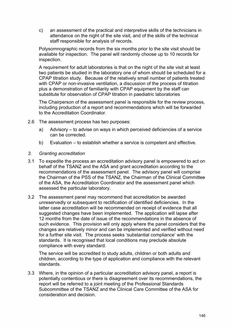
c) an assessment of the practical and interpretive skills of the technicians in attendance on the night of the site visit, and of the skills of the technical staff responsible for analysis of records.
Polysomnographic records from the six months prior to the site visit should be available for inspection. The panel will randomly choose up to 10 records for inspection. A requirement for adult laboratories is that on the night of the site visit at least two patients be studied in the laboratory one of whom should be scheduled for a CPAP titration study. Because of the relatively small number of patients treated with CPAP or non-invasive ventilation, a discussion of the process of titration plus a demonstration of familiarity with CPAP equipment by the staff can substitute for observation of CPAP titration in paediatric laboratories The Chairperson of the assessment panel is responsible for the review process, including production of a report and recommendations which will be forwarded to the Accreditation Coordinator.
2.6 The assessment process has two purposes: a) Advisory � to advise on ways in which perceived deficiencies of a service
can be corrected. b) Evaluation � to establish whether a service is competent and effective.
3. Granting accreditation 3.1 To expedite the process an accreditation advisory panel is empowered to act on
behalf of the TSANZ and the ASA and grant accreditation according to the recommendations of the assessment panel. The advisory panel will comprise the Chairman of the PSS of the TSANZ, the Chairman of the Clinical Committee of the ASA, the Accreditation Coordinator and the assessment panel which assessed the particular laboratory.
3.2 The assessment panel may recommend that accreditation be awarded unreservedly or subsequent to rectification of identified deficiencies. In the latter case accreditation will be recommended on receipt of evidence that all suggested changes have been implemented. The application will lapse after 12 months from the date of issue of the recommendations in the absence of such evidence. This provision will only apply where the panel considers that the changes are relatively minor and can be implemented and verified without need for a further site visit. The process seeks �substantial compliance� with the standards. It is recognised that local conditions may preclude absolute compliance with every standard. The service will be accredited to study adults, children or both adults and children, according to the type of application and compliance with the relevant standards.
3.3 Where, in the opinion of a particular accreditation advisory panel, a report is potentially contentious or there is disagreement over its recommendations, the report will be referred to a joint meeting of the Professional Standards Subcommittee of the TSANZ and the Clinical Care Committee of the ASA for consideration and decision.
146
3.4 A recommendation against accreditation will normally be referred to the Presidents and Executives of the TSANZ and ASA for confirmation before the report is issued.
3.5 A Certificate of Accreditation will be issued on behalf of the TSANZ and ASA once the recommendation for accreditation has been made by the Accreditation Advisory Panel. The certificate will be signed by the Chairs of the Professional Standards Subcommittee and the Clinical Committee of the TSANZ and ASA respectively, and by the Presidents of the TSANZ and ASA. Accreditation is granted for a period of five years.
3.6 Laboratories that fail accreditation will be advised of the reasons for the decision. If the laboratory wishes to challenge the decision it must do so in writing to the Accreditation Coordinator within 14 days of receiving the decision, stating the reasons for appeal. The appeal will then be considered by a joint meeting of the Professional Standards subcommittee of the TSANZ and the Clinical Care Committee of the ASA to be convened within six weeks of receipt of the appeal. A recommendation against accreditation following appeal will be referred to the Presidents and Executives of the TSANZ and ASA for confirmation before the report is issued. The Accreditation Coordinator will advise the laboratory of the decision on the appeal and the reasons for the decision. A laboratory that fails accreditation may reapply at any time that it believes its standards have met those required for accreditation.
3.7 Each accreditation report will be seen in full by the PSS of the TSANZ and the Clinical Committee of the ASA.
3.8 The Accreditation Coordinator will provide the Executive of the TSANZ with an Annual Report, a copy of which will be sent to the Executive of the ASA.
4. Re-accreditation of an accredited service 4.1 No less than 12 months before the end of the five-year accreditation period the
Executive Officer of the Society will provide to the Medical Director of the Service: ! a copy of the previous assessment panel report. ! the current accreditation guidelines and application for initial accreditation. ! a request for re-accreditation.
4.2 The request for re-accreditation will elicit information regarding the laboratory and the investigations or measurements it performs. Additionally, the Medical Director will be asked to detail changes to the service since the previous accreditation. Emphasis will be placed on the implementation of recommendations suggested by the previous assessment panel report. The service completes the request for re-accreditation and returns only these forms to the Executive Officer, along with the site visit fee current at the time of application. The service must also complete the application for initial accreditation and update laboratory manuals to reflect current practice. The application for initial accreditation and laboratory manuals are to be retained by the service for review at any subsequent site visit. Copies of all correspondence will be sent to the Accreditation Co-ordinator.
147
On receipt of the request for re-accreditation the Accreditation Coordinator will appoint an assessment panel and its Chairperson as described in paragraph 2.3. Where practicable at least one member of the assessment panel will be from the previous assessment panel.
4.3 The Chairperson of the assessment panel will arrange for the request for re-accreditation and the previous assessment panel report to be reviewed by the panel members. The result of this review will be provided to the service within six weeks of receipt of the request. If the request for re-accreditation is unacceptable then reasons for the decision will be provided and the site visit fee refunded. If the request for re-accreditation demonstrates that the service has adequately addressed the recommendations contained in the previous assessment panel report a site visit will be arranged.
4.4 Normally, the site visit will occur at least three months before the end of the current five year accreditation period. At the site visit attention will focus on problems or deficiencies identified during the previous accreditation. Compliance with any new or revised standards introduced since the previous accreditation will also be examined as may any aspect of the service�s operations. The Chairperson of the assessment panel is responsible for the review process including producing a report and recommendations that will be forwarded to the Accreditation Coordinator.
4.5 The process of granting re-accreditation will be as described in Section 3 (Granting Accreditation).
5. Confidentiality of assessment procedures 5.1 All information provided by a service in relation to preliminary enquiries or to an
application for Accreditation and all information obtained in the course of, or in connection with, an assessment of the service is considered by the TSANZ and the ASA to be completely confidential. Such information is received and studied only by members of the AAC, the AAC assessors and the TSANZ and ASA Executives, and these persons are all made aware of the confidential nature of this information.
5.2 The TSANZ and the ASA requires that all documents associated with accreditation of a service be maintained in strict confidence. This requirement imposes particular obligations on assessors. An assessor must not disclose any information gained during an assessment to any person other than a member of the AAC. Under normal circumstances there is little need for an assessor to retain a copy of the briefing notes provided for an assessment or a copy of his or her report.
5.3 It may be prudent for an assessor to keep a copy of the report temporarily to obviate loss in the mail, but this copy should be destroyed once acknowledgement of receipt is received by the AAC. If an assessor retains copies of briefing notes or reports, they must be kept in a secure place. They are not to be incorporated into the general records system of the assessors� employer in a manner which would allow unauthorised access by others.
148
Standards for accreditation This document outlines the minimum standards required for accreditation of a sleep disorders service. It must be read in conjunction with the questionnaire in the application for accreditation. It should be referred to when completing the application to ensure that the laboratory is likely to meet the requirements before submission. 1. Identifying information That information identifying the applicant service be specified at the front of the application (Form A). 2. Historical overview That a brief overview of the history of the development of the service be provided with the application. 3. Organisation and administration That the service is organised and administered to meet its objectives and the needs of the population it serves.
3.1 Goals and objectives: That the service�s goals and objectives are specified and that they reflect its role and responsibilities.
3.2 Relationship to host institution, other laboratories: That the relationship(s) of the service to its host institution and to related laboratories are appropriate to the discharge of its responsibilities. These relationships must be specified and clearly defined. There should be evidence of commitment by the host institution to its support.
3.3 Relationships with other specialities: Services are encouraged to develop a broad range of skills in the management of sleep disorders. Where this is limited (eg, to sleep breathing disorders) the service should have established appropriate relationships and communication with other specialities with a common interest in sleep disorders to ensure that clinical problems are directed to clinicians with relevant expertise and to facilitate advancement in clinical standards. This applies, for example, to the management of a patient referred with excessive somnolence to a �respiratory� sleep disorders service in whom sleep disordered breathing is subsequently excluded as a cause.
3.4 Referrals: That the sources and types of referrals to the service are relevant to the services provided. Each patient should have had an appropriate clinical evaluation prior to a diagnostic study. While the service can perform tests requested by other clinicians without direct consultation with the patient, one of the reporting consultant medical staff of the service must obtain and review sufficient information prior to the test to ensure that it is appropriate to the patient�s condition. Polysomnography should only be performed for those disorders of sleep for which it is of established diagnostic value. In the case of children, each patient should be evaluated by a paediatrician with expertise in sleep and or respiratory medicine prior to a diagnostic study.
3.5 Workload: That the service�s resources (staffing, equipment, facilities and finances) are sufficient to meet its workload without compromising the minimum standards set elsewhere in this document and in the TSANZ guidelines.239
149
3.6 Demand: That the service attempts to adequately cope with the demand for its services. Where demand exceeds capacity, the service should have a system for prioritising cases perceived to be urgent. Urgent cases should be assessed and studied in less than two weeks.
3.7 Budget: That the service�s financial plan and budget covers it�s operational costs or that there is a commitment by the host institution or company to underwrite budget deficits.
4. Staffing and direction The service is directed and staffed to achieve its objectives.
4.1 Staff structure and direction: ! That the service has a medical director responsible for overall clinical
standards and development of policies governing the service. These should be ratified by other committees in the host institution as necessary.
! That there are clear, documented lines of accountability/responsibility between medical director and all staff members. These must represent the actual manner in which the service is organised, be regularly reviewed and readily available to all staff.
! That a single designated consultant is responsible for each patient�s investigation and advice regarding management.
4.2 Staff qualifications and experience: ! That staff members are appropriately qualified for their tasks by education,
training, and or experience, and that their roles and responsibilities are specified by job description. The medical director should have specific, detailed training in sleep disorders and meet the criteria set by TSANZ guidelines.239 Consistent with the criterion set by TSANZ guidelines for advanced trainees240 wishing to practice predominantly in sleep disorders medicine, the medical director is expected to have the equivalent of two years� full-time training in sleep disorders. Other reporting consultant medical staff are expected to have completed the equivalent of at least one year full-time training in sleep disorders medicine, consistent with the guidelines for trainees wishing to make sleep disorders medicine an important area of their practice.
! There is currently no recognised tertiary training programme in sleep disorders evaluation for scientific/technical staff, hence individuals must receive �on the job� training in an established service. Basic qualifications will depend on the local requirements for classification as either scientific or technical staff (eg, a tertiary degree). Scientific or technological staff are responsible for accurate performance of sleep studies and other tests, equipment maintenance, evaluation and development of new equipment and techniques, and patient safety during performance of tests. A minimum of two years experience in a sleep disorders service and a tertiary degree in biological or physical sciences, or equivalent qualification, is desirable for a scientist or technologist to be able to function in a supervisory capacity under medical direction.
150
! Paediatric laboratories must have the facility to care for sick children or children with complex conditions. For most laboratories this will require nurses with paediatric experience trained in polysomnography with additional paediatric nursing support as necessary. All staff must be certified as competent in paediatric cardiopulmonary resuscitation.
4.3 Staff numbers: ! That sufficient medical, technical and clerical staff are employed to
adequately meet service needs. This will depend on the workload, organisation, and type of equipment and circumstances of the individual hospital. A four-bed sleep laboratory should employ approximately two full-time equivalent medical staff. For sleep studies rostering must allow for the following conditions: ! a technologist must be in attendance throughout the study (see below for
further explanation or qualification) ! the study must be of at least eight hours duration including at least one
hour for the preparation of each patient prior to study and a further hour for completion of duties following termination of the study
! in general, a ratio of no less than one technologist to three patients should be allowed for overnight
! at least two hours should be allowed for analysis of each study. ! A higher staff per patient ratio is required when studies of extra complexity
are undertaken, for example non-invasive or invasive ventilation trials, or titration of CPAP in patients with respiratory failure. Rosters must also allow for equipment calibration and maintenance, preparation and processing of reports, and in service education or professional development. Rosters must meet relevant award requirements for meal breaks, shift work, public holidays and leave.
! In the case of paediatric laboratories additional nursing requirements necessitate a higher staff�patient ratio: no less than one nurse or technologist to two patients. Rosters should be flexible to allow study commencement at or close to the child�s normal bedtime.
Explanatory notes: �A technologist must be in attendance throughout the study.� Short absences from the facility (eg, toilet breaks taken away from the laboratory) during a routine diagnostic or CPAP titration study may be covered by staff who have limited technical expertise in sleep studies but nevertheless are able to attend to the needs of the patient and are trained in emergency procedures. More prolonged absences from the facility (eg, meal breaks) in such cases, or short absences during complex studies (eg, nasal IPPV trials), should be covered by another sleep technologist. In a free standing laboratory (that is a laboratory located away from a hospital that has emergency back up) two staff trained in emergency procedures should be in attendance for the duration of the study to ensure safety and security of patients and staff.
151
In the case of paediatric laboratories a team trained in paediatric cardiopulmonary resuscitation must be available on site for the duration of the study.
4.4 Staff appraisal: That a staff appraisal system is in operation, that a written report is produced, that the staff member involved is aware of the contents of the report and that a plan to address deficiencies is defined.
4.5 Training of staff in cardiopulmonary resuscitation: That all medical, technological and nursing staff are trained in cardiopulmonary resuscitation, and that a basic level of competence is maintained and evaluated regularly. In the case of paediatric laboratories, special training in paediatric cardiopulmonary resuscitation is required.
5. Policies and procedures That the service has documented policies and procedures that reflect current knowledge and practice in the conduct of a sleep disorders service and, where relevant, comply with statutory requirements.
5.1 Patient referral, handling, documentation, follow-up: ! That procedures exist for prompt, efficient handling of patient referrals, initial
consultations and follow-up, documentation, communication with the referring doctor, and protection of patient confidentiality, and that these are consistent with good professional practice. It is expected that patients be clinically evaluated prior to sleep study. A patient record should be maintained, which is well ordered and contains all laboratory reports, records of consultations and procedures, copies of correspondence, working and or final diagnoses and, where appropriate, clearly defined treatment or follow-up recommendations.
! Recommended treatments must be consistent with current knowledge and practice. Correspondence should be completed promptly (within five working days) following each patient contact. Special care should be taken with transmission of information by facsimile. Use of the fax for this purpose should be minimised and any information transmitted by these means should be accompanied by a suitably worded warning regarding the confidential nature of the enclosed information. Patient records should be kept for a period of time that complies with legislative requirements and is consistent with good professional practice.
5.2 Sleep studies: types, methods of measurement: That methods for the conduct of sleep studies, are consistent with recognised standards, including the relevant TSANZ guidelines239 and, where applicable, paediatric guidelines.241 Types of sleep studies performed and the parameters measured must be specified. Sleep studies must allow full disclosure of the raw signals, which must be adequately labelled and calibrated. Standard physical calibrations should be used wherever possible. Where electrical calibration is used it must be checked against physical calibration regularly. Calibrations should be done prior to each study and whenever accuracy is in doubt. Each calibration procedure should be repeated at least twice to ensure reproducibility. Calibration results should be clearly labelled. The equipment must conform to specifications (linearity, sensitivity, frequency response, signal to noise ratio,
152
stability) that ensure collection of meaningful, interpretable results. Overnight visual monitoring of patients (by infra-red or low light video) is a desirable feature.
5.3 Sleep studies: analysis and interpretation: ! That methods for the analysis of sleep studies are consistent with recognised
standards, including the relevant TSANZ guidelines.239 Scoring and interpretation of the data should conform to American Thoracic Society242 and American Sleep Disorders Association243,244 recommendations. In the case of paediatric laboratories, scoring and interpretation should conform with ATS and ASDA recommendations for the analysis of polysomnographic studies in children and be age appropriate.241,245
! While analysis of the sleep study may be performed by a well-trained technologist, interpretation is the responsibility of the patient�s clinician. Computerised analysis systems are considered aids to the process: final analysis must be performed manually and involve reference to the raw data, as must interpretation by the responsible clinician. The report must clearly identify the Service, and the Patient and the date of the study. It should be consistent with TSANZ guidelines,239 containing the study results along with an interpretive summary statement signed by the interpreting clinician. The laboratory should have established methods for assessing the quality of measurements and analysis including periodic assessment of inter-observer variability in analysis of sleep studies (see also 9, Quality Assurance).
5.4 Other procedures: That the methods for multiple sleep latency testing (MSLT) and related studies are consistent with established standards246, including the relevant TSANZ guidelines239. It is expected that sleep services be able to perform MSLTs, or have an affiliation with a service with that capacity, to enable the further investigation and diagnostic refinement of the sleepy patient eg. to confirm or exclude the presence of pathological daytime sleepiness in difficult cases, or to assist in the diagnosis of narcolepsy.
5.5 Nasal continuous positive airway pressure (nCPAP) and other respiratory appliances: That procedures for the prescription and supply of CPAP therapy and its follow-up are consistent with good professional practice. This requires a diagnostic study prior to prescription of CPAP and a CPAP titration study. Early follow-up after prescription (within one month) is required to determine whether problems affecting compliance exist.
5.6 Safety: That the laboratory meets standards of laboratory safety consistent with State occupational health and safety regulations, including infection control, handling of gas cylinders, fire and electrical safety and general safety procedures. Electrical supply to the monitoring room and the bedrooms of the laboratory should be at minimum body protected standard (class B [AS specification]). Monitoring equipment should be supported by a certificate of type testing to AS 3200.1 (1990) or AS 3200 (1986) or equivalent.
5.7 Laboratory manual: ! That each type of test performed by the laboratory be described in detail in a
laboratory manual. Each test should be separately described with the following detail included or cross-referenced from other sources, preferably under appropriate subheadings.
153
! The purpose of the test. a) A description of the equipment used, with special reference to its
specifications and their applicability to the measurement. b) The calibration procedure. c) The procedure for performance of the test. d) Troubleshooting. Problems which may be encountered in the
performance of each test and their appropriate remedies. e) Specific quality assurance. Details of quality control steps required for
the method. f) Cleaning and maintenance. g) Infection control and other safety requirements. h) Records and reports (with samples, including interpretation of the
results). i) Normal values and prediction equations used to interpret the results. j) References. If the test is based on unpublished work, relevant details
of this work should be included. k) The date of issue of and alterations to the method. l) The signature of the senior laboratory officer � this indicates that the
method. ! Appropriate cross-referencing (eg, to manufacturer�s manual) under each
subheading could minimise redundancy while ensuring that all issues relevant to each test have been addressed.
! Ideally this laboratory manual should be part of a service policy and procedures manual (see 11 below).
6. Staff development, teaching, research That staff have access to education programmes which maintain and develop their knowledge and skills.
6.1 Staff development: ! That programmes exist to orientate new staff, and for continuing education of
existing staff taking into account results of performance appraisal (see 4.4 above), service objectives and quality assurance activities (see 9 below).
! That opportunities exist for senior staff to attend relevant professional meetings (state, national, international).
6.2 Teaching: That where the service operates in a teaching hospital environment it offers education programmes for undergraduates and postgraduates.
6.3 Research: That where the service operates in a teaching hospital environment it has a commitment to research. This can be demonstrated by reference to current projects, recent presentations (abstracts) and publications.
154
7. Facilities and equipment That adequate facilities and equipment exist for the service to meet its objectives and comply with statutory requirements.
7.1 Consulting rooms: That the reception area, waiting room, offices and consultation rooms conform to generally accepted standards for medical suites in size, appearance, privacy, lighting, furniture and provision of other equipment.
7.2 Sleep laboratory: ! That the sleep laboratory has comfortably furnished bedrooms conducive to
sleep and of sufficient size (minimum approximately 2.5 x 3.5 metres) to allow access in an emergency, with adequate lighting, sound-proofing, exclusion of light during the study, air conditioning, emergency oxygen and suction, resuscitation equipment, and security.
! That the rooms conform to local regulations with respect to entrances, exits and fire precautions.
! That there is a separate bedroom for each patient with comfortable bedding, wardrobe, chair and bedside lamp. In the case of paediatric laboratories the bedroom must be child-safe and age-appropriate, with age-appropriate bedding for each patient. Facilities for a parent to sleep in the child�s bedroom should be available.
! That there are conveniently located and adequate toilet and shower facilities. ! That the monitoring room is located in close proximity to the bedrooms and
that a patient call system is available from bedrooms to monitoring room. ! That office space exists with adequate space, furniture, lighting and privacy
for analysis of sleep studies. ! That the facilities are regularly cleaned.
7.3 Equipment: That the equipment used for the conduct of respiratory sleep studies and related tests is suitable for the purpose (see 5.2, 5.3, 5.4 above) and is regularly maintained and safety checked. In paediatric laboratories the sensors and other equipment interfaced with the patient should be appropriately sized and a range of sizes should be available for each study.
7.4 Identification: That the service is identified by signage, telephone and stationery so that it can be easily found and/or accessed.
155
8. Provision for emergencies 8.1 Medical emergencies:
! That adequate provision is made for medical emergencies. These should include an on-call roster for medical staff, CPR training for all staff, availability of resuscitation equipment, oxygen and suction, and easy access to the laboratory and the patient.
! In the case of paediatric laboratories and a team trained in paediatric cardiopulmonary resuscitation must be available on-site for the duration of the study. All staff must be trained in paediatric cardiopulmonary resuscitation. A complete range of age-appropriate resuscitation equipment must be available in the laboratory for the duration of the studies and oxygen and suction must be available at the bedside.
8.2 Non-medical emergencies: That provisions complying with relevant site and statutory requirements are made for non-medical emergencies (fire and safety).
9. Quality assurance programme That procedures exist to evaluate the quality of the service provided, correct identified problems, and advance the service�s standards. The process must include the following elements: ! Monitoring. Regular collection of data relevant to important aspects of service
delivery. ! Assessment. Periodic assessment of the data to identify problems or
opportunities to improve. ! Action. Action to address such problems or opportunities. ! Evaluation. Evaluation of the effects of such action. ! Feedback. Regular communication to the staff of the results of these activities. The process must be documented and patient confidentiality must be protected. 10. Meetings That regular scheduled meetings occur, at no greater than monthly intervals, for the purposes of laboratory function and planning, quality assurance and clinical review, in-service education, and, where applicable, research. There should be records of these meetings. Action statements are encouraged where applicable. 11. Policies and procedures manual That the department maintains a policies and procedures manual which specifies its organisation and administration, staffing and direction, policies and procedures (see 5.7, Laboratory Manual), staff development and education, facilities and equipment, and quality assurance programme.
156
3. Accreditation of respiratory function assessment services
Introduction 1. Preamble Accreditation of respiratory function services is, at present, voluntary. The Thoracic Society of Australia and New Zealand (TSANZ) has established an accreditation process to foster excellence in the approach to assessment of respiratory function. The process seeks to define uniform minimum standards for services in Australia and New Zealand. It is intended that, while rigorous, the process be �user friendly�. It will be revised periodically and constructive suggestions for improvement are welcomed by the TSANZ Professional Standards Subcommittee. The process assesses the service�s organisation and administration, staffing and direction, policies and procedures, staff development and education, facilities and equipment, and quality assurance programmes. Its general approach is influenced by programmes established by the Australian Council of Healthcare Standards (ACHS). It is hoped that consistency with ACHS guidelines will decrease the amount of work necessary to prepare the application for those laboratories that have already been involved in ACHS accreditation procedures (eg, hospital accreditation) and help prepare the way for ACHS accreditation where this is anticipated. The first phase of the process involves answering a detailed questionnaire which has been designed to assess the laboratory�s readiness for accreditation. Self Assessment is a key feature of this phase of the process. Ability to satisfactorily respond to the questionnaire, guided by the �Standards for Accreditation� detailed below, should indicate to the applicant service its likely ability to comply with the requirements for accreditation. If satisfied that its responses are adequate the service submits a completed application. If the TSANZ Assessment Panel is satisfied that the application meets the required standard a site visit follows. These procedures are detailed under �Administration� below. 2. Definition Accreditation is the process whereby the professional standards and competence of a respiratory function assessment service, hereafter referred to as the service, is formally recognised by the TSANZ. 3, Purpose ! To encourage appropriate standards of medical and technical practice to ensure
that a service is effective. ! To grant recognition to services which achieve these standards. ! To foster the standards of service by consultation and advice rather than by
regulation, consistent with the voluntary nature of accreditation.
157
Administration 1. Coordinator 1.1 The process of Accreditation will be administered on behalf of the Professional
Standards Subcommittee (PSS) by an Accreditation Coordinator.
1.2 The Accreditation Coordinator will be a member of the PSS with expertise in clinical respiratory physiology and sleep disorders.
1.3 The Accreditation Coordinator will be elected yearly by the PSS at the time of the TSANZ Annual Scientific Meeting.
1.4 The minimum term of office of the Accreditation Coordinator is one year, the maximum term is four consecutive years. An individual is eligible for re-election after a minimum period of two years during which he or she has not held the office of Accreditation Coordinator.
1.5 The Accreditation Coordinator will be responsible for administering the process of accreditation including receipt of applications, appointment of an assessment panel, supervision of each accreditation process including production of a report which is clear and reasonable in its comments and recommendations.
1.6 The Chairman of the PSS will act on behalf of the Accreditation Coordinator in his or her absence. The Accreditation Coordinator will be the Vice-Chairman of the PSS.
2. Categories of respiratory function assessment services 2.1 Category 1: Basic assessment of respiratory function including, as a minimum,
measurement of static lung volumes (total lung capacity, residual volume, functional residual capacity and vital capacity); maximum expiratory flow rates before and after bronchodilator (maximum expiratory flow volume curves); carbon monoxide gas transfer; and maximum respiratory pressures measured at the mouth.
2.2 Category 2: Measurements as in Category 1 plus arterial blood gas analysis.
2.3 Category 3: Standard assessment of respiratory function including measurements as in Categories 1 or 2 plus pharmacologic and non-pharmacologic bronchial provocation tests and exercise tests.
2.4 Category 4: Comprehensive assessment of respiratory function including measurements in Category 3 plus any of the following: ! measurements of the control of breathing ! of lung mechanics ! of chest wall mechanics ! of pulmonary gas exchange ! of nasal resistance ! simulated altitude measurements ! any other complex measurements of respiratory function.
158
3. Process 3.1 Applications for accreditation will be received by the Executive Secretary of the
Society. The application should specify the category of respiratory function assessment for which accreditation is sought and a list of the individual tests for which accreditation is sought. Accreditation will be granted only in relation to those categories or tests for which application is made. The Executive Secretary will respond to all applications by providing applicant laboratories with accreditation guidelines and the application forms which seek information regarding the laboratory and investigations/measurements that it performs (�the accreditation package�). These forms include questions designed to indicate the laboratory�s readiness for accreditation. Self-assessment is a key feature of this phase of the process. Once satisfied it can respond to the questions adequately the laboratory completes the forms and returns them to the Executive Secretary, along with an �initial assessment fee� which covers the cost of the initial assessment of submitted material. Copies of all correspondence will be sent to the Accreditation Coordinator. A further fee (the �site visit� fee) will be charged if the application is found to be acceptable, and a site visit is arranged (see below). These fees, which are set to recover costs, will be determined by the Society and revised from time to time. The current schedule of fees is obtainable from the Society office.
3.2 On receipt of the accreditation fee and application forms the Accreditation Coordinator will appoint an assessment panel and its Chairperson. The assessors will be recognised experts in the physiological and/or technical aspects of respiratory function assessment and its application to the diagnosis and management of respiratory disease. The assessment panel will normally have three members, at least one of whom will be from a city other than the one in which the service undergoing accreditation is located. While individual assessors need not necessarily be members of the TSANZ, at least two members of the assessment panel will be. Where practicable one member will be a respiratory scientist/technologist. The Chairperson of the assessment panel will cause the documentation supplied by the applicant laboratory to be reviewed by the panel members and seek supplementary information where necessary. The result of the initial assessment will be given to the applicant within eight weeks of receipt of the application. If the application is unacceptable the reasons for the decision will be provided. If the application is acceptable a site visit will be arranged at a mutually convenient time (within two to three months of notice of approval). The site visit is a critical step in the accreditation process. At the site visit the veracity of answers provided in the application is examined, and specific questions raised by these responses and by inspection of the facilities addressed. The Chairperson of the assessment panel is responsible for the review process including producing a report and recommendations which will be forwarded to the Accreditation Coordinator.
159
3.3 The assessment process has two purposes: ! Advisory � to advise on ways in which perceived deficiencies of a service can
be corrected. ! Evaluation � to establish whether a service is competent and effective.
4. Granting accreditation 4.1 To expedite the process an accreditation advisory panel is empowered to act on
behalf of the Society and grant accreditation according to the recommendations of the assessment panel. The advisory panel will comprise the Chairman of the PSS, the Accreditation Coordinator and the assessment panel which assessed the particular laboratory.
4.2 The assessment panel may recommend that accreditation be awarded unreservedly or subsequent to rectification of identified deficiencies. In the latter case accreditation will be granted on receipt of evidence that all suggested changes have been implemented. The application will lapse after 12 months from the date of issue of the recommendations in the absence of such evidence. This provision will only apply where the panel considers that the changes are relatively minor and can be implemented and verified without need for a further site visit. The process seeks �substantial compliance� with the standards. It is recognised that local conditions may preclude absolute compliance with every standard.
4.3 Where, in the opinion of a particular accreditation advisory panel, a report is potentially contentious or there is disagreement over its recommendations, the report will be referred to the President and the Executive of the Society for comment and decision.
4.4 A recommendation against accreditation will normally be referred to the President and Executive of the Society for confirmation before the report is issued.
4.5 A Certificate of Accreditation will be issued once the recommendation for accreditation is ratified by the accreditation advisory panel or, where applicable, the Executive. The certificate will be signed by the Chairman of the PSS and the President on behalf of the Society. Accreditation is granted for a period of five years.
4.6 Laboratories that fail accreditation will be advised of the reasons for the decision. If the laboratory wishes to challenge the decision it must do so in writing to the Accreditation Coordinator within 14 days of receiving the decision stating the reasons for appeal. The appeal will then be considered by a meeting of the Professional Standards Subcommittee to be convened within six weeks of receipt of the appeal. A recommendation against accreditation following appeal will be referred to the President and Executive of the Society for confirmation before the report is issued. The Accreditation Coordinator will advise the laboratory of the decision on the appeal and the reasons for the decision. A laboratory that fails accreditation may reapply at any time that it believes its standards have met those required for accreditation.
160
4.7 Each accreditation report will be seen in full by the PSS.
4.8 The Accreditation Coordinator will provide the Executive of the Society with an Annual Report.
5. Re-accreditation of an accredited service 5.1 No less than 12 months before the end of the five year accreditation period the
Executive Officer of the Society will provide to the Medical Director of the service: ! a copy of the previous assessment panel report ! the current accreditation guidelines and application for initial accreditation ! a request for re-accreditation (see Forms C, D and E).
5.2 The request for re-accreditation will ask for the category of respiratory function assessment for which accreditation is sought, a list of the individual tests for which accreditation is sought and staffing information. Additionally, the Medical Director will be asked to detail changes to the service since the previous accreditation. Emphasis will be on the implementation of recommendations suggested by the previous assessment panel report.
5.3 The service completes the request for re-accreditation and returns �only these forms� to the Executive Officer, along with the site visit fee current at the time of application. The service must also complete the application for initial accreditation and update laboratory manuals to reflect current practice. The application for initial accreditation and laboratory manuals are to be retained by the service for review at any subsequent site visit. Copies of all correspondence will be sent to the Accreditation Co-ordinator.
5.4 On receipt of a request for re-accreditation the Accreditation Coordinator will appoint an assessment panel and its Chairperson as described in paragraph 3.3. Where practicable at least one member of the assessment panel will be from the previous assessment panel. The Chairperson of the assessment panel will arrange for the request for re-accreditation and the previous assessment panel report to be reviewed by the panel members. The result of this review will be provided to the service within six weeks of receipt of the request. If the request for re-accreditation is unacceptable then reasons for the decision will be provided and the site visit fee refunded. If the request for re-accreditation demonstrates that the service has adequately addressed the recommendations contained in the previous assessment panel report a site visit will be arranged. Normally, the site visit will occur at least three months before the end of the current five year accreditation period. At the site visit attention will focus on problems or deficiencies identified during the previous accreditation. Compliance with any new or revised standards introduced since the previous accreditation will also be examined as may any aspect of the service�s operations. The Chairperson of the assessment panel is responsible for the review process including producing a report and recommendations that will be forwarded to the Accreditation Coordinator.
5.6 The process of granting re-accreditation will be as described in Section 4 (granting accreditation).
161
6. Confidentiality of assessment procedures All information provided by a testing laboratory in relation to preliminary enquiries or to an application for accreditation and all information obtained in the course of or in connection with an assessment of the service is considered by the TSANZ to be completely confidential. Such information is received and studied only by members of the AAC, the AAC assessors and TSANZ Executive and these persons are all made aware of the confidential nature of this information. The TSANZ requires that all documents associated with accreditation of a service be maintained in strict confidence. This requirement imposes particular obligations on assessors. An assessor must not disclose any information gained during an assessment to any person other than a member of the AAC. Under normal circumstances there is little need for an assessor to retain a copy of the briefing notes provided for an assessment or a copy of his or her report. It may be prudent for an assessor to keep a copy of the report temporarily to obviate loss in the mail, but this copy should be destroyed once acknowledgement of receipt is received by the AAC. If an assessor retains copies of briefing notes or reports, they must be kept in a secure place. They are not to be incorporated into the general records system of the assessor�s employer in a manner which would allow unauthorised access by others. Standards for accreditation This document outlines the minimum standards required for accreditation of a respiratory function assessment service. It must be read in conjunction with the questionnaire in the application for accreditation. It should be referred to when completing the application to ensure that the laboratory is likely to meet the requirements before submission. 1. Identifying information That information identifying the applicant service be specified at the front of the application (Form A). 2. Historical overview That a brief overview of the history of the development of the service be provided with the application. 3. Organisation and administration That the service is organised and administered to meet its objectives and the needs of the population it serves.
3.1 Goals and objectives: That the service�s goals and objectives are specified and that they reflect its role and responsibilities.
3.2 Relationship to host institution, other laboratories: That the relationship(s) of service to its host institution and to related laboratories are appropriate to the discharge of its responsibilities. These relationships must be specified and clearly defined. There should be evidence of commitment by the host institution to its support.
162
3.3 Relationships with other specialities: That the service has established appropriate relationships and communication with other specialities with a common interest in respiratory disease to ensure that clinical problems are directed to clinicians with relevant expertise and to facilitate advancement in clinical standards.
3.4 Referrals: That the sources and types of referrals to the service are relevant to the services provided. Referrals to the service should be related to respiratory function assessment or other matters for which there is local expertise.
3.5 Workload: That the service�s resources (staffing, equipment, facilities finances) are sufficient to meet its workload without compromising the minimum standards set elsewhere in this document. A regular updated audit of the laboratory�s workload in terms of numbers of tests of each particular type should be kept and these records should be available on a quarterly or yearly basis.
3.6 Demand: That the service attempts to adequately cope with the demand for its services. Where demand exceeds capacity, the service should have a system for prioritising cases perceived to be urgent. The service should be able to assess new patients within two weeks of referral. Urgent cases should be assessed within two days.
3.7 Budget: That the service�s budget covers its operational costs or that there is a firm commitment by the host institution or company to underwrite budget deficits.
4. Staffing and direction The service is directed and staffed to achieve its objectives.
4.1 Staff structure and direction: ! That the service has a medical director responsible for overall standards and
development of policies governing the service. These should be ratified by other committees in the host institution as necessary.
! That there are clear, documented lines of accountability and responsibility between medical director and all staff members. These must represent the actual manner in which the service is organised, be regularly reviewed and readily available to all staff.
4.2 Staff qualifications and experience247,240,248 ! That staff members are appropriately qualified for their tasks by education,
training, and/or experience, and that their roles and responsibilities are specified by job description. The medical director should have specific, detailed training in clinical respiratory physiology and meet the criteria set by the relevant TSANZ position paper.247 Other consultant medical staff are expected to have completed the equivalent of one years full time training in respiratory physiology, consistent with the TSANZ guidelines for advanced trainees240 wishing to make respiratory physiology an important area of their practice.
163
! Scientific or technological staff are responsible for accurate performance of tests, equipment maintenance, continuing quality assurance of both equipment and techniques and patient safety during performance of tests. The basic qualification for classification as either scientific or technical staff depends on local requirements (eg, two years tertiary training in biological or physical science for technologists, Bachelor of Science or equivalent for scientists).
! Until 1995 there was no recognised tertiary training programme in respiratory physiology or function assessment for scientific or technological staff in Australia. Hence most staff received their professional or vocational training through experience in an established service. However formal training in respiratory science is now offered by several courses. These are strongly endorsed, particularly for new staff and those without substantial experience in respiratory function assessment. It is recommended that scientific or technical staff acquire the Certified Respiratory Function Scientist (CRFS) credential based on examination by the Australian and New Zealand Society of Respiratory Science (ANZRS).
! To function in a supervisory capacity under medical direction a scientist/ technologist should have a Bachelor of Science in biological or physical sciences, and between three and five years experience, depending on the nature of that experience and the range of tests performed by the laboratory. In addition acquisition of the CRFS credential is strongly recommended.
4.3 Staff numbers: That sufficient medical, technical and clerical staff are employed to adequately meet service needs. This will depend on the workload, organisation, type of equipment and circumstances of the individual hospital. Where three or more scientific or technological staff are employed it is advisable that there be a designated chief scientist or technologist to assist the Medical Director to administer the service.
4.4 Staff appraisal: That a staff appraisal system is in operation, that a written report is produced, that the staff member involved is aware of the contents of the report and that a plan to address deficiencies is defined.
4.5 Training of staff in cardiopulmonary resuscitation: That all medical, technological and nursing staff are trained in cardiopulmonary resuscitation, and that a basic level of competence is maintained.
5. Policies and procedures That the service has documented policies and procedures that reflect current knowledge and practice in the conduct of a respiratory function assessment service and, where relevant, comply with statutory requirements.
5.1 Patient referral, handling, documentation, follow-up: That procedures exist for prompt, efficient handling of patient referrals, documentation, communication with the referring doctor, and that these are consistent with good professional practice. A patient record should be maintained which is well ordered and contains all laboratory test results and reports (see 5.5 and 5.6), records of consultations and copies of correspondence. Reports and correspondence should be completed promptly (within five working days) following each patient
164
contact. Records should be kept for a period of time that complies with legislative requirements and is consistent with good professional practice.
5.2 Respiratory function tests: equipment and methods (see also 5.3 Quality Control, below): That the methods for conducting respiratory function tests are consistent with recognised standards, including relevant TSANZ guidelines. 5.2.1 Equipment
! That the equipment used for the conduct of respiratory function and related tests is suitable for the purpose (see also 5.3) and is regularly maintained and safety checked.
! The choice of equipment will depend on the required accuracy of particular measurements, the workload, ease of use, servicing and economic considerations as well as biochemical and electrical safety standards. ! Arterial blood gas analysis. Most equipment used for this procedure
is now self-calibrating. A quality control procedure must be used regularly to ensure the validity of the results.
! Spirometry and gas transfer. Equipment used for spirometry and gas transfer and the use of computers for data collection and analysis should meet published standards of the American Thoracic Society.249,250,251
! Equipment and systems used for other measurements should have linearity, sensitivity, signal to noise and frequency response characteristics which are appropriate to the particular measurement and should meet currently accepted, published criteria which help to ensure accurate measurement.
! Each service must purchase and maintain the equipment necessary to perform routine calibration of all equipment used in the performance of the above tests.
5.2.2 Methods ! In the field of respiratory function testing, rigid insistence on the use of
particular published methods is inappropriate and would detract from versatility and originality of expertise which reflects the competence of the service. However, test procedures should be validated. The TSANZ does not specify which methods a service may or should use for any particular test.
! Accreditation will involve examination of the documentation of laboratory methods and their availability to staff working within the service. To this end each service should have a procedures manual which contains not only the essential procedural elements of the method but also information on problems that may be encountered and details of any equipment checks and calibrations or other aspects of quality control which may be necessary for that method (see 5.3, below). Personnel performing tests should have ready access to the methods manual and should be encouraged to refer to it frequently.
165
! For a general description of commonly employed methodology for most respiratory function tests, reference to standard texts may be useful.252,253,254,255,256 Specific information summarising current or standard practices for performing the commonly employed respiratory function tests and investigations is available from the current literature.252,249,250,251,257,258,259.
5.2.3 Computer-aided respiratory function tests ! The basic principles of assessment of computerised testing systems are
the same as for any other kind of testing equipment. By their very nature however, computer or microprocessor-aided equipment often requires additional procedures for testing and quality control. Details of any such additional procedures should be documented in the laboratory manual (see 5.2.4 below). Microprocess or controlled systems are by nature prone to subtle errors not normally encountered in purely manual systems.
! Laboratories using such systems should demonstrate that the methods used and results obtained are valid. A general approach to the use of computers in respiratory laboratories is given by Clausen JL252 (Chapter 27). Specific guidelines for the general use of computers are also given in: Guide to Assessment of Laboratories, National Association of Testing Authorities, Australia, February 1984.260 These principles will be followed in the assessment of services in so far as they relate to respiratory function testing.
5.2.4 Laboratory procedures manual (LPM) ! Laboratory procedures should be described in detail in a laboratory
manual. Each test should be separately described with the following detail included or cross referenced from other sources, preferably under appropriate subheadings: LPM 1) The purpose of the test. LPM 2) A description of the equipment used, with special reference
to its specifications and their applicability to the measurement.
LPM 3) The calibration procedure. LPM 4) The procedure for performance of the test. LPM 5) Troubleshooting. Problems which may be encountered in
the performance of each test and their appropriate remedies. LPM 6) Specific quality assurance. Details of quality control steps
required for the method. LPM 7) Cleaning and maintenance LPM 8) Infection control and other safety requirements. LPM 9) Records and reports (with samples, including interpretation
of the results). LPM 10) Normal values and prediction equations used to interpret the
results. LPM 11) References. If the test is based on unpublished work,
relevant details of this work should be included.
166
LPM 12) The date of issue of and alterations to the method. LPM 13) The signature of the senior laboratory officer. This indicates
that the method section has been checked by a senior staff member for procedural and typographical errors before being included in the manual.
! Appropriate cross-referencing (eg, to manufacturer�s manual) under each subheading could minimise redundancy while ensuring that all issues relevant to each test have been addressed.
! Special requirements for computer-aided respiratory function tests: Whether written in the laboratory or purchased �off the shelf�, computer programmes should also be adequately documented in the laboratory manual. Minimum requirements for this documentation include: ! details of programme flow and logic checks performed when
implementing the system ! for programmes written in the laboratory including:
(i) an outline of the basic structure and logic of the programme (ii) an up-to-date listing of the programme
! details of comparisons between computer derived results and manually derived results
! operating instructions for running the programme, including details on restoration of the computer to running condition in the event of computer failure.
! Documentation of procedures should be reviewed regularly so that any alterations to methods can be dealt with before the accumulation of such alterations requires the entire manual to be revised. These reviews should be carried out at least annually.
! Personnel performing tests should have ready access to the methods manual and should be encouraged to refer to it frequently.
! Ideally this laboratory manual should be part of a service policy and procedures manual (see 11 below).
5.3 Quality control of measurements155 (see also 9, quality assurance, below): ! That regular monitoring of the accuracy of measurement is undertaken, using
appropriate calibrations, internal quality control procedures and, where possible, participation in appropriate inter-laboratory test programmes.
! Appropriate quality control procedures and equipment for performing them are an essential component of any respiratory function laboratory. (a) Calibration:
! Services should purchase and maintain the equipment necessary to perform routine calibration checks of all testing equipment and set aside non-patient time for equipment calibration. Standard physical calibration should be used routinely. Where electrical calibration is utilised, it must be checked against physical calibration regularly. Calibration results should be labelled, dated and filed for at least the last two years of use of the instrument. Each calibration procedure should be repeated at least twice to ensure reproducibility.
167
Calibration procedures should be done on a regular basis, or whenever accuracy is in doubt.
! Following the calibration of equipment, normal individuals may be utilised to verify the overall performance of the equipment, keeping in mind the reproducibility of the test within a given individual. With more complex tests involving multi-component systems, normal subjects should be employed more frequently.
! Guidelines for the calibration and quality control of all individual lung function tests are available from the literature.252,258,155 Further specific information regarding quality control and calibration guidelines for spirometry, lung volume and diffusing capacity measurements is also available.250,257,155
(b) Internal quality control: ! Internal quality control procedures are the responsibility of the senior
laboratory personnel and should be practised in association with each test method at appropriate levels. Details of such procedures must be recorded as part of each test method in the laboratory manual. When routine quality assurance testing indicates that the method is moving out of control, written protocols are helpful in specifying the courses of action to be followed for diagnosis and correction. Graphic records assist the monitoring of internal quality control procedures.
! Routine preventive maintenance of equipment used should be documented in the laboratory manual.
(c) External quality control: ! External quality control, that is participation in inter-hospital
proficiency testing programmes, assists in monitoring the effectiveness of internal quality control procedures.
5.4 Predicted values: ! That the laboratory utilises appropriate predicted values for comparison with
the results of each respiratory function test it performs. ! The purpose of such a comparison is to help determine whether respiratory
function is normal or abnormal. There are a great number of published �predicted� values for many respiratory function tests. Unfortunately, the variability among them is often large, making the choice of predicted values difficult. Frequently there is no objective means of singling out one set of data as being superior to others particularly when different equipment and methodologies have been used.
! When a laboratory chooses a set of predicted values, the following recommendations should be taken into account: ! predictions of expected normal values should be based on studies with
large numbers of subjects of both sexes and covering a wide range of ages, heights and weights
! the equipment and techniques used by a service and those used to obtain predicted values should, to the extent possible, be similar
168
! the population samples should be heterogeneous, going across socioeconomic groups. Surveys should be of communities or towns, not of professional groups. Homogenous groups � religious groups, miners, subjects in sanatoria, and so on, should be avoided unless a special purpose population is sought
! ethnic factors, smoking habits and respiratory symptoms should be accounted for if possible
! the equipment and methods used to obtain and analyse the data should be described.
! It is highly recommended that the appropriateness of any chosen predicted values should be checked by comparing predicted data with data obtained from a representative sample of �normal� people.
! For general information regarding normal reference values is refer to Clausen JL (Chapter 6),252 Cotes254 and the relevant ATS statement.261
5.5 Laboratory test records: ! That the laboratory maintains a record of all respiratory function
measurements performed. ! The TSANZ�s basic requirement is that the results are recorded in a clear and
unambiguous way, are complete in respect to the performance of the test and can be checked against the original data obtained at the time of measurement. The application of these concepts will vary from one laboratory to another and will depend upon the extent to which the records system is computerised, but the following guidelines will generally be applicable.
! If a manual recording system is used, all test data should be recorded clearly and permanently on pro-forma sheets, on test cards or in work books. Sheets of plain paper should not be used because they are easily lost and because they engender a less disciplined approach to recording the information required. Test records should contain all information needed to show unambiguously what has been done, by whom and when. This will usually mean the following details: LPM 14) Date and time of test. LPM 15) Identity of the testing officer. LPM 16) Identity of the test method. LPM 17) Any variations from the standard method. LPM 18) Identification of the subject. LPM 19) Reference standard employed (where different from usual). LPM 20) All test data (including units). LPM 21) Any necessary calculations. LPM 22) The final results. LPM 23) The rounded test result. LPM 24) Any other information required by test method. LPM 25) Any pertinent observations of the testing officer. LPM 26) Signature or initials of the testing officer. LPM 27) Signature or initials of the checking officer.
169
! Corrections to the recorded data should be made without obliterating the original data, the reasons for the corrections recorded, and the corrections initialled by the person making them.
! The whole records system should be organised in an orderly manner so that any element (sample records, test data, copies of test reports, and so on) may be readily retrieved.
! In the case of traditional processes of recording, calculation and typing, printed pro-forma test documents are useful for routine tests. It is the responsibility of the signatory to ensure that all calculations and data transfers have been checked before he or she signs the report.
! If test reports are produced by electronic data processing, in some cases remotely from the site of the test, such systems should have built in safeguards as described under computer-aided respiratory function tests (see section 5.5).
! A copy of all test results and reports should be maintained in a readily accessible state in the records system of the service. Apart from spirometry, hard copies of original tracings and working sheets should ideally be kept for at least seven years.
5.6 Laboratory test reports and interpretation of results: ! That the test reports provide a clear unambiguous statement of test results. ! Each report should contain:
LPM 28) Name of service. LPM 29) Unique identification of report for reference. LPM 30) Unique identification of patient. LPM 31) Test results accompanied by interpretive summary statement(s)
relating to clinical significance of test data and addressing any specific questions raised in the request for the test.
LPM 32) Signature, identification and position of the approved signatory. LPM 33) Date of report separately from date of test. LPM 34) The reasons for test request should be on, or attached to, the
report. ! In examining reporting practice the assessment team will take account of the
number of test reports being issued in relation to the number and availability of people authorised to sign them. The number of test reports expected to be signed by any one officer of the laboratory should not exceed that person�s capacity to review and check them adequately before issue.
5.7 Confidentiality of records and reports: ! That the service protects confidentiality of the patient records and reports. ! The service should maintain circumspection regarding the availability of test
reports. Generally this should be restricted to the medical and paramedical staff directly involved in requesting the test and in the assessment and management of the particular patient. Information requested by the patient himself or herself should normally be dealt with by the medical officer responsible for the service or by the medical officer/s responsible for the patient�s management.
170
! In teaching institutions, access to individual patients� results should be made available to individual students involved in that particular patient�s management under the supervision of the medical laboratory staff and the individual patient�s attending medical staff. Test reports used for teaching purposes of a general nature (eg, lectures) should have specific patient identifying information removed before use.
! Special care should be taken with regard to transmission via facsimile of information which identifiably pertains to individual patient(s). Use of the fax for this purpose should be minimised and any information transmitted by this means should be accompanied by a suitably worded warning regarding the confidential nature of the enclosed information.
5.8 Safety: ! That the laboratory meets standards of laboratory safety4 consistent with
State occupational health and safety regulations. ! The areas to be covered by documented safety procedures should include
infection control, sterilisation, performance of arterial blood gas sampling and handling of samples, handling of hazardous material, handling of gas cylinders, electrical safety and general safety procedures. Electrical supply to the electric monitoring equipment attached directly to patients should be at minimum body protected standard (class B [AS specification]). Laboratory equipment should be supported by a certificate of type testing to AS 3200.1 (1990) or AS 3200 (1986) or equivalent (where available).
6. Staff development, teaching, research That staff have access to education programmes which maintain and develop their knowledge and skills.
6.1 Staff development: ! That programmes exist to orientate new staff, and for continuing education of
existing staff taking into account results of performance appraisal (see 4.4 above), service objectives and quality assurance activities (see 9 below).
! That opportunities exist for senior staff to attend relevant professional meetings (state, national, international).
6.2 Teaching: That where the service operates in a teaching hospital environment it offers education programmes for undergraduates and postgraduates.
6.3 Research: That where the service operates in a teaching hospital environment it has a commitment to research. This can be demonstrated by reference to current projects, recent presentations (abstracts) and publications.
171
7. Facilities That adequate space and facilities exist for the service to meet its objectives and comply with statutory requirements.
7.1 Space allocation: That adequate space exists for the service to function efficiently and effectively. The requirements of a respiratory function service for space will depend on the type of service provided and in general will consist of: ! Primary service areas: These will include the areas set aside for test
systems, scientific/technologist staff, and work space for the performance of specific tests or procedures.
! Support areas: Will include waiting room for patients, patient toilet facilities and adequate storage areas for equipment, consumable stores, gas cylinders and records of investigations.
! Administrative space: These facilities will depend upon the relationship of the unit to other units, its overall size, and the number of personnel employed in it, as well as occupational health and safety regulations applicable to the location of the unit. This space may include a Medical Director�s office, service supervisor�s office (if applicable), clerical space, record storage facilities, conference room and staff lockers and facilities.
7.2 Facilities: That the facilities should conform to generally accepted standards for medical suites in size, appearance, privacy, lighting, furniture and provision of other equipment, including office equipment, telephone and other equipment for internal/external communications. Some modern equipment is highly sensitive to ambient temperature making air-conditioning of primary service areas essential.
7.3 Identification: That the service is identified by signage, telephone and stationery so that it can be easily found and/or accessed.
8. Provision for emergencies 8.1 Medical emergencies: That adequate provision is made for medical
emergencies. These should include an on-call roster for medical staff, CPR training for all staff, availability of resuscitation equipment, oxygen and suction, and easy access to the laboratory and the patient.
8.2 Non-medical emergencies: That provisions complying with relevant site and statutory requirements are made for non-medical emergencies (fire and safety).
9. Quality assurance programme That procedures exist to evaluate the quality of the service provided, correct identified problems, and advance the service�s standards. The process must include the following elements: ! Monitoring. Regular collection of data relevant to important aspects of service
delivery. ! Assessment. Periodic assessment of the data to identify problems or
opportunities to improve. ! Action. Action to address such problems or opportunities.
172
! Evaluation. Evaluation of the effects of such action. ! Feedback. Regular communication to the staff of the results of these activities. The process must be documented and patient confidentiality must be protected. 10. Meetings That regular scheduled meetings occur, at no greater than monthly intervals, for the purposes of laboratory function and planning, quality assurance and clinical review, in-service education, and, where applicable, research. There should be records of these meetings. Action statements are encouraged where applicable. 11. Policies and procedures manual That the department maintains a policies and procedures manual which specifies its organisation and administration, staffing and direction, policies and procedures (see 5.7, Laboratory Manual), staff development and education, facilities and equipment, and quality assurance programme.
173
174
References
1 Murray CJL, Lopez AD. The Global Burden of Disease: A comprehensive assessment of mortality and disability from diseases, injuries and risk factors in 1990 and projected to 2020. Harvard School of Public Health on behalf of WHO 1996. Harvard University Press
2 Tobias M, Borman B, Forbus G, Hemono P. New Zealand Burden of Disease Study. Ministry of Health, 1999
3 The Burden of Disease and Injury in New Zealand. Public Health Intelligence Occasional Bulletin No 1. Ministry of Health, New Zealand 2001, 1-67
4 The Burden of Lung Disease, A statistics Report from the British Thoracic Society, November 2001
5 Cigarette smoking and Health. Am J Resir Crit Care Med 1996; 153:861-5.
6 Eaton T, Withy S, Garrett, et al. Spirometry in Primary Care Practice Chest 1999; 116: 2: 416-23.
7 Skills of the Health Team involved in out-of-hospital care for patients with COPD. Am Rev Respir Dis 1986; 133:948-9
8 Clark TJH. Requirements for thoracic medicine. Br Med J 1984; 289:1246-7.
9 Editorial. Requirements for thoracic medicine. A document for the guidance of those planning thoracic medicine services or consultant posts. Br J Dis Chest 1984; 78:303-4.
10 Editorial. Requirements for thoracic medicine. Thorax 1984; 39:400.
11 Bates JH. Editorial. Pulmonary manpower surplus in 1990: a challenge for analysis and planning in 1981. Am Rev Respir Dis 1981;124:671-2.
12 Bates JH, Addington WW, Briggs DD, Davis AL, Hudson LD, Kronenberg RS, Miller RD. Pulmonary Manpower Report. Am Rev Respir Dis 1983; 127:665-70.
13 Simpson FG, Wilson J, Peake MD, Cooke NJ. Audit of work-load of Physicians with a special interest in respiratory medicine. Br J Dis Chest 1987; 81:150-4
14 Meeting the needs of adult patients in the UK. A report of the Royal College of Physicians 1996
15 Fellowship Affairs. A publication of the Royal Australasian College of Physicians, 1997;16:17-30
16 Selected morbidity data for Publicly funded Hospitals 1997/8. NZ Health Interaction Service 1999
17 Anderson HR. Respiratory Diseae in Childhood. Br Med Bulletin 1986; 42:167-71
18 The Burden of Asthma in New Zealand, Asthma and Respiratory Foundation 2002
19 Barbee RA, Dodge R, Lebowitz ML, Burrows B. The epidemiology of asthma. Chest 1985; 87:521-25
20 Garrett JE, Richards GN, Kolbe J, Rea HH. Major reduction in asthma morbidity and continued reduction in asthma mortality in New Zealand: What lessons have been learned? Thorax 1995; 50:303-11
21 Sears MR, Rea HH, Beaglehole R, Gillies AJD, Holst PE, O�Donnell TV, Rothwell RPG, Sutherland DC. Asthma mortality in New Zealand: a two year national study. NZ Med J 1985; 98:271-5
22 Broad, J and Jackson R. Chronic Obstructive Pulmonary Disease and Lung Cancer in New Zealand. Commissioned report, School of Population Health, University of Auckland, 2003.
23 Terán-Santos J et al. The association between sleep apnea and the risk of traffic accidents. New Engl J Med 1999: 340;847-851.
24 Kolbe J, Wells AU. Bronchiectasis: a neglected cause of respiratory morbidity and mortality. Respirology 1996; 1:221-5
25 Hay DR. Smoking and Health � New Zealand statistics, 1984. National Heart Foundation of New Zealand 1984; Report No 40
26 Report on the Notifiable Occupational Disease System. Occupational Safety and Health Service, 2000
175
27 Wilson N, Hampson A, Jennings L. The Evidence for Bencht firm Influenza Immunisation for specific populations aged under 65 years. Report Commissioned by Northern RHA 1997.
28 Blackwelder WC, et al. Association of excess mortality from chronic non-specific lung disease with epidemics of influenza.Am Rev Respir Dis 1982; 125:511-6
29 Martin I. Barriers to Primary Health Care in New Zealand. Commissioned by National Asthma Advisory Committee 2000
30 Pattemore PK. The Paediatric Society of New Zealand: passive exposure of children to environmental tobacco smoke. NZ Med J 1995; 108:196-7
31 Citron KM, McNicol M, Raynes R, Thomas GO. Control and prevention of tuberculosis: a scope of practice. Br Med J 1983; 287:1118-21
32 Harrison AC. New Tuberculosis Control in New Zealand. NZ Public Health Report 1996;3; 17:89-92
33 Guidelines for Tuberculosis Control in New Zealand. 1996 Health Department Report
34 Russell MAH, Stapleton JA, Jackson PH, Hajek P, Belcher M. District Programme to Reduce Smoking: Effect of clinic supported brief intervention by general practitioners. Br Med J 1987; 295:1240-4
35 Counties Manukau Health Profile � Counties Manukau District Health Board May 2001
36 Marwick JC, Scott KM, and Crampton PR. Utilisation of general practitioner services in New Zealand and its relationship with income, ethnicity and government subsidy � In Press (results presented to RNZCGP annual conference, June 2000)
37 Cross T, Bazron B, Dennis K and Isaacs M. (1989). Towards a culturally competent system of care, volume I. Washington, D.C.: Georgetown University Child Development Center, CASSP Technical Assistance Center.
38 Penn NE, Kar S, Kramer J, Skinner J and Zambrana RE. Ethnic minorities, health care systems, and behavior. Health Psychology, 1995 Dec, 14(7):641-6
39 A survey of determinants of patient satisfaction with GP in Auckland � The Radford group: report to ProCare Health Ltd. 1996
40 Jansen P, Improving Consultations with Maori Clients, NZFamPhys 1998 April; 25(2)
41 Isaacs M and Benjamin M. (1991). Towards a culturally competent system of care, volume II, programmes which utilize culturally competent principles. Washington, D.C.: Georgetown University Child Development Center, CASSP Technical Assistance Center.
42 Prescription Presentation Rates in South Auckland. Unpublished study by ProCare IPA, Healthcare Management Associates and the Northern Regional Health Authority, 1996
43 Karalus NC, Cursons RT, Long RA, et al. Community acquired pneumonia: aetiology and prognostic index evaluation. Thorax 1991; 46:413-8
44 American Thoracic Society. Guidelines for the Initial Management of Adults with Community Acquired Pneumonia: Diagnosis Assessment of Severity and Initial Antimicrobial Therapy. Am Rev Resp Dis 1993; 148:1418-26
45 The Thoracic Society of Australia and New Zealand. AIDS and Infection Control for Thoracic Physicians (1994)
46 Wesley AW, Asher MI, Gillies JD, Pattemore PK, Kerr A, Hewitt C. Proposals for standards of cystic fibrosis management in New Zealand. A position statement by the respiratory committee of the Paediatric Society of New Zealand. NZ Med J 1996; 106:140-1
47 Guidelines for the Approach to the patient with severe hereditary alpha-1-antitrypsin deficiency. Am Rev Resp Dis 1989; 140:1494-7
48 Fiel SB. Clinical management of pulmonary disease in cystic fibrosis. Lancet 1993; 341:1070-4
49 Webb K. Editorial on Cystic Fibrosis. Thorax 1994; 49:291-2
50 Treatment of Tuberculosis and Tuberculosis Infection in Adults and Children. ATS guidelines. Am J Respir Crit Care Med 1994; 149:1359-74
176
51 Brett W, Harrison AC, Breed MC, Brett A. Tuberculosis at Green Lane Hospital 1980-1982. NZ Med J 1986; 705-8
52 Garrett JE,Rea HH, Fenwick J, et al. Prospective controlled evaluation of the effect of a community-based education centre on a multi-racial working class neighbourhood of asthmatics. Thorax 1994; 24:521-9
53 Kolbe J, Garrett J, Vamos M, Rea H. Symposium on Asthma Education: The New Zealand Experience. Chest 1994; 104; 4; 211-215
54 Beasley R, Cashley M, Holgate S. A Self-Management Plan in the Treatment of Asthma. Thorax 1989; 44:200-4
55 D�Souza W, Crane J, Burgess C et al. Community based asthma care: trial of a �credit card� asthma self-management plan. Eur Respir J 1994; 7:1260-5
56 Charlton I et al. Evaluation of peak flow and symptoms only self-management plans for management of asthma in general practice. BMJ 1990; 301:1355-9
57 Garrett JE et al. Reasons for racial differences in A & E attendance rates for asthma. NZ Med J 1989; 102:121-4
58 Wilson N. The Cost of Burden of Asthma in New Zealand, NZ Med J 2001; 114:416-7
59 Indications and Standards for Use of Nasal Continuous Positive Airways Pressure (cPAP) in Sleep Apnoea Syndromes. ATS guidelines. Am J Respir Crit Care Med, 1994; 150:1738-45
60 Whyte K et al. Prevalence of OSH in Maori and Pacific Islanders referred to a sleep service
61 Kramer, et al, 1995
62 Brochard, et al, 1995
63 Bolt, et al, 1993
64 Vitacca M, et al. Non-invasive modalities of positive pressure ventilators improve the outcome of acute exacerbations in COLD patients. Intensive Care Med 1993; 19(8):450-5
65 Adult Domicillary Oxygen Therapy: A position paper of the Thoracic Society of Australia and New Zealand, Thoracic Society News, June 1991; 11-13
66 Williams BT, Nicholl JP. Prevalence of hypoxaemic chronic obstructive lung disease with reference to long term oxygen therapy. Lancet 1985; 2:369-72
67 Sivakumaran S, Garrett JE. Review of the prescription of domiciliary long term oxygen therapy in Auckland. NZ Med J 1996; 109:439-42
68 Global Initiative for Chronic Obstructive Lung Disease. National Heart, Lung and Blood Institute and WHO. 2001
69 Hetzel MR, Smith SGT. Endoscopic palliation of tracheobronchial malignancies. Thorax 1991; 46:325-333
70 Clinical practice guidelines for the management of lung cancer. Developed by the ACN Working Party on the management of cancer 2002.
71 Gregor A, et al. Radical radiotherapy and chemotherapy in localised inoperable non small cell lung cancer: a randomised trial. J Natl Cancer Inst 1993;85:997-9).
72 Schiller JH, et al. Comparison of four chemotherapy regimens for advanced non-small cell lung cancer. N Engl J Med 2002; 346:92-8.
73 Lung Transplantation. Report of the ATS Workshop. Am Rev Respir Dis 1993; 147:772-6
74 Mechanical Ventilation from Intensive Care to Home Care. ERS monograph, Vol 3; 8:1998
75 Kinnear WJM. Assisted ventilation at home: Is it worth considering? Br J Dis Chest 1985; 19:315-51
76 Ellis ER. Treatment of respiratory failure during sleep in patients with neuromuscular disease. Positive Pressure Ventilation through a nose mask. Am Rev Respir Dis 1987; 135(1):148-52
77 Jamieson SW, et al. Experience and Results with 150 pulmonary thromboendarterectomy operations over a 29 month period. J Thorax Cardiovascular Surg 106:116-26
177
78 Snell G, Peacock M, Garrett J. Lung Volume Reduction Surgery (LVRS): The Thoracic Society of Australia and New Zealand Internal Medicine Journal 2001; 31:112-5
79 Nebulised Tobramycin (Toby) in the treatment of cystic fibrosis and bronchiectasis
80 IFN gamma in the management of ILD
81 Anti IgE Antibodies
82 Anzuct A, et al. Changes in FEV1 over time in 1 year clinical trials of Tiotropium in COPD. Am J Respir Crit Care Med 2001; 163(5): A280
83 Laugesen M. Tobacco Statistics 2000. Wellington: Cancer Society of NZ Inc. 2000
84 Tobacco Facts. Ministry of Health, May 2001, http://www.Ministry of Health.govt.nz/
85 Jansen D, Jansen P Medical school pass rates: a survey of Maori doctors and medical students. FOHPEd Nov 2001 3(1)
86 Rea H, McAuley S, et al. A chronic disease management programme can reduce days in hospital for patients with chronic obstructive pulmonary disease. Internal Medicine Journal 2004; 34: 608-614
87 Bailey JJ, Black M, Wilkin D. Specialist outreach clinics in general practice. BMJ 1994; 308: 1083-6
88 Davies PDO. Use of mortality rates and hospital activity analysis as performance indicators of consultant staffing in thoracic medicine in Wales. Thorax 1988; 43:475-7
89 International Consensus Report on Diagnosis and Treatment of Asthma. US Department of Health and Human Services, June 1992
90 Asthma Management Handbook 1996; National Asthma Campaign (Australia)
91 The British Guidelines on Asthma Management � 1995 Review and Position Statement. Thorax 1997; 52:1
92 Thoracic Society of Australia and New Zealand. Guidelines for the management of chronic obstructive pulmonary disease (1995)
93 Standards for the Diagnosis and Care for Patients with Chronic Obstructive Pulmonary Disease. ATS Statement. Am J Respir Crit Care Med 1995; 152:577-120
94 Lung Cancer Staging Classifications. Clifton F Mountain. Clinics in Chest Medicine 1993; Vol 14, No 1:43-51
95 Cystic Fibrosis � From the Gene to the Cure. Am J Respir Crit Care Med 1995; 151:545-87
96 The Cystic Fibrosis Foundation Center Committee, Guidelines Sub-committee. Guidelines for patient services, evaluation and monitoring in cystic fibrosis. Am J Dis Child 1990; 144:1311-2
97 Levels of Laboratory Services for Mycobacterial Diseases. ATS. Am Rev Resp Dis 1983; 128:213-200
98 Chappell AG. Regional Thoracic Centres. Br J Dis Chest 1987; 81:3-5
99 Working Party of the Regional Representatives Committee, British Thoracic Society. Thoracic medicine in Great Britain. British Thoracic Society 1987; 1-67
100 Respiratory Care Services. Accreditation manual for hospitals, 1984; 169-74
101 Toward Clinical Excellence, A Framework for the Credentialling of Senior Medical Officers in NZ 2001
102 Dische S, Gibson D, et al. Time course from first symptom to treatment in patients with non-small cell lung cancer referred for radiotherapy: a report by the CHART Steering Committee. Thorax 1996; 51:1262-5
103 Dasgupta K et al. Comparison of Cost-Effectiveness of Tuberculosis Screening of Close contacts and Foreign-born Peoples.
104 Medical Director of Respiratory Care. Am Rev Respir Dis 1988; 1082-3
178
105 Standards of Nursing Care for Adult Patients with Pulmonary Dysfunction. Am Rev Resp Dis 1990; 142:1459-64
106 Cochrane GM. The difficult chronic asthmatic. Br J Dis Chest 1987; 81:313-20
107 Bucknall CE, Robertson C, Moran F, Stevenson RD. Differences in hospital asthma management. Lancet 1988 1988; 1:748-50
108 British Thoracic Society Recommendations of Lung Cancer Management 2000
109 The Thoracic Society of Australia and New Zealand (New Zealand Branch). Position statement on Fibreoptic Bronchoscopy (1994)
110 Section on Bronchoscopy, American College of Chest Physicians. Guidelines for competency and training in fibreoptic bronchoscopy. Chest 1982; 81-739
111 Guidelines for Fibreoptic Bronchoscopy in Adults. Am Rev Respir Dis 1986; 136:1066
112 Standardisation of Spirometry. 1994 update. Am J Respir Crit Care Med 1995; 152:1107-1136
113 Lung Function Testing: Selection of Reference Values and Interpretative Strategies. ATS Guidelines. Am Rev Respir Dis 1991; 144:1202-18
114 Margin MBL, Quirk FM, Singh SJ. Rehabilitation for people with Chronic Lung Disease. Purchasing for Quality: the Providers View. Quality in Health Care, 1995; 4:284-8
115 The Thoracic Society of Australia and New Zealand. Accreditation of Respiratory Function Assessment Services, 1995
116 TSANZ Standards. Qualifications of Respiratory Function Laboratory Scientific and Technical Staff. 2000
117 Sawyer S, Robertson C, Bowes G. Cystic Fibrosis: a changing clinical perspective. Aust NZ J Med 1997; 27:6-10
118 Meredith SK, Taylor VM, McDonald JC. Occupational respiratory disease in the United Kingdom 1989: a report to the British Thoracic Society and the Society of Occupational Medicine by the SWORD project group. Br J Indust Med 1991; 48:292-8
119 Korn RJ, Dockery DW, Speizer FE, Ware JH, Ferris BG. Occupational exposures and chronic respiratory symptoms. A population-based study. Am Rev Respir Dis 1987; 136:298-304
120 The Diagnosis of Non-malignant Disease Related to Asbestos: Am Rev Resp Dis 1986; 134:363-8
121 Occupational Safety and Health Information Series. A guide to the management of occupational asthma. 1994
122 Sleep Apnoea, Sleepiness and Driving Risk. Am J Respir Crit Care Med 1994; 150:1463-73
123 Indications and Standards for Cardiopulmonary Sleep Studies. Am Rev Respir Dis 1989; 139:559-568
124 Baldwin D, Kolbe J, Troy K, Belcher J, Frankel et al. Comparative Clinical and Physiological Features of Maori, Pacific Islanders and Europeans with Sleep Related Breathing Disorders. Eur J Resp Med (In press)
125 McLaughlin JK, et al. Smoking and cancer mortality among US veterans: a 26 year follow up. Journal INT J Cancer 1995;60:190-3
126 Silagy C, Stead LF. Physician advice for smoking cessation (Cochrane Review). Cochrane Database Syst Rev JID 1902;2:CD000165).
127 Lancaster T, Stead L, Silagy C and Sowden A. Effectiveness of interventions to help people stop smoking. Findings from the Cochrane Library. BMJ JID 2000;321:355-8).
128 Richardson GE et al. Application of an algorhythm for staging small cell lung cancer can save one third of the initial evaluation costs. Arch Intern Med 1993;153:329-337
129 Patterson GA. Lung Cancer Staging. Chest 1991; 100:520-2
130 Izbicki JR, et al. Effectiveness of radical systematic mediastinal lymphadenectomy in patients with resectable non-small cell lung cancer: results of a prospective randomised trial. Ann Surg JID, 1998;227:138-144
179
131 Roth JA, et al. A randomised trial comparing peri-operative chemotherapy and surgery with surgery alone in resectable Stage IIIA non-small cell lung cancer. J Natl Cancer Inst 1994;86:673-680
132 Anonymous. Chemotherapy in non-small cell lung cancer: A meta-analysis using updated data on individual patients from 52 randomised clinical trials. Non-small Cell Lung Cancer Collaborative Group. BMJ 1995;311:899-909
133 Wolf J, et al. Controlled study of survival of patients with clinically inoperable lung cancer treated with radiation therapy. Am J Med 1996;40;360-7
134 Mascaux C, et al. A systematic review of the role of etoposide and cisplatin in the chemotherapy of small cell lung cancer with methodology assessment and meta-analysis. Lung Cancer 2000;30:23-36
135 Stahel RA, Ginsberg R, Havemann, et al. Staging and Prognostic Factors in Small Cell Lung Cancer: A Consensus Report. Lung Cancer 5:119,1989
136 Auperin A, et al. Prophylactic cranial irradiation for patients with small cell lung cancer in complete remission. Prophylactic Cranial Irradiation Overview Collaborative Group. N Engl J Med 1999;341:476-484
137 National Heart and Lung Institute. Respiratory Disease. Report on problems, research, approach and needs. Washington: DHEW (NIH) 73-432, 1972
138 Johnston IDA, et al. British Thoracic Society study of cryptogenic fibrosing alveolitis. Current presentation and management. Thorax1997;52:38-44
139 Lake F, Chapman S, Ryan G, Scicchitano R, Wilsher M. Management of Cryptogenic Fibrosing Alveolitis(CFA) in Australia and New Zealand: treatment patterns. Aust NZ J Med (proceedings) 2000
140 Standards of Care Committee,British Thoracic Society. The diagnosis, assessment and treatment of diffuse parenchymallung disease in adults. BTS Recommendations. Thorax 1999;54:supplement
141 Wells AU. Clinical utility of thin section CT in the diffuse interstitial lung diseases. Clinical Pulmonary Medicine 1994;1:154-164.
142 King T. New approaches to managing idiopathic pulmonary fibrosis. Monograph Americal Thoracic Society 2000
143 Gray HH, Firoozan S. Management of pulmonary embolism. Thorax 1992; 47:825-32
144 Kirkland MA, Gallus AS. Clinical implications of laboratory testing for the thrombophilic disorders. Aust NZ J Med 1993; 23:291-5
145 Report of the Consensus Conference on Asthma Services, 1993. Ministry of Health
146 The Smokescreen Programme: University of New South Wales, 1991
147 The Thoracic Society of Australia and New Zealand. Advanced Training Programme in Respiratory Medicine Special Society (1995)
148 Garrett JE, Breed C. Changes in outcome for patients with bronchial carcinoma: The Green Lane experience. NZ Med J 1986; 99:455-8
149 Borrie J, et al. Cancer of the Lung in New Zealand, Department of Health Special Report Series, 1973
150 Nursing Council of New Zealand (2001). The Nurse Practitioner, responding to health needs in New Zealand
151 University of Auckland, Division of Nursing 2001
152 Wood-Baker R, Burdon J. Fibre-optic bronchoscopy in adults: a position paper of the Thoracic Society of Australia and New Zealand Int Med J; 31:479-87
153 The Thoracic Society of Australia and New Zealand. Sedation for Bronchoscopy (1995)
154 Laser Safety Australian Standard 2211, 1981. Published by Standards Association of Australia. Adopted by Standards Association of NZ, NZS 5821.1, 1981
180
155 Gardner RM, Clausen JL, Crapo RO, Epler GR, Hankinson JL, Johnson RL, Plummer AL. Quality assurance in pulmonary function laboratories. Am Rev Respir Dis 1986; 133:625-7
156 Rucsi EW, Stammberger U, Webster W. Lung volume reduction surgery for emphysema. Eur Respir J 1997; 10:208-18
157 Brantigan OC, Mueller E, Kress MB. A surgical approach to pulmonary emphysema. Am Rev Respir Dis 1957; 80:194-204
158 Cooper ID, Trulock EP. Triantafillou AN, et al. Bilateral pneumectomy (volume reduction) for chronic obstructive pulmonary disease. J Thorac Cardiovasc Surg 1995; 109:106-19.
159 Krucylak PE. Key references: lung volume reduction surgery. Ann Thorac Surg 1997; 64: 1514-15
160 Voelker R. Lung volume reduction surgery puts a new twist on an old technique. JAMA 1997; 277: 1830-1
161 American Thoracic Society Position Paper. Lung reduction surgery. New York: American Thoracic Society, 1996
162 Rationale and design of The National Emphysema Treatment Trial: a prospective randomized trial of lung volume reduction surgery. Chest 1999; 116: 1750-61
163 Miller ID, Malthaner RA, Goldsmith CH, et al. Lung volume reduction for emphysema and the Canadian Lung Volume Reduction Surgery Project. Can Respir J 1999; 6: 26-32
164 Criner GJ, Cordova FC, Furukawa S, et al. Prospective randomized trial comparing bilateral lung volume reduction surgery to pulmonary rehabilitation in severe chronic obstructive pulmonary disease. Am J Respir Crit Care Med 1999; 160: 2018-27
165 Geddes D, Davies M, Koyama H, et al. Effect of lung-volume reduction surgery in patients with severe emphysema. N Engi J Med 2000; 343: 239-45
166 Meyers B, Yusen R, Lefrak S, et al. Outcome of Medicare patients with emphysema selected for, but denied, a lung volume reduction operation. Ann Thorac Surg 1998; 66:331-6
167 Sneil GI, Solin P, Chin W, et al. Lung volume reduction surgery for emphysema. Med J Aust 1997;167: 529-33
168 Porter SH, Ruffin RE, Pfitzner J, et al. Videoscopic lung volume reduction surgery in an Australian public teaching hospital. Aust NZ J Med 2000; 30: 202-9
169 Reports of the Australian & New Zealand Lung Volume Reduction Surgery Database, 2000. Available from Dr G Snell, Alfred Hospital, Melbourne
170 Review of Lung Volume Reduction Surgery, Australian Safety and Efficacy Register of New Interventional Procedures � Surgical. Adelaide: Royal Australasian College of Surgeons, 1999
171 Stirling GR, Babidge WJ , Peacock MI, et al. Lung volume reduction surgery in emphysema: a systematic review. Ann Thorac Surg 2001; (in press).
172 Sciurba FC, Rogers RM, Keenan RI, et al. Improvement in pulmonary function and elastic recoil after lung-reduction surgery for diffuse emphysema. N Engl J Med 1996; 334:1095-9
173 Moy ML, Ingenito EP, Mentzer SI, Evans RB, Reilly JJ. Health-related quality of life improves following pulmonary rehabilitation and lung volume reduction surgery. Chest 1999;1 15: 383-9
174 Naunheim KS, Kaiser LR, Bavaria IF, et al. Long-term survival after thoracoscopic lung volume reduction: a multi-institutional review. Ann Thorac Surg 1999; 68: 2026-32
175 Pohl MS, Lefrack SS, Yusen RD, et al. Functional results of 200 consecutive bilateral Lung Volume Reduction Surgery Candidates. Am I Respir Crit Care Med 1999; 159: A924
176 Gelb AF, Brenner M, McKenna RI, et al. Lung function 4 years after lung volume reduction surgery (LVRS) for emphysema is limited. Chest 1999; 116: 1608-15
177 Fessler HE, Wise RA. Lung volume reduction surgery, is less really more? Am J Respir Crit Care Med 1999; 159: 1031-5
178 Szekely LA, Oelberg DA, Wright C, et al. Preoperative predictors of operative morbidity and mortality in COPD patients undergoing bilateral lung volume reduction surgery. Chest 1997; 111: 550-8
181
179 Slone RM, Gierada DS. Radiology of pulmonary emphysema and lung volume reduction surgery. Semin Thorac Cardiovasc Surg 1996; 8: 61-82
180 Hensley M, Coughlan JL, Gibson P. Lung volume reduction surgery for diffuse emphysema (Cochrane Review). In The Cochrane Library, issue 3; 2000. Oxford: Update Software
181 National Commission on Sleep Disorders Research. Report of the National Commission on Sleep Disorders Research. Wake up America: A National Sleep Alert. Washington DC: US Department of Health and Human Services; 1993.
182 Mitler MM, Dement WC & Dinges OF. Sleep Medicine, Public Health Policy, and Public Health. In: Principles and Practice of Sleep Medicine; 3rd edition; Eds. Kryger, Roth & Dement; WB Saunders Co., Philadelphia.
183 Raphael JC, Cheveret S, Chastane C, et al. French multi-centre trial of prophylactic nasal ventilation in Duchenne�s Muscular Dystrophy. Lancet 1994;343:1600-1604.
184 Simonds AK, Elliot MW. Outcome of domiciliary nasal intermittent positive pressure ventilation in restrictive and obstructive disorders. Thorax 1995;50:604-609.
185 Leger P, Bedican JM, Cornette A, et al. Nasal intermittent positive pressure ventilation: Long term follow up in patient with severe chronic respiratory insufficiency. Chest 1994;105:100-105.
186 Bach JR, Rajaraman R, Ballanger F. et al. Neuromuscular ventilatory insufficiency: effect of home mechanical ventilator use v oxygen therapy on pneumonia and hospitalization rates. Am J Phys Med Rehab 1998;77:8-19.
187 Pehrsson K, Olofson J, Larsson S, et al. Quality of life of patients treated with home mechanical ventilation due to restrictive ventilatory disorders. Resp Med 1994;88:21-26.
188 Keenan SP, Gregor J, Sibblad WJ, et al. Non-invasive positive pressure ventilation in the setting of seven acute exacerbations of chronic obstructive pulmonary disease. Critical Care Med 2000.
189 Ronald J, Delaive K, Roos L, et al. Obstructive sleep apnoea patients use more health care resources ten years prior to diagnosis. Sleep Res Online 1998; 1:71-74.
190 Teran-Santos J, Jimenez-Gomez A, Cordero-Guevera J, et al. The association between sleep apnoea and the risk of traffic accidents. N Engl J Med 1999;340:847-851.
191 Young T, Blustein J, Finn L, Palta M. Sleep Disordered breathing and motor vehicle accidents in a population based sample of employed adults. Sleep 1997;20:608-613.
192 Barbe F, Pericas J, Munoz A, et al. Automobile accidents in patient with sleep apnoea syndrome: an epidemiological and mechanistic study. Am J Respir Crit Care Med 1998;158:18-22.
193 Findley L, Smith C, Hooper J, et al. Treatment with Nasal CPAP decreases automobile accidents in patients with sleep apnoea. Am J Respir Crit Care Med, 2000;161:857-859.
194 Peppard Pe, Young T, Palta M, Skatrud J. Prospective study of the association between sleep-disordered breathing and hypertension. N Eng J Med 2000; 342:1378-1384.
195 Nieto FJ, Young TB, Lind BK, et al. Association of sleep-disordered breathing, sleep apnoea and hypertension in a large community based study. JAMA 2000; 283:1829-1836.
196 Zwillich CW. Sleep apnoea and autonomic function. Thorax 1998; 53(Suppl):S20-24.
197 Peker Y, Hedner J, Kraiczi H, Loth S. Respiratory Disturbance Index: an independent predictor of mortality in coronary artery disease. Am J Respir Crit Care Med 2000; 162:81-86.
198 Quan SF, Howard BV, Iber C, et al. The Sleep Heart Health Study: design, rationale and methods. Sleep 1997; 20:1077-1085.
199 Young T, Palta M, Dempsey J, et al. The occurrence of sleep-disordered breathing amount middle aged adults. N Engl J Med 1993; 328:1230-1235.
200 Baldwin O, Kolbe J, Troy K, et al. Comparative clinical and physiological features of Maori, Pacific Islanders and Europeans with sleep related breathing disorders. Respiratory 1998; 3:253-260.
201 Coltman RD, Taylor DR, Whyte KF, et al. Craniofacial form and obstructive sleep apnoea in polynesian and Caucasian men. Sleep (In Press).
182
202 Egger G, Swinburn B. The fat loss handbook: a guide for professionals. Allen & Unwin, 1996.
203 Young T, Evans L, Finn L, Palta M. Estimation of the clinically diagnosed proportion of sleep apnoea syndrome in middle-aged men and women. Sleep 1997; 20:705-706.
204 Jokic R, Klimaszewski A, Crossley M, et al. Postional treatment vs continuous positive airway pressure in patients with positional obstructive sleep apnoea syndrome. Chest 1999; 115:771-778.
205 National Health Advisory Committee of the Australian Ministry of Health: The effectiveness and cost-effectiveness of nasal continuous positive airway pressure (nCPAP) in the treatment of obstructive sleep apnoea in adults. 1998.
206 Ferguson KA, Ono T, Lowe AA, et al. A short term controlled trial of an adjustable oral appliance for the treatment of mild to moderate obstructive sleep apnoea. Thorax 1997; 52:362-368.
207 Sher A, Schechtman KB, Piccirillo JF. The efficacy of surgical modifications of the upper airway in adults with obstructive sleep apnea syndrome. An American Sleep Disorders Association Review. Sleep 1996; 19:156-177.
208 Bridgman SA, Dunn KM. Surgery for obstructive sleep apnoea (Cochrane Review). In: The Cochrane Library, Issue 3, 2000. Oxford: Update Software.
209 Conradt R, Hochban W, et al. Long term follow up after surgical treatment of obstructive sleep apnoea by maxillomandibular advancement. Eur Respir J 1997; 10:123-128.
210 Mahowald M, Schenck CH. Parasomnias including the restless legs syndrome. Clinics in Chest Medicine 1998; 19:183-202.
211 Fry JM. Current issues in the diagnosis and management of narcolepsy. Neurology 1998; 50 (Supplement 1): S2-48.
212 Nishino S, Okura M, Mignot E. Narcolepsy: genetic predisposition and neuropharmacological mechanisms. Sleep Medicine Reviews 2000; 4:57-99.
213 Hoffstein V, Szalai JP. Predictive value of clinical features in diagnosing obstructive sleep apnea. Sleep 1993; 165:118-122.
214 Douglas NJ. How to reach a diagnosis in patients who may have the sleep apnoea/hypopnea syndrome. Thorax 1995; 50:883-886.
215 ASDA Standards of Practice Committee Practice parameters for the indications for polysomnography and related procedures. An American Sleep Disorders Association Report. Sleep 1997; 20:406-422.
216 Hillman DR, et al. Guidelines for respiratory Sleep Studies. Thoracic Society of Australia and New Zealand 1994.
217 Redline S, Snaders MH, Lind BK, et al. Methods for obtaining and analysing unattended polysomnography data for a multicenter study. Sleep 1998; 21:1-10.
218 Pepin JL, Krieger J, Rodenstein D, et al. Effective compliance during the first 3 months of continuous positive airway pressure. A European prospective study of 121 patients. AM J Respir Crit Care Med 1999; 160:1124-1129.
219 Hauri JP. Insomnia. Clinics in Chest Medicine 1998; 19:157-168.
220 Kripke DF. Chronic hypnotic use: deadly risks, doubtful benefit. Sleep Medicine Reviews 2000; 4:5-20.
221 Morin CM, Hauri PJ, Espie CA, et al. Non-pharmacological treatment of chronic insomnia. An American Academy of Sleep Medicine Review. Sleep 1999; 22:1134-1156.
222 Wedzicha JA, Meecham Jones DJ. Domicillary ventilation in chronic obstructive pulmonary disease: where are we? Thorax 1996; 51:455-457.
223 Hancox B, Whyte KF, Baxter JM. Home Ventilation: the Greenlane Experience. New Zealand Medical Journal (In Press).
224 Deegan PC, McNicholas WT. Predictive values of clinical features for the obstructive sleep apnoea syndrome. EurRespirJ 1996; 9:117-124.
183
225 Herer B, Roche N, Carton M, et al. Value of clinical, functional and oximetric data for the prediction of obstructive sleep apnea in obese patients. Chest 1999; 116:1537-1544.
226 Asher MI, Barry D, Clayton T, et al. The International Study of Asthma and Allergies (ISAAC) Phase One. The burden of symptoms of asthma, allergic rhinoconjunctivitis and atopic eczema in children and adolescents in six New Zealand centres. NZ Med J 2001; 114: 114-20.
227 Wilson N. The cost burden of asthma in New Zealand. Asthma and Respiratory Foundation of New Zealand and Health Funding Authority, 2000.
228 The burden of disease and injury in New Zealand. Public Health Intelligence Occasional Bulletin No 1. Ministry of health, New Zealand 2001, 1-67.
229 Garrett J, et al. Reasons for Racial differences in A&E attendance rates for asthma. NZ Med J 1989; 102:121-4.
230 Garrett J, et al. Characteristics of asthmatics using and urban accident and emergecy department. NZ Med J 1988;101:359-361.
231 Garrett J. The relationship between social and economic deprivation and asthma morbidity/mortality in New Zealand. Eur Resp J 1999 (Abstract).
232 Levy M. Running an Asthma Service. Primary Care. Manual of Asthma Management. WB Saunders and Company 2000. Editors O�Byrne P, Thompson NC. Hospital Practice 613-629.
233 Garrett J, Kolbe J. Running an Asthma Service. Hospital Practice. Manual of Asthma Management . WB Saunders and Company 2000. Editors O�Byrne P, Thompson NC. Hospital Practice 631-642.
234 BTS guidelines on asthma management. 1995 review and position statement. Thorax 1997; 52 (Suppl 1).
235 Allen RM, Jones MP, Oldenburg B. Randomised trial of an asthma self-management plan for adults. Thorax 1995; 50: 731-8.
236 Gibson PG, Coughlan J,Wilson AJ, et al. The effects of self-management education and regular practitioner review in adults with asthma. The Cochrane Database of systeatic Reviews. The Cochrane Library, Volume 4, 1998.
237 Strand LM, West R. Health Databases in Saskatchewan. Pharmacoepidemiology. BL Strom Churchill Livingstone, 1989; pp189-200.
238 Toward Clinical Excellence. A Framework for the credentialling of Senior Medical Officers in New Zealand, 2001
239 Guidelines for Respiratory Sleep Studies. Thoracic Society of Australia and New Zealand, 1994
240 Training Programme in Thoracic Medicine. Thoracic Society of Australia and New Zealand, 1997
241 Loughlin GM, Broullette RT, co-chairs. American Thoracic Society: Standard and indications for cardiopulmonary sleep studies in children. Am J Respir Crit Care Med 1996; 15:866-878
242 Phillipson EA, Remmers JE, chairmen. American Thoracic Society consensus conference on indications and standards for cardiopulmonary sleep studies. Am Rev Resp Dis 1989; 139:559-68
243 Sleep Disorders Atlas Task force of the American Sleep Disorders Association. EEG arousals: scoring rules and examples. Sleep 1992; 15:173-184
244 Rectschaffen A, Kales A. A manual of standardized terminology, techniques and scoring system for sleep staging of human subjects. Washington DC: US Government Printing Office, 1968
245 Anders T, Emde R, Parmelee A, editors. A manual of standardized terminology, techniques and criteria for scoring of states of sleep and wakefulness in newborn infants. Los Angeles CA: UCLA Brain Information Service/Brain Research Institute, 1971
246 Carskadon MA, et al. Guidelines for the Multiple Sleep Latency Test. Sleep 1986; 9:519-24
247 Director of a Respiratory Function Laboratory. Thoracic Society of Australia and New Zealand, 1996
184
248 Gardner RM, chairman. American Thoracic Society: Pulmonary function laboratory personnel qualifications. Am Rev Respir Dis 1986; 134:623-4
249 Gardner RM, chairman. American Thoracic Society. Standardization of spirometry � 1987 Update. Am Rev Respir Dis 1987; 136:1285-1298
250 Crapo RO, chair. American Thoracic Society: Single breath carbon monoxide diffusing capacity (transfer factor) � recommendations for a standard technique, 1995 update. Am J Respir Crit Care Med 1995; 152:2185-2198
251 American Thoracic Society. Computer guidelines for pulmonary laboratories. Am Rev Respir Dis 1986; 134:628-629
252 Clausen J, ed. Pulmonary Function Testing, Guidelines and Controversies. New York: Academic Press, 1982
253 Laszlo G, Sudlow MF, eds. Measurement in Clinical Respiratory Physiology. London: Academic Press, 1982
254 Cotes JE. Lung Function. Assessment and Application in Medicine. 5th edition, London: Blackwell Scientific Publications, 1993
255 Bates DV. Respiratory Function in Disease. 3rd edition. Philadelphia: WB Saunders, 1989
256 Sackner M, ed. Diagnostic Techniques in Pulmonary Disease. New York: Marcel Dekker, 1980
257 Gardner RM, chair. American Thoracic Society. Standardization of spirometry. Am J Rev Respir Dis 1979; 119:831-838
258 Quanjer PH, ed. Standardized Lung Function Testing. European Resp J 1993; 6, Suppl 16:1-100
259 Jones NL. Clinical Exercise Testing. 3rd edition. Philadelphia: WB Sauders, 1988
260 National Association of Testing Authorities: Guide to assessment of laboratories. Melbourne: National Association of Testing Authorities, 1984
261 Becklake M, Crapo RO, co-chairs. American Thoracic Society: Lung Function Testing � selection of reference values and interpretative strategies. Am Rev Respir Dis 1991; 144:1202-1218