Standard Infectious Disease Case Classifications - KDHE · KDHE Bureau of Epidemiology and Public...
36
KDHE Bureau of Epidemiology and Public Health Informatics Infectious Disease Epidemiology and Response Section Standard Infectious Disease Classifications Updated JAN 2013 1 KDHE Infectious Disease and Epidemiology and Response (IDER) section created this table to aid in classifying infectious diseases according to the CSTE approved case definitions. All diseases have a CSTE case definition for ‘confirmed’ but not all diseases have case definitions for all of the other classifications (probable, suspect, and not a case). KDHE IDER developed definitions for most of the classifications not populated by CSTE case definitions. These definitions are the grey-shaded boxes. The most current case definitions can be found at the website for “Case Definitions for Infectious Conditions Under Public Health Surveillance” http://www.cdc.gov/osels/ph_surveillance/nndss/phs/infdis.htm#top *Note: Updates in case definitions have been highlighted in blue.
Transcript of Standard Infectious Disease Case Classifications - KDHE · KDHE Bureau of Epidemiology and Public...
KDHE Bureau of Epidemiology and Public Health Informatics Infectious Disease Epidemiology and Response Section
Standard Infectious Disease Classifications
Updated JAN 2013
1
KDHE Infectious Disease and Epidemiology and Response (IDER) section created this table to aid in classifying infectious diseases according to the CSTE approved case definitions. All diseases have a CSTE case definition for ‘confirmed’ but not all diseases have case definitions for all of the other classifications (probable, suspect, and not a case). KDHE IDER developed definitions for most of the classifications not populated by CSTE case definitions. These definitions are the grey-shaded boxes. The most current case definitions can be found at the website for “Case Definitions for Infectious Conditions Under Public Health Surveillance” http://www.cdc.gov/osels/ph_surveillance/nndss/phs/infdis.htm#top *Note: Updates in case definitions have been highlighted in blue.
KDHE Bureau of Epidemiology and Public Health Informatics Infectious Disease Epidemiology and Response Section
Standard Infectious Disease Classifications
Updated JAN 2013
2
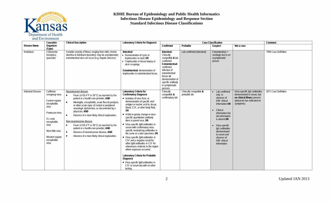
Disease Name
Causative Organism (Type)
Clinical Description Laboratory Criteria for Diagnosis Case Classification Comment Confirmed Probable Suspect Not a case
Amebiasis
Entamoeba histolytica, (parasite)
Variable severity of illness ranging from mild, chronic diarrhea to fulminant dysentery; may be asymptomatic; extraintestinal also can occur (e.g. hepatic abscess)
Intestinal: • Demonstration of cysts or
trophozoites in stool; OR • Trophozoites in tissue biopsy or
ulcer scrapings. Extraintestinal: demonstration of trophozoites in extraintestinal tissue.
Intestinal: Clinically compatible & lab confirmed Extraintestinal: confirmed infection of extraintestinal tissue; or demonstration of specific antibody in symptomatic persons
Lab confirmed (intestinal) Extraintestinal: + serologic test in an asymptomatic person
1990 Case Definition
Arboviral Disease California serogroup virus Eastern equine encephalitis virus Powassan virus St. Louis encephalitis virus West Nile virus Western equine encephalitis virus
Neuroinvasive disease • Fever (≥100.4°F or 38°C) as reported by the
patient or a health-care provider, AND • Meningitis, encephalitis, acute flaccid paralysis,
or other acute signs of central or peripheral neurologic dysfunction, as documented by a physician, AND
• Absence of a more likely clinical explanation.
Non-neuroinvasive disease • Fever (≥100.4°F or 38°C) as reported by the
patient or a health-care provider, AND • Absence of neuroinvasive disease, AND • Absence of a more likely clinical explanation.
Laboratory Criteria for Confirmatory Diagnosis • Isolation of virus from, or
demonstration of specific viral antigen or nucleic acid in, tissue, blood, CSF, or other body fluid, OR
• 4-fold or greate change in virus-specific quantitative antibody titers in paired sera, OR
• Virus-specific IgM antibodies in serum with confirmatory virus-specific neutralizing antibodies in the same or a later specimen, OR
• Virus-specific IgM antibodies in CSF and a negative result for other IgM antibodies in CSF for arboviruses endemic to the region where exposure occurred.
Laboratory Criteria for Probable Diagnosis • Virus-specific IgM antibodies in
CSF or serum but with no other testing
Clinically compatible & confirmatory lab
Clinically compatible & probable lab
• Lab confirmed only in absence of ANY clinical information OR
• Clinical information but lab information is absent OR
• Virus-specific
IgG antibodies demonstrated in serum and absence of ANY clinical information
Virus-specific IgG antibodies demonstrated in serum, but no clinical illness present (physician has indicated no symptoms)
2011 Case Definition
KDHE Bureau of Epidemiology and Public Health Informatics Infectious Disease Epidemiology and Response Section
Standard Infectious Disease Classifications
Updated JAN 2013
3
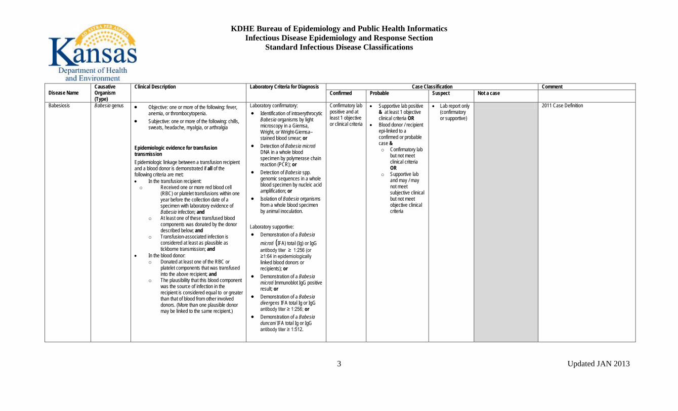
Disease Name
Causative Organism (Type)
Clinical Description Laboratory Criteria for Diagnosis Case Classification Comment Confirmed Probable Suspect Not a case
Babesiosis Babesia genus • Objective: one or more of the following: fever, anemia, or thrombocytopenia.
• Subjective: one or more of the following: chills, sweats, headache, myalgia, or arthralgia
Epidemiologic evidence for transfusion transmission Epidemiologic linkage between a transfusion recipient and a blood donor is demonstrated if all of the following criteria are met: • In the transfusion recipient: o Received one or more red blood cell
(RBC) or platelet transfusions within one year before the collection date of a specimen with laboratory evidence of Babesia infection; and
o At least one of these transfused blood components was donated by the donor described below; and
o Transfusion-associated infection is considered at least as plausible as tickborne transmission; and
• In the blood donor: o Donated at least one of the RBC or
platelet components that was transfused into the above recipient; and
o The plausibility that this blood component was the source of infection in the recipient is considered equal to or greater than that of blood from other involved donors. (More than one plausible donor may be linked to the same recipient.)
Laboratory confirmatory: • Identification of intraerythrocytic
Babesia organisms by light microscopy in a Giemsa, Wright, or Wright-Giemsa–stained blood smear; or
• Detection of Babesia microti DNA in a whole blood specimen by polymerase chain reaction (PCR); or
• Detection of Babesia spp. genomic sequences in a whole blood specimen by nucleic acid amplification; or
• Isolation of Babesia organisms from a whole blood specimen by animal inoculation.
Laboratory supportive: • Demonstration of a Babesia
microti (IFA) total (Ig) or IgG antibody titer ≥ 1:256 (or ≥1:64 in epidemiologically linked blood donors or recipients); or
• Demonstration of a Babesia microti Immunoblot IgG positive result; or
• Demonstration of a Babesia divergens IFA total Ig or IgG antibody titer ≥ 1:256; or
• Demonstration of a Babesia duncani IFA total Ig or IgG antibody titer ≥ 1:512.
Confirmatory lab positive and at least 1 objective or clinical criteria
• Supportive lab positive & at least 1 objective clinical criteria OR
• Blood donor / recipient epi-linked to a confirmed or probable case & o Confirmatory lab
but not meet clinical criteria OR
o Supportive lab and may / may not meet subjective clinical but not meet objective clinical criteria
• Lab report only (confirmatory or supportive)
2011 Case Definition
KDHE Bureau of Epidemiology and Public Health Informatics Infectious Disease Epidemiology and Response Section
Standard Infectious Disease Classifications
Updated JAN 2013
4
Disease Name
Causative Organism (Type)
Clinical Description Laboratory Criteria for Diagnosis Case Classification Comment Confirmed Probable Suspect Not a case
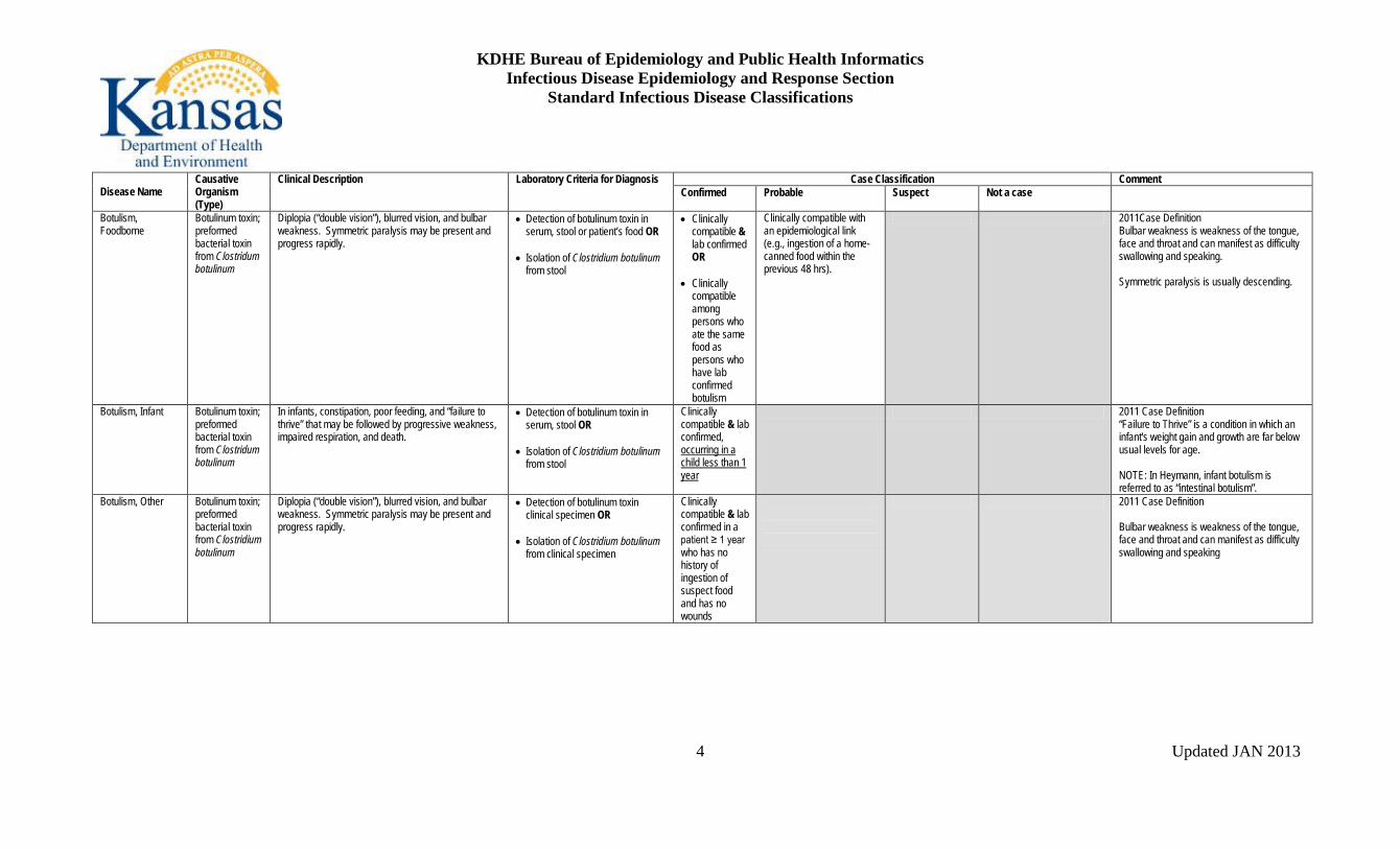
Botulism, Foodborne
Botulinum toxin; preformed bacterial toxin from Clostridum botulinum
Diplopia (“double vision”), blurred vision, and bulbar weakness. Symmetric paralysis may be present and progress rapidly.
• Detection of botulinum toxin in serum, stool or patient’s food OR
• Isolation of Clostridium botulinum
from stool
• Clinically compatible & lab confirmed OR
• Clinically
compatible among persons who ate the same food as persons who have lab confirmed botulism
Clinically compatible with an epidemiological link (e.g., ingestion of a home-canned food within the previous 48 hrs).
2011Case Definition Bulbar weakness is weakness of the tongue, face and throat and can manifest as difficulty swallowing and speaking. Symmetric paralysis is usually descending.
Botulism, Infant Botulinum toxin; preformed bacterial toxin from Clostridum botulinum
In infants, constipation, poor feeding, and “failure to thrive” that may be followed by progressive weakness, impaired respiration, and death.
• Detection of botulinum toxin in serum, stool OR
• Isolation of Clostridium botulinum
from stool
Clinically compatible & lab confirmed, occurring in a child less than 1 year
2011 Case Definition “Failure to Thrive” is a condition in which an infant's weight gain and growth are far below usual levels for age. NOTE: In Heymann, infant botulism is referred to as “intestinal botulism”.
Botulism, Other
Botulinum toxin; preformed bacterial toxin from Clostridium botulinum
Diplopia (“double vision”), blurred vision, and bulbar weakness. Symmetric paralysis may be present and progress rapidly.
• Detection of botulinum toxin clinical specimen OR
• Isolation of Clostridium botulinum
from clinical specimen
Clinically compatible & lab confirmed in a patient ≥ 1 year who has no history of ingestion of suspect food and has no wounds
2011 Case Definition Bulbar weakness is weakness of the tongue, face and throat and can manifest as difficulty swallowing and speaking

KDHE Bureau of Epidemiology and Public Health Informatics Infectious Disease Epidemiology and Response Section
Standard Infectious Disease Classifications
Updated JAN 2013
5
Disease Name
Causative Organism (Type)
Clinical Description Laboratory Criteria for Diagnosis Case Classification Comment Confirmed Probable Suspect Not a case
Botulism, Wound Botulinum toxin; preformed bacterial toxin from Clostridium botulinum
Diplopia (“double vision”), blurred vision, and bulbar weakness. Symmetric paralysis may be present and progress rapidly.
• Detection of botulinum toxin in serum OR
• Isolation of Clostridium botulinum
from wound
Clinically compatible & lab confirmed in a patient who has no suspected exposure to contaminated food & who has history of a fresh, contaminated wound during the 2 weeks before onset.
Clinically compatible in a patient who has no suspected exposure to contaminated food & who has either a history of a fresh, contaminated wound during the 2 weeks before onset of symptoms, or a history of injection drug use within the 2 weeks before onset of symptom
2011 Case Definition
Brucellosis Brucella spp. including B. melitensis, B. suis, B. abortus, and B. canis (bacteria)
Acute or insidious onset of fever & one or more of the following: night sweats, arthralgia, headache, fatigue, anorexia, myalgia, weight loss, arthritis/spondylitis, meningitis, or focal organ involvement (endocarditis, orchitis/epididymitis, hepatomegaly, splenomegaly).
Definitive • Culture+ OR • 4X change of agglutination titer in
paired sera OR Presumptive • Antibody titer of >160 in one or
more serum specimens) OR • PCR
Clinically compatible & definitive labs
Clinically compatible & • epi-linked to lab
confirmed case (human or animal) OR
• presumptive lab only
Positive definitive or presumptive lab results without any clinical information
2010 Case Definition
Campylobacter infection
Campylobacter spp. (bacteria)
Diarrheal illness
Confirmed: Isolation of Campylobacter spp. in a clinical specimen. Suspect: Detection of Campylobacter spp. in a clinical specimen using non-culture based laboratory methods
Case that meets the confirmed laboratory criteria for diagnosis
Clinically compatible & epi-linked to lab confirmed case
Case that meets the suspect laboratory criteria for diagnosis
2012 Case Definition The use of culture independent methods as standalone tests for the direct detection of Campylobacter in stool appears to be increasing. Data available about the performance characteristics of these assays indicates there is variability in the sensitivity, specificity and positive predictive value of these assays depending on the test (EIA test format -lateral flow or – microplate) and manufacturer. It is therefore useful to collect information on which type of EIA test and manufacturer are used to diagnose a case. Culture confirmation of culture independent (e.g. EIA) test positive specimens is ideal.
KDHE Bureau of Epidemiology and Public Health Informatics Infectious Disease Epidemiology and Response Section
Standard Infectious Disease Classifications
Updated JAN 2013
6
Disease Name
Causative Organism (Type)
Clinical Description Laboratory Criteria for Diagnosis Case Classification Comment Confirmed Probable Suspect Not a case
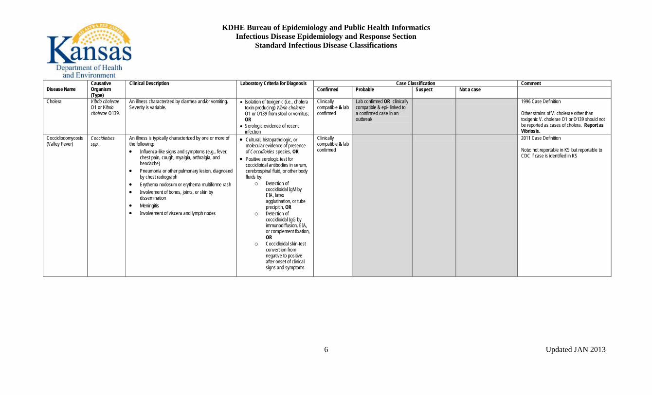
Cholera Vibrio cholerae O1 or Vibrio cholerae O139.
An illness characterized by diarrhea and/or vomiting. Severity is variable.
• Isolation of toxigenic (i.e., cholera toxin-producing) Vibrio cholerae O1 or O139 from stool or vomitus; OR
• Serologic evidence of recent infection
Clinically compatible & lab confirmed
Lab confirmed OR clinically compatible & epi- linked to a confirmed case in an outbreak
1996 Case Definition Other strains of V. cholerae other than toxigenic V. cholerae O1 or O139 should not be reported as cases of cholera. Report as Vibriosis.
Coccidiodomycosis (Valley Fever)
Coccidioises spp.
An illness is typically characterized by one or more of the following: • Influenza-like signs and symptoms (e.g., fever,
chest pain, cough, myalgia, arthralgia, and headache)
• Pneumonia or other pulmonary lesion, diagnosed by chest radiograph
• Erythema nodosum or erythema multiforme rash • Involvement of bones, joints, or skin by
dissemination • Meningitis • Involvement of viscera and lymph nodes
• Cultural, histopathologic, or molecular evidence of presence of Coccidioides species, OR
• Positive serologic test for coccidioidal antibodies in serum, cerebrospinal fluid, or other body fluids by:
o Detection of coccidioidal IgM by EIA, latex agglutination, or tube precipitin, OR
o Detection of coccidioidal IgG by immunodiffusion, EIA, or complement fixation, OR
o Coccidioidal skin-test conversion from negative to positive after onset of clinical signs and symptoms
Clinically compatible & lab confirmed
2011 Case Definition Note: not reportable in KS but reportable to CDC if case is identified in KS

KDHE Bureau of Epidemiology and Public Health Informatics Infectious Disease Epidemiology and Response Section
Standard Infectious Disease Classifications
Updated JAN 2013
7
Disease Name
Causative Organism (Type)
Clinical Description Laboratory Criteria for Diagnosis Case Classification Comment Confirmed Probable Suspect Not a case
Cryptosporidiosis (symptomatic &asymptomatic)
Cryptosporidium parvum (Parasite)
A gastrointestinal illness characterized by diarrhea and one or more of the following: diarrhea duration of 72 hours or more, abdominal cramping, vomiting, or anorexia.
Confirmed: Evidence of Cryptosporidium organisms or DNA in stool, intestinal fluid, tissue samples, biopsy specimens, or other biological sample by certain laboratory methods with a high positive predictive value (PPV), e.g., • direct fluorescent antibody [DFA]
test, • polymerase chain reaction [PCR], • enzyme immunoassay [EIA], or • light microscopy of stained
specimen.
Probable: The detection of Cryptosporidium antigen by a screening test method, such as immunochromatographic card/rapid card test; or a laboratory test of unknown method.
Diagnosed by lab confirmed criteria
Clinically compatible & epi-linked to confirmed case OR Diagnosed by lab probable criteria
2012 Case Definition Comment: Persons who have a diarrheal illness and are epidemiologically linked to a probable case because that individual was only diagnosed with cryptosporidiosis by an immunocard/rapid test/ or unknown test method cannot be classified as probable cases. These epi-links can be considered suspect cases only.
Cyclosporiasis, (symptomatic & asymptomatic)
Cyclospora cayetanensis (Parasite)
Watery diarrhea, loss of appetite, weight loss, abdominal bloating and cramping, increased flatus, nausea, fatigue, low-grade fever, vomiting
Detection of Cyclospora organisms or DNA in stool, intestinal fluid/aspirate, or intestinal biopsy specimens
Clinically compatible & lab confirmed
Clinically compatible & epi-linked to lab confirmed case
2010 Case Definition
KDHE Bureau of Epidemiology and Public Health Informatics Infectious Disease Epidemiology and Response Section
Standard Infectious Disease Classifications
Updated JAN 2013
8
Disease Name
Causative Organism (Type)
Clinical Description Laboratory Criteria for Diagnosis Case Classification Comment Confirmed Probable Suspect Not a case
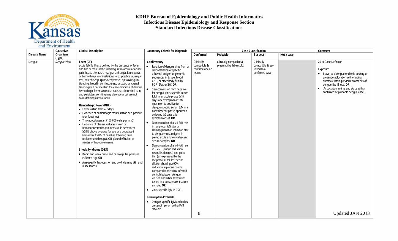
Dengue Dengue Virus Fever (DF) acute febrile illness defined by the presence of fever and two or more of the following, retro-orbital or ocular pain, headache, rash, myalgia, arthralgia, leukopenia, or hemorrhagic manifestations (e.g., positive tourniquet test, petechiae; purpura/ecchymosis; epistaxis; gum bleeding; blood in vomitus, urine, or stool; or vaginal bleeding) but not meeting the case definition of dengue hemorrhagic fever. Anorexia, nausea, abdominal pain, and persistent vomiting may also occur but are not case-defining criteria for DF Hemorrhagic Fever (DHF) • Fever lasting from 2-7 days • Evidence of hemorrhagic manifestation or a positive
tourniquet test • Thrombocytopenia (≤100,000 cells per mm3) • Evidence of plasma leakage shown by
hemoconcentration (an increase in hematocrit ≥20% above average for age or a decrease in hematocrit ≥20% of baseline following fluid replacement therapy), OR pleural effusion, or ascites or hypoproteinemia
Shock Syndrome (DSS) • Rapid and weak pulse and narrow pulse pressure
(<20mm Hg), OR • Age-specific hypotension and cold, clammy skin and
restlessness
Confirmatory • Isolation of dengue virus from or
demonstration of specific arboviral antigen or genomic sequences in tissue, blood, CSF, or other body fluid by PCR, IFA, or IHC OR
• Seroconversion from negative for dengue virus-specific serum IgM in an acute phase (≤ 5 days after symptom onset) specimen to positive for dengue-specific serum IgM in a convalescent-phase specimen collected ≥5 days after symptom onset, OR
• Demonstration of a ≥4-fold rise in reciprocal IgG titer or Hemagglutination inhibition titer to dengue virus antigens in paired acute and convalescent serum samples, OR
• Demonstration of a ≥4-fold rise in PRNT (plaque reduction neutralization test) end point titer (as expressed by the reciprocal of the last serum dilution showing a 90% reduction in plaque counts compared to the virus infected control) between dengue viruses and other flaviviruses tested in a convalescent serum sample, OR
• Virus-specific IgM in CSF.
Presumptive/Probable • Dengue-specific IgM antibodies
present in serum with a P/N ratio ≥2.
Clinically compatible & confirmatory lab results
Clinically compatible & presumptive lab results
Clinically compatible & epi-linked to a confirmed case
2010 Case Definition Exposure • Travel to a dengue endemic country or
presence at location with ongoing outbreak within previous two weeks of dengue-like illness, OR
• Association in time and place with a confirmed or probable dengue case.

KDHE Bureau of Epidemiology and Public Health Informatics Infectious Disease Epidemiology and Response Section
Standard Infectious Disease Classifications
Updated JAN 2013
9
Disease Name
Causative Organism (Type)
Clinical Description Laboratory Criteria for Diagnosis Case Classification Comment Confirmed Probable Suspect Not a case
Diphtheria Corynebacterium diphtheriae (Bacteria)
Upper respiratory tract illness characterized by sore throat, low-grade fever, and adherent membrane of the tonsil(s), pharynx, and/or nose
• Isolation of Corynebacterium diphtheriae, OR,
• Histopathologic diagnosis of diphtheria
Clinically compatible &
• Lab confirmed OR
• Epi- linked to lab confirmed case
Clinically compatible case that is not lab confirmed & not epi- linked to a lab confirmed case
Lab confirmed only 1995 Case Definition Do not report cutaneous diphtheria. Respiratory disease caused by nontoxigenic C. diphtheriae should be reported.
Anaplasmosis, human granulocytic (HGE) Ehrlichiosis, human monocytic (HME)
Anaplasma phagocytophilum Ehrlichia chaffeensis
Clinical presentation Acute onset of fever and one or more of the following symptoms or signs: headache, myalgia, malaise, anemia, leukopenia, thrombocytopenia, or elevated hepatic transaminases. Nausea, vomiting, or rash may be present in some cases. Intracytoplasmic bacterial aggregates (morulae) may be visible in the leukocytes of some patients. Clinical evidence Any reported fever and one or more of the following: headache, myalgia, anemia, leukopenia, thrombocytopenia, or any hepatic transaminase elevation.
Lab Confirmed • 4X change of IgG by IFA in paired
sera (specimens collected in 1st week of illness and 2-4 weeks later) OR
• PCR + OR • Immunostain of antigen OR • Identification of morulae in
leukocytes, and a positive IFA titer
• Culture + Lab Supportive • Serological evidence of elevated
IgG or IgM antibody by IFA, ELISA, dot-ELISA, or assays in other formats (CDC uses an IFA IgG cutoff of ≥1:64 and does not use IgM test results independently as diagnostic support criteria.), or • Identification of morulae in the
cytoplasm of monocytes or macrophages by microscopic examination
Clinically compatible & lab confirmed
Clinically compatible & supportive lab
Lab confirmed only 2008 Case Definition

KDHE Bureau of Epidemiology and Public Health Informatics Infectious Disease Epidemiology and Response Section
Standard Infectious Disease Classifications
Updated JAN 2013
10
Disease Name
Causative Organism (Type)
Clinical Description Laboratory Criteria for Diagnosis Case Classification Comment Confirmed Probable Suspect Not a case
Ehrlichiosis, human other
Ehrlichia ewingii Clinical presentation Acute onset of fever and one or more of the following symptoms or signs: headache, myalgia, malaise, anemia, leukopenia, thrombocytopenia, or elevated hepatic transaminases. Nausea, vomiting, or rash may be present in some cases. Intracytoplasmic bacterial aggregates (morulae) may be visible in the leukocytes of some patients. Clinical evidence Any reported fever and one or more of the following: headache, myalgia, anemia, leukopenia, thrombocytopenia, or any hepatic transaminase elevation.
• PCR Clinically compatible & lab confirmed
2008 Case Definition
Escherichia coli enteric infection
Escherichia coli O157:H7
Refer to STEC information Refer to STEC information Refer to STEC information
Refer to STEC information Refer to STEC information
Refer to STEC information
Giardiasis Giardia lamblia (parasite)
diarrhea, abdominal cramps, bloating, weight loss, or malabsorption
Detection of Giardia organisms, antigen, or DNA in stool, intestinal fluid, tissue samples, biopsy specimens or other biological sample
Clinically compatible & lab confirmed
Clinically compatible & epi-linked to lab confirmed case
Lab confirmed only 2011 Case Definition
Haemophilus influenzae (invasive disease)
Haemophilus influenzae (bacteria)
Invasive disease caused by Haemophilus influenzae may produce any of several clinical syndromes, including meningitis, bacteremia, epiglottitis, or pneumonia
• Isolation of H. influenzae from a normally sterile site
Clinically compatible & lab confirmed
Meningitis with detection of H. influenzae type b antigen in CSF
Lab confirmed only 1997 Case Definition Positive antigen test results from urine or serum samples are unreliable for diagnosis of H. influenzae disease
KDHE Bureau of Epidemiology and Public Health Informatics Infectious Disease Epidemiology and Response Section
Standard Infectious Disease Classifications
Updated JAN 2013
11
Disease Name
Causative Organism (Type)
Clinical Description Laboratory Criteria for Diagnosis Case Classification Comment Confirmed Probable Suspect Not a case
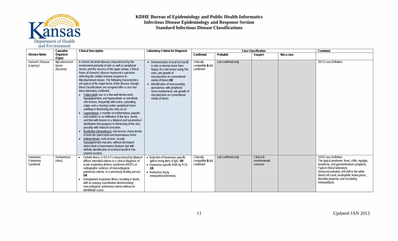
Hansen’s Disease (Leprosy)
Mycobacterium leprae (Bacteria)
A chronic bacterial disease characterized by the involvement primarily of skin as well as peripheral nerves and the mucosa of the upper airway. Clinical forms of Hansen's disease represent a spectrum reflecting the cellular immune response to Mycobacterium leprae. The following characteristics are typical of the major forms of the disease, though these classifications are assigned after a case has been laboratory confirmed. • Tuberculoid: one or a few well-demarcated,
hypopigmented, and hypoesthetic or anesthetic skin lesions, frequently with active, spreading edges and a clearing center; peripheral nerve swelling or thickening also may occur
• Lepromatous: a number of erythematous papules and nodules or an infiltration of the face, hands, and feet with lesions in a bilateral and symmetrical distribution that progress to thickening of the skin, possibly with reduced sensation.
• Borderline (dimorphous): skin lesions characteristic of both the tuberculoid and lepromatous forms
• Indeterminate: early lesions, usually hypopigmented macules, without developed tuberculoid or lepromatous features but with definite identification of acid-fast bacilli in Fite stained sections
• Demonstration of acid fast bacilli in skin or dermal nerve from biopsy of a skin lesion using Fite stain, w/o growth of mycobacteria on conventional media (if done) OR
• Identification of noncaseating granulomas with peripheral nerve involvement, w/o growth of mycobacteria on conventional media (if done)
Clinically compatible & lab confirmed
Lab confirmed only 2013 Case Definition
Hantavirus Pulmonary Syndrome
Hantaviruses (virus)
• Frebrile illness (>101.0 F) characterized by bilateral diffuse interstitial edema or a clinical diagnosis of acute respiratory distress syndrome (ARDS) or radiographic evidence of noncardiogenic pulmonary edema, in a previously healthy person; OR
• Unexplained respiratory illness resulting in death, with an autopsy examination demonstrating noncardiogenic pulmonary edema without an identifiable cause.
• Detection of hantavirus-specific IgM or rising titers of IgG: OR
• Hantavirus-specific RNA by PCR; OR
• Hantavirus Ag by immunohistochemistry
Clinically compatible & lab confirmed
Lab confirmed only Clinical & environmental concerns
2010 Case Definition The typical prodrome: fever, chills, myalgia, headache, and gastrointestinal symptoms. Typical clinical laboratory: hemoconcentration, left shift in the white blood cell count, neutrophilic leukocytosis, thrombocytopenia, and circulating immunoblasts.

KDHE Bureau of Epidemiology and Public Health Informatics Infectious Disease Epidemiology and Response Section
Standard Infectious Disease Classifications
Updated JAN 2013
12
Disease Name
Causative Organism (Type)
Clinical Description Laboratory Criteria for Diagnosis Case Classification Comment Confirmed Probable Suspect Not a case
Hemolytic Uremic Syndrome, post-diarrheal
Acute onset of microangiopathic hemolytic anemia, renal injury, and low platelet count. Thrombotic thrombocytopenic purpura (TTP) is also characterized by these features, but can include central nervous system involvement and fever and may have a more gradual onset. Most cases of HUS (but few cases of TTP) occur after an acute gastrointestinal illness (usually diarrheal).
• Anemia with microangiopathic changes (i.e., schistocytes, burr cells, or helmet cells) on peripheral blood smear; AND
• Renal injury evidenced by either hematuria, proteinuria, or elevated creatinine level (i.e., greater than or equal to 1.0 mg/dl in a child aged less than 13 years or greater than or equal to 1.5 mg/dl in an adult, or greater than or equal to 50% increase over baseline)
NOTE: A low platelet count can usually, but not always, be detected early in the illness, but it may then become normal or even high. If a platelet count obtained within 7 days after onset of the acute gastrointestinal illness is not less than 150,000/mm3, other diagnoses should be considered.
• Acute illness diagnosed as HUS or TTP, that is lab confirmed and
• Began within 3 weeks of onset of an acute or bloody diarrhea
• Acute illness diagnosed as HUS or TTP that is lab confirmed, but circumstances of onset have not been determined; OR
• Acute illness diagnosed as HUS or TTP, with onset within 3 weeks of onset of acute or bloody diarrhea, that meets lab criteria except that microangiopathic changes are not confirmed
1996 Case Definition Some investigators consider HUS and TTP to be part of a continuum of disease. Therefore, criteria for diagnosing TTP on the basis of central nervous system involvement and fever are not provided because cases diagnosed clinically as postdiarrheal TTP should also meet the criteria for HUS. These cases are reported as postdiarrheal HUS.
Hepatitis A Hepatitis A virus An acute illness with a discrete onset of any sign or symptom consistent with acute viral hepatitis (e.g., fever, headache, malaise, anorexia, nausea, vomiting, diarrhea, and abdominal pain), and either • jaundice, or • elevated serum aminotransferase (ALT or AST)
levels.
• IgM anti-HAV Clinically compatible & • lab
confirmed OR • epi-linked to
lab confirmed hepatitis A case
Lab confirmed only Total anti-HAV positive
2012 Case Definition

KDHE Bureau of Epidemiology and Public Health Informatics Infectious Disease Epidemiology and Response Section
Standard Infectious Disease Classifications
Updated JAN 2013
13
Disease Name
Causative Organism (Type)
Clinical Description Laboratory Criteria for Diagnosis Case Classification Comment Confirmed Probable Suspect Not a case
Hepatitis B, acute Hepatitis B virus (virus)
An acute illness with a discrete onset of any sign or symptom* consistent with acute viral hepatitis (e.g., fever, headache, malaise, anorexia, nausea, vomiting, diarrhea, and abdominal pain), and either • jaundice, or • elevated serum alanine aminotransferase (ALT)
levels >100 IU/L. *A documented negative HBsAg laboratory test result within 6 months prior to a positive test (either HBsAg, HBeAg, or HBV NAT including genotype) result does not require an acute clinical presentation to meet the surveillance case definition.
• HBsAg positive AND • IgM anti-HBc positive (if done)
Clinically compatible & lab confirmed And is not known to have chronic hepatitis B
IgM anti-HBc positive & missing / incomplete clinical information
2012 Case Definition
Hepatitis B, chronic Hepatitis B virus (virus)
No symptoms are required. Persons with chronic HBV infection may have no evidence of liver disease or may have a spectrum of disease ranging from chronic hepatitis to cirrhosis or liver cancer.
• IgM anti-HBc negative AND a positive result on one of the following tests: HBsAg, HBeAg, or HBV DNA (including qualitative, quantitative and genotype testing)
OR • HBsAg positive or HBV DNA
positive (including qualitative, quantitative and genotype testing) or HBeAg positive two times at least 6 months apart (Any combination of these tests performed 6 months apart is acceptable.)
Lab confirmed Case with a single HBsAg positive or HBV DNA positive (including qualitative, quantitative and genotype testing) or HBeAg positive lab result & does not meet the case definition for acute
Lab testing not meeting criteria for either confirmed or probable classifications
Labs indicating a past infection w/ recovery Anti-HBc total (only), Anti-HBs (only), or both tests .
2012 Case Definition Multiple lab tests indicative of chronic HBV infection may be performed simultaneously on the same patient specimen as part of a “hepatitis panel”. Testing performed in this manner may lead to seemingly discordant results, e.g. HBsAg-negative AND HBV DNA-positive. Any positive result among the 3 laboratory tests mentioned is acceptable, regardless of other results. Negative HBeAg results & HBV DNA levels below positive cutoff level do not confirm the absence of HBV infection.
Hepatitis B, perinatal infection
Hepatitis B virus (virus)
Perinatal hepatitis B in the newborn may range from asymptomatic to fulminant hepatitis
Hepatitis B surface antigen (HBsAg) positive
HBsAg positivity in any infant aged >1-24 months who was born in the United States or in U.S. territories to an HBsAg-positive mother
1995 Case Definition HBsAg + infants 24 months and younger and NOT born in US should be classified as HBV chronic

KDHE Bureau of Epidemiology and Public Health Informatics Infectious Disease Epidemiology and Response Section
Standard Infectious Disease Classifications
Updated JAN 2013
14
Disease Name
Causative Organism (Type)
Clinical Description Laboratory Criteria for Diagnosis Case Classification Comment Confirmed Probable Suspect Not a case
Hepatitis C, acute Hepatitis C virus (virus)
An acute illness with a discrete onset of any sign or symptom* consistent with acute viral hepatitis (e.g., fever, headache, malaise, anorexia, nausea, vomiting, diarrhea, and abdominal pain), and either • jaundice, or • elevated serum alanine aminotransferase (ALT)
levels >400 IU/L. *A documented negative HCV antibody laboratory test result followed within 6 months by a positive test (as described in the laboratory criteria for diagnosis) result does not require an acute clinical presentation to meet the surveillance case definition.
One or more of the following 3 criteria: • Anti-HCV screening-test-positive
with a signal to cut-off ratio predictive of a true positive as determined for the particular assay as defined by CDC. OR
• HCV RIBA positive OR • NAT positive for HCV RNA
(including qualitative, quantitative or genotype testing) AND, if done meets the following 2 criteria
• IgM antibody to hepatitis A virus (IgM anti-HAV) negative AND
• IgM antibody to hepatitis B core antigen (IgM anti-HBc) negative
Meets the clinical case definition, is laboratory confirmed, and is not known to have chronic hepatitis C
Lab confirmed and only partially meeting the clinical criteria
2012 Case Definition URL for the signal to cut-off ratios: http://www.cdc.gov/ncidod/diseases/hepatitis/c/sc_ratios.htm
Hepatitis C, chronic Hepatitis C virus (virus)
No symptoms are required. Most HCV-infected persons are asymptomatic; however, many have chronic liver disease, which can range from mild to severe.
One or more of the following 3 criteria (except in persons less than 18 months of age, for whom only criteria 3 would meet the case classification criteria): • Antibodies to hepatitis C virus
(anti-HCV) screening-test-positive with a signal to cut-off ratio predictive of a true positive as determined for the particular assay as defined by CDC. OR
• Hepatitis C Virus Recombinant Immunoblot Assay (HCV RIBA) positive, OR
• Nucleic Acid Test (NAT) for HCV RNA positive (including qualitative, quantitative or genotype testing)
Lab confirmed & does not meet the case definition for acute hepatitis C.
Case not meet the case definition for acute hepatitis C, is anti-HCV positive (repeat reactive) by EIA, and has alanine aminotransferase (ALT or SGPT) values above the upper limit of normal, but the anti-HCV EIA result has not been verified by an additional more specific assay or the signal to cut-off ratio is unknown.
Anti- HCV+ only 2012 Case Definition URL for the signal to cut-off ratios: http://www.cdc.gov/ncidod/diseases/hepatitis/c/sc_ratios.htm

KDHE Bureau of Epidemiology and Public Health Informatics Infectious Disease Epidemiology and Response Section
Standard Infectious Disease Classifications
Updated JAN 2013
15
Disease Name
Causative Organism (Type)
Clinical Description Laboratory Criteria for Diagnosis Case Classification Comment Confirmed Probable Suspect Not a case
Influenza deaths in children <18 years of age
Influenza virus An influenza-associated death is defined for surveillance purposes as a death resulting from a clinically compatible illness that was confirmed to be influenza by an appropriate laboratory or rapid diagnostic test. There should be no period of complete recovery between the illness and death.
Pre- or post-mortem clinical specimens • Influenza virus isolation in
tissue cell culture from respiratory specimens OR;
• (RT-PCR testing of respiratory specimens OR;
• IFA staining (direct or indirect) of respiratory specimens;
• Rapid influenza diagnostic testing of respiratory specimens;
• IHC staining for influenza viral antigens in respiratory tract tissue from autopsy specimens;
• Four-fold rise in influenza hemagglutination inhibition (HI) antibody titer in paired acute and convalescent sera
A death meeting the clinical case definition that is laboratory confirmed.
2004 Case Definition A death should not be reported if: • There is no laboratory confirmation of
influenza virus infection. • The influenza illness is followed by full
recovery to baseline health status prior to death.
• The death occurs in a person 18 years or older.
• After review and consultation there is an alternative agreed upon cause of death.

KDHE Bureau of Epidemiology and Public Health Informatics Infectious Disease Epidemiology and Response Section
Standard Infectious Disease Classifications
Updated JAN 2013
16
Disease Name
Causative Organism (Type)
Clinical Description Laboratory Criteria for Diagnosis Case Classification Comment Confirmed Probable Suspect Not a case
Legionellosis
Legionella pneumophila (Bacteria)
Legionnaires’ disease: Fever, myaligia, cough, and clinical/radiographic pneumonia Pontiac Fever: Milder illness without pneumonia (also called “Nonpneumonic Legionellosis”)
Laboratory Criteria for Suspect Diagnosis: • 4X or > rise in antibody titer to
specific species or serogroups of Legionella other than L. pneumophila serogroup 1 (e.g., L. micdadei, L. pneumophila serogroup 6) OR
• 4X or > rise in antibody titer to multiple species of Legionella using pooled antigen and validated reagents OR
• Detection of specific Legionella antigen or staining of the organism in respiratory secretions, lung tissue, or pleural fluid by DFA staining, IHC or other similar method, using validated reagents OR
• Detection of Legionella species by a nucleic acid assay
Laboratory Criteria for Confirmatory Diagnosis: • Isolation of any Legionella
organism from respiratory secretions, lung tissue, pleural fluid, or other normally sterile fluid OR
• Detection of Legionella pneumophila serogroup 1 antigen in urine using validated reagents OR
• 4X or > rise in specific serum antibody titer to Legionella pneumophila serogroup
Clinically compatible & meets at least 1 of the confirmatory lab criteria.
Clinically compatible case that meets at least one of the suspect laboratory criteria
Lab confirmed only. *Enter into the surveillance system & request follow-up
2005 Case Definition Travel associated: A case that has a history of spending at least one night away from home, either in the same country of residence or abroad, in the 10 days before onset of illness
KDHE Bureau of Epidemiology and Public Health Informatics Infectious Disease Epidemiology and Response Section
Standard Infectious Disease Classifications
Updated JAN 2013
17
Disease Name
Causative Organism (Type)
Clinical Description Laboratory Criteria for Diagnosis Case Classification Comment Confirmed Probable Suspect Not a case
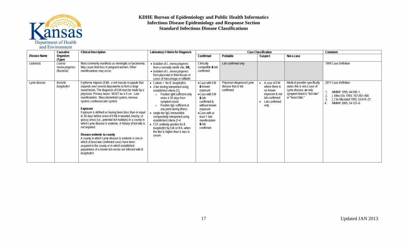
Listeriosis Listeria monocytogenes (Bacteria)
Most commonly manifests as meningitis or bacteremia. May cause fetal loss in pregnant women. Other manifestations may occur.
• Isolation of L. monocytogenes from a normally sterile site, OR,
• Isolation of L. monocytogenes from placental or fetal tissues in cases of miscarriage or stillbirth
Clinically compatible & lab confirmed
Lab confirmed only 1999 Case Definition
Lyme disease
Borrelia burgdorferi
Erythema migrans (EM) - a red macule or papule that expands over several days/weeks to form a large round lesion. The diagnosis of EM must be made by a physician. Primary lesion MUST be ≥ 5 cm. Late manifestation: Musculoskeletal system, nervous system, cardiovascular system. Exposure Exposure is defined as having been (less than or equal to 30 days before onset of EM) in wooded, brushy, or grassy areas (i.e., potential tick habitats) in a county in which Lyme disease is endemic. A history of tick bite is not required. Disease endemic to county A county in which Lyme disease is endemic is one in which at least two confirmed cases have been acquired in the county or in which established populations of a known tick vector are infected with B. burgdorferi.
• Culture + for B. burgdorferi, • 2-tier testing interpreted using
established criteria [1], o Positive IgM sufficient only
when ≤ 30 days from symptom onset
o Positive IgG sufficient at any point during illness
• single-tier IgG immunoblot seropositivity interpreted using established criteria [1-4
• CSF antibody positive for B. burgdorferi by EIA or IFA, when the titer is higher than is was in serum
• Case with EM & known exposure • Case with EM
& lab confirmed & without known exposure • Case with at
least 1 late manifestation & lab confirmed
Physician-diagnosed Lyme disease that is lab confirmed
• A case of EM where there is no known exposure & not lab confirmed
• Lab confirmed only
Medical provider specifically states this is not a case of Lyme disease, or only symptom listed is "tick bite" or "insect bite."
2011 Case Definition
1. MMWR 1995; 44:590–1. 2. J Infect Dis 1993; 167:392–400. 3. J Clin Microbiol 1995; 33:419–27. 4. MMWR 2005; 54:125–6.

KDHE Bureau of Epidemiology and Public Health Informatics Infectious Disease Epidemiology and Response Section
Standard Infectious Disease Classifications
Updated JAN 2013
18
Disease Name
Causative Organism (Type)
Clinical Description Laboratory Criteria for Diagnosis Case Classification Comment Confirmed Probable Suspect Not a case
Malaria Plasmodium spp.
Acute or subacute febrile disease, usually associated with episodes of chills and fever every 2-3days, persistent headache, muscle ache, weakness, vomiting or diarrhea, separated by afebrile periods. May progress to jaundice, shock, renal failure, coma, and death.
• Detection of circulating malaria-specific antigens using rapid diagnostic test (RDT), OR
• Detection of species specific parasite DNA in a sample of peripheral blood by PCR, OR
• Detection of malaria parasites in thick or thin peripheral blood films
• Detection & specific identification of malaria parasites by microscopy on blood films in a lab with appropriate expertise OR
• Detection of Plasmodium species by nucleic acid test
in any person (symptomatic or asymptomatic) diagnosed in the US, regardless of whether the person experienced previous episodes of malaria while outside the country
Clinical and travel to malaria endemic area
Detection of Plasmodium species by rapid diagnostic antigen testing without confirmation by microscopy or nucleic acid testing in any person
2010 Case Definition A subsequent attack experienced by the same person but caused by different Plasmodium species is counted as an additional case. Cases also are classified according to WHO categories: • Autochthonous: o Indigenous or Introduced • Imported: • Induced • Relapsing • Cryptic

KDHE Bureau of Epidemiology and Public Health Informatics Infectious Disease Epidemiology and Response Section
Standard Infectious Disease Classifications
Updated JAN 2013
19
Disease Name
Causative Organism (Type)
Clinical Description Laboratory Criteria for Diagnosis Case Classification Comment Confirmed Probable Suspect Not a case
Measles (Rubeola) Measles virus (virus)
Illness characterized by ALL of the following: • Generalized rash lasting ≥3 days & • Temperature ≥101.0°F (38.3°C) & • Cough, coryza, or conjunctivitis
• Isolation of measles virus from a clinical specimen, OR
• Detection of measles-virus-specific nucleic acid by PCR, OR
• IgG seroconversion or a significant rise in measles IgG using any evaluated and validated method
• Positive measles IgM
Acute febrile rash illness (fever does not need to reach ≥101.0°F (38.3°C) ) & • Lab
confirmed OR • Epi linked to
a confirmed case.
Meets clinical description with • No epi linkage to lab-
confirmed case; & • Noncontributory or no
measles lab testing
Acute illness with fever ≥101.0°F (38.3°C) & generalized maculopaular rash & no other diagnosis
2013Case Definition • Internationally imported: exposure to
measles virus outside the US as evidenced by at least some of the exposure period (7–21 days before rash onset) occurring outside the US & rash onset occurring within 21 days of entering the US and there is no known exposure to measles in the US during that time.
• U.S.-acquired case o Import-linked case o Imported-virus case o Endemic case o Unknown source case
Meningoccemia Neisseria meningitidis
Meningococcal disease manifests most commonly as meningitis and/or meningococcemia that may progress rapidly to purpura fulminans, shock, and death. However, other manifestations might be observed.
Confirmed: • Isolation of Neisseria meningitides
from normally sterile site or skin scrapings of purpuric lesions
Probable: • PCR+ OR • Antigen +
• Formalin-fixed tissue by IHC OR
• CSF by latex agglutination Suspect: • Gram negative diplococci from
normally sterile site OR • Clinical purpura w/o blood culture
positive
Llab confirmed Probable lab positive • Clinical purpura fulminans w/o blood culture + OR
• Clinically compatible and suspect lab positive
2010 Case Definition

KDHE Bureau of Epidemiology and Public Health Informatics Infectious Disease Epidemiology and Response Section
Standard Infectious Disease Classifications
Updated JAN 2013
20
Disease Name
Causative Organism (Type)
Clinical Description Laboratory Criteria for Diagnosis Case Classification Comment Confirmed Probable Suspect Not a case
Mumps Mumps virus, Paramyxoviridae family (similar to Parainfluenza viruses)
Clinical evidence for confirmed, probable and suspect: • Acute parotitis or other salivary gland swelling
lasting at least 2 days • Orchitis • Oophoritis Additional clinical evidence for confirmed: • Aseptic meningitis • Encephalitis • Hearing loss • Mastitis • Pancreatitis
Confirmed: • Isolation of mumps virus in cell
culture from a clinical specimen OR
• Detection of mumps virus with RT-PCR
Probable: • Positive test for serum anti-mumps
IgM antibody
• Lab confirmed by culture or RT-PCR and
• Acute illness characterized by any of the clinical evidence
Acute illness characterized by the clinical evidence for a probable case AND EITHER • Mumps IgM positive OR • Epi-link to another
confirmed or probable cases or link to group/community during an outbreak
• Clinically compatible unexplained by another more likely diagnosis, OR
• Lab confirmed with no mumps clinical symptoms (with or without epi-link to confirmed / probable case)
2012 Case Definition With previous contact with mumps virus either through vaccination (particularly with 2 doses) or natural infection, serum mumps IgM test results may be negative; IgG test results may be positive at initial blood draw; and viral detection in RT-PCR or culture may have low yield if the buccal swab is collected too long after parotitis onset. Therefore, mumps cases should not be ruled out by negative laboratory results. Serologic tests should be interpreted with caution, as false positive and false negative results are possible with IgM tests.
Pertussis Bordetella pertussis (Bacteria)
A cough illness lasting at least 2 weeks with one of the following: • paroxysms of coughing, OR • inspiratory "whoop," OR • post-tussive vomiting, without other apparent cause
(as reported by a health professional)
• Isolation of Bordetella pertussis from clinical specimen OR
• Positive polymerase chain reaction (PCR) for B. pertussis
• Case that is culture + & with cough illness of any duration; OR
• Clinically compatible and PCR+ OR
• Clinically compatible & epi-linked to a case either culture + or PCR +
Clinically compatible & not lab confirmed & not epidemiologically linked to a lab-confirmed case
PCR+ but does NOT meet the clinical case definition PCR- but does NOT meet the clinical case definition
2010 Case Definition DFA testing of nasopharyngeal secretions should not be relied for lab confirmation. Serologic testing is available but is not standardized and, therefore, should not be lab confirmation. Separate laboratory tests within 3 months should be considered part of the same event.

KDHE Bureau of Epidemiology and Public Health Informatics Infectious Disease Epidemiology and Response Section
Standard Infectious Disease Classifications
Updated JAN 2013
21
Disease Name
Causative Organism (Type)
Clinical Description Laboratory Criteria for Diagnosis Case Classification Comment Confirmed Probable Suspect Not a case
Plague Yersinia pestis (Bacteria)
Fever, chills, headache, malaise, prostration, leukocytosis that manifests in one or more of the following principal clinical forms: • Regional lymphadenitis (bubonic plague) • Septicemia without an evident bubo (septicemic
plague) • Plague pneumonia, resulting from hematogenous
spread in bubonic or septicemic cases (secondary pneumonic plague) or inhalation of infectious droplets (primary pneumonic plague)
• Pharyngitis and cervical lymphadenitis resulting from exposure to larger infectious droplets or ingestion of infected tissues (pharyngeal plague)
Confirmed Lab Criteria for Diagnosis • Isolation of Y. pestis from a
clinical specimen OR • ≥4 time change in serum Ab titer
to Y. pestis F1 (fraction 1) Ag Presumptive Lab Criteria for Diagnosis: • Elevated serum Ab titer(s) to
Yersinia pestis fraction 1 (F1) Ag in patients with no history of plague vaccination; OR
• Detection of F1 Ag in a clinical specimen
Clinically compatible & lab confirmed
Clinically compatible case with presumptive laboratory results
Clinically compatible case without presumptive or confirmatory laboratory results
1996 Case Definition
Poliovirus infection, nonparalytic
Poliovirus Most poliovirus infections are asymptomatic or cause mild febrile disease. Poliovirus infections occasionally cause aseptic meningitis and one out of 200 infections from poliovirus type 1 results in paralytic poliomyelitis, characterized by acute onset of flaccid paralysis that is typically asymmetric and associated with a prodromal fever.
Poliovirus isolate identified in an appropriate clinical specimen (e.g., stool, cerebrospinal fluid, oropharyngeal secretions), with confirmatory typing and sequencing performed by the CDC Poliovirus Laboratory, as needed.
Lab confirmed Clinically compatible case 2007 Case Definition Applies only to poliovirus infections found in asymptomatic persons or those with mild, nonparalytic disease. Isolation of polioviruses from persons with acute paralytic poliomyelitis should continue to be reported as “paralytic poliomyelitis.”
Poliomyelitis, paralytic
Poliovirus (virus)
Acute onset of a flaccid paralysis of one or more limbs with decreased or absent tendon reflexes in the affected limbs, without other apparent cause, and without sensory or cognitive loss
Clinically compatible & has a neurologic deficit 60 days after onset of initial symptoms, has died, or has unknown follow-up status
Clinically compatible 1997 Case Definition All suspected cases of paralytic poliomyelitis are reviewed by a panel of expert consultants before final classification occurs. Confirmed cases are then further classified based on epidemiologic & lab criteria.

KDHE Bureau of Epidemiology and Public Health Informatics Infectious Disease Epidemiology and Response Section
Standard Infectious Disease Classifications
Updated JAN 2013
22
Disease Name
Causative Organism (Type)
Clinical Description Laboratory Criteria for Diagnosis Case Classification Comment Confirmed Probable Suspect Not a case
Psittacosis / Ornithosis
Chlamydia psittaci (Bacteria)
Fever, chills, headache, myalgia, and a dry cough with pneumonia often evident on chest x-ray. Severe pneumonia requiring intensive-care support, endocarditis, hepatitis, and neurologic complications occasionally occur.
• Isolation of C. psittaci from respiratory specimens or blood,OR
• ≥ 4 X increase in antibody IgG against C. psittaci by complement fixation (CF) or microimmunofluorescence (MIF) between paired acute- and convalescent-phase serum specimens obtained at least 2-4 weeks apart , OR
• Supportive serology (IgM) of ≥ 32 in at least one serum specimen obtained after onset of symptoms, OR
• Detection of C. psittaci DNA in a respiratory specimen by PCR
Clinically compatible & lab confirmed by one of the following • Isolation of C.
psittaci from respiratory specimens or blood, OR
≥ 4 X increase in antibody (IgG) against C. psittaci by CF or MIF between paired acute- & convalescent-phase serum specimens obtained at least 2-4 weeks apart.
Clinically compatible & lab confirmed by one of the following: • Supportive serology
(IgM) of ≥ 32 in at least one serum specimen obtained after onset of symptoms, OR
• Detection of C. psittaci DNA in a respiratory specimen by PCR
2010 Case Definition Serologic findings by complement fixation also may occur as a result of infection with Chlamydia pneumoniae or Chlamydia trachomatis.

KDHE Bureau of Epidemiology and Public Health Informatics Infectious Disease Epidemiology and Response Section
Standard Infectious Disease Classifications
Updated JAN 2013
23
Disease Name
Causative Organism (Type)
Clinical Description Laboratory Criteria for Diagnosis Case Classification Comment Confirmed Probable Suspect Not a case
Q Fever, acute Coxiella burnetii Clinical evidence Acute fever and one or more of the following: rigors, severe retrobulbar headache, acute hepatitis, pneumonia, or elevated liver enzyme levels Clinical Description fever illness usually accompanied by rigors, myalgia, malaise, and retrobulbar headache. Severe disease can include acute hepatitis, atypical pneumonia with abnormal radiograph, and meningoencephalitis. Clinical laboratory findings may include elevated liver enzymes, leukocytosis, and thrombocytopenia. Asymptomatic infections may also occur
Laboratory confirmed: • Serological evidence of a 4X
change in IgG-specific antibody titer to C. burnetii phase II antigen by indirect IFA between paired serum samples, (CDC suggests one taken during the first week of illness and a second 3-6 weeks later, antibody titers to phase I antigen may be elevated or rise as well), or
• Detection of C. burnetii DNA in a clinical specimen via amplification of a specific target by PCR assay, or
• Demonstration of C. burnetii in a clinical specimen by IHC, or
• Isolation of C. burnetii from a clinical specimen by culture.
Laboratory supportive: • Single supportive IFA IgG titer of
≥1:128 to phase II antigen (phase I titers may be elevated as well).
• Serologic evidence of elevated phase II IgG or IgM antibody reactive with C. burnetii antigen by ELISA, dot-ELISA, or latex agglutination.
Lab confirmed & • Clinically
compatible or
• Epi-linked to confirmed case
Clinically compatible that has lab supportive results for past or present disease (antibody to Phase II antigen) but not laboratory confirmed.
Lab confirmation only or Single supportive IgG or IgM titier
2009 Case Definition Note: Serologic profiles of pregnant women infected with acute Q fever during gestation may progress frequently and rapidly to those characteristic of chronic infection.

KDHE Bureau of Epidemiology and Public Health Informatics Infectious Disease Epidemiology and Response Section
Standard Infectious Disease Classifications
Updated JAN 2013
24
Disease Name
Causative Organism (Type)
Clinical Description Laboratory Criteria for Diagnosis Case Classification Comment Confirmed Probable Suspect Not a case
Q Fever, chronic Coxiella burnetii Chronic infection: Infection that persists for more than 6 months. Potentially fatal endocarditis may evolve months to years after acute infection, particularly in persons with underlying valvular disease. Infections of aneurysms and vascular prostheses have been reported. Immunocompromised individuals are particularl y susceptible. Rare cases of chronic hepatitis without endocarditis, osteomyelitis, osteoarthritis, and pneumonitis have been described.
Laboratory confirmed: • Serological evidence of IgG to C.
burnetii phase I antigen ≥ 1:800 by IFA (while phase II IgG titer will be elevated as well; phase I titer is higher than the phase II titer), or
• Detection of C. burnetii DNA in a clinical specimen via PCR ,or
• Demonstration of C. burnetii antigen in a clinical specimen by IHC, or
• Isolation of C. burnetii from a clinical specimen by culture
Laboratory supportive: • Has an antibody titer to C. burnetii
phase I IgG antigen ≥1:128 and < 1:800 by IFA
Clinically compatible case of chronic illness & lab confirmed for chronic infection
Clinically compatible case of chronic illness & lab supportive results for past or present chronic infection
2009 Case Definition

KDHE Bureau of Epidemiology and Public Health Informatics Infectious Disease Epidemiology and Response Section
Standard Infectious Disease Classifications
Updated JAN 2013
25
Disease Name
Causative Organism (Type)
Clinical Description Laboratory Criteria for Diagnosis Case Classification Comment Confirmed Probable Suspect Not a case
Rabies (human) Lyssavirus Acute encephalomyelitis that almost always progresses to coma or death within 10 days after first symptom
• detection of Lyssavirus antigens in a clinical specimen (preferably the brain or the nerves surrounding hair follicles in the nape of the neck) by DFA test, OR
• isolation (in cell culture or in a laboratory animal) of a Lyssavirus from saliva or central nervous system tissue, OR
• identification of Lyssavirus specific antibody (i.e. by IFA test or complete rabies virus neutralization at 1:5 dilution) in the CSF, or
• identification of Lyssavirus specific antibody (i.e. by IFA test or complete rabies virus neutralization at 1:5 dilution) in the serum of an unvaccinated person, OR
• detection of Lyssavirus viral RNA (using reverse transcriptase-polymerase chain reaction [RT-PCR]) in saliva, CSF, or tissue.
Clinically compatible & lab confirmed
2011 Case Definition Lab confirmation by ALL methods is strongly recommended.

KDHE Bureau of Epidemiology and Public Health Informatics Infectious Disease Epidemiology and Response Section
Standard Infectious Disease Classifications
Updated JAN 2013
26
Disease Name
Causative Organism (Type)
Clinical Description Laboratory Criteria for Diagnosis Case Classification Comment Confirmed Probable Suspect Not a case
Rubella (German measles)
Rubella virus (virus)
Illness characterized by ALL of the following: • Acute onset of generalized maculopapular rash & • Temperature ≥99.0°F (37.2°C), if measured & • Arthralgia/arthritis, lymphadenopathy, or
conjunctivitis
• Isolation of rubella virus, OR • Detection of rubella-virus specific
nucleic acid by PCR • IgG seroconversion or significant
rise between acute- and convalescent- phase titers of rubella IgG, OR
• Positive serologic test for rubella IgM
• Laboratory confirmed (with or w/o symptoms), OR
• Meets clinical description & epi-linked to lab-confirmed case
Meets clinical description & not epi-linked to a lab confirmed case & Noncontributory or no serologic or virologic testing
Any generalized rash illness of acute onset that does not meet criteria for confirmed or probable rubella or any other illness
2013 Case Definition • Internationally imported: case in which
rubella results from exposure to rubella virus outside the US as evidenced by at least some of the exposure period (12–23 days before rash onset) occurring outside the US & onset of rash within 23 days of entering the US and no known exposure to rubella in the US during that time
• U.S.-acquired case o Import-linked case o Imported-virus case o Endemic case o Unknown source case
Rubella, Congenital Syndrome
Rubella virus (virus)
• • 2010 Case Definition
SARS (see CDC case definition on web)
Salmonellosis Salmonella spp. Various serotypes, particularly S. Typhimurium and S. Enteritidis
An illness of variable severity commonly manifested by diarrhea, abdominal pain, nausea, and sometimes vomiting. Asymptomatic infections may occur, and the organism may cause extraintestinal infections.
Confirmed: Isolation of Salmonella from a clinical specimen (culture) Suspect: Detection of Salmonella from a clinical specimen using a non-culture based method
A case that meets the confirmed laboratory criteria for diagnosis. When available, serotype characterization should be reported.
Clinically compatible & epi-linked to lab confirmed case
a case that meets the suspect laboratory criteria for diagnosis
2012 Case Definition Both asymptomatic infections and infections at sites other than the gastrointestinal tract, if laboratory confirmed, are considered confirmed cases that should be reported.
KDHE Bureau of Epidemiology and Public Health Informatics Infectious Disease Epidemiology and Response Section
Standard Infectious Disease Classifications
Updated JAN 2013
27
Disease Name
Causative Organism (Type)
Clinical Description Laboratory Criteria for Diagnosis Case Classification Comment Confirmed Probable Suspect Not a case
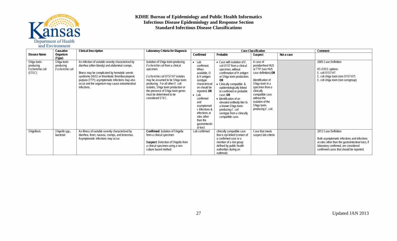
Shiga toxin-producing Escherichia coli (STEC)
Shiga-toxin producing Escherichia coli
An infection of variable severity characterized by diarrhea (often bloody) and abdominal cramps. Illness may be complicated by hemolytic uremic syndrome (HUS) or thrombotic thrombocytopenic purpura (TTP); asymptomatic infections may also occur and the organism may cause extraintestinal infections.
Isolation of Shiga toxin-producing Escherichia coli from a clinical specimen. Escherichia coli O157:H7 isolates may be assumed to be Shiga toxin-producing. For all other E. coli isolates, Shiga toxin production or the presence of Shiga toxin genes must be determined to be considered STEC.
• Lab confirmed. When available, O & H antigen serotype characterization should be reported; OR
• Lab confirmed and asymptomatic infections & infections at sites other than the gastrointestinal tract
• Case with isolation of E. coli O157 from a clinical specimen, without confirmation of H antigen or Shiga toxin production; OR
• Clinically compatible & epidemiologically linked to confirmed or probable case; OR
• Identification of an elevated antibody titer to a known Shiga toxin-producing E. coli serotype from a clinically compatible case.
A case of postdiarrheal HUS or TTP (see HUS case definition),OR Identification of Shiga toxin in a specimen from a clinically compatible case without the isolation of the Shiga toxin-producing E. coli.
2005 Case Definition KS-EDSS options: E. coli O157:H7 E. coli shiga toxin (non O157:H7) E. coli shiga toxin (non serogroup)
Shigellosis
Shigella spp., bacterial
An illness of variable severity characterized by diarrhea, fever, nausea, cramps, and tenesmus. Asymptomatic infections may occur.
Confirmed: Isolation of Shigella from a clinical specimen Suspect: Detection of Shigella from a clinical specimen using a non-culture based method
Lab confirmed clinically compatible case that is epi linked (contact of a confirmed case or a member of a risk group defined by public health authorities during an outbreak)
Case that meets suspect lab criteria
2012 Case Definition Both asymptomatic infections and infections at sites other than the gastrointestinal tract, if laboratory confirmed, are considered confirmed cases that should be reported.

KDHE Bureau of Epidemiology and Public Health Informatics Infectious Disease Epidemiology and Response Section
Standard Infectious Disease Classifications
Updated JAN 2013
28
Disease Name
Causative Organism (Type)
Clinical Description Laboratory Criteria for Diagnosis Case Classification Comment Confirmed Probable Suspect Not a case
Spotted Fever Rickettsiosis (aka Rocky Mountain Spotted Fever)
Rickettsia rickettsii (most common) but includes other Rickettsia spp.
Any reported fever and one or more of the following: rash, headache, myalgia, anemia, thrombocytopenia, or any hepatic transaminase elevation. A macular or maculopapular rash appears 4-7 days following onset in many (~80%) patients, often present on the palms and soles.
Laboratory confirmed: • Serological evidence of a 4X
change in IgG-specific antibody titer reactive with Ri. rickettsii or other spotted fever group antigen by indirect IFA between paired serum specimens (one taken in the first week of illness and a second 2-4 weeks later), OR
• Detection of R. rickettsii or other spotted fever group DNA in a clinical specimen by PCR , OR
• Demonstration of spotted fever group antigen in a biopsy or autopsy specimen by IHC, OR
• Isolation of R. rickettsii or other spotted fever group rickettsia from a clinical specimen in cell culture.
Laboratory supportive: • Serologic evidence of elevated
IgG or IgM reactive with R. rickettsii or other spotted fever group antigen by IFA, ELISA, dot-ELISA, or latex agglutination.
Clinically compatible & lab confirmed
Clinically compatible case & supportive laboratory results.
A case with laboratory evidence of past or present infection but no clinical information available
2010 Case Definition Exposure Having been in potential tick habitats within the past 14 days before onset of symptoms. Occupation should be recorded if relevant to exposure. A history of a tick bite is not required.
Streptococcal disease, invasive, Group A
Group A Streptococcus (Streptococcus pyogenes)
Invasive Group A streptococcal infections may manifest as any of several clinical syndromes, including pneumonia, bacteremia in association with cutaneous infection (e.g., cellulitis, erysipelas, or infection of a surgical or nonsurgical wound), deep soft-tissue infection (e.g., myositis or necrotizing fasciitis), meningitis, peritonitis, osteomyelitis, septic arthritis, postpartum sepsis (i.e., puerperal fever), neonatal sepsis, and nonfocal bacteremia.
Isolation of group A Streptococcus (Streptococcus pyogenes) by culture from a normally sterile site (e.g., blood or cerebrospinal fluid, or, less commonly, joint, pleural, or pericardial fluid)
Laboratory confirmed
Clinically compatible case
1995 Case Definition Nationally notifiable condition, but reportable in KS only if drug-resistant.

KDHE Bureau of Epidemiology and Public Health Informatics Infectious Disease Epidemiology and Response Section
Standard Infectious Disease Classifications
Updated JAN 2013
29
Disease Name
Causative Organism (Type)
Clinical Description Laboratory Criteria for Diagnosis Case Classification Comment Confirmed Probable Suspect Not a case
Streptococcus pneumoniae, invasive disease, drug resistant
Streptococcus pneumoniae
Depends of site of infection • Isolation of S. pneumoniae from a normally sterile site AND
• “nonsusceptible” isolate (intermediate or high-level resistance to at least 1 antimicrobial agent currently approved for use in treating pneumococcal infection)
Clinically compatible & lab confirmed
Clinically compatible & lab confirmed but oxacilin is the only agent screened
2007 Case Definition Antimicrobial agents include: Oxacillin Penicillin Cephalosporins & others as clinically indicated If isolated from CSF, other appropriate criteria are met, and diagnosis is meningitis, case should still be reported as Streptococcus pneumoniae, invasive disease, drug resistant
Streptococcus pneumoniae, invasive disease
Streptococcus pneumoniae
Depends on site of infection Isolation of S. pneumoniae from a normally sterile site (e.g., blood, cerebrospinal fluid, or, less commonly, joint, pleural, or pericardial fluid)
Lab confirmed Isolation of S. pneumoniae lacking the confirmation from a normally sterile site
2010 Case Definition If isolated from CSF, other appropriate criteria are met, and diagnosis is meningitis, case should still be reported as Streptococcus pneumoniae, invasive disease

KDHE Bureau of Epidemiology and Public Health Informatics Infectious Disease Epidemiology and Response Section
Standard Infectious Disease Classifications
Updated JAN 2013
30
Disease Name
Causative Organism (Type)
Clinical Description Laboratory Criteria for Diagnosis Case Classification Comment Confirmed Probable Suspect Not a case
Streptococcal Toxic Shock Syndrome (STSS)
Group A Streptococcus (Streptococcus pyogenes)
Illness with the following clinical manifestations: Hypotension: systolic blood pressure ≤90 mmHg for adults or <5th percentile by age for children <16 years Multi-organ involvement characterized by ≥2 of the following: • Renal impairment: creatinine ≥2 mg/dL
(≥177mmol/L) for adults or ≥2 times the upper limit of normal for age. In patients with preexisting renal disease, ≥2 times elevation over baseline level
• Coagulopathy: Platelets ≤100,000/mm3 or disseminated intravascular coagulation
• Liver involvement: AST, ALT or total bilirubin levels ≥2 times the upper limit of normal for the patient’s age. In patients with preexisting liver disease, ≥2 times increase over baseline level
• Acute respiratory distress syndrome: defined by acute onset of diffuse pulmonary infiltrates and hypoxemia in the absence of cardiac failure or by evidence of diffuse capillary leak manifested by acute onset of generalized edema, or pleural or peritoneal effusions with hypoalbuminemia
• A generalized erythematous macular rash that may desquamate
• Soft-tissue necrosis, including necrotizing fasciitis or myositis, or gangrene
Isolation of Group A Streptococcus
• Clinically compatible &
Isolation of group A Streptococcus from normally sterile site
• Clinically compatible in the absence of another identified etiology for the illness and
• isolation of group A Streptococcus from a nonsterile site
2010 Case Definition
Tetanus Acute onset of hypertonia and/or painful muscular contractions (usually of the muscles of the jaw and neck) and generalized muscle spasms without other apparent medical cause
None Acute illness with • Muscle spasms or
hypertonia & • Dx by health care
provider OR Death with tetanus listed as cause of death or contributing cause
1996 Case Definition
KDHE Bureau of Epidemiology and Public Health Informatics Infectious Disease Epidemiology and Response Section
Standard Infectious Disease Classifications
Updated JAN 2013
31
Disease Name
Causative Organism (Type)
Clinical Description Laboratory Criteria for Diagnosis Case Classification Comment Confirmed Probable Suspect Not a case
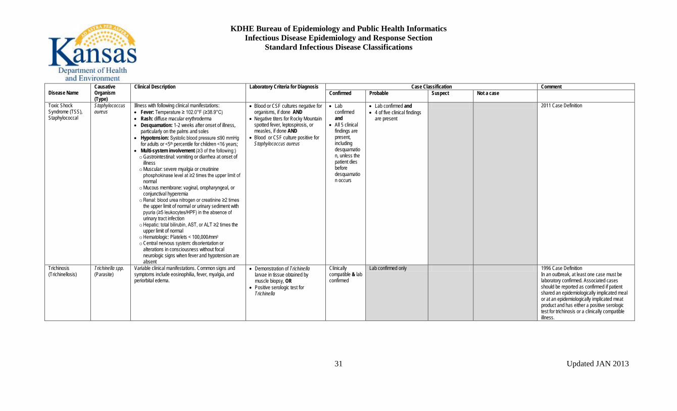
Toxic Shock Syndrome (TSS), Staphylococcal
Staphylococcus aureus
Illness with following clinical manifestations: • Fever: Temperature ≥ 102.0°F (≥38.9°C) • Rash: diffuse macular erythroderma • Desquamation: 1-2 weeks after onset of illness,
particularly on the palms and soles • Hypotension: Systolic blood pressure ≤90 mmHg
for adults or <5th percentile for children <16 years; • Multi-system involvement (≥3 of the following:) o Gastrointestinal: vomiting or diarrhea at onset of
illness o Muscular: severe myalgia or creatinine
phosphokinase level at ≥2 times the upper limit of normal
o Mucous membrane: vaginal, oropharyngeal, or conjunctival hyperemia
o Renal: blood urea nitrogen or creatinine ≥2 times the upper limit of normal or urinary sediment with pyuria (≥5 leukocytes/HPF) in the absence of urinary tract infection
o Hepatic: total bilirubin, AST, or ALT ≥2 times the upper limit of normal
o Hematologic: Platelets < 100,000/mm3 o Central nervous system: disorientation or
alterations in consciousness without focal neurologic signs when fever and hypotension are absent
• Blood or CSF cultures negative for organisms, if done AND
• Negative titers for Rocky Mountain spotted fever, leptospirosis, or measles, if done AND
• Blood or CSF culture positive for Staphylococcus aureus
• Lab confirmed and
• All 5 clinical findings are present, including desquamation, unless the patient dies before desquamation occurs
• Lab confirmed and • 4 of five clinical findings
are present
2011 Case Definition
Trichinosis (Trichinellosis)
Trichinella spp. (Parasite)
Variable clinical manifestations. Common signs and symptoms include eosinophilia, fever, myalgia, and periorbital edema.
• Demonstration of Trichinella larvae in tissue obtained by muscle biopsy, OR
• Positive serologic test for Trichinella
Clinically compatible & lab confirmed
Lab confirmed only 1996 Case Definition In an outbreak, at least one case must be laboratory confirmed. Associated cases should be reported as confirmed if patient shared an epidemiologically implicated meal or at an epidemiologically implicated meat product and has either a positive serologic test for trichinosis or a clinically compatible illness.

KDHE Bureau of Epidemiology and Public Health Informatics Infectious Disease Epidemiology and Response Section
Standard Infectious Disease Classifications
Updated JAN 2013
32
Disease Name
Causative Organism (Type)
Clinical Description Laboratory Criteria for Diagnosis Case Classification Comment Confirmed Probable Suspect Not a case
Tularemia Francisella. tularensis (Bacteria)
Clinical diagnosis is supported by evidence or history of a tick or deerfly bite, exposure to tissues of a mammalian host of Francisella tularensis, or exposure to potentially contaminated water. • Ulceroglandular: cutaneous ulcer with regional
lymphadenopathy • Glandular: regional lymphadenopathy with no
ulcer • Oculoglandular: conjunctivitis with preauricular
lymphadenopathy • Oropharyngeal: stomatitis or pharyngitis or
tonsillitis and cervical lymphadenopathy • Intestinal: intestinal pain, vomiting, and diarrhea • Pneumonic: primary pleuropulmonary disease • Typhoidal: febrile illness without early localizing
signs and symptoms
Confirmatory: isolation of F. tularensis in clinical specimen; or ≥ 4X change in serum Ab titer to F. tularensis Ag Presumptive: ↑ serum Ab titer to F. tularensis Ag with no hx tularemia vaccination; or detection of F. tularensis in clinical specimen fluorescent assay
Clinically compatible & lab confirmed
Clinically-compatible case with lab results indicative of presumptive infection.
Positive polymerase chain reaction (PCR) for Francisella. tularensi
1999 Case Definition
Typhoid Fever Salmonella typhi Insidious onset of sustained fever, headache, malaise, anorexia, relative bradycardia, constipation or diarrhea, and nonproductive cough. However, many mild and atypical infections occur. Carriage of S. typhi may be prolonged.
Isolation of S. typhi from blood, stool, or other clinical specimen
Clinically compatible & lab confirmed
Clinically compatible case that is epidemiologically linked to a confirmed case in an outbreak
1997 Case Definition Serologic evidence alone is not sufficient for diagnosis. Asymptomatic carriage should not be reported as typhoid fever. Isolates of S. typhi are reported to the Foodborne and Diarrheal Branch through the PHLIS.
Varicella Varicella-zoster virus (VZV) (virus)
An illness with acute onset of diffuse (generalized) maculo-papulovesicular rash without other apparent cause.
• Isolation of varicella virus from clinical specimen, OR
• DFA, OR • PCR, OR • significant rise in serum varicella
IgG Ab level by any standard serologic assay
• Lab confirmed OR
• Clinically compatible & epi-linked to a confirmed or probable case
Clinically compatible & not lab confirmed & not epi-linked to a confirmed or probable case
2010 Case Definition

KDHE Bureau of Epidemiology and Public Health Informatics Infectious Disease Epidemiology and Response Section
Standard Infectious Disease Classifications
Updated JAN 2013
33
Disease Name
Causative Organism (Type)
Clinical Description Laboratory Criteria for Diagnosis Case Classification Comment Confirmed Probable Suspect Not a case
Vibriosis (Vibrio spp. not V. cholerae)
Vibrio spp. other than toxigenic Vibrio cholerae O1 or O139
An infection of variable severity characterized by watery diarrhea, primary septicemia, or wound infection. Asymptomatic infections may occur, and the organism may cause extra-intestinal infection.
Isolation of a species of the family Vibrionaceae (other than toxigenic Vibrio cholerae O1 or O139, which are reportable as cholera) from a clinical specimen. *Infections due to toxigenic Vibrio cholerae O1 or O139 are reportable as cholera.
Lab confirmed Clinically-compatible epidemiologically linked to a confirmed case.
Clinically compatible, symptomatic case that is not epi-linked to a confirmed case.
2012 Case Definition Genera in the family Vibrionaceae (not all have been recognized to cause human illness) currently include: Aliivibrio Allomonas Catenococcus Enterovibrio Grimontia Listonella Photobacterium Salinivibrio Vibrio

KDHE Bureau of Epidemiology and Public Health Informatics Infectious Disease Epidemiology and Response Section
Standard Infectious Disease Classifications
Updated JAN 2013
34
Disease Name
Causative Organism (Type)
Clinical Description Laboratory Criteria for Diagnosis Case Classification Comment Confirmed Probable Suspect Not a case
Viral Hemorrhagic Fever
Filoviruses (Ebola, Marburg) Lassa Virus New World Arenaviruses (Guanarito, Machupo, Junin, Sabia) Crimean-Congo Hemorrhagic Fever (Nairovirus)
Acute onset with ALL the following clinical findings: • A fever > 40°C, AND • One or more of the following clinical findings:
o Severe headache o Muscle pain o Erythematous maculopapular rash on the
trunk with fine desquamation 3–4 days after rash onset
o Vomiting o Diarrhea o Pharyngitis (arenavirus only) o Abdominal pain o Bleeding not related to injury o Retrosternal chest pain (arenavirus only) o Proteinuria (arenavirus only) o Thrombocytopenia
Epidemiologic Linkage Criteria • One or more of the following exposures within 3
weeks before onset of symptoms: o Contact with blood or other body fluids of
patient with VHF o Residence in / travel to a VHF endemic
area o Work in lab that handles VHF specimens o Work in lab that handles bats, rodents, or
primates from endemic areas o Exposure to semen from a confirmed
acute / convalescent case of VHF within 10 weeks of that’s person’s onset of symptoms
• Detection of VHF viral antigens in blood by ELISA antigen detection
• VHF viral isolation in cell culture for blood or tissues
• Detection of VHF viral genes using reverse transcriptase with polymerase chain reaction amplification (RT-PCR) from blood or tissues
• Detection of VHF viral antigens in tissues by immunohistochemistry
Clinically compatible & lab confirmed
Clinically compatible and epi linkage criteria
2010 Case Definition Exposure • One or more of the following exposures
within the 3 weeks before onset of symptoms: o Contact with blood or other body fluids
of a patient with VHF o Residence in—or travel to—a VHF
endemic area o Work in a laboratory that handles VHF
specimens o Work in a laboratory that handles
primates from endemic areas OR • Exposure within the past 3 weeks to
semen from a confirmed acute or convalescent case of VHF within the 10 weeks of onset of symptoms

KDHE Bureau of Epidemiology and Public Health Informatics Infectious Disease Epidemiology and Response Section
Standard Infectious Disease Classifications
Updated JAN 2013
35
Disease Name
Causative Organism (Type)
Clinical Description Laboratory Criteria for Diagnosis Case Classification Comment Confirmed Probable Suspect Not a case
Yellow Fever Yellow fever virus (virus)
Acute onset of fever, headache, and myalgia. Recurrence of fever may follow initial remission of symptoms. Renewal of symptoms may be accompanied by hepatitis, albuminuria, renal failure, shock, and generalized hemorrhages.
• ≥4 times rise in yellow fever Ab titer in a patient who has no history of recent yellow fever vaccination and cross-reactions to other flaviviruses have been excluded, OR,
• Demonstration of yellow fever
virus, Ag or genome in tissue blood, or other body fluid
Clinically compatible & lab confirmed
Clinically compatible with supportive serology. Cross-reactive serologic reactions to other flaviviruses must be excluded. Patient must not have had yellow fever vaccination.
1997 Case Definition

KDHE Bureau of Epidemiology and Public Health Informatics Infectious Disease Epidemiology and Response Section
Standard Infectious Disease Classifications
Updated JAN 2013
36
Box Colors White CSTE case classification Gray KDHE OSE case classification Green refer to the CDC website for the case classification Blue Updated definition List of terms ARDS acute respiratory disease syndrome CNS central nervous system CSF cerebrospinal fluid DFA direct fluorescent antibody DNA deoxyribonucleic acid EIA enzyme immunoassay ELISA enzyme-linked immunosorbent assay IFA immunofluorescent antibody analysis IHC immunohistochemical NAT nucleic acid test Normally sterile site Includes blood, CSF, joint, pleural, or pericardial fluid PCR Polymerase chain reaction -- Used to replicate DNA/RNA .
If lab results indicated that levels are not detectable, the results are NOT to be classified as negative.
RIBA recombinant immunoblot assay RT-PCR reverse-transcriptase polymerase chain reaction WHO World Health Organization Epi-Linked: A case in which a) the patient has had contact with one or more persons who either have/had the disease or have been exposed to a point source of infection (i.e., a single source of infection, such as an event leading to a foodborne-disease outbreak, to which all confirmed case-patients were exposed) and b) transmission of the agent by the usual modes of transmission is plausible. A case may be considered epidemiologically linked to a laboratory-confirmed case if at least one case in the chain of transmission is laboratory confirmed.