Stability of Mandibular Incisor Position
43
University of Connecticut OpenCommons@UConn SoDM Masters eses School of Dental Medicine June 1991 Stability of Mandibular Incisor Position Jin-Myung Kim Follow this and additional works at: hps://opencommons.uconn.edu/sodm_masters Recommended Citation Kim, Jin-Myung, "Stability of Mandibular Incisor Position" (1991). SoDM Masters eses. 69. hps://opencommons.uconn.edu/sodm_masters/69
Transcript of Stability of Mandibular Incisor Position

University of ConnecticutOpenCommons@UConn
SoDM Masters Theses School of Dental Medicine
June 1991
Stability of Mandibular Incisor PositionJin-Myung Kim
Follow this and additional works at: https://opencommons.uconn.edu/sodm_masters
Recommended CitationKim, Jin-Myung, "Stability of Mandibular Incisor Position" (1991). SoDM Masters Theses. 69.https://opencommons.uconn.edu/sodm_masters/69

THE STABILITY OF MANDIBULAR INCISOR POSITION
Dr. Jin-Myung Kim
In Partial Fulfillment of the Requirementsof Certificate in Orthodontics
Major Advisor- Dr. Ravindra Nanda
Co-Advisors- Dr. Charles J. Burstone
Dr. Joseph Burleson
Division of Orthodontics
The University of Connecticut Health Center
Farmington, Connecticut 06032
1991

APPROVAL PAGE
Certificate in Orthodontics Thesis,
The Stability of Mandibular Incisor Position
presented by
Dr. Jin-Myung Kim
Major Advisor " ..-Dr. Ravindra Nanda
Co AdvisorsDr. Crl-e-J. Burstone
oseph Burleson
The University of Connecticut Health Center
1991

ABSTRACT
This retrospective study was performed to investigate the
stability of the mandibular incisor position following orthodontic
treatment. Thirty-four patients were selected. Three time periods
were studied; pretreatment (TI), post-treatment (T2) and post-
retention(T3) The changes of dental, hard and soft tissue at TI-
T2 and T2-T3 were measured using study models and cephalograms.
The correlation coefficient between the incisor irregularity at T3
and various pretreatment variables was performed in addition to
standard statistical methods. The results of the present study
showed a significant increase in incisor irregularity at post-
retention along with other significant dental and soft tissue
.changes. The increase in incisor irregularity during post-
retention phase was found to be correlated with the amount of
crowding prior to the treatment, upper and lower lip protrusion at
the end of the treatment, the degree of retroinclination of the
mandibular incisors at the start of the treatment, and the amount
of the mandibular growth. Often mentioned factors such as upper
incisor inclination, overbite, arch length, overjet, intermolar and
intercanine widths, mandibular rotation and lip thickness and
length did not show a correlation with the post-retention
mandibular incisor irregularity. The result suggests that
treatment mechanics may play a significant role in the stability of
the mandibular incisor position.

INTRODUCTION
The position of the mandibular incisors has been the focus of
numerous clinical reports as they have served as guides for
treatment planning and stability of attained results. A number of
studies in recent years have investigated the stability of the
mandibular incisors following orthodontic treatment.
The final position of mandibular incisors following treatment
can change due to various causes acting alone or together. Some of
the factors reported in the literature are, differential
dentofacial growth, patient age and sex, incisor position over the
basal bone, tooth size-arch dimension, anterior component of the
occlusal force, eruptive force of the third molars, soft tissue and
musculature, physiologic recovery, inappropriate mechanics,
periodontal fibers, treatment procedures, and the length of
retention period. However, the nature of the correlation between
the post-treatment changes in the mandibular incisor position and
some of the above mentioned predisposing factors is still unclear.

REVIEW OF THE LITERATURE
Dental Changes
A number of long term studies have been conducted to
determine the stability of mandibular incisor position in untreated
growing individuals. The crowding of the lower incisors increases
after the permanent dentition especially between the ages of 14 to
1,2,3,420 years. Furthermore, intercanine width increases markedly
until canines erupt, and decreases slightly afterwards .4,5
Untreated individuals also show a decrease in arch length during
the premolar eruption which thereafter decreases steadily. 1,4
Intermolar width also decreases with age,4,6 however, Moorrees
reported an increase between the ages of 9 to 14 years.
Post-retention studies of dental changes indicate a different
pattern compared to the untreated groups. Littlez reported an
increase in incisor irregularity in extraction patients at post-
retention period. Glenn et al. 8 found relatively stable incisor
position in nonextraction post-retention patients as compared to
patients who had extractions. However, Uhde et al. 9 reported no
different relapse tendencies between extraction and nonextraction
patients. FastlichtI found more crowding in untreated individuals
as compared to treated patients.
Mandibular intercanine width generally decreases in both

extraction and nonextraction patients during post-retention, z,s09,11,12
Intermolar width decreases 9,12 or it remains relatively stable
during post-retention. 8,11
Overbite has been shown to increase from mixed dentition to
permanent dentition and decreases subsequently during maturation of
untreated occlusion. I04,6 Overbite tends to increase slightly
during postretention stage both in extraction and nonextraction
groups .013 However, Glenn et. al. 8 found overbite remained
remarkably stable in nonextraction patients Little et al z and
Hernandez4 have reported a significant relapse of overbite in
extraction patients.
Incisor position and adjacent hard tissue variables
The final tooth position at maturity is influenced by the
relative amount and direction of anteroposterior and vertical
facial growth. 5,6 Bjork and Skieller15 have indicated that the
dentitional changes in tooth position and axial inclination were
the result of a balance between the facial, skeletal and dental
relationships. Other studies have mentioned following factors;
anterior growth of the mandibular base3, axial inclination of
mandibular incisor relation to mandibular molar inclinationz, a
relationship of mandibular inclination to the maxillary base8,
downward mandibular growth and forward mandibular rotation I,0 and
a relationship of lower incisor size to the size of the face and

jaws21. A longitudunal investigation22 based on the Denver growth
study shows relatively constant mandibular incisor angular
measurement with age such as the interincisal angle and mandibular
incisor to mandibular plane. Sinclair and Little16 found that
incisor angulation to the cranial base was relatively stable in
untreated patients but failed to correlate with lower incisor
position and factors relating to growth pattern. Miethke: also
indicated no correlation between lower incisor crowding and either
skeletal morphology or mandibular incisor position.
In a postretention study using cephalometric variables El-
Mangoury24 found orthodontic relapse associated with a decrease in
the palatal plane and mandibular plane angle. Huggins and Birch5
reported that an early relapse in upper incisor position could
cause a new incisal relationship. Shields26 and Schulhofz reported
no significant correlation between cephalometric variables such as
incisor position, facial growth and long-term mandibular
irregularity.
Soft tissue factors
Several studies have shown that the position of lower incisor
is influenced by the surrounding musculature, z8’9 Luffingham3, and
Thuer and Ingerval131 found that different malocclusion group have
different lip and cheek pressure. Riedel reported that the soft
tissue profile is closely related to the skeletal and dental

structures that comprise the bony profile. Winders33 indicated
stable tooth position has minimal lingually directed muscle
pressure against the incisors. Subtenly34"5 reported that all
components of the soft tissue musculature do not directly follow
the underlying skeletal profile, although lip posture was found to
be closely related with the position of the underlying dental and
alveolar structures. Burstone6,3z noted that the postural position
of the lower lip influences the maintenance of the original
mandibular incisor position. He also mentioned that although tooth
stability and facial esthetics are closely related to soft tissue
morphology and posture of lips, relaxed postural lip position is
partially independent of tooth position.
The lip change during growth is sexually dimorphic. Lenard8
showed a slight retrusion of upper and lower lip during growth in
males and females. Males showed a larger change in the upper and
lower lip length growth whereas females showed only a small
change. Upper and lower lip thickness also increased significantly
in the male group,but only slightly in the female group. Nanda et
al. 9 agreed with different male and female patterns and added that
most of the soft tissue measurements in females had attained their
adult size at the age of 15 years, whereas in males, several
measurements appeared to be on the increase even at the age of 18
years.
Rudee4 reported, the ratio of soft tissue profile response
concomitant to dental or skeletal changes during treatment. Bloom41
also found the possibility of predicting perioral soft tissue
changes in relation to the expected amount of anterior tooth
movement like lower incisor. However, Burstone3z and Hershey42
indicated that perioral soft tissues may be self-supporting and
that factors other than dental movement may cause the wide variety
of individual response. Oliver43 also showed that soft tissue may
vary enough in thickness, length and postural tone to cause the
response of soft tissue to incisor retraction to be different in
persons with thick upper lips as compared to those with thin upper
lips. Anderson et ai.44 showed that the upper lip thickness
increases considerably following maxillary incisor retraction while
the lower lip was not found to be affected by orthodontic
treatment. Raines and Nanda45 indicated that the change in
mandibular incisor position does not correlate with lip changes,
and the lower lip response is influenced by various other factors
such as the direction of the growth of the mandible.
OBJECTIVES
The objective of this retrospective study is to investigate
the stability of the mandibular incisor position following
orthodontic treatment. The study attempted to establish the
correlation between mandibular incisor position and several dental,

skeletal and soft tissue factors. The dental parameters including
anteroposterior and width changes were measured on occlusograms of
the study casts. The differences in skeletal and soft tissue
values were evaluated by cephalometric measurements.
MATERIALS AND METHODS
Patient Selection
Thirty-four Caucasian patients treated at the Orthodontic
Clinic of the University of Connecticut Health Center and from the
orthodontic practices of two part-time faculty members were
selected for the study. The mean age at the start of the treatment
was 12.9 years. All patients received edgewise orthodontic
therapy. The records were obtained from three time intervals
namely, pretreatment (TI), at the end of treatment (T2) and at
least one year out of retention (T3). The subjects were further
categorized by extraction or nonextraction, sex and Angle’s dental
classification (Table i).
Dental Measurements
Dental casts of different periods were collected and recorded
using the occlusograms46. The following measurements were analyzed
by computer-aided digitizing program (Fig. I)

i. Mandibular incisor irregularity index The sum of displacement
of the anatomic contact points of the lower anterior teeth as
suggested by Little4z.
2. Mandibular intercanine width The distance between the cusp
tips of left and right canine.
3. Mandibular intermolar width The distance between the central
fossae of the left and right first molars.
4. Mandibular arch length The sum of the distances between the
mesial contact point of the first molar and the contact or midpoint
of the mandibular central incisors (Nance48).
Cephalometric measurements
Lateral cephalometric headfilms in centric occlusion and with
relaxed lips were taken at pretreatment (TI), post-treatment (T2)
and at post-retention (T3) periods. Tracings of lateral
cephalometric headfilms were analyzed by a computer aided
cephalometric analysis program (Fig 2). Lateral tracings were
superimposed using anterior cranial base and ethmoid triad
structures. An x- and y-coordinate system was utilized for
analyzing horizontal and vertical changes (Fig 2). A horizontal
(x) reference line was used as Constructed Frankfort Horizontal
line (CFH,SN-7) A perpendicular line to CFH drawn at point sella
served as the vertical reference line (y).
Maxillary superimpostion based on the zygomatic process,
inferior orbital rim and suborbital structures was used to examine

the maxillary dental changes. Changes are also noted using nasal
floor (NF) as x’-coordinate and a perpendicular line to NF served
as the y’-coordinate(Fig 2)
Mandibular superimposition was based on the inner structures
of the symphysis, cortical outlines of the anterior mandibular body
and the mandibular canal. Original plane of occlusion was also
used as x"-coordinate and perpendicular line of OP was used as the
y"- coordinate(Fig 2)
Three types of changes between TI-T2 and T2-T3 were recorded,
a. absolute change
b. positive change anterior or extrusive change
c. negative change posterior or intrusive change
The following 20 linear and ii angular measurements were used
for cephalometric analysis (Fig.2)
Mandibular Incisor Measurements
I. LI-CFH() .The angle between axial inclination of mandibular
central incisor(Ll) and Conctructed Frankfort Horizontal(CFH).
2. LI-MP() -The angle between axial inclination of mandibular
central incisor and mandibular plane(Go-Me).
3. LI-OP() "The angle between axial inclination of mandibular

central incisor and occlusal plane.
L6-OP() -The angle between axial inclination of mandibular
first molar(L6) and occlusal plane.
Ii-CFH (mm, y-coordinate) -The perpendicular distance between
mandibular central incisal edge and CFH.
Ii-CFH (mm, x-coordinate) The perpendicular distance between
mandibular central incisal edge and perpendicular line of CFH.
7. Ii-OP (mm, y" -coordinate -The perpendicular distance between
mandibular central incisal edge and occlusal plane.
8. Ii-OP (mm, x"-coordinate) -The perpendicular distance between
mandibular central incisal edge and perpendicular line of occlusal
plane.
Maxillary Incisor Measurements
9. Ul-CFH() -The angle between axial inclination of maxillary
central incisor(Ul) and CFH.
i0. UI-NF() -The angle between axial inclination of maxillary
central incisor and nasal floor(ANS-PNS).
ii. UI-LI(O):The angle between axial inclinations of maxillary and
mandibular incisor.
12. UI-LI (CFH,mm, overjet) -The horizontal distance between maxillary
and mandibular central incisal edges parallel to CFH.
13. UI-LI (CFH,mm, overbite) -The Vertical distance between maxillary
and mandibular central incisal edges perpendicular to CFH.
Ul-CFH (mm,y-coordinate) The perpendicular distance between
maxillary central incisal edge and CFH.

15. Ul-CFH (mm, x-coordinate) The perpendicular distance between
maxillary central incisal edge and perpendicular line of CFH.
UI-NF (mm, y’-coordinate) -The perpendicular distance between
maxillary central incisal edge and nasal floor.
UI-NF (mm, x’-coordinate) The perpendicular distance between
maxillary central incisal edge and perpendicular line of NF
Soft Tissue Measurements
18. Pls(Sn-Pg’ ,mm,upper lip protrusion)-The perpendicular distance
from most anterior part of upper lip(Prolabale superius,Pls) to
sbnasale(Sn)-soft tissue pogonion(Pg’) line.
19. Pli (Sn-Pg’ ,mm, lower lip protrusion) -The perpendicular distance
from most anterior part of lower lip(Prolabale inferius,Pli) to Sn-
20. Cm-Sn-Pls (nasolabial angel) The angle formed by a line
connecting columella(Cm)-Sn to the line from Sn-Pls.
21. Upper lip thickness(CFH,mm)-The horizontal line from A point to
Sn parallel to CFH.
22. Lower lip thickness (CFH,mm) -The horizontal distance from Pli to
contact point between labial surface of mandibular central incisor
and inner margin of lower lip parallel to CFH.
23. Upper lip length(CFH,mm)-The vertical distance from Sn to lower
most point of upper lip(Stms) perpendicular to CFH.
24. Lower lip length(CFH,mm):The vertical distance from upper most
point of lower lip(Stmi) to mentolabial sulcus (Mls) perpendicular
to CFH.

25. Lip to tooth contact(mm)"The distance from mandibular incisal
edge to contact point between labial surface of mandibular central
incisor and inner margin of lower lip.
Occlusal Plane Measurements
26. A-B(OP,mm) -The distance between perpendicular line from A and
B point to occlusal plane.
27. OP-CFH() :The angle between occlusal plane and CFH.
Mandibular Rotation Measurements
28. Y-axis():The angle between CFH and the sella-gnathion(Gn)
line.
29. Ar-Go-Me (gonial angle) :The angle between articulare (At)
gonion(Go) and Go-menton(Me) line.
Growth Measurements
30. Ar-Pg(mm)-The distance between Ar and Pg.
31. ANS-Me(mm)-The distance between ANS and Me.
Statistical Analysis and Measurement Reliability
Statistical analyses were performed by standard methods. Mean
and standard deviation were calculated for each variable at TI,
TI-T2, and T2-T3. Paired t-test was performed for assessing
significance of the change from T1 to T2, and from T2 to T3.
Pearson correlation coefficient analysis was used to analyze the
12

relationship between the changes at TI-T2 with those at T2-T3 to
estimate the effects of treatment magnitude to relapse. Pearson
correlation coefficients were also calculated between the changes
in incisor irregularity at T2-T3 and absolute, positive and
negative changes at TI-T2 and T2-T3 ,respectively.
Student t-tests were used for assessing the significant differences
between extraction and nonextraction, males and females, Angle
dental classification Class I and Class II, respectively.
To assess the reliability of measurements in tracing and
digitizing procedures, five cephalograms and occlusograms were
randomly selected. Five angular and five linear measurements were
selected from original tracing, and were compared with each other
using Pearson correlation coefficient. The average correlation
coefficient was r= 0.85 (p<0.01) in linear measurements and r=
0.95(p<0.005) in angular measurements. For the digitizing error
all dental measurements and five linear and five angular
measurements were redigitized and compared to original data. The
average Pearson correlation coefficient was r= 0. 91(p<0. 001) All
measurement reliability analyses in the present study indicated
high accuracy.

RESULTS
Dental Changes
There was a significant decrease in the incisor irregularity
during treatment (X= -5.30mm, p<0.01) which increased during the
post-retention period (X= i. 7mm, p<0.01) Intermolar width
decreased significantly in extraction patients (X= -2.20, p<0.01),
whereas nonextraction patients maintained their width during
treatment. Both groups remained stable during the post-retention
period. There was an increase in the intercanine width during
treatment (X= 0.69mm, p<0.01) which decreased slightly at T3 (X=
0.4mm, p<0.01). Arch length decreased significantly in extraction
patients but remained relatively unchanged in nonextraction
patients at T2. It also showed a further decrease at T3 although
the difference between extraction and nonextraction patients was
not significant(Table II).
Mandibular Incisal Position
Angular measurements showed no significant changes in both
treatment and post-retention period except for the first molar
uprighted in relation to the occlusal plane angle. In the linear
measurements, extrusion of the incisal edge of the mandibular
incisor was significant at T2 (X= -l.81mm, p<0.01) and it moved
further superiorly at T3 (X= -0.81mm, p<0.01) Mandibular incisor
also moved superiorly relative to the occlusal plane both at T2 (X=
1.36mm, p<0.01) and T3 periods(X= 0.81mm, p<0.01). Anteroposterior
14

changes were not significant in both TI-T2 and T2-T3 period. There
was a significant difference between extraction and nonextraction
patients(p<0.01) in horizontal movement of mandibular incisors at
T2, but no significant difference was found at T3(Table II).
Maxillary incisor position
Mean maxillary incisor position did not change significantly
in the anteroposterior direction at T2 but did change in anterior
direction at postretention period(X= 2.27 to CFH, p<0.05, X= 1.72
to NF, p<0.05,). Overjet decreased significantly during treatment
(X= -i. 14mm, p<0.01) but increased during postretention period (X=
0.54mm, p<0.01) Overbite decreased during treatment (X= -0.79mm,
p<0.05) but remained stable during the post-retention period.
Vertical position of the maxillary incisor showed a significant
downward movement (X= i. 69mm to CFH, p<0.01, X= i. 14mm to
NF,p<0.01) at T2 and continued to move downward at T3 (X= 1.30mm to
CFH, p<0.01, X= 1.14mm to NF, p<0.01) (Table II)
Soft Tissue
A significant decrease in upper and lower lip protrusion was
found during the treatment period (Ls-X= -i. 20mm, p<0.01, Li:X=
1.22mm, p<0.01). Upper lip remained stable whereas the lower lip
protrusion continued to decrease during the post-retention
period. (X= -0.50mm, p<0.05) (Table III). Nasolabial angle (X=
3.55 p<o. 01) and upper lip thickness (X= 0.70ram, p<0.05mm)
increased significantly at T2, but remained stable during the
15

post-retention period. Upper and lower lip length increased
signficantly at T2, however, only the lower lip length showed an
increase at T3. Lip to tooth contact measured from the incisal
edge to the inner lower margin of the lower lip decreased slightly
during treatment (X= -0.99mm, p<0.05) but remained stable during
the postretention period. Upper lip length, lip thickness, lower
lip protrusion, lip to tooth contact showed significant differences
between extraction and nonextraction at T2, whereas no differences
were found at T3(Table III).
Occlusal Plane
No significant changes of the denture base relationship and
occlusal plane were found in both T2 and T3 observation period
(Table III).
Mandibular rotation and growth
Y-axis decreased slightly during the postretention period
(X= -0.50, p<0.01) Gonial angle also decreased a small amount at
both T2 (X= -0 93 p<0 01) and T3 periods (X=-i 06 p<0 01)
Significant growth of the mandible and lower facial height was
found between TI-T2 and T2-T3 periods (Table III).
Interparameter correlations
The following correlations were studied(Table IV-VI).
i. Correlation between treatment effects(Ti-T2) and post-retention
chanqes (T2-T3)
16

Maxillary and mandibular incisors, and soft tissue changes
during treatment show a significant negative correlation with post-
retention change, indicating that dental and soft tissues have a
tendency to return to their pretreatment position. The angular
change of lower incisors correlated with its rebound after
treatment. Similarly anteroposterior movements of mandibular
incisor also had a correlation, whereas vertical change did not
affect any relapse tendency. Maxillary incisor position including
overjet had a negative correlation with its post-retention change,
and overbite represented a weak correlation. Lip protrusion and
thickness showed a tendency to return to pretreatment position.
However, lip length did not show a significant correlation with the
post-retention changes(Table IV).
2. Correlation between pretreatment variables and the chanqes in
incisor irreqularity at post-retention
Table V and VI show the relationship between the post-
retention changes of mandibular incisors and the pretreatment
measurements. Pretreatment incisor crowding, interincisal angle,
overjet, lower lip protrusion were not significantly correlated
with the post-retention change in incisor irregularity. Axial
inclination of mandibular incisors before treatment correlated
with post-retention change of incisor irregularity.
3. Correlation between post-treatment (TI-T2) post-retention
chanqe(T2-T3) and the chanqes of incisor irreqularity at
17

post-retention
The retroinclination of mandibular incisors at T3 showed a
correlation with an increase in incisor irregularity. Extrusion of
the mandibular incisor and an increase in overbite also showed a
weak correlation with incisor crowing at T3. Increases in upper and
lower lip protrusion including nasolabial angle at T2 were found to
be correlated with incisor irregularity at T3. The amount of
mandibular and lower facial height growth also affected mandibular
incisor crowding at post-retention(Tables V and VI).

DISCUSSION
In the present study, incisor irregularity was significantly
increased at the post-retention period. The amount of incisor
retraction and post-retention crowding showed a positive
correlation. Little et al. z reported a considerable increase of
incisor irregularity in sixty-five patients who had four first
premolar extractions. Glenn et al. 8 showed that the amount of
relapse in nonextraction patients was less than the extraction
patients and more than untreated normals in a eight year post-
retention study. Although the present study had a relatively short
observation period, there was no significant difference between the
extraction and non-extraction patients. Uhde et al.9 examined
post-retention stability with 20 years post-treatment and reported
similar findings.
Mandibular intermolar width decreased relatively more in the
extraction patients after treatment,and it remained unchanged
during the post-retention period. Shapiro12, also reported that the
intermolar width decreased at T2 in the extraction group but a
decrease was also noted at T3. Nonextraction patients showed a
stable intermolar width at T2 and T3. 8,12 Gardner and Chaconas
also reported minimal post-retention change in the intermolar width
regardless of the type of treatment. The small differences noted
in the above mentioned studies may be due to the method of
measuring intermolar width. For example, Shapiro used the tip of
19
mesiobuccal cusp, Gardner and Chaconas11 measured it from the buccal
groove, and the present study used the central fossa of the molar.
The method used in the present study was not influenced by the
rotational changes of the molars.
Intercanine width showed a slight increase during treatment
and it significantly decreased during the post-retention period.
Lombardi49 reported that the crowding of the mandibular incisors
occured with an increase in the mandibular intercanine width
during the post-retention period. On the other hand, Little et
al. z reported that intercanine width change during TI-T2 and T2-T3
was a poor predictor of long-term crowding. Similarly, the degree
of expansion or constriction had little association with the post-
retention incisor alignment. In this study, intercanine width
change was not found to be correlated with an increase in incisor
irregularity during post-retention. The results support the
observation of Little et al. z
Mandibular arch length also decreased significantly at T3.
There were no significant differences at the post-retention period
between the extraction and non-extraction groups even though
considerable more reduction was found in extraction patients.
These findings are consistent with those of other studies, z,8,9,11012
The axial inclination and anteroposterior position of
mandibular incisor was found to be stable at post-retention. This

finding is important since it shows that the incisor position was
in harmony with the soft tissues at the end of the treatment. 28
Since the irregularity of mandibular incisors increased
significantly during post-retention, it also indicates that axial
inclination of the lower incisor is not one of the causative
factors. These results support the observation of Shields et al. 26
and Schulhof et al..z The present study did show a positive
correlation between the amount of flaring done during the treatment
to correct retrusive incisors and an increase in crowding at the
post-retention phase.
In the present study, maxillary incisor inclination and
anteroposterior position changed significantly in the anterior
direction during the post-retention period. This supports the
observation of Huggins and Birch5 who studied eighty-two patients,
up to three years after treatment. On the other hand, Shields et.
al. 26 reported that the upper incisors remained relatively stable
ten years post-retention. In the latter study, a significant
incisor uprighting was accomplished during treatment.
A majority of the studies have shown that the overbite
increases during the retention period. The present study found
that there was no significant change at post-retention, although
overbite changed significantly during treatment. Since the average
post-retention period occurred during adolescence, vertical growth
of the mandible and the stability of the intruded teeth might have

been contributing factors. This study also could not find
significant differences in the relapse of the overbite whether
extraction was performed or not. These results support the
findings of Uhde et al.9 Simons and Joondeph13, and Magill5. The
relationship between the overbite change and incisor irregularity
during the post-retention showed weak correlations, supporting the
finding of Little et al. z
Soft tissues of the face play a major role in the stability of
upper and lower incisors in both untreated and orthodontically
treated individuals.35,3z’44 Proffit29 concluded that pressure of
tongue and lips, as well as the forces created within the
periodontal membrane are the primary factors in attainment of
dental equilibrium. This has been supported by other studies. 2’30’31
Orthodontic treatment in majority of the patients significantly
alters, though temporarily, the soft tissue-hard tissue equilibrium
especially due to retraction or flaring of the incisors, expansion
or constriction of the arches and shortening or lengthening of the
arch length.
In the present study significant observations were made
regarding the status of upper and lower lips at the end of the
treatment and at the post-retention phase. Upper lip length
increased at T2 and remained stable thereafter. The lower lip
length, however, continued to increase into T3 period although less
than T2 period. These changes can be attributed to a combination

of incisor retraction and normal growth. Nanda et al. 39 in a recent
longitudinal study on untreated individuals have shown a
significant lengthening of the upper(X= 2.7mm in males; X= l.lmm in
females) and lower lip(X= 4.2mm in males;X= i. 5mm in females)
between the ages of 7 to 18 years.
The thickness of the upper and lower lip showed a different
pattern of change than the length. The upper lip thickened (X=
0.70mm) at the end of the treatment, and thereafter, almost no
change took place. The lower lip, on the other hand, did not show
any appreciable change in thickness at both TI-T2 and T2-T3
observation periods. These results support the observations of
Angelle. 51 Our findings do not support the finding of Anderson et
al. 44 who noted continued thickening of the upper lip during post-
retention. This difference can be explained due to a much longer
retention period(10 years) in their study. Similarly, the present
results as well as the findings of Anderson et ai.44 Angelle51 and
Ricketts52 do not support the finding of Roos53 who noted a decrease
in the lower lip thickness following treatment. This difference in
results is probably due to inclusion of only Class II division 1
patients in Roos’ study as well as his measurement technique which
was differnt.
The relationship of soft tissue measurements with dental
changes showed an overall weak correlation except a relatively
higher correlation for the upper and lower lip protrusion at the

end of the treatment. This indicated that if the lips are more
protrusive at the end of the treatment, more incisor crowding can
be expected at post-retention.
The occlusal plane was stable during the post-treatment and
post-retention period. This does not support the results of Simons
and Joondeph13 who reported an increase in the occlusal plane angle
at the end of the orthodontic treatment. This difference is
probably due to the use of Class II elastics and resultant
elevation of the posterior teeth in their studies. The use of
Class II elastics was none or minimal in the present study. The
overbite correction was performed by the use of incisor intrusion
arches54 rather than extrusive mechanics.
The correlation of the mandibular growth and incisor
irregularity was also studied for various measurements. The
direction of the mandibular growth as noted from gonial angle and
Y-axis did not show any correlation with the incisor irregularity
at the post-retention phase. These results support Lundstrom3,
Sinclair and Little16 and Solow55. However, the amount of mandibular
growth of patients in the present study showed a positive
correlation with the incisor crowding. This observation suggests
that patients with large amount of mandibular growth during post-
treatment would also exhibit an increased amount of incisor
crowding.
24

SUMMARY
The present study showed that mandibular incisor irregularity
increased following the removal of retaining devices. Various
parameters reported to be associated with the mandibular crowding
were studied, and correlations, if any, were .sought. No single
causative factor could be determined. The results suggest that the
amount of crowding prior to the treatment, upper and lower lip
protrusion at the end of the treatment, the amount of the growth at
the end of the treatment and the degree of retroinclination of the
lower incisors at the start of the treatment, correlate with the
increase in mandibular incisor irregularity during the post-
retention phase. Often mentioned factors such as upper incisor
inclination, overbite, arch length, overjet, intermolar and
intercanine width, mandibular rotation and lip thickness and length
were not found not to be correlated with the post-retention incisor
crowding.
These result should be considered with caution since they only
describe the patients studied and the specific treatment mechanics
performed. The present study suggests that treatment mechanics
may play a significant role in the stability of the mandibular
incisor alignment following orthodontic treatment.

REFERENCES
i. Moorrees, C.F.A.- The dentition of the growing child. Cambridge,
Mass., Harvard University Press.1959.
2. Foster, T.D., Hamilton, M.C., and LaVelle, C.L.B. A study of
dental arch crowding in four age-groups. Dent. Practit. 21-9-12,
1970.
3. Lundstrom, A.- A study of correlation between mandibular growth
direction and changes in incisor inclination, overjet, overbite and
crowding. Trans. Eur. Orthod. Soc. 131-140, 1975.
4. Sinclair, P.M., Little, R.M.- Maturation of untreated normal
occlusions. Am. J. Orthod. 83-114-123, 1983.
5. Morrees, C.F.A., Gron, A., Lebret, L.M., Yen,P.K.J., Fronlich,
F.J." Growth studies of the dentition- A review. Am. J. Orthod. 55-
600-616.
6. Barrow, G.V., and White, J.R.- Developmental changes of the
maxillary and mandibular dental arches. Angle Orthod. 22- 41-
45,1952.
7. Little, R.M., Wallen, T.R., and Riedel R.A.- Stability and
relapse of mandibular anterior alignment first premolar
extraction cases treated by traditional edgewise orthodontics. Am.
J. Orthod. 80-349-365, 1981.
8. Glenn, G., Sinclair, P.M., Alexander, R.G. Nonextraction
orthodontic therapy- Posttreatment and skeletal stability. Am. J.
Orthod. 92-321-328, 1987.
9. Uhde, M.D., Sadowsky, C., BeGole, E.A.- Long-term stability of

dental relationships after orthodontic treatment. Angle Orthod.
53-240-252, 1983.
i0. Fastlicht. J." Crowding of mandibular incisors. Am. J. Orthod.
58- 156-163 1970.
11. Gardner, S.D., and Chaconas, S.J. Posttreatment and
postretention changes following orthodontic therapy. Angle Orthod.
46-151-161, 1976.
12. Shapiro, P.A." Mandibular dental arch form and dimension. Am.
J. Orthod. 66-58-70, 1974.
13. Simons, M.E., Joondeph, D.R. Change in overbite" A ten-year
postretention study. Am. J. Orthod. 64-349-367, 1973.
14. Hernandez, J. L. Mandibular bicanine width relative to
overbite. Am. J. Orhod. 56-455-467, 1969.
15. Bjork, A., and Skieller, V. Facial development and tooth
eruption. An implant study at the age of puberty. Am. J. Orthod.
62-339-383, 1972.
16. Sinclair, P.M., Little, R.M. Dentofacial maturation of
untreated normals. Am. J. Orthod. 88:146-156, 1985.
17. Sanin, C., Savara, B.C.- Factors that affect the alignment of
the mandibular incisors- A longitudinal study. . J. Orthod.
64-248-257, 1973.
18. Norderval., K., Wisth, P. J., Boe, O. E." Mandibular anterior
crowding in relation to tooth size and craniofacial morphology.
Scand. J. Dent. Res. 83-267-273,1975.
19. Richardson, M.E. Late lower arch crowding, the role of facial
morphology. Angle Orthod. 56- 244-254,1986.
27

20. Schudy, G.F. Posttreatment craniofacial growth" Its
implications in orthodontic treatment. Am J. Orthod. 65- 39-57,
1974.
21. Keene, A., Engel, G. The mandibular dental arch, part IV-
prediction and prevention of lower anterior relapse, Angle Orthod.
49-173-180, 1979.
22. Burstone, C. J., Hickman, J.- University of Connecticut Health
Center, Monograph. Denver growth study data. 1968.
23. Miethke, R., Behm-Menthel, A. Correlation between lower
incisor crowding and lower incisor position and lateral
craniofacial morphology. Am. J. Orthod. Dentofac. Orthop. 94-231-
239,1988.
24. Ei-Mangoury, N.H.- Orthodontic relapse in subjects with varying
degrees of anteroposterior and vertical dysplasia. Am. J. Orthod.
75-548-561, 1979.
25. Huggins, D.G., Birch,R.H.- A cephalometric investigation of
stability of upper incisor following their retraction. Am. J.
Orthod. 50-852-856, 1964.
26. Shields, T.E., Little, R.M., Chapko, M.K.- Stability and
relapse of mandibular anterior alignment" A cephalometric appraisal
of first-premolar-extraction cases treated by traditional edgewise
orthodontics. Am. J. Orthod. 87"27-38, 1985.
27. Schulhof, R.J., Allegw R.W., Walters, R.D., Dreskin, M.- The
mandibular dental arch- part I, lower incisor position. Angle
Orthod. 47-280-287, 1977.
28. Weinstein, S., Haack, D.C., Morris, N.Y., Snyder, B.B.,

Attaway, H.E." On an equilibrium theory of tooth position. Angle
Orthod. 33:1-26, 1963.
29. Proffit, W.R.- Equilibrium theory revisied. Angle Orthod.
48:175-186, 1978.
30. Luffingham, J.K.- Lip and cheek pressure exerted upon teeth in
three adult groups with different occlusions. Archs. Oral. Biol.
14:337-350, 1969.
31. Thuer, U., Ingervall, B.- Pressure from the lips on the teeth
and malocclusion. Am. J. Orthod. Dentofac. Orthop. 90"234-242,1990.
32. Riedel, R. A." An analysis of dentofacial relationships. Am.
J. Orthod. 43:103-119,1957.
33. Winders, R.V. An electronic technique to measure the forces
exerted on the dentition by perioral and lingual musculature. Am.
J. Orthod. 42:645-657, 1956.
34. Subtenly, J.D.- The soft tissue profile, growth, and treatment
changes. Angle Orthod. 31:105-122, 1961.
35. Subtenly, J.D. A longitudinal study of soft tissue facial
structures and their profile characteristics defined in relation to
underlying skeletal structures. Am. J. Orthod. 45- 481-507, 1959.
36. Burstone, C. The integumental profile. Am. J. Orthod. 44-1-
25,1958.
37. Burstone, C.J. Lip posture and its significance in treatment
planning. Am. J. Orthod. 53:262-332, 1967.
38. Lenard, J. A.- A longitudinal study of integumental profile
growth increments of normal males and females from eihgt to
eighteen years of age. Thesis, University of Connecticut. 1984.

39. Nanda, R.S., Meng, H., Kapila, S., Goorhuis, J.- Growth changes
in the soft tissue facial profile. Angle Orthod. 60:177-190, 1990.
40. Rudee, D.A. Proportional profile changes concurrent with
orthodontic therapy. Am. J. Orthod. 50- 421-434, 1964.
41. Bloom, L.A.- Perioral profile changes in orthodontic treatment.
Am. J. Orthod. 47:371-379, 1961.
42. Hershey, H. G.- Incisor tooth retraction and subsequent profile
change in postadolescent female patients. Am. J. Orthod. 61" 45-
54 1972.
43. Oliver, B.M.- The influence of lip thickness and strain on
upper lip response to incisor retraction. Am. J. Orthod. 82-141-
149, 1982.
44. Anderson, J. P.., Joondeph, D.R., and Turpin, D.L. A
cephalometric study of the profile changes in orthodontically
treated cases ten years out of retention. Angle Orthod. 43:324-336,
1973.
45. Rains, M.D., and Nanda, R.- Soft-tissue changes associated with
maxillary incisor retraction. Am. J. Orthod. 81-481-488, 1982.
46. Marcotte, M. R.- The use of occlusogram in planning orthodontic
treatment. Am. J. Orthod. 69:655-667,1976.
47. Little, R. M.- The irregularity index- A quantitative score of
mandibular anterior alignment. Am. J. Orthod. 68-554-563,1975.
48. Nance, H.N. The limitations of orthodontic treatment. Am. J.
Orthod. 33:177-223, 1947.
49. Lombardi, A.R.- Mandibular incisor crowding in completed cases.
Am. J. Orthod. 61:374-383,1972.

50. Magill, J.M." Changes in the anterior overbite relationship
following orthodontic treatment in extraction cases. Am. J. orthod.
46:755-788, 1960.
51. Angelle, P. L. A cephalometric study of the soft tissue
changes during and after orthodontic treatment. Trans. Eur. Orthod.
Soc. 49:267-280,1973.
52. Rickettes, R. M." The influence of orthodontic treatment on
facial growth and development. Angle Orthod. 30- 103-133,1960.
53. Roos, N- Soft-tissue profile changes in Class II treatment. Am.
J. Orthod. 72:165-175, 1977.
54. Burstone, C. J.- Deep overbite correction by intrusion. Am. J.
Orthod. 72:437-436,1977.
55. Solow, B.- The pattern of craniofacial associations. Acta.
Odont. Scand. 24:Suppl. 46, 1966.

Table I. Sample Characteristics
Mean (years) Range (years)
Pre-treatment agePost-treatment agePost-retention period
Treatment rocedure
12.9114.86I.Ii
Number
9.08 23.17ii.83 24.721.05 1.75
Extraction 16Nonextract ion 18
Percent
47.152.9
Sex
Male 13Female 21
38.261.8
AnGle’s Dn%,,.l Classification
Class I IiClass I I 23
32.467.6
Total 34 i00.0
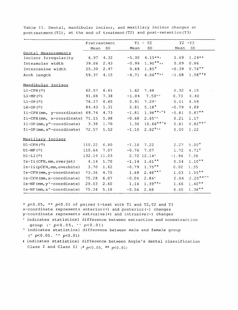
Table II. Dental, mandibular incisor, and maxillary incisor changes at
pretreatment (TI), at the end of treatment (T2) and post-retention(T3)
Dentl MeasurementsIncisor Irregularity
Intermolar width
Intercanine width
Arch length
Mandibular Incisor
LI-CFH o
LI-MP (o)
LI-OP (o)
L6-OP (o)
Ii-CFH(mm, y-coordinate)
Pretreatment T1 T2 T2 -T3
Mean SD Mean SD Mean SD
6.97 4.32 -5.30 4.15"*+ i. 69 1.24"*
39.06 2.43 -0 99 I. 90**++ 0.09 0 86
25.30 2.47 0.69 1.85" -0.39 0.76**59.37 4.15 -4.71 6.06 **++ -1.08 1.58 **#
62.57 6.61 1.62 7.48 0.32 4.15
91.08 7.38 -1.04 7.50 ++ 0.73 4.40
76.17 6.65 0.91 7.29 + 0.ii 4.59
84.43 5.31 2.81 5.18" -0.79 4.89
69.74 4.73 -1.81 1.96 **+^# -0.81 0.87**Ii-CFH(mm, x-coordinate) 71.15
Ii-OP (ram, y" -coordinate 3.38
Ii-OP (ram, x" -coordinate 72.57
5.98 -0 68 2 65 ++ 0.21 1 17
1.70 1.36 10.66 **^# 0.81 0.82 **^
5.52 -I.I0 2.82 *++ 0.00 1.22
Maxillry Incisor
UI-CFH o
UI-NF (o)
UI-LI o
Is-Ii (CFH, ram, overjet)Is-Ii (pCFH, ram, overbite)
Is-CFH (ram, y-coordinate)
Is-CFH (ram, x-coordinate)
Is-NF (ram, y’ -coordinate)Is-NF (ram, x’ -coordinate)
110.22 6.80 -!.I0 7.22 2.27 5.00*110.64 7.07 -0.76 7.07 1.72 4.71"132.19 ii.03 2.72 12.14 + -1.94 7.36
4.14 1.70 -1.14 1.61"* 0.54 i.I0"*3.70 1.62 -0.79 1.75"* 0.02 1.35
73.36 4.70 1.69 2.48 **^ 1.03 1.55"*75.28 6.07 -0.04 2.84 + 2.04 2.20 **^^
29.03 2.60 1.14 1.39 **+ 1.66 1.40"*75.34 5.18 -0.56 2.68 0.65 1.38"*
* p<0.05, ** p<0.01 of paired t-test with T1 and T2,T2 and T3x-coordinate represents anterior(+) and posterior(-) changesy-coordinate represents extrusive(+) and intrusive(-) changes+ indicates statistical difference between extraction and nonextraction
group (+ p<0. 05, ++ p<0. 01)indicates statistical difference between male and female group
(^ p<0.05. p<0.01)# indicates statistical difference between Angle’s dental classification
Class I and Class II (# p<0.05, ## p<0.01)

Table II. Soft tissue, occlusal plane, mandibular rotation, and growth
ohanges at pretreatment(Tl), at the end of treatment(T2) and post-
retention (T3)
Soft Tissue
Ls (Sn-Pg ’, mm)Li Sn-Pg ’, mm)Nasolabial angle
Upper lip thickness (mm)Lower lip thickness (mm)
Upper lip length(mm)Lower lip length(mm)
Lip to tooth contact (mm)
occlusal Plane
A-B (OP, mm)
OP-CFH (o)
OP change (o)
Mandibular RotatioY-axis (o)
Gonial angle
GrowthAr-Pg (ram)
ANS-Me (mm)
Pretreatment T1 T2 T2 T3
Mean SD Mean SD Mean SD
3.31 2.74
3.02 2.68
109.16 12.04
12.99 2.5214.77 2.21
21.10 2.71
16.66 1.97
5.39 2.25
-1.20 1.45"* -0.28 1.57
-1.22 1.49 **++ -0.49 1.37"3.55 4.87** -0.37 4.95 #
0.70 1.99 *+ -0.08 1.92-0.28 1 89+ -0 36 1 47
0.74 1.65"++ -0 35 1. Ii
1.03 1.64"* 0.58 1.37"-0.99 2.16 *++ -0.48 1.80
2.04 2.60
13.69 4.28
0.24 2.34 0.13 2.17 +
-0.71 3.00 -0.17 3.12
-0.97 2.93 0.23 2.54 +
60 18 3.48 0 06 1 Ii -0 50 1 05"*125.06 6.12 -0 93 1 80** -I. 06 2 06**
107.17 6.17 4.18 3.67 **^ 2.37 2.09**67.87 5.70 2.33 2.81 **^^ 1.33 1.76"*
* p<0.05, ** p<0.01 of paired t-test with T1 and T2,T2 and T3/ indicates statistical difference between extraction and nonextraction
group (+ p<0. 05, ++ p<0.01)indicates statistical difference between male and female group(^ p<0.05. p<0.01)
# indicates statistical difference between Angle’s dental classificationClass I and Class II (# p<0.05, ## p<0.01)

Table IV. Correlation between treatment effects(Ti-T2) and the change at
post-retention (T2-T3)
Dental MeasurementsIncisor IrregularityIntermolar widthIntercanine widthArch length
-0.31"-0.33*-0.47**-0.43**
Mandibular Incisor
LI-CFH (o)
LI-MP (o)
LI-OP (o)
L6-OP (o)Ii-CFH (ram, y-coordinate)Ii-CFH (ram, x-coordinate)Ii-OP (mm, y" -coordinateIi-OP (mm, x"-coordinate)
-0.56**
-0.63**
-0.36*
-0.52**0.08
-0.45**0.07
-0.42**
Maxillary Incisor
UI-CFH (o)
UI-NF (o)
UI-LI (o)Is-Ii (CFH, mm, overjet)Is-Ii (pCFH, ram, overbite)Is-CFH (ram, y-coordinate)Is-CFH (ram, x-coordinate)Is-NF (mm, y -coordinateIs-NF (mm, x -coordinate
-0.55**
-0.57**
-0.61"*-0.46**-0.34**0.16
-0.44**0.ii
-0.18
Soft TissueLs (Sn-Pg’)Li (Sn-Pg’)Nasolabial angleUpper lip thickness (mm)Lower lip thickness (ram)Upper lip length (mm)
Lower lip length(ram)Lip to tooth contact(mm)
-0.54**-0.54**-0.56**-0.57**-0.48**-0.20-0.09-0.29*
Occlusal PlaneA-B (OP, mm) -0.20
OP-CFH o -0.07OP change 0.04Mandibular Rotation
Y-axis (o) -0.34"Gonial angle -0.28
Ar-Pg (ram) 0.22ANS-Me (mm) -0.07
** p<0 01 of Pearson correlation coefficient* p<0.05,

Table V. Changes of incisor irregularity correlation coefficient withpretreatment variables, absolute, positive and negative changes at TI-T2and T2-T3(dental, maxillary and mandibular measurements)
Pretreatemnt
Dental MeasurementsIncisor IrregularityIntermolar widthIntercanine widthArch length
0.27-0.01-0.i0-0.19
Mandibu!a r_ Inc iso_rI-CFH (o) 0.39i-Mp (o) -0.30l-OP (o) 0.316-OP (o) -0.09Ii-CFH(mm, y coordinate) -0.87li-CFH(mm, x coordinate) -0.13Ii-OP (ram, y" coordinate -0.19Ii-OP (mm, x"coordinate) -0.14
Maxillary. IncisorI-CFH (o) -0.02I-NF (o)
0.02i-i (o)
0.25Is-Ii (CFH, ram, overjet) 0.24Is-Ii (pCFH, ram, overbite) 0.19Is-CFH(mm, y coordinate) -0.08Is-CFH(mm, x coordinate) 0.01Is-NF (mm, y coordonate) -0.34Is-NF (mm, x’ coordinate) 0.03
Absolute Change Positive change Negative change
TI-T2 T2-T3 TI-T2 T2-T3 TI-T2 T2-T3
0.30" 0.30*0.03 0.25 0.34 0.21 0.15 -0.40*
-0.04 0.13 -0.12 0.28 0.04 -0.09-0.21 -0.28 -0.20 0.23 0.23 -0.20
-0.17 -0.36* -0.24 -0.39* 0.03 0.29-0.12 -0.33* 0.28 -0.18 0.38* 0.40*-0.17 -0.18 -0.41" -0.41 -0.08 -0.17-0.20 -0.12 -0.14 0.17 0.42 0.270.18 -0.37* 0.35 0.35 0.27 -0.380.32* -0.16 -0.51 0.08 0.36* 0.190.07 0.33* -0.23 0.41" -0.18 0.810.32" -0.i0 -0.48 0.03 0.35 0.08
-0.06 0.01 -0.21 0.08 -0.15 0.24-0.08 -0.02 -0.21 -0.05 -0.06 0.06-0.03 -0.07 -0.04 -0. i0 -0.08 0.120.20 0.22 -0.79"* 0.24 -0.24 0.070.01 0.31" -0.ii 0.14 0.i0 -0.410.21 0.19 0.30 0.13 -0.01 0.410.03 0.05 -0.08 0.02 -0.20 -0.640.14 0.01 0.i0 0.00 -0.36 1.00
-0.14 -0.20 0.05 0.30 -0.14 -0.18
*** p<0 05, p<0 01 of Pearson correlation coefficient

Table VI. Changes of incisor irregularity correlation coefficient with
pretreatment variables, absolute, positive and negative changes at TI-T2and T2-T3(soft tissue, occlusal plane, mandibular rotation and growthmeasurements
Pretreatemnt
Soft Tissue
Pls (Sn-Pg’)Pli (Sn-Pg’)Nasolabial angleUpper lip thickness(ram)
Lower lip thickness(mm)Lower lip length(mm)Upper lip length (ram)Lip contact (mm)
Occlusal Plane
A-B (OP, mm)
OP-CFH oOP change
Mandibular RotationY-axis (o)Gonial angle
Ar-Pg (ram)
ANS-Me (ram)
Absolute Change Positive change Negative change
TI-T2 T2-T3 TI-T2 T2-T3 TI-T2 T2-T3
-0.II -0.17 -0.23 -0.68* -0.21 0.07 0.14-0.28 -0.25 0.05 -0.66* 0.35 0.15 -0.010.06 0.15 -0.001 0.33* -0.02 0.25 -0.060.04 0.04 0. Ii 0. 14 0. 46* 0. 18 0. 190 .22 -0.03 -0. 06 -0.21 0. 17 -0. 15 0.08
-0.24 0.23 -0.004 0.37 0.27 0.002 -0.030.03 -0.07 -0.13 -0.06 -0. 18 0.58* 0.330.16 0.12 0.06 -0.20 0.12 -0.16 -0.25
-0 001 0 03 0 19 -0 01 0 41" -0 07 0 34-0 13 0.08 -0 I0 -0 03 0 08 -0 .27 0 30
0.ii 0.I0 0.07 0.40* 0.ii 0.i0
-0 19 -0 .28 -0 ii -0 .20 -0 13 0 41 0 ii
0 16 0.08 0. 002 -0 .28 0. 04 -0 .25 -0. 07
-0.02 0.01 0.39* 0.20 0.45** 0.09 -0.14-0.20 --0.05 0.35* 0.07 0.43* -0.20 0.78*
* p<0.05, ** p<0.01 of Pearson correlation coefficient

Fig. 1 Landmarks for dental measurements

X" (OP)
Fig. 2. The hard and soft tissue landmarks along with horizontaland vertical reference planes in anterior cranial base,maxillaryand mandibular superimposition.