Srinivasan and Lawrence Spine 213 S2 pine Journal … · Srinivasan and Lawrence Spine 213 S2 DOI:...
8
Srinivasan and Lawrence, J Spine 2013, S2 DOI: 10.4172/2165-7939.S2-007 Review Article Open Access J Spine Degenerative Spinal Disorders ISSN: 2165-7939, an open access journal J o u r n a l o f S p i n e ISSN: 2165-7939 Journal of Spine Posterior Osteophyte Evolution and its Impact in Cervical Spondylosis: A Literature Review Srinivasan US 1 * and Radhi Lawrence 2 1 Chief Neurosurgeon, Madras Institute of Orthopaedics and Traumatology Hospitals, India 2 Chief Pathologist, Madras Institute of Orthopaedics and Traumatology Hospitals, India Abstract Introduction: In cervical disc disease, the exact location of the posterior osteophyte with relationship to the bodies of the adjacent cervical vertebra at each disc space level, as observed during surgery under operating microscope and correlating it with the histopathological development of the posterior osteophyte has not been documented in literature. A detailed review of literature on the development of posterior osteophytes and its impact on cervical spinal cord is also being reviewed. Materials and Methods: 1 st Phase: In a prospective study conducted over first 5 years (2007-2012), intraoperative observations of 294 disc spaces in 201 patients who were operated for cervical disc disease using the standard anterior cervical microdiscectomy were analyzed. We observed under operating microscope 32 C3-C4 disc spaces, 62 C4-C5, 123 C5- C6, 74 C6-C7, 2 C7- D1 and 1 C2- C3 disc space during the study period. 2 nd Phase: The above prospective study was extended into the 2nd phase over next 3 1/2 years (2009-2012), and clinico-radiological (MRI findings) - intraoperative observations of 118 disc spaces in 70 patients who were operated for cervical disc disease were analyzed. These observations were correlated with the histopathological characteristics of the excised disc material. Results: 1 st Phase study: At C3-C4 disc space, posterior osteophyte originates from the upper vertebral body of C3 in 78.13% [25/32] disc spaces. In mobile segments of C4-C5 in 70.96% [44/62] and C5-C6 in 84.55% [104/123] disc spaces, the osteophyte arises from posterior margins of both the vertebral bodies. At C6 –C7, it arises from lower vertebral body of C7 in 71.62% [53/74]) disc spaces. 2 nd Phase: It was observed histopathologically and intraoperatively, that posterior osteophytes formation goes through three stages. Posterior osteophyte formation is of Fibrocartilage in 10.17% (12/118), Mixed variety 7.62% (9/118) and Bony type in 82.20% [97/118]. In patients who had Bony type, 89.4% had myelopathy, 75 % had radiculopathy while 89.4% patients had hyperintense signal within the spinal cord. Discussion: Our results show that there is a definite pattern in the formation of the posterior osteophyte within the cervical disc spaces. At junctional areas like C3-C4 and C6 –C7 the posterior osteophyte originates from the relatively fixed vertebra like C3 and C7. In mobile segments like C4-C5 and C5- C6 the posterior osteophyte originates from both the bodies of adjacent vertebra. We also observed that the posterior osteophyte formation in cervical disc disease goes through the following three stages. 1 st stage: Fibrocartilage, 2 nd stage: Mixed type consisting of both the fibrocartilage and partially bony and 3 rd stage: Bony type. These stages we feel evolve over a span of few years. Patients presenting with myeloradiculopathy and hyperintense signal within the spinal cord are likely to harbor bony posterior osteophytes compressing the thecal sac and requires surgical intervention. This is the first document of its kind in literature. Detailed reviews of literature on experimental models showing the development of posterior osteophyte supporting our observations, the pathological and radiological impact of the posterior osteophytes on cervical spinal cord, natural history of cervical spondylotic myelopathy and fate of the posterior osteophytes after anterior fusion surgery are being dealt upon in this paper. *Corresponding author: Srinivasan US, Senior Consultant Neurosurgeon, Ground Floor 1, Vignesh Apartments, 44B 3rd Main Road, Gandhinagar, Adyar 600020, Chennai, Tamil Nadu, India, Tel: 91-98412-94495; E-mail: [email protected] Received October 09, 2013; Accepted November 22, 2013; Published November 25, 2013 Citation: Srinivasan US, Lawrence R (2013) Posterior Osteophyte Evolution and its Impact in Cervical Spondylosis: A Literature Review. J Spine S2: 007. doi:10.4172/2165-7939.S2-007 Copyright: © 2013 Srinivasan US, et al. This is an open-access article distributed under the terms of the Creative Commons Attribution License, which permits unrestricted use, distribution, and reproduction in any medium, provided the original author and source are credited. Keywords: Cervical disc disease; Posterior osteophyte; Cervical spondylosis; Cervical spondylotic myelopathy; Anterior cervical discectomy Introduction Cervical spondylosis refers to an osteoarthritic degeneration of the cervical spine. “Wear and Tear” due to years of motion and activity is the common etiology for the cause of degeneration to occur in cervical spine [1]. Various studies have shown that excessive motion and repetitive microtrauma accelerates degenerative changes [2- 4]. ese degenerative changes affect the canal diameter and sagittal mobility of the cervical spine which is pronounced when one has a congenitally narrow spinal canal [5-7]. e spondylotic degenerative changes usually begin at a single level in the lower spine which is the most mobile segment and over a course of time progresses to involve multiple spinal levels [8]. ese changes which occur in the cervical spine may result in direct compressive and ischemic dysfunction of the spinal cord resulting in cervical spondylotic myelopathy [CSM]. Brain et al. [9,10] suggested that clinical picture occurring in CSM resulted from disc protrusion, osteophytes formation and associated soſt tissue abnormalities.
Transcript of Srinivasan and Lawrence Spine 213 S2 pine Journal … · Srinivasan and Lawrence Spine 213 S2 DOI:...

Srinivasan and Lawrence, J Spine 2013, S2DOI: 10.4172/2165-7939.S2-007
Review Article Open Access
J Spine Degenerative Spinal Disorders ISSN: 2165-7939, an open access journal
Journal of Spine
ISSN: 2165-7939
Journal of Spine
Posterior Osteophyte Evolution and its Impact in Cervical Spondylosis: A Literature ReviewSrinivasan US1* and Radhi Lawrence2
1Chief Neurosurgeon, Madras Institute of Orthopaedics and Traumatology Hospitals, India2Chief Pathologist, Madras Institute of Orthopaedics and Traumatology Hospitals, India
AbstractIntroduction: In cervical disc disease, the exact location of the posterior osteophyte with relationship to the bodies
of the adjacent cervical vertebra at each disc space level, as observed during surgery under operating microscope and correlating it with the histopathological development of the posterior osteophyte has not been documented in literature. A detailed review of literature on the development of posterior osteophytes and its impact on cervical spinal cord is also being reviewed.
Materials and Methods: 1st Phase: In a prospective study conducted over first 5 years (2007-2012), intraoperative observations of 294 disc spaces in 201 patients who were operated for cervical disc disease using the standard anterior cervical microdiscectomy were analyzed. We observed under operating microscope 32 C3-C4 disc spaces, 62 C4-C5, 123 C5- C6, 74 C6-C7, 2 C7- D1 and 1 C2- C3 disc space during the study period.
2nd Phase: The above prospective study was extended into the 2nd phase over next 3 1/2 years (2009-2012), and clinico-radiological (MRI findings) - intraoperative observations of 118 disc spaces in 70 patients who were operated for cervical disc disease were analyzed. These observations were correlated with the histopathological characteristics of the excised disc material.
Results: 1st Phase study: At C3-C4 disc space, posterior osteophyte originates from the upper vertebral body of C3 in 78.13% [25/32] disc spaces. In mobile segments of C4-C5 in 70.96% [44/62] and C5-C6 in 84.55% [104/123] disc spaces, the osteophyte arises from posterior margins of both the vertebral bodies. At C6 –C7, it arises from lower vertebral body of C7 in 71.62% [53/74]) disc spaces.
2nd Phase: It was observed histopathologically and intraoperatively, that posterior osteophytes formation goes through three stages. Posterior osteophyte formation is of Fibrocartilage in 10.17% (12/118), Mixed variety 7.62% (9/118) and Bony type in 82.20% [97/118]. In patients who had Bony type, 89.4% had myelopathy, 75 % had radiculopathy while 89.4% patients had hyperintense signal within the spinal cord.
Discussion: Our results show that there is a definite pattern in the formation of the posterior osteophyte within the cervical disc spaces. At junctional areas like C3-C4 and C6 –C7 the posterior osteophyte originates from the relatively fixed vertebra like C3 and C7. In mobile segments like C4-C5 and C5- C6 the posterior osteophyte originates from both the bodies of adjacent vertebra. We also observed that the posterior osteophyte formation in cervical disc disease goes through the following three stages. 1st stage: Fibrocartilage, 2nd stage: Mixed type consisting of both the fibrocartilage and partially bony and 3rd stage: Bony type. These stages we feel evolve over a span of few years. Patients presenting with myeloradiculopathy and hyperintense signal within the spinal cord are likely to harbor bony posterior osteophytes compressing the thecal sac and requires surgical intervention. This is the first document of its kind in literature.
Detailed reviews of literature on experimental models showing the development of posterior osteophyte supporting our observations, the pathological and radiological impact of the posterior osteophytes on cervical spinal cord, natural history of cervical spondylotic myelopathy and fate of the posterior osteophytes after anterior fusion surgery are being dealt upon in this paper.
*Corresponding author: Srinivasan US, Senior Consultant Neurosurgeon, Ground Floor 1, Vignesh Apartments, 44B 3rd Main Road, Gandhinagar, Adyar 600020, Chennai, Tamil Nadu, India, Tel: 91-98412-94495; E-mail: [email protected]
Received October 09, 2013; Accepted November 22, 2013; Published November 25, 2013
Citation: Srinivasan US, Lawrence R (2013) Posterior Osteophyte Evolution and its Impact in Cervical Spondylosis: A Literature Review. J Spine S2: 007. doi:10.4172/2165-7939.S2-007
Copyright: © 2013 Srinivasan US, et al. This is an open-access article distributed under the terms of the Creative Commons Attribution License, which permits unrestricted use, distribution, and reproduction in any medium, provided the original author and source are credited.
Keywords: Cervical disc disease; Posterior osteophyte; Cervical spondylosis; Cervical spondylotic myelopathy; Anterior cervical discectomy
IntroductionCervical spondylosis refers to an osteoarthritic degeneration of the
cervical spine. “Wear and Tear” due to years of motion and activity is the common etiology for the cause of degeneration to occur in cervical spine [1]. Various studies have shown that excessive motion and repetitive microtrauma accelerates degenerative changes [2-4]. These degenerative changes affect the canal diameter and sagittal mobility of the cervical spine which is pronounced when one has a congenitally narrow spinal canal [5-7]. The spondylotic degenerative changes usually begin at a single level in the lower spine which is the most mobile segment and over a course of time progresses to involve multiple spinal levels [8]. These changes which occur in the cervical spine may result in direct compressive and ischemic dysfunction of the
spinal cord resulting in cervical spondylotic myelopathy [CSM]. Brain et al. [9,10] suggested that clinical picture occurring in CSM resulted from disc protrusion, osteophytes formation and associated soft tissue abnormalities.

Citation: Srinivasan US, Lawrence R (2013) Posterior Osteophyte Evolution and its Impact in Cervical Spondylosis: A Literature Review. J Spine S2: 007. doi:10.4172/2165-7939.S2-007
Page 2 of 8
J Spine Degenerative Spinal Disorders ISSN: 2165-7939, an open access journal
Development of the Posterior Osteophyte in Cervical Spondylosis
Increased mechanical stress on Sharpe’s fibers inserted into the margins of vertebral bodies adjacent to an intervertebral disc undergoing resorptive changes is considered to be the pathogenesis of posterior osteophyte [11]. The posterior osteophyte begins to form and project posteriorly and postero-laterally into the spinal canal or intervertebral foramina, which progressively exert pressure on the spinal cord and cervical nerve roots [12,13]. In cervical spondylosis, the increased tension of the attachments of the anterior longitudinal ligament and posterior longitudinal ligament to bone leads to a periosteal reaction with bone growth in the form of osteophyte. The cephalo-caudal extent of the midline posterior osteophyte along the posterior margin of the vertebral body should also be included. In most instances, this extends only few millimeters from each body [14].
Pattern of Development of Cervical Posterior Osteophyte in Human Beings and its Surgical Importance
In cervical disc disease, prior knowledge of the exact location of the posterior osteophytes within the intervertebral disc space at each disc space level as observed during surgery under operating microscope would greatly aid the surgeon in achieving good decompression in symptomatic cases subjected to surgery. Srinivasan et al. [15] in a study involving 201 patients 294 disc spaces operated using standard anterior cervical microdiscectomy [16] observed that there is a definite pattern in the formation of the posterior osteophytes within the cervical disc spaces (Table 1). Among 201 patients with cervical disc disease who were prospectively studied, in 294 disc spaces the characteristics of the disc space were surveyed under operating microscope. This included the presence or absence of posterior osteophyte and the site of origin of the posterior osteophyte from the body of vertebra. Percentage analysis was applied for analysis of data (Table 1). Results of 1st phase of study showed that at C3-C4 disc space, posterior osteophyte originates from the upper vertebral body of C3 in 78.13% [25/32] cases. In mobile segments of C4-C5 in 70.96% [44/62] and C5-C6 in 84.55% [104/123]
disc spaces, the osteophyte arises from the posterior margins of both the vertebral bodies. At C6 –C7, it arises from the lower vertebral body of C7 in 71.62% [53/74] disc spaces (Table 1).
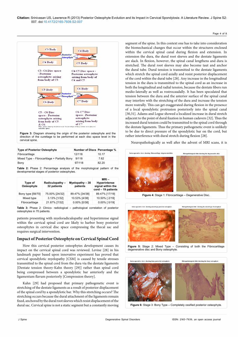
The C4-C5 and C5-C6 disc space is generally considered to have the greatest range of motion especially in the sagittal plane [17,18]. In these segments it was observed that the posterior osteophytes originates from the bodies of both the vertebra in the majority of cases (Figure 1-3) (Table 1). At junctional areas like C3-C4 and C6 –C7 the posterior osteophyte originates from the relatively fixed vertebra like C3 and C7 (Table 1). The C3 and C7 vertebral bodies are labelled as relatively fixed vertebra, since C3 articulate with the C1-C2 vertebras which are structurally different and they in turn articulate with the fixed skull and lower down C7 articulates with dorsal vertebra which is fixed by the chest cage and hence are relatively immobile vertebral bodies [17,18].
The surgical importance of this is the edge of the posterior osteophytes lies in the center within the C4-C5, C5-C6 intervertebral disc spaces while it is close to the inferior C4 body at C3-C4 disc space and close to the C6 superior body at C6-C7 disc space. Hence when the surgeon starts to remove the posterior osteophytes either by drilling or curetting the most important step is to identify the edge of the posterior osteophytes within the intervertebral disc space. At C4-C5, C5-C6 disc spaces since the edge lies in the center, one has to curette upwards to remove the posterior osteophytes arising from the superior body and then come back and curette downwards towards the inferior body to excise the inferiorly placed posterior osteophytes. While at C3- C4 disc space in majority of cases since the edge of the osteophytes is close to the body of C4 one has to angulate the operating microscope upwards and has to start curetting upwards from the edge of the osteophytes towards the C3 body. At C6-C7 disc space it is vice versa and the operating microscope has to be angulated inferiorly towards C7 body since the edge of the osteophytes is close to the C6 body and the curetting should be downwards towards C7 body. This also applies when one uses high speed microdrill for removing the posterior osteophytes.
Indiscriminate drilling of the osteophytes leads to detachment of the osteophytes from the body, after which it is extremely difficult to
Table 1: Phase 1: Showing the percentage of origin of the posterior osteophyte at each cervical intervertebral disc space level in 294 disc spaces in 201 patients as observed under operating microscope.
Disc Space/ Number of Discs Upper Vertebra Lower Vertebra Both VertebraC2-C3 [1] 100 % [1/1] 0.00% [0/1] 0.00% [0/1]C3-C4 [32] 78.13% [25/32] 18.75% [6/32] 3.12% [1/32]C4-C5 [62] 25.80% [16/62] 3.23% [2/62] 70.96% [44/62]C5-C6 [123] 9.75% [12/123] 5.69% [7/123] 84.55% [104/123]C6-C7 [74] 6.75% [5/74] 71.62% [53/74] 21.62% [16/74]C7 –D1 [2] 0.00% [0/2] 100.00% [2/2] 0.00% [0/2]
MRI showing the origin of theposterior osteophyte from thebody of C3.
C3 – C4 DISC SPACEIntraoperative photo - Posterior osteophyte from the Body of C3.
C4- C5 DISC SPACEIntraoperative photo - Posterior osteophyte arising from bodies of C4 & C5
Posterior Osteophyte
Posterior Osteophyte
C3 BODY
C4 BODY
C5 BODYC4 BODY
MRI showing the Multiple levelcompression - even at C4- C5.
Figure 1: MRI – Intraoperative Photographs showing the Origin of the Posterior Osteophytes at Cervical Disc Space Levels C3- C4, C4 –C5.
Citation: Srinivasan US, Lawrence R (2013) Posterior Osteophyte Evolution and its Impact in Cervical Spondylosis: A Literature Review. J Spine S2: 007. doi:10.4172/2165-7939.S2-007
Page 3 of 8
J Spine Degenerative Spinal Disorders ISSN: 2165-7939, an open access journal
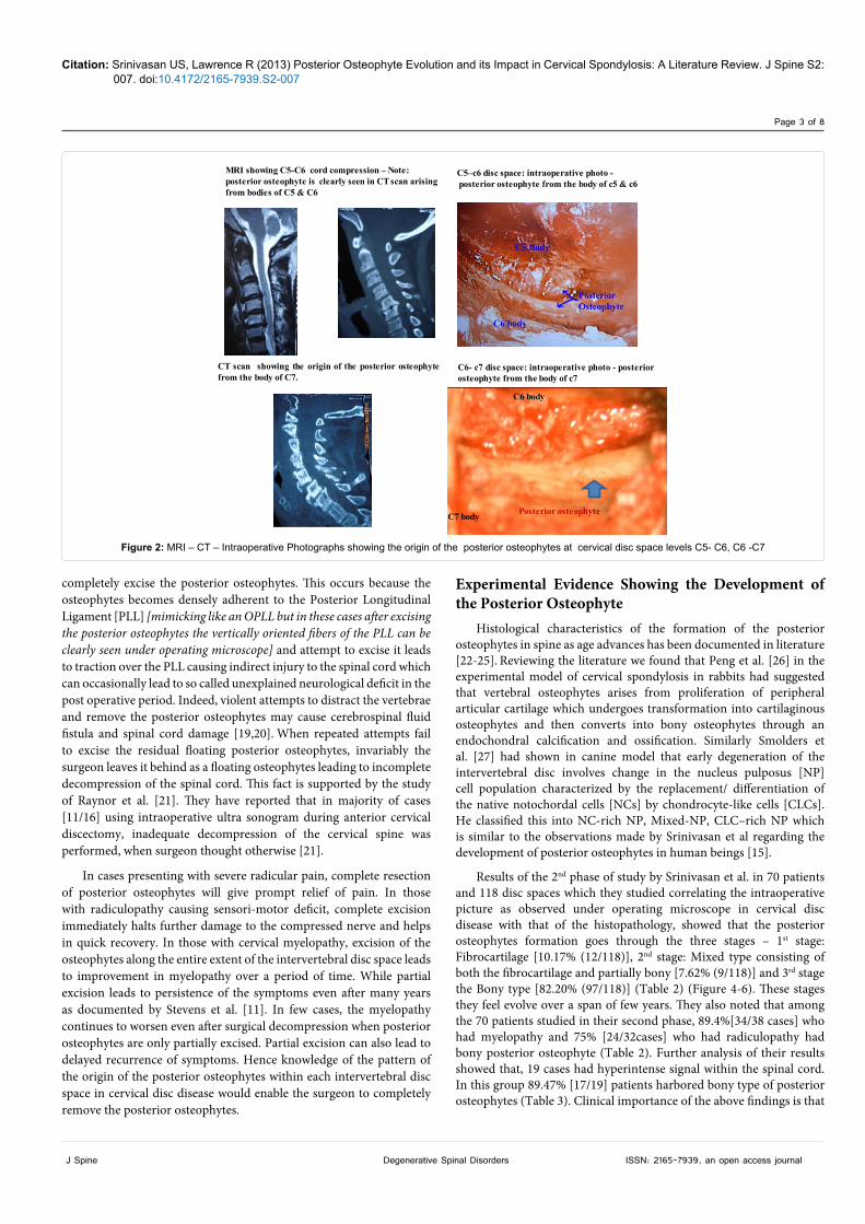
C5–c6 disc space: intraoperative photo -posterior osteophyte from the body of c5 & c6
C6- c7 disc space: intraoperative photo - posterior osteophyte from the body of c7
CT scan showing the origin of the posterior osteophytefrom the body of C7.
MRI showing C5-C6 cord compression – Note: posterior osteophyte is clearly seen in CT scan arising from bodies of C5 & C6
Posterior osteophyteC7 body
C6 body
Figure 2: MRI – CT – Intraoperative Photographs showing the origin of the posterior osteophytes at cervical disc space levels C5- C6, C6 -C7
completely excise the posterior osteophytes. This occurs because the osteophytes becomes densely adherent to the Posterior Longitudinal Ligament [PLL] [mimicking like an OPLL but in these cases after excising the posterior osteophytes the vertically oriented fibers of the PLL can be clearly seen under operating microscope] and attempt to excise it leads to traction over the PLL causing indirect injury to the spinal cord which can occasionally lead to so called unexplained neurological deficit in the post operative period. Indeed, violent attempts to distract the vertebrae and remove the posterior osteophytes may cause cerebrospinal fluid fistula and spinal cord damage [19,20]. When repeated attempts fail to excise the residual floating posterior osteophytes, invariably the surgeon leaves it behind as a floating osteophytes leading to incomplete decompression of the spinal cord. This fact is supported by the study of Raynor et al. [21]. They have reported that in majority of cases [11/16] using intraoperative ultra sonogram during anterior cervical discectomy, inadequate decompression of the cervical spine was performed, when surgeon thought otherwise [21].
In cases presenting with severe radicular pain, complete resection of posterior osteophytes will give prompt relief of pain. In those with radiculopathy causing sensori-motor deficit, complete excision immediately halts further damage to the compressed nerve and helps in quick recovery. In those with cervical myelopathy, excision of the osteophytes along the entire extent of the intervertebral disc space leads to improvement in myelopathy over a period of time. While partial excision leads to persistence of the symptoms even after many years as documented by Stevens et al. [11]. In few cases, the myelopathy continues to worsen even after surgical decompression when posterior osteophytes are only partially excised. Partial excision can also lead to delayed recurrence of symptoms. Hence knowledge of the pattern of the origin of the posterior osteophytes within each intervertebral disc space in cervical disc disease would enable the surgeon to completely remove the posterior osteophytes.
Experimental Evidence Showing the Development of the Posterior Osteophyte
Histological characteristics of the formation of the posterior osteophytes in spine as age advances has been documented in literature [22-25]. Reviewing the literature we found that Peng et al. [26] in the experimental model of cervical spondylosis in rabbits had suggested that vertebral osteophytes arises from proliferation of peripheral articular cartilage which undergoes transformation into cartilaginous osteophytes and then converts into bony osteophytes through an endochondral calcification and ossification. Similarly Smolders et al. [27] had shown in canine model that early degeneration of the intervertebral disc involves change in the nucleus pulposus [NP] cell population characterized by the replacement/ differentiation of the native notochordal cells [NCs] by chondrocyte-like cells [CLCs]. He classified this into NC-rich NP, Mixed-NP, CLC–rich NP which is similar to the observations made by Srinivasan et al regarding the development of posterior osteophytes in human beings [15].
Results of the 2nd phase of study by Srinivasan et al. in 70 patients and 118 disc spaces which they studied correlating the intraoperative picture as observed under operating microscope in cervical disc disease with that of the histopathology, showed that the posterior osteophytes formation goes through the three stages – 1st stage: Fibrocartilage [10.17% (12/118)], 2nd stage: Mixed type consisting of both the fibrocartilage and partially bony [7.62% (9/118)] and 3rd stage the Bony type [82.20% (97/118)] (Table 2) (Figure 4-6). These stages they feel evolve over a span of few years. They also noted that among the 70 patients studied in their second phase, 89.4%[34/38 cases] who had myelopathy and 75% [24/32cases] who had radiculopathy had bony posterior osteophyte (Table 2). Further analysis of their results showed that, 19 cases had hyperintense signal within the spinal cord. In this group 89.47% [17/19] patients harbored bony type of posterior osteophytes (Table 3). Clinical importance of the above findings is that
Citation: Srinivasan US, Lawrence R (2013) Posterior Osteophyte Evolution and its Impact in Cervical Spondylosis: A Literature Review. J Spine S2: 007. doi:10.4172/2165-7939.S2-007
Page 4 of 8
J Spine Degenerative Spinal Disorders ISSN: 2165-7939, an open access journal
patients presenting with myeloradiculopathy and hyperintense signal within the cervical spinal cord are likely to harbor bony posterior osteophytes in cervical disc space compressing the thecal sac and requires surgical intervention.
Impact of Posterior Osteophyte on Cervical Spinal CordHow this cervical posterior osteophytes development causes its
impact on the cervical spinal cord was reviewed. Levine [28] in his landmark paper based upon innovative experiment has proved that cervical spondylotic myelopathy [CSM] is caused by tensile stresses transmitted to the spinal cord from the dura via the dentate ligaments [Dentate tension theory-Kahn theory [29]] rather than spinal cord being compressed between a spondylotic bar anteriorly and the ligamentum flavum posteriorly [Compression theory].
Kahn [29] had proposed that primary pathogenetic event is stretching of the dentate ligaments as a result of posterior displacement of the spinal cord by a spondylotic bar. Why this stretching occurs? The stretching occurs because the dural attachment of the ligaments remain fixed, anchored by the dural root sleeves which resist displacement of the dural sac. Cervical spine is not a static segment but a constantly moving
segment of the spine. In this context one has to take into consideration the biomechanical changes that occur within the structures enclosed within the cervical spinal canal during flexion and extension. In extension the dura, the dural root sleeves and the dentate ligaments are slack. In flexion, however, the spinal canal lengthens and dura is stretched. The dural root sleeves may also become taut and anchor the dural tube. Dural tension is transmitted to the dentate ligaments which stretch the spinal cord axially and resist posterior displacement of the cord within the dural tube [28]. Any increase in the longitudinal tension in the dura is transmitted to the spinal cord as an increase in both the longitudinal and radial tension, because the dentate fibers run medio-laterally as well as rostrocaudally. It has been speculated that tension between the dura and the anterior surface of the spinal canal may interfere with the stretching of the dura and increase the tension more rostrally. This can get exaggerated during flexion in the presence of a local spondylotic protrusion posteriorly into the spinal canal [30,31]. Adams and Logue showed a localized increase in dural stretch adjacent to the point of dural fixation in human cadavers [32]. Thus the increased dural tension could be transmitted to the spinal cord through the dentate ligaments. Thus the primary pathogenetic event is unlikely to be due to direct pressure of the spondylotic bar on the cord, but rather interference with dural stretch during flexion [28].
Neuropathologically as well after the advent of MRI scans, it is
Table 2: Phase 2: Percentage analysis of the morphological pattern of the developmental stages of posterior osteophytes.
Type of Posterior Osteophyte Number of Discs Percentage %Fibrocartilage 12/118 10.17Mixed Type – Fibrocartilage + Partially Bony 9/118 7.62Bony 97/118 82.20
Table 3: Phase 2: Clinico- radiological – pathological correlation of posterior osteophytes in 70 patients.
Type of Osteophyte
Radiculopathy – 32 patients
Myelopathy – 38 patients
MRI – Hyperintense
signal within the cord – 19 patients
Bony type [58/70] 75.00% [24/32] 89.47% [34/38] 89.47% [17/19]Mixed type 3.13% [1/32] 10.53% [4/38] 10.50% [ 2/19]
Fibrocartilage 21.87% [7/32] 0.00% [0/38] 0.00% [ 0/19]
Figure 3: Diagram showing the origin of the posterior osteophyte and the direction of the curettage to be performed at each disc space level in the cervical spine.
Histopathological slide showing the degenerative discIntra-operative view showing fibrocartilage -degenerated disc
Figure 4: Stage 1: Fibrocartilage – Degenerative Disc.
Intra-operative view showing mixed type posterior osteophyte Histopathological slide showing the mixed type of osteophyte
Figure 5: Stage 2: Mixed Type – Consisting of both the Fibrocartilage degenerative disc and Bony osteophyte.
Intra-operative view showing bony posterior osteophyte Histopathological slide showing the bony osteophyte
Figure 6: Stage 3: Bony Type – Completely ossified posterior osteophyte.

Citation: Srinivasan US, Lawrence R (2013) Posterior Osteophyte Evolution and its Impact in Cervical Spondylosis: A Literature Review. J Spine S2: 007. doi:10.4172/2165-7939.S2-007
Page 5 of 8
J Spine Degenerative Spinal Disorders ISSN: 2165-7939, an open access journal
observed that there is pronounced flattening of the spinal cord in the anteroposterior dimension at the level of the spondylotic bar [33-35]. This observation could be explained by the transmission of increased lateral and longitudinal tension from the dentate ligaments to the spinal cord. The nervous tissue is relatively incompressible and hence the resulting longitudinal and transverse elongations will be accompanied by a compensatory contraction in the remaining anteroposterior dimension. It has also been observed that in follow up MRI scans, even after complete removal of the compressing spondylotic bar still the normal contour of the cervical spinal cord is not restored in the anteroposterior sections. At autopsy studies it is clearly documented that, there is thickening and fibrosis in the dentate ligaments and there is permanent flattening and widening of the spinal cord even after it is removed and freed of the stress [28]. This non-restoration of the normal contour of the cervical spinal cord in antero-posterior direction seen in MRI scans even after removal of the spondylotic bar is probably due to the above fibrosis of the dentate ligaments [35]. This fibrosis represents a physiological response to chronically raised levels of stress in these tissues [36]. Thus a spondylotic bar can increase dentate tension by displacing the spinal cord dorsally and also increase dentate tension by interfering locally with dural stretch during neck flexion with the resultant stress being transmitted to the spinal cord. Levine also has given convincing explanation supporting Breig’s vascular theory [35]
which states that transversely running blood vessels become elongated as the spinal cord is stretched transversely with resulting narrowing of the vessels in cross section. Such narrowing might cause ischemia which could contribute to the neuropathology [28].
Radiological Evidence of Impact of Posterior OsteophyteChronic pressure over the cervical spinal cord by the posterior
osteophytes leads to focal ischemia opposite to the disc space. These ischemic changes noted in the cervical spinal cord is seen in MRI as hyperintense signal. Intramedullary spinal cord changes in signal intensity in patients with CSM can be reversible (hyperintensity on T2-weighted imaging) or nonreversible (hypointensity on T1-weighted imaging). The regression of areas of hyperintensity on T2-weighted imaging is associated with a better prognosis. The T1-weighted hypointensity is an expression of irreversible damage and, therefore, the worst prognosis [37]. Vedantam et al. have classified this intermittent signal intensity [ISI] changes in T2 weighted MRI images into three types: No Change [Type 0], Fuzzy [Type 1] and Sharp [Type 2] [38]
(Figure 7). Both multisegmental T2W ISI and sharp, intense T2W ISI are associated with poorer surgical outcome (Class II evidence). The regression of T2W ISI postoperatively correlates with better functional outcomes (Class II).
An “absolute stenosis” in cervical spinal canal has been defined as a sagittal canal diameter of less than10mm, a “relative stenosis” as a canal diameter of less than 13mm and a normal sagittal diameter in the mid-cervical spine as 14-18mm [39]. However, these measurements are subject to genetic variations between different ethnic groups. Boden in a prospective study of asymptomatic individuals noted a high false positive rate of stenosis using these absolute measurements. He observed cervical stenosis in asymptomatic individuals using MRI imaging, in 14% of individuals less than the age of 40 and 28% over the age of 40. Disc degeneration was noted in 60% of those over the age of 40 [40]. Muhle et al. [41] have shown that significant increase of spinal stenosis have been observed in extension more so than in flexion. Dynamic MRI studies have shown that patients with a congenitally narrow spinal canal [<13mm] predispose to the development of radiographic dynamic cord compression and potentially clinical myelopathy [42].
Analysis of our results in 201 patients show that at a sagittal diameter of less than 8mm in MRI scan opposite to the disc space patient develop early clinical evidence of cervical myelopathy (Figure 8). This narrowing of the sagittal diameter occurs due to the development of the posterior osteophytes which projects into the cervical spinal canal causing indentation over the spinal cord. As such the diagnosis of CSM requires consideration of history, physical examination and imaging studies for each individual patient [43].
Fate of the Posterior Osteophyte after Anterior Fusion Surgery and Recent Prognostic Indicators of Outcome
Majority of spine surgeons opine that after good interbody fusion the posterior osteophytes get resorbed in the long term. But what the evidence based medicine states? Stevens et al. [11] and Seo et al. [44] have shown that there was no evidence of remodeling or resorption of osteophytes and persistent posterior osteophytes continue to deform the spinal cord for up to 5 to 12 years after achieving good anterior interbody fusion following anterior cervical disc surgery (Figure 9). Indeed in Seo et al. series in 5 patients among 31 cases it increased in size and in 19 it remained the same [45]. The importance of this in relation to cervical spinal surgery is that every effort should be made to remove posterior osteophytes during anterior interbody fusion [11,45].
MRI showing Type 2 sharp ISIMRI showing the Type 1 fuzzy
Figure 7: MRI of cervical spine showing the various types of hyperintense signal within the spinal cord.
Figure 8: MRI of cervical spine showing antero-posterior spinal cord diameter less than 8mm.

Citation: Srinivasan US, Lawrence R (2013) Posterior Osteophyte Evolution and its Impact in Cervical Spondylosis: A Literature Review. J Spine S2: 007. doi:10.4172/2165-7939.S2-007
Page 6 of 8
J Spine Degenerative Spinal Disorders ISSN: 2165-7939, an open access journal
We agree with them and in all cases we had excised the posterior osteophytes to achieve good results. These were documented by following up of our patients for periods greater than 5 years along with post operative MRI scans. Lack of expansion of the spinal cord even after 6 months post surgery indicates poor prognosis [45]. Thus we opine it is probably due to inadequate decompression of the spinal cord which has occurred due to partial removal of the posterior osteophytes or non-removal of it.
Rajasekaran et al. [46] in their study using MRI spinal tractography and diffusion tensor imaging [DTI] indices Apparent Diffusion Coefficient [ADC], Fractional Anatomy [FA] and Eigen Values [E1, E2 and E3] in pre and post decompression cervical myelopathic patients had stated that the postoperative DTI values [ADC, E1 and E2] of patients with Nurick grading 2 and 3 were observed to approach normal values indicating restoration of linear diffusion in neuronal tracts. DTI indices in patients with Nurick Grade 4 and 5 remained same or worsened more. This could probably indicate persistent axonal damage. These results suggest that DTI indices especially ADC and Eigen vectors can be promising indicators in the evaluation of prognostic assessment of myelopathy patients. Alkhatib et al. [47] have identified a biomarker for disk degeneration called Chondroadherin [CHAD] fragmentation from analyzing the healthy intervertebral discs [IVD] obtained through organ donations and from patients with degenerative disc disease and scoliosis and in future it is likely to be the biomarker of disc degeneration even before MRI changes occur.
Natural History of CSMIn this era of evidence based medicine with information pouring
into patient’s smart phone through internet what if we are questioned on natural history of CSM. Let us have a look at it. Matz et al. [48] have suggested that patients with CSM experience stable quiescent periods with an intervening slow and stepwise decline in function. Oshima et al. [49] have suggested that 62% of patients with mild CSM will not deteriorate or undergo surgery at 10 years. But systematic review of literature by Lebl et al. [43] suggest a mixed course with many patients having quiescent disease for long periods of time while
others experiencing a slow, stepwise decline. Thus review of literature shows that the clinical course of cervical myelopathy is variable and that conservative management may result in stability or improvement of symptoms in the majority of the patients with mild symptoms [50-53]. Predicting the clinical course of the individual patient is difficult but some evidence suggests that younger patients and those with mild symptoms are more likely to improve [1,53].
Take Home Message• There is a definite macroscopic and histopathological pattern
in the formation of cervical posterior osteophytes which is supported both by the animal studies and in vivo human being studies. It goes through three stages of formation: 1: Fibrocartilage 2: Mixed type 3: Bony posterior osteophytes.
• Patients presenting with myeloradiculopathy and hyperintense signal within the cervical spinal cord are likely to harbor bony posterior osteophytes compressing the thecal sac and requires surgical intervention.
• In mobile segments like C4-C5, C5- C6 intervertebral disc spaces the posterior osteophytes arises from bodies of both the adjacent vertebra while at C3-C4, C6-C7 it arises from relatively fixed vertebra like C3 and C7 respectively.
• The edge of the posterior osteophytes lies in the center within the C4-C5, C5-C6 intervertebral disc spaces while it is close to the inferior C4 body at C3-C4 disc space and close to the C6 superior body at C6-C7 disc space.
• Posterior osteophytes definitely produces an impact over the cervical spinal cord and causes cervical spondylotic myelopathy most probably by producing tension over the dentate ligaments and shearing force over the cervical spinal cord. It also causes changes in the small intramedullary vessels leading to ischemia which could explain sudden deterioration as observed in natural history of the disease. Radiological evidence for the above is available at present as seen in MRI scans.
• Even though operative treatment especially the anterior cervical discectomy remains the standard care for moderate to severe CSM, it is clearly evident that the cervical posterior osteophytes has to be excised to achieve good outcome.
• Posterior osteophytes do not get resorbed even in the long run even after achieving good bony fusion.
• Intramedullary spinal cord changes in signal intensity in patients with CSM can be reversible (hyperintensity on T2-weighted imaging) or nonreversible (hypointensity on T1-weighted imaging). The regression of areas of hyperintensity on T2-weighted imaging is associated with a better prognosis. Lack of expansion of the spinal cord after surgical decompression even after 6 months indicate poor prognosis.
• Diffusion Tractography Indices using MRI spinal tractography especially Apparent Diffusion Coefficient [ADC] and Eigen Vectors can be promising indicators in the evaluation and prognostic assessment of myelopathy patients.
• Natural history of CSM suggests a mixed course with many patients having quiescent disease for long periods of time while others experiencing a slow, stepwise decline.
• In future biochemical marker for disc degeneration is likely to
Figure 9: CT scan showing the presence of posterior osteophyte even after good fusion at C5-C6.

Citation: Srinivasan US, Lawrence R (2013) Posterior Osteophyte Evolution and its Impact in Cervical Spondylosis: A Literature Review. J Spine S2: 007. doi:10.4172/2165-7939.S2-007
Page 7 of 8
J Spine Degenerative Spinal Disorders ISSN: 2165-7939, an open access journal
be available to identify the patients who are likely to develop cervical spondylotic myelopathy at an early stage even before radiological evidence occurs.
Acknowledgements
We wish to acknowledge the support and encouragement given by Prof.Dr.P.V.A. Mohandas – Managing Director of MIOT Hospitals, Chennai, India.
References
1. Yarbrough CK, Murphy RK, Ray WZ, Stewart TJ (2012) The natural history and clinical presentation of cervical spondylotic myelopathy. Adv Orthop 2012: 480643.
2. Berge J, Marque B, Vital JM, Sénégas J, Caillé JM (1999) Age-related changes in the cervical spines of front-line rugby players. Am J Sports Med 27: 422-429.
3. Quarrie KL, Cantu RC, Chalmers DJ (2002) Rugby union injuries to the cervical spine and spinal cord. Sports Med 32: 633-653.
4. Wada E, Ebara S, Saito S, Ono K (1992) Experimental spondylosis in the rabbit spine. Overuse could accelerate the spondylosis. Spine (Phila Pa 1976) 17: S1-6.
5. Morishita Y, Naito M, Wang JC (2011) Cervical spinal canal stenosis: the differences between stenosis at the lower cervical and multiple segment levels. Int Orthop 35: 1517-1522.
6. Edwards WC, LaRocca H (1983) The developmental segmental sagittal diameter of the cervical spinal canal in patients with cervical spondylosis. Spine (Phila Pa 1976) 8: 20-27.
7. Gore DR (2001) Roentgenographic findings in the cervical spine in asymptomatic persons: a ten-year follow-up. Spine (Phila Pa 1976) 26: 2463-2466.
8. Hayashi H, Okada K, Hashimoto J, Tada K, Ueno R (1988) Cervical spondylotic myelopathy in the aged patient. A radiographic evaluation of the aging changes in the cervical spine and etiologic factors of myelopathy. Spine (Phila Pa 1976) 13: 618-625.
9. Brain WR, Knight GC, Bull JW (1948) Discussion of rupture of the intervertebral disc in the cervical region. Proc R Soc Med 41: 509-516.
10. Brain WR, Northfield D, Wilkinson M (1952) The neurological manifestations of cervical spondylosis. Brain 75: 187-225.
11. Stevens JM, Clifton AG, Whitear P (1993) Appearances of posterior osteophytes after sound anterior interbody fusion in the cervical spine: a high-definition computed myelographic study. Neuroradiology 35: 227-228.
12. Wilkinson M (1967) Pathology. In: Cervical spondylosis and other disorders of the cervical spine. (1stedn). Brain L and Wilkinson M. William Heinemann medical books limited. London.
13. Torrens MJ (1992) Cervical disc disease. In: Surgery of the spine: a combined orthopedic and neurosurgical approach. Findlay G, Owen R Blackwell scientific publications, London.
14. Truumees E (2005) Pain and neurologic dysfunction. In: The cervical spine. (4thedn) Ed. The cervical spine research society editorial committee Clark CR. Lippincott William & Wilkins, Philadelphia.
15. Srinivasan US, Radhi L (2012) Developmental pattern of the posterior osteophytes in cervical disk disease-an intraoperative, histopathological study. Global Spine Journal 2: Suppl S1: S74-S75.
16. Robinson RA, Smith GW (1955) Anterolateral cervical disc removal and interbody fusion for cervical disc syndrome. Bull Johns Hopkins Hosp 96: 223-224.
17. Penning L (1989) Functional anatomy of joints and discs. In: The cervical spine. (2ndedn) Ed. The cervical spine research society editorial committee-Sherk HH, Dunn EJ, Eismont FJ et al. J.B. Lippincott Company, Philadelphia.
18. Jofe MH, White AA, Panjabi MH (1989) Clinically relevant kinematics of the cervical spine. In: The cervical spine (2ndedn). The cervical spine research society editorial committee, Philadelphia.
19. Saunders RL, Batzdorf U, Raynor RB (1999) Cervical discectomy. In: Spine surgery, techniques, complication avoidance and management. Benzel EC, Churchill Livingstone, New York.
20. Hoff JT, Papdopoulous SM (1996) Cervical disc disease and cervical spondylosis. In: Neurosurgery. Ed. Wilkins RH, Rengachary SS, McGraw-Hill, New York.
21. Raynor RB (1997) Intraoperative ultrasound for immediate evaluation of anterior cervical decompression and discectomy. Spine (Phila Pa 1976) 22: 389-395.
22. Gen H (1990) [A clinicopathological study of cervical intervertebral discs--Part 2: On morphological and roentgenologic findings]. Nihon Seikeigeka Gakkai Zasshi 64: 572-582.
23. Prescher A (1998) Anatomy and pathology of the aging spine. Eur J Radiol 27: 181-195.
24. Longo UG, Ripalda P, Denaro V, Forriol F (2006) Morphologic comparison of cervical, thoracic, lumbar intervertebral discs of cynomolgus monkey (Macaca fascicularis). Eur Spine J 15: 1845-1851.
25. Benjamin M, Toumi H, Suzuki D, Hayashi K, Mc Gonagle D (2009) Evidence for a distinctive pattern of bone formation in enthesophytes. Ann Rheum Dis 68: 1003-1010.
26. Peng B, Hou S, Shi Q, Jia L (2000) Experimental study on mechanism of vertebral osteophyte formation. Chin J Traumatol 3: 202-205.
27. Smolders L, Meij B, Onis D, Reimers F, Bergknut N, et al. Gene expression profiling of early intervertebral disk degeneration. Global Spine Journal 2012: Suppl S1: S17–S18.
28. Levine DN (1997) Pathogenesis of cervical spondylotic myelopathy. J Neurol Neurosurg Psychiatry 62: 334-340.
29. Kahn EA (1947) The role of the dentate ligaments in spinal cord compression and the syndrome of lateral sclerosis. J Neurosurg 4: 191–199.
30. Reid JD (1960) Effects of flexion-extension movements of the head and spine upon the spinal cord and nerve roots. J Neurol Neurosurg Psychiatry 23: 214-221.
31. Tencer AF, Allen BL Jr, Ferguson RL (1985) A biomechanical study of thoracolumbar spine fractures with bone in the canal. Part III. Mechanical properties of the dura and its tethering ligaments. Spine (Phila Pa 1976) 10: 741-747.
32. Adams CB, Logue V (1971) Studies in cervical spondylotic myelopathy. I. Movement of the cervical roots, dura and cord, and their relation to the course of the extrathecal roots. Brain 94: 557-568.
33. Bedford PD, Bosanquet FD (1952) Degeneration of the spinal cord associated with cervical spondylosis. Lancet 2: 55-59.
34. Wilkinson M (1960) The morbid anatomy of cervical spondylosis and myelopathy. Brain 83: 589-617.
35. Breig A, Turnbull I, Hassler O (1966) Effects of mechanical stresses on the spinal cord in cervical spondylosis. A study on fresh cadaver material. J Neurosurg 25: 45-56.
36. Cusick JF, Ackmann JJ, Larson SJ (1977) Mechanical and physiological effects of dentatotomy. J Neurosurg 46: 767-775.
37. Mastronardi L, Elsawaf A, Roperto R, Bozzao A, Caroli M, et al. (2007) Prognostic relevance of the postoperative evolution of intramedullary spinal cord changes in signal intensity on magnetic resonance imaging after anterior decompression for cervical spondylotic myelopathy. J Neurosurg Spine 7: 615-622.
38. Vedantam A, Rajshekhar V (2013) Does the type of T2-weighted hyperintensity influence surgical outcome in patients with cervical spondylotic myelopathy? A review. Eur Spine J 22: 96-106.
39. Wolf BS, Khilnani, Malis L (1956) The sagittal diameter of the bony cervical spinal canal and its significance in cervical spondylosis. J Mt Sinai Hosp N Y 23: 283-292.
40. Boden SD, McCowin PR, Davis DO, Dina TS, Mark AS, et al. (1990) Abnormal magnetic-resonance scans of the cervical spine in asymptomatic subjects. A prospective investigation. J Bone Joint Surg Am 72: 1178-1184.
41. Muhle C, Weinert D, Falliner A, Wiskirchen J, Metzner J, et al. (1998) Dynamic changes of the spinal canal in patients with cervical spondylosis at flexion and extension using magnetic resonance imaging. Invest Radiol 33: 444-449.
42. Morishita Y, Naito M, Hymanson H, Miyazaki M, Wu G, et al. (2009) The relationship between the cervical spinal canal diameter and the pathological changes in the cervical spine. Eur Spine J 18: 877-883.
43. Lebl DR, Hughes A, Cammisa FP Jr, O’Leary PF (2011) Cervical spondylotic myelopathy: pathophysiology, clinical presentation, and treatment. HSS J 7: 170-178.

Citation: Srinivasan US, Lawrence R (2013) Posterior Osteophyte Evolution and its Impact in Cervical Spondylosis: A Literature Review. J Spine S2: 007. doi:10.4172/2165-7939.S2-007
Page 8 of 8
J Spine Degenerative Spinal Disorders ISSN: 2165-7939, an open access journal
Submit your next manuscript and get advantages of OMICS Group submissionsUnique features:
• User friendly/feasible website-translation of your paper to 50 world’s leading languages• Audio Version of published paper• Digital articles to share and explore
Special features:
• 300 Open Access Journals• 25,000 editorial team• 21 days rapid review process• Quality and quick editorial, review and publication processing• Indexing at PubMed (partial), Scopus, EBSCO, Index Copernicus and Google Scholar etc• Sharing Option: Social Networking Enabled• Authors, Reviewers and Editors rewarded with online Scientific Credits• Better discount for your subsequent articles
Submit your manuscript at: http://www.omicsgroup.info/editorialtracking/spine/
This article was originally published in a special issue, Degenerative Spinal Disorders handled by Editor. Dr. Ely Steinberg, Sourasky Tel-Aviv Medical Center, Israel
Citation: Srinivasan US, Lawrence R (2013) Posterior Osteophyte Evolution and its Impact in Cervical Spondylosis: A Literature Review. J Spine S2: 007. doi:10.4172/2165-7939.S2-007
44. Seo JY, Ha KY (2012) Fate of posterior osteophytes in fused segments after anterior cervical discectomy and fusion. Spine (Phila Pa 1976) 37: 741-747.
45. Arvin B, Kalsi-Ryan S, Karpova A, Mercier D, Furlan JC, et al. (2011) Postoperative magnetic resonance imaging can predict neurological recovery after surgery for cervical spondylotic myelopathy. A prospective study with blinded assessments. Neurosurgery 69: 362-368.
46. Rajasekaran S, Prasad V, Shetty J, Kanna R (2012) Diffusion tensor imaging as a prognostic investigation in cervical myelopathy-comparison of pre- and post surgical decompression DTI indices and Tractography in 56 patients. Global Spine Journal 2: S101–S102.
47. Alkhatib B, Gawri R, Onnerfjord P, Ouellet J, Roughley P, et al. (2012) Chondroadherin fragmentation as a biochemical marker for early stage disk degeneration. Global Spine Journal 2: S16– S17.
48. Matz PG (2006) Does nonoperative management play a role in the treatment of cervical spondylotic myelopathy? Spine J 6: 175S-181S.
49. Oshima Y, Seichi A, Moni J, Chikuda H, Kawaguchi H, et al. (2010) Natural course and prognostic factors for mid cervical spondylotic myelopathy. Cervical spine research society: Annual Meeting 38: 83.
50. Kadanka Z, Mares M, Bednaník J, Smrcka V, Krbec M, et al. (2002) Approaches to spondylotic cervical myelopathy: conservative versus surgical results in a 3-year follow-up study. Spine (Phila Pa 1976) 27: 2205-2210.
51. Kadanka Z, Mares M, Bednarík J, Smrcka V, Krbec M, et al. (2005) Predictive factors for mild forms of spondylotic cervical myelopathy treated conservatively or surgically. Eur J Neurol 12: 16-24.
52. Shimomura T, Sumi M, Nishida K, Maeno K, Tadokoro K, et al. (2007) Prognostic factors for deterioration of patients with cervical spondylotic myelopathy after nonsurgical treatment. Spine (Phila Pa 1976) 32: 2474-2479.
53. Nakamura K, Kurokawa T, Hoshino Y, Saita K, Takeshita K, et al. (1998) Conservative treatment for cervical spondylotic myelopathy: achievement and sustainability of a level of “no disability”. J Spinal Disord 11: 175-179.