SRCT Fisher 3 - USCAP · 4/13/2016 1 Small Round Cell Tumors in Soft Tissue and Bone...
15
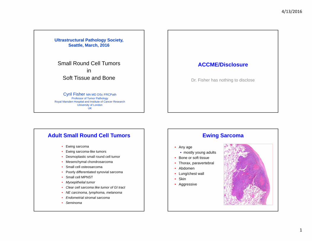
4/13/2016 1 Small Round Cell Tumors in Soft Tissue and Bone Ultrastructural Pathology Society, Seattle, March, 2016 Cyril Fisher MA MD DSc FRCPath Professor of Tumor Pathology Royal Marsden Hospital and Institute of Cancer Research University of London UK ACCME/Disclosure Dr. Fisher has nothing to disclose Adult Small Round Cell Tumors • Ewing sarcoma • Ewing sarcoma-like tumors • Desmoplastic small round cell tumor • Mesenchymal chondrosarcoma • Small cell osteosarcoma • Poorly differentiated synovial sarcoma • Small cell MPNST • Myoepithelial tumor • Clear cell sarcoma like tumor of GI tract • NE carcinoma, lymphoma, melanoma • Endometrial stromal sarcoma • Seminoma Ewing Sarcoma • Any age • mostly young adults • Bone or soft tissue • Thorax, paravertebral • Abdomen • Lung/chest wall • Skin • Aggressive
Transcript of SRCT Fisher 3 - USCAP · 4/13/2016 1 Small Round Cell Tumors in Soft Tissue and Bone...
4/13/2016
1
Small Round Cell Tumors in
Soft Tissue and Bone
Ultrastructural Pathology Society, Seattle, March, 2016
Cyril Fisher MA MD DSc FRCPathProfessor of Tumor Pathology
Royal Marsden Hospital and Institute of Cancer Research University of London
UK
ACCME/Disclosure
Dr. Fisher has nothing to disclose
Adult Small Round Cell Tumors
• Ewing sarcoma• Ewing sarcoma-like tumors• Desmoplastic small round cell tumor• Mesenchymal chondrosarcoma• Small cell osteosarcoma • Poorly differentiated synovial sarcoma• Small cell MPNST• Myoepithelial tumor • Clear cell sarcoma like tumor of GI tract• NE carcinoma, lymphoma, melanoma• Endometrial stromal sarcoma• Seminoma
Ewing Sarcoma
• Any age• mostly young adults
• Bone or soft tissue • Thorax, paravertebral• Abdomen• Lung/chest wall• Skin• Aggressive
4/13/2016
2
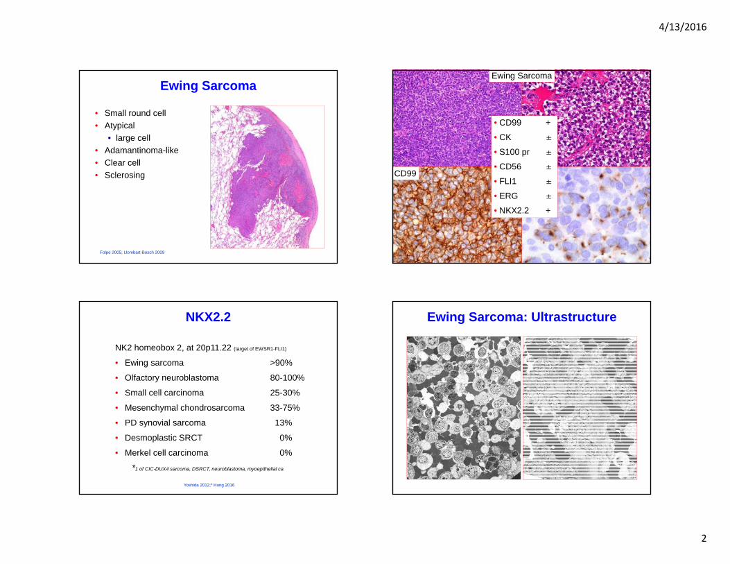
Ewing Sarcoma
• Small round cell• Atypical
• large cell• Adamantinoma-like• Clear cell• Sclerosing
Folpe 2005; Llombart-Bosch 2009
Ewing Sarcoma Ewing Sarcoma
CD99 CK
• CD99 +
• CK ±
• S100 pr ±
• CD56 ±
• FLI1 ±
• ERG ±
• NKX2.2 +
NKX2.2
NK2 homeobox 2, at 20p11.22 (target of EWSR1-FLI1)
• Ewing sarcoma >90%
• Olfactory neuroblastoma 80-100%
• Small cell carcinoma 25-30%
• Mesenchymal chondrosarcoma 33-75%
• PD synovial sarcoma 13%
• Desmoplastic SRCT 0%
• Merkel cell carcinoma 0%
*1 of CIC-DUX4 sarcoma, DSRCT, neuroblastoma, myoepithelial ca
Yoshida 2012;* Hung 2016
Ewing Sarcoma: Ultrastructure
4/13/2016
3
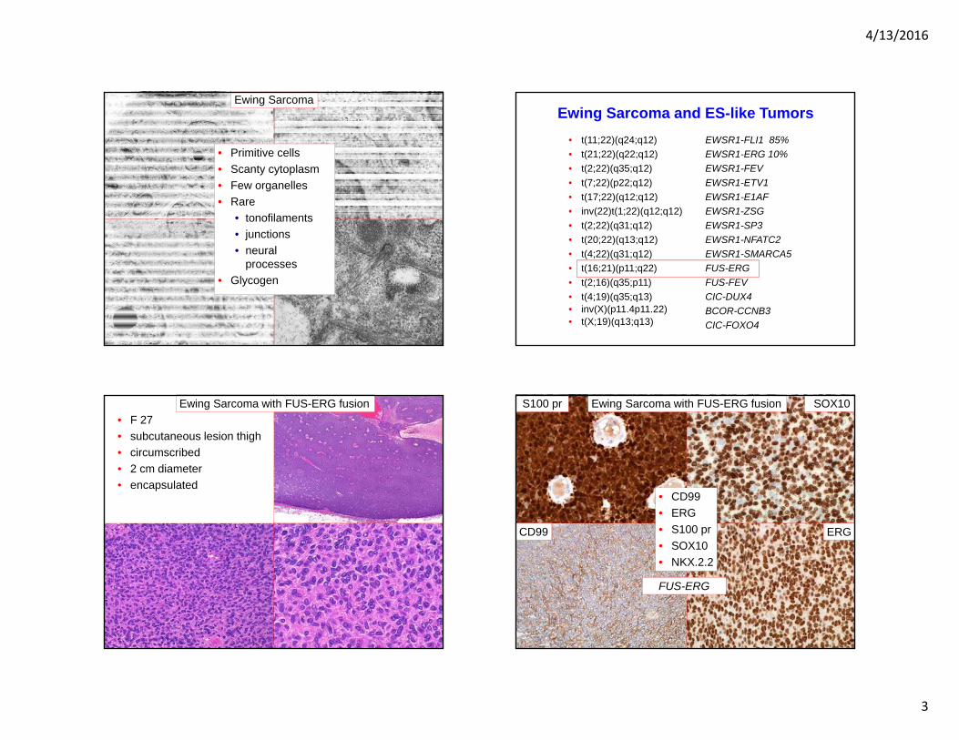
Ewing Sarcoma
• Primitive cells• Scanty cytoplasm• Few organelles• Rare
• tonofilaments• junctions• neural
processes• Glycogen
Ewing Sarcoma and ES-like Tumors• t(11;22)(q24;q12)• t(21;22)(q22;q12)• t(2;22)(q35;q12)• t(7;22)(p22;q12)• t(17;22)(q12;q12) • inv(22)t(1;22)(q12;q12)• t(2;22)(q31;q12)• t(20;22)(q13;q12)• t(4;22)(q31;q12)• t(16;21)(p11;q22)• t(2;16)(q35;p11)• t(4;19)(q35;q13)• inv(X)(p11.4p11.22)• t(X;19)(q13;q13)
EWSR1-FLI1 85%EWSR1-ERG 10% EWSR1-FEV EWSR1-ETV1EWSR1-E1AF EWSR1-ZSGEWSR1-SP3EWSR1-NFATC2EWSR1-SMARCA5FUS-ERGFUS-FEVCIC-DUX4BCOR-CCNB3CIC-FOXO4
• F 27 • subcutaneous lesion thigh• circumscribed• 2 cm diameter• encapsulated
Ewing Sarcoma with FUS-ERG fusion S100 pr Ewing Sarcoma with FUS-ERG fusion SOX10
ERG
• CD99• ERG• S100 pr• SOX10• NKX.2.2
CD99
FUS-ERG
4/13/2016
4
‘Promiscuous’ Fusions• EWSR1-CREB1
angiomatoid FH, clear cell sarcoma, CCSLTGIT, PPMS• EWSR1-ATF1
angiomatoid FH, clear cell sarcoma, CCSLTGITmyoepithelial tumor, angiosarcoma, salivary HCCC, CCOC
• ETV6-NTRK3infantile fibrosarcoma, inflammatory myofibroblastic tumor, mesoblastic nephroma, AML, secretory ca breast,mammary analogue secretory carcinoma of salivary glands, GIST
• ASPSCR1-TFE3alveolar soft part sa, juvenile renal cell carcinoma
• TMP3-ALKinflammatory myofibroblastic tumor, anaplastic large cell lymphoma
• YWHAE-NUTM22A/Bendometrial stromal sarcoma, clear cell sarcoma of kidney
• FUS-ERGEwing sarcoma, AML
• BRD4-NUTEwing-like sarcoma, thymic & other carcinomas
Ewing Sarcoma and ES-like Tumors• t(11;22)(q24;q12)• t(21;22)(q22;q12)• t(2;22)(q35;q12)• t(7;22)(p22;q12)• t(17;22)(q12;q12) • inv(22)t(1;22)(q12;q12)• t(2;22)(q31;q12)• t(20;22)(q13;q12)• t(4;22)(q31;q12)• t(16;21)(p11;q22)• t(2;16)(q35;p11)• t(4;19)(q35;q13)• inv(X)(p11.4p11.22)• t(X;19)(q13;q13)
EWSR1-FLI1 85%EWSR1-ERG 10% EWSR1-FEV EWSR1-ETV1EWSR1-E1AF EWSR1-ZSGEWSR1-SP3EWSR1-NFATC2EWSR1-SMARCA5FUS-ERGFUS-FEVCIC-DUX4BCOR-CCNB3CIC-FOXO4
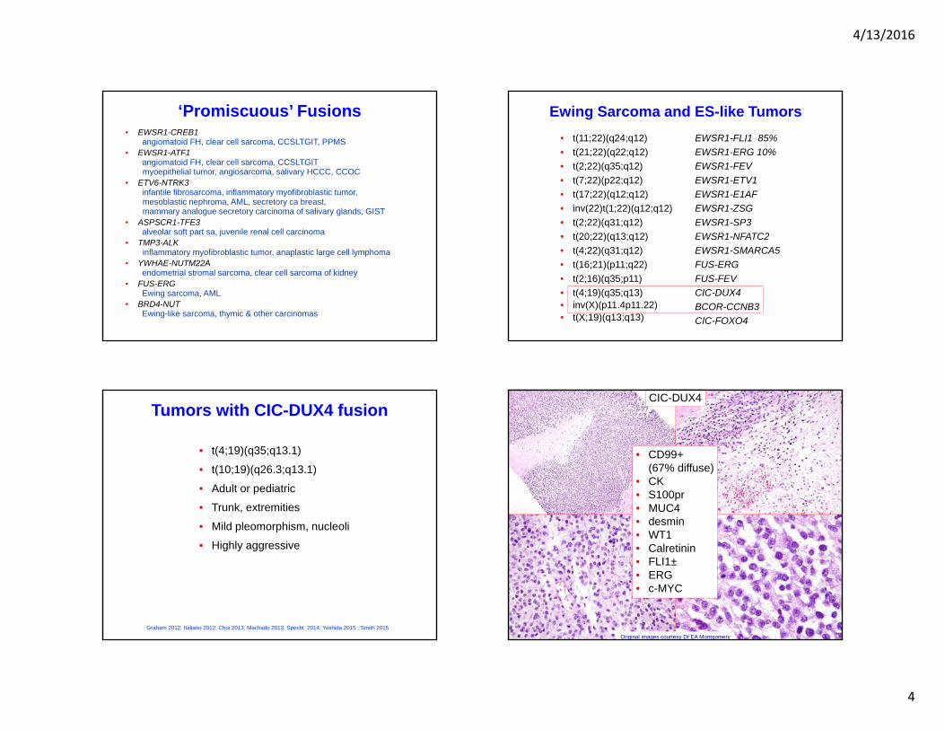
Tumors with CIC-DUX4 fusion
• t(4;19)(q35;q13.1)
• t(10;19)(q26.3;q13.1)
• Adult or pediatric
• Trunk, extremities
• Mild pleomorphism, nucleoli
• Highly aggressive
Graham 2012; Italiano 2012; Choi 2013; Machado 2013; Specht 2014; Yoshida 2015 ; Smith 2015
CIC-DUX4
• CD99+ (67% diffuse)
• CK• S100pr• MUC4• desmin• WT1• Calretinin• FLI1±• ERG• c-MYC
Original images courtesy Dr EA Montgomery
4/13/2016
5
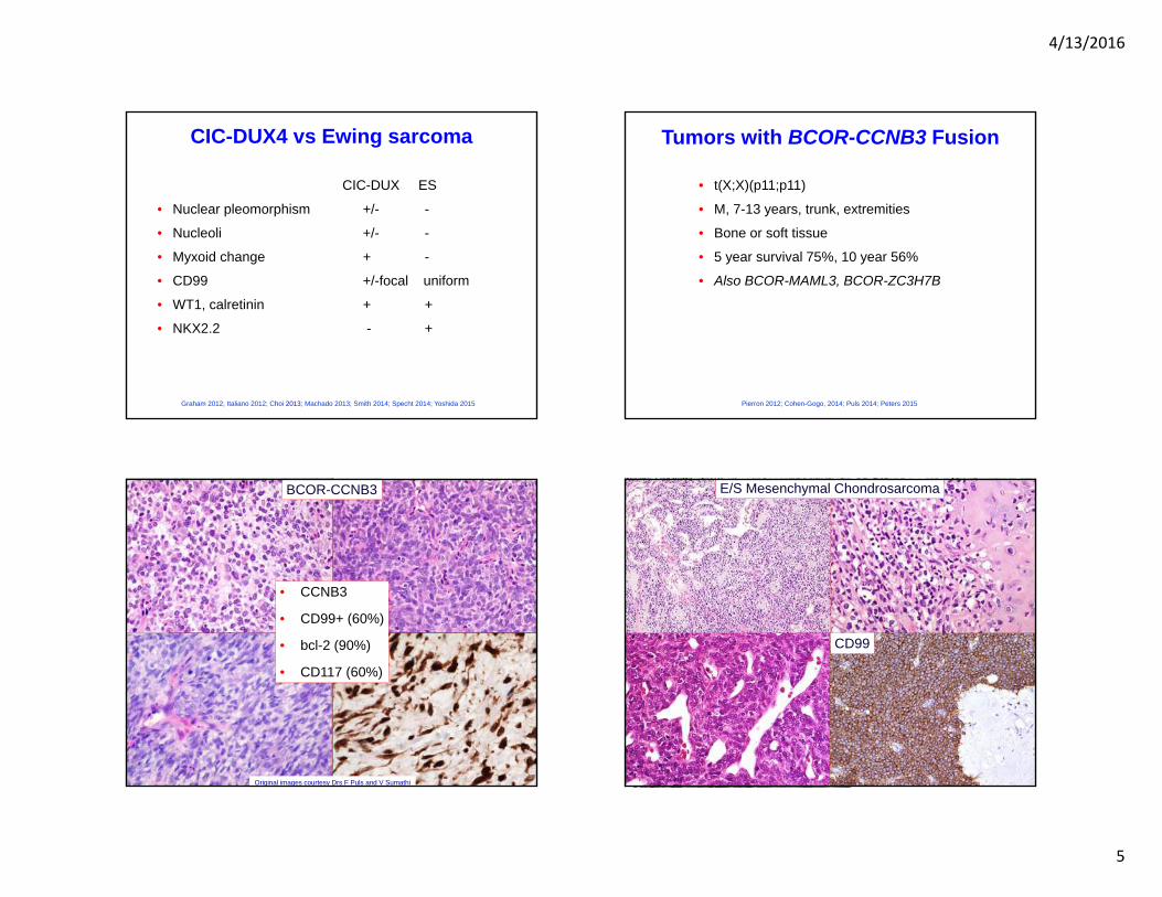
CIC-DUX4 vs Ewing sarcoma
CIC-DUX ES
• Nuclear pleomorphism +/- -
• Nucleoli +/- -
• Myxoid change + -
• CD99 +/-focal uniform
• WT1, calretinin + +
• NKX2.2 - +
Graham 2012; Italiano 2012; Choi 2013; Machado 2013; Smith 2014; Specht 2014; Yoshida 2015
Tumors with BCOR-CCNB3 Fusion
• t(X;X)(p11;p11)
• M, 7-13 years, trunk, extremities
• Bone or soft tissue
• 5 year survival 75%, 10 year 56%
• Also BCOR-MAML3, BCOR-ZC3H7B
Pierron 2012; Cohen-Gogo, 2014; Puls 2014; Peters 2015
BCOR-CCNB3
CD99 CCNB3
Original images courtesy Drs F Puls and V Sumathi
• CCNB3
• CD99+ (60%)
• bcl-2 (90%)
• CD117 (60%)
CD99
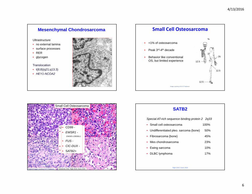
E/S Mesenchymal Chondrosarcoma
4/13/2016
6
Mesenchymal Chondrosarcoma
Ultrastructure• no external lamina• surface processes• RER• glycogen
Translocation• t(8;8)(q21;q13.3)• HEY1-NCOA2
Small Cell Osteosarcoma
• <1% of osteosarcoma
• Peak 3rd-4th decade
• Behavior like conventionalOS, but limited experience 12.5
%
12.5
37
12.5
25
Image courtesy of Dr R Tirabosco
Small Cell Osteosarcoma
Original images courtesy Dr R Tirabosco
Ultrastructure• fibroblastic
• RER• no external
lamina• glycogen• pre-osteoid
Debelenko 2011; Righi 2015, Davis 2015
• CD99 -
• EWSR1 -EWSR1-CREB3L1
• FUS -
• CIC-DUX -
• SATB2+
SATB2
Special AT-rich sequence binding protein 2 2q33
• Small cell osteosarcoma 100%
• Undifferentiated pleo. sarcoma (bone) 50%
• Fibrosarcoma (bone) 45%
• Mes chondrosarcoma 23%
• Ewing sarcoma 10%
• DLBC lymphoma 17%
Righi 2015; Davis 2015
4/13/2016
7
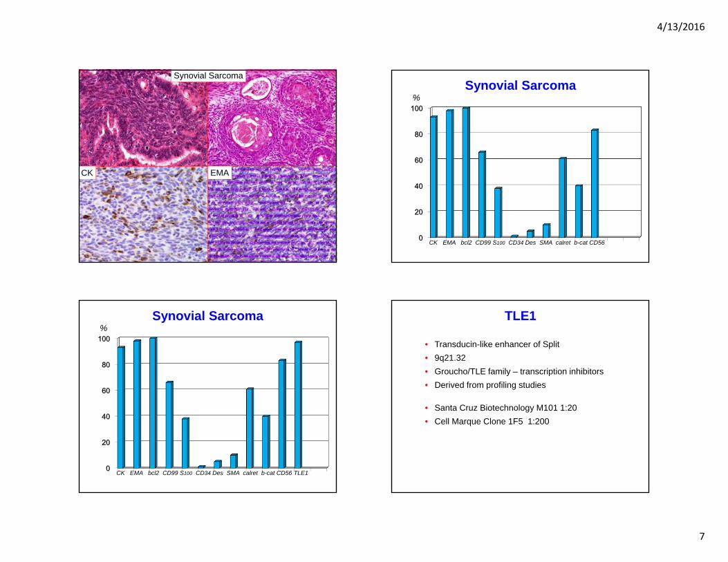
Synovial Sarcoma
CK EMA
0
20
40
60
80
100
Synovial Sarcoma
CK EMA bcl2 CD99 S100 CD34 Des SMA calret b-cat CD56
%
0
20
40
60
80
100
Synovial Sarcoma
CK EMA bcl2 CD99 S100 CD34 Des SMA calret b-cat CD56 TLE1
%TLE1
• Transducin-like enhancer of Split• 9q21.32• Groucho/TLE family – transcription inhibitors• Derived from profiling studies
• Santa Cruz Biotechnology M101 1:20• Cell Marque Clone 1F5 1:200
4/13/2016
8
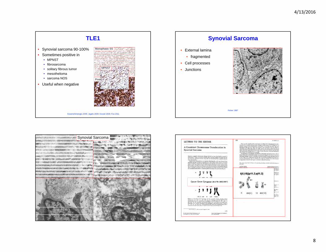
TLE1
• Synovial sarcoma 90-100% • Sometimes positive in
• MPNST• fibrosarcoma • solitary fibrous tumor • mesothelioma • sarcoma NOS
• Useful when negative
Kosemehmetoglu 2009; Jagdis 2009; Knosel 2009; Foo 2011
Monophasic SS
Poorly Differentiated SS
MPNST
Synovial Sarcoma
• External lamina
• fragmented
• Cell processes
• Junctions
Fisher 1987
Synovial Sarcoma
4/13/2016
9
Synovial Sarcoma
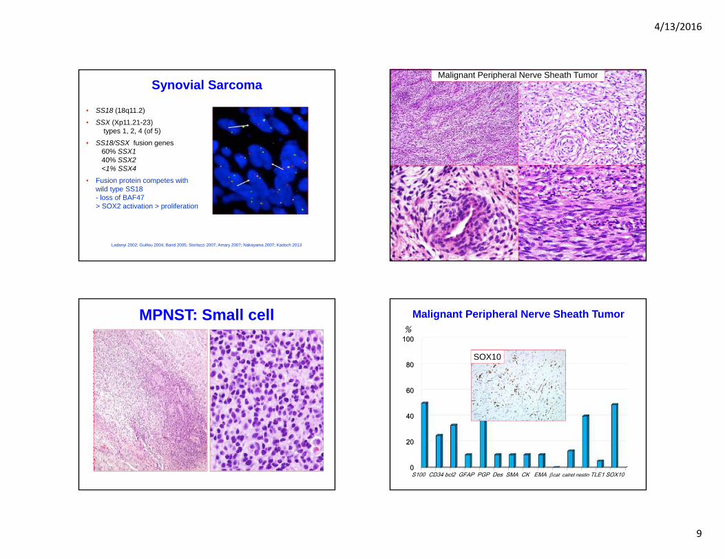
• SS18 (18q11.2)
• SSX (Xp11.21-23) types 1, 2, 4 (of 5)
• SS18/SSX fusion genes60% SSX140% SSX2<1% SSX4
• Fusion protein competes with wild type SS18 - loss of BAF47> SOX2 activation > proliferation
Ladanyi 2002; Guillou 2004; Baird 2005; Storlazzi 2007; Amary 2007; Nakayama 2007; Kadoch 2013
Malignant Peripheral Nerve Sheath Tumor
MPNST: Small cell
0
20
40
60
80
100
Malignant Peripheral Nerve Sheath Tumor
S100 CD34 bcl2 GFAP PGP Des SMA CK EMA cat calret nestin TLE1 SOX10
%
S100 prSOX10
4/13/2016
10
SOX10
• SRY-related HMG Box 10 (22q13.1)• Melanocytes, Schwann cells, myoepithelial cells, mast cells
• BPNST• Schwannoma, neurofibroma, granular cell tumor
• MPNST 49% (SS 3%)• more sensitive than S100 protein
• Clear cell sarcoma of soft tissue• Myoepithelial tumors • Salivary gland tumors
Nonaka 2008; Ordonez 2013; Miettinen 2015
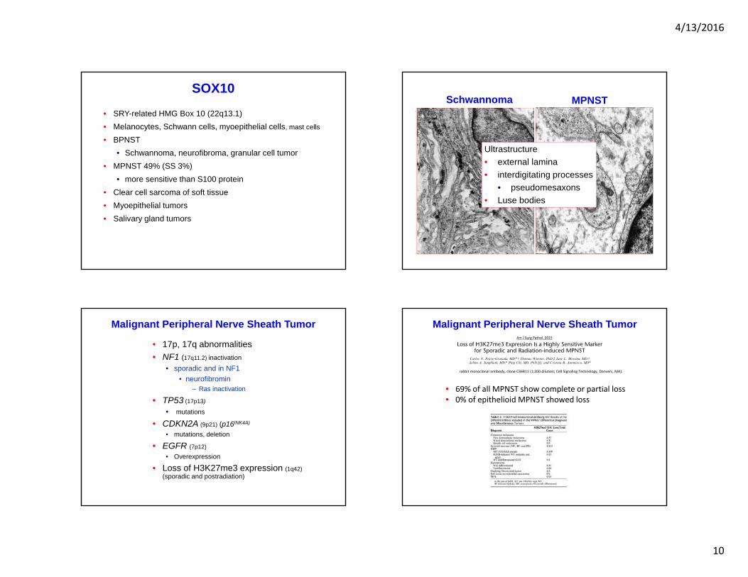
Schwannoma MPNST
Ultrastructure• external lamina• interdigitating processes
• pseudomesaxons• Luse bodies
Malignant Peripheral Nerve Sheath Tumor
• 17p, 17q abnormalities• NF1 (17q11.2) inactivation
• sporadic and in NF1• neurofibromin
– Ras inactivation
• TP53 (17p13)
• mutations
• CDKN2A (9p21) (p16INK4A)
• mutations, deletion
• EGFR (7p12)
• Overexpression
• Loss of H3K27me3 expression (1q42)(sporadic and postradiation)
Carroll 2012; Prieto-Grenada 2015
Malignant Peripheral Nerve Sheath Tumor
rabbit monoclonal antibody, clone C36B11 (1:200 dilution; Cell Signaling Technology, Danvers, MA).
• 69% of all MPNST show complete or partial loss• 0% of epithelioid MPNST showed loss
Am J Surg Pathol. 2015
4/13/2016
11
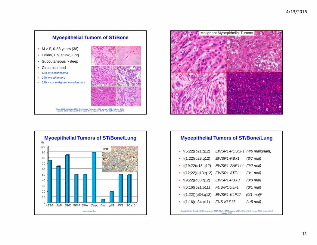
Myoepithelial Tumors of ST/Bone
• M > F, 0-83 years (38)
• Limbs, HN, trunk, lung
• Subcutaneous > deep
• Circumscribed • 42% myoepithelioma
• 25% mixed tumors
• 32% ca or malignant mixed tumors
Stout 1959; Kilpatrick 1997; Fernandez-Figueras 1998; Michal 1999; Kutzner 2001; Mentzel 2003; Hornick 2003; Flucke 2011; Agaram 2014; Puls 2014; Huang 2015
Malignant Myoepithelial Tumors
0
10
20
30
40
50
60
70
80
90
100
Myoepithelial Tumors of ST/Bone/Lung
AE1/3 EMA S100 GFAP SMA Calpo Des p63 INI1 SOX10
%
Aggregated Data
EMAINI1
Myoepithelial Tumors of ST/Bone/Lung
• t(6;22)(p21;q12) EWSR1-POU5F1 (4/6 malignant)
• t(1;22)(q23;q12) EWSR1-PBX1 (3/7 mal)
• t(19:22(q13;q12) EWSR1-ZNF444 (2/2 mal)
• t(12;22)(q13;q12) EWSR1-ATF1 (0/1 mal)
• t(9;22)(q33;q12) EWSR1-PBX3 (0/3 mal)
• t(6;16)(p21;p11) FUS-POU5F1 (0/1 mal)
• t(1;22))(p34;q12) EWSR1-KLF17 (0/1 mal)*
• t(1;16)(p34;p11) FUS-KLF17 (1/5 mal)
Brandal 2008; Brandal 2009; Antonescu 2010; Flucke 2012; Agaram 2014; Puls 2014; Huang 2015; Leduc 2016 *kidney 2015
4/13/2016
12
Myoepithelial Tumors of ST/Bone/Lung
• Morphology variable
• IHC variable
• EMA, S100 protein
• Genetics variable
• EWSR1, FUS
• Many partners
• Gold standard EM
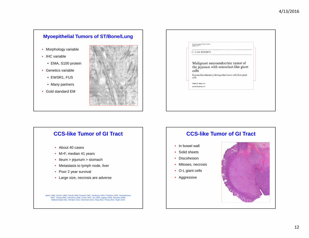
CCS-like Tumor of GI Tract
• About 40 cases• M>F, median 41 years• Ileum > jejunum > stomach• Metastasis to lymph node, liver• Poor 2 year survival• Large size, necrosis are adverse
Alpers 1985; Donner 1998; Fukuda 2000; Pauwels 2002; Zambrano 2003; Friedrichs 2005; Venkataraman2005; Huang 2006;; Antonescu 2006; Comin 2007; Joo 2009; Lagmay 2009; Terazawa 2009; Balkaransingh 2011; Shenjere 2012; Stockman 2012; Yang 2012; Thway 2014; Yegen 2015
CCS-like Tumor of GI Tract
• In bowel wall
• Solid sheets
• Discohesion
• Mitoses, necrosis
• O-L giant cells
• Aggressive
4/13/2016
13
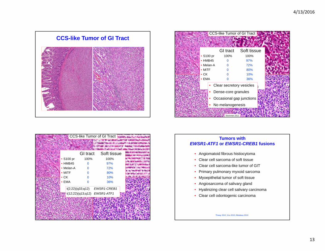
CCS-like Tumor of GI TractCCS-like Tumor of GI Tract
• Uniform clear cells in nests • Sheets of relatively small cells• Pseudoglandular/pseudopapillary• Spindled, plasmacytoid, rhabdoid• Osteoclast-like giant cells
GI tract Soft tissue• S100 pr 100% 100%• HMB45 0 97%• Melan-A 0 72%• MiTF 0 80%• CK 0 10%• EMA 0 36%
(CD56, SYN, NF, NB84, SOX10)
Stockman 2012
• Clear secretory vesicles
• Dense-core granules
• Occasional gap junctions
• No melanogenesis
CCS-like Tumor of GI Tract
• Uniform clear cells in nests • Sheets of relatively small cells• Pseudoglandular/pseudopapillary• Spindled, plasmacytoid, rhabdoid• Osteoclast-like giant cells
GI tract Soft tissue• S100 pr 100% 100%• HMB45 0 97%• Melan-A 0 72%• MiTF 0 80%• CK 0 10%• EMA 0 36%
(CD56, SYN, NF, NB84, SOX10)t(2;22)(q33;q12) EWSR1-CREB1t(12;22)(q13;q12) EWSR1-ATF1
Tumors withEWSR1-ATF1 or EWSR1-CREB1 fusions
• Angiomatoid fibrous histiocytoma• Clear cell sarcoma of soft tissue• Clear cell sarcoma-like tumor of GIT• Primary pulmonary myxoid sarcoma• Myoepithelial tumor of soft tissue• Angiosarcoma of salivary gland• Hyalinizing clear cell salivary carcinoma• Clear cell odontogenic carcinoma
Thway 2012; Gru 2013; Bilodeau 2013

4/13/2016
14
Case 19 Endometrial Stromal Sarcoma
• CD10• ER• PgR• SMA• CK• H-caldesmon -
JAZF1-SUZ12
Ultrastructure • Fibroblastic
• RER, Golgi, few desmosomes, cilia
Seminoma
• Middle aged males
• Midline in abdomen
• Primary or metastatic
• Lymphocytes/granulomas
• Raised serum HCG
CK
OCT3/4CD117
Seminoma
• CD117• OCT3/4• SALL4• podoplanin• PLAP• CK• HCGUltrastructure
• smooth nuclei• rope-like nucleoli,, • glycogen, few junctions• annulate lamellae
Small Round Cell Tumors
• Alveolar rhabdomyosarcoma desmin, myogenin
• Desmoplastic round cell tumorWT1, EMA, CK, desmin, NSE, NF, CD56
• Ewing sarcoma CD99, FLI1, ERG, CK, desmin
• CIC-DUX4 tumors• CD99, ERG, MUC4,• PD synovial sarcoma
TLE1, EMA, CK, CD99, CD56, bcl-2 • Mesenchymal chondrosarcoma
CD99• Small cell neuroendocrine carcinoma
TTF1, CK, CD56, CG
4/13/2016
15
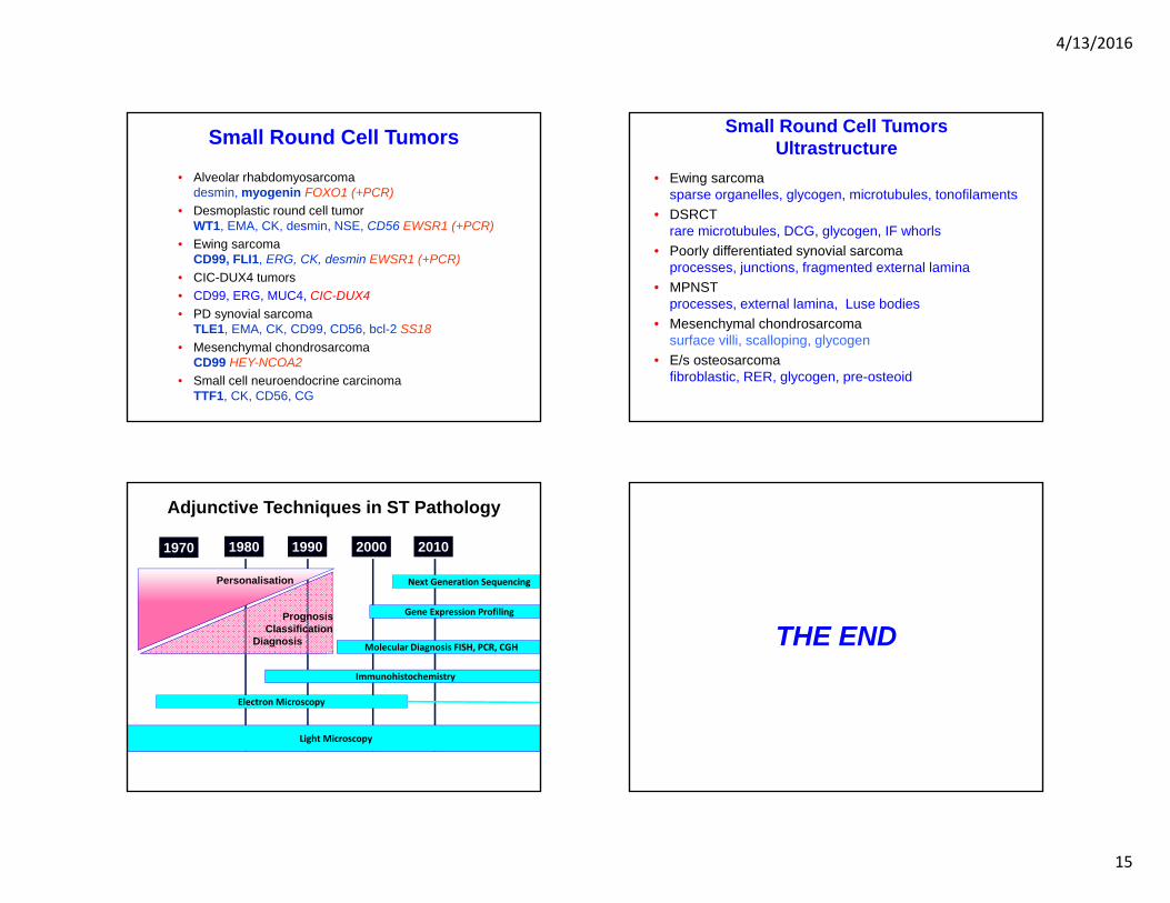
Small Round Cell Tumors
• Alveolar rhabdomyosarcoma desmin, myogenin FOXO1 (+PCR)
• Desmoplastic round cell tumorWT1, EMA, CK, desmin, NSE, CD56 EWSR1 (+PCR)
• Ewing sarcoma CD99, FLI1, ERG, CK, desmin EWSR1 (+PCR)
• CIC-DUX4 tumors• CD99, ERG, MUC4, CIC-DUX4• PD synovial sarcoma
TLE1, EMA, CK, CD99, CD56, bcl-2 SS18• Mesenchymal chondrosarcoma
CD99 HEY-NCOA2• Small cell neuroendocrine carcinoma
TTF1, CK, CD56, CG
Small Round Cell TumorsUltrastructure
• Ewing sarcomasparse organelles, glycogen, microtubules, tonofilaments
• DSRCT rare microtubules, DCG, glycogen, IF whorls
• Poorly differentiated synovial sarcoma processes, junctions, fragmented external lamina
• MPNST processes, external lamina, Luse bodies
• Mesenchymal chondrosarcomasurface villi, scalloping, glycogen
• E/s osteosarcoma fibroblastic, RER, glycogen, pre-osteoid
Light Microscopy
Electron Microscopy
Immunohistochemistry
Molecular Diagnosis FISH, PCR, CGH
1970 1980 1990 2000 2010
Next Generation Sequencing
Adjunctive Techniques in ST Pathology
Gene Expression Profiling
Personalisation
Prognosis Classification
Diagnosis THE END