SpyGlass - Boston Scientific- US · T he SpyGlaSS® DireCT ViSualizaTiOn SySTem is an intuitive...
12
One System. One Operator. One Solution. SpyGlass ® Direct Visualization System
Transcript of SpyGlass - Boston Scientific- US · T he SpyGlaSS® DireCT ViSualizaTiOn SySTem is an intuitive...

One System. One Operator. One Solution.
SpyGlass®
Direct Visualization System

T a b l e O f C O n T e n T S
1 SpyGlass® Direct Visualization
System Overview
2 SpyScope® Access and Delivery Catheter
4 SpyGlass Direct Visualization Probe
6 SpyBite® Biopsy Forceps
8 SpyGlass Direct Visualization System
Capital Components

The SpyGlaSS® DireCT ViSualizaTiOn SySTem is an
intuitive platform that enables simple, single-operator, direct
visualization cholangioscopy for detection and treatment of large
stones and strictures throughout in the pancreatico-biliary system.
By attaching the single-use SpyScope® Access and Delivery Catheter to
a standard duodenoscope and inserting through the working channel, the
SpyGlass Direct Visualization System allows for:
• Site-specific examination of the entire lumen, made possible by 4-way
deflection and fiber optic lighting and imaging technology.
• improved diagnosis by visualization and Spybite® forceps biopsy,
allowing for adequate tissue acquisition for histological examination in 88%
of patients.*
• Treatment of large biliary stones by either electrohydraulic lithotripsy
(EHL) or Holmium Laser lithotripsy through the 1.2mm working channel,
under direct visualization for focused treatment.
Each year more than one million people throughout the world undergo
endoscopic retrograde cholangiopancreatography (ERCP) for conditions related
to the liver, gallbladder, pancreas and bile ducts.* * The benefits of direct
visualization cholangioscopy are well known and documented but traditional
approaches limited adoption due to the need for two operators and continual,
costly capital scope repairs.
The SpyGlass System addresses both of these limitations with a single-operator,
single-use device that is redefining cholangioscopy with over 30,000 worldwide
cases completed to date.* *
1
SpyBite Forceps
EHL Fiber
Laser Fiber
* Meining A, Chen Y, Pleskow D, et al; Direct visualization of indeterminate pancreticobiliary strictures with probe-based confocal laser endomicroscopy: a multicenter experience, Gastrointestinal Endoscopy, November 2011.
**Data as of January 2012.

2 SpyScope®
Access and Delivery Catheter
➢ Distal Tip’s 10Fr outer diameter facilitates access to the duct system with 1.2mm accessory channel, optical channel and 2 dedicated irrigation channels.
➢ Dual Controls allow for 4-way deflection to navigate tortuous anatomy.
➢ Locking mechanism ensures direct visualization of target is easily maintained throughout entire procedure.

Optic Port
Device Delivery Port
Irrigation Port
The SpySCOpe aCCeSS anD DeliVery CaTheTer is a single-use, single-operator controlled device designed to facilitate access to the pancreatico-biliary anatomy for
both diagnostic and therapeutic procedures. The SpyScope Catheter is comprised of a control handle that attaches to any standard duodenoscope and a long, flexible delivery catheter that is inserted down the working channel of the duodenoscope and passed directly past the ampulla into the pancreatico-biliary anatomy. This design allows for a single physician to simultaneously operate both scopes for a greatly simplified cholangioscopy procedure.
handle
• Dial knob controls with locking mechanism facilitate deflection up to 30° in all four directions for greater control.
• Dedicated ports for delivery and independent control of optics, irrigation and SpyBite Forceps or lithotripsy accessories.
Catheter
• A 10Fr (3.3mm) outer diameter, 230cm length catheter fits down standard duodenoscope working channel and allows for easy access to non-dilated pancreatico-biliary anatomy.
• Dedicated 1.2mm accessory channel for significant biopsy specimen retrieval with SpyBite Forceps and large biliary stone management with EHL or Holmium Laser lithotripsy.*
• Four-way tip deflection allows for enhanced directional control and more precise navigation.
• 1mm optical channel for SpyGlass Direct Visualization Probe facilitates both illumination and endoscopic visualization.
• Dual, independent irrigation channels for fluid aspiration to maintain visualization.
SpySCOpe aCCeSS anD DeliVery CaTheTer
Order Number Description
M00546230 SpyScope Access and Delivery Catheter
Recommended Guidewire: 0.035” (0.89mm), 450cm Jagwire® Guidewire or 450cm Hydra Jagwire® Guidewire
* The SpyGlass System has also been shown to be compatible with the Northgate® 1.9Fr Biliary Probe (9-195-25) and the Northgate Autolith® Intracorporeal Electrohydraulic Lithotripsy iEHL Generator (9-201-00) for EHL applications.
The SpyGlass System has also been shown to be compatible using the Lumenis® SlimLine™ 365 micron Laser probe (M0068408420) and the Lumenis® VersaPulse PowerSuite 20 Watt Holmium Laser Generator (M0068408220). The testing followed the laser probe placement instructions.
Northgate and Autolith are trademarks of Northgate Technologies, Inc; Lumenis and Slimline are trademarks of Lumenis Ltd.
3SpyScope®
Access and Delivery Catheter
SpyScope®
Access and Delivery Catheter
Three dedicated ports for:
➢ Device Delivery: SpyBite®
Biopsy Forceps, EHL or Holmium Laser probes for large biliary stone management.
➢ Optics: SpyGlass® Probe for delivery of light and transmission of image.
➢ Irrigation: Flush and aspirate anatomy to clear the field of view and optimize visualization.

The SpyGlaSS® DireCT ViSualizaTiOn prObe is a fiber optic device designed to optimize light delivery to the anatomy and to acquire and transmit endoscopic images back to the
camera. The probe connector on the proximal end attaches to the light guide and ocular. The SpyGlass Probe, when delivered through the SpyGlass Catheter, provides complete circumferential visualization in the pancreatico-biliary anatomy, which may expand options for visual diagnosis and targeted tissue acquisition, potentially leading to accelerated diagnosis and altered patient treatment.
• 6,000 pixel image bundle surrounded by 225 light fibers optimize illumination and image quality in a .9mm diameter probe.
• Outer sheath engineered for maximum flexibility and pushability to facilitate access to any target within the pancreatico-biliary anatomy.
• Distal lens provides 70° field of view to capture complete circumferential visualization within the lumen.
• Durable, re-useable design for up to 20 cases.
The probe should be reprocessed and stored and protected in the large or small SpyGlass System storage tray.2
4
SpyGlass®
Direct Visualization Probe
Optic Port
Probe Connector
1Please refer to the SpyGlass Direct Visualization Probe Directions for Use.2 Large Storage Trays are recommended whenever possible to maintain a larger loop radius for the coiled probe, which puts less strain on the probe fibers.
SpyGlaSS DireCT ViSualizaTiOn prObe
Order Number Description
M00546030 SpyGlass Direct Visualization Probe1
M00546040 Ocular
M00546050 Probe Small Storage Tray2
M00546060 Probe Large Storage Tray2
M00546210 Light Cable
Working Length – 231cm; Field of View – 70°; Maximum Insertion Portion – 0.81mm; Maximum Diameter – 0.9mm; Minimum Required Working Channel – 1mm
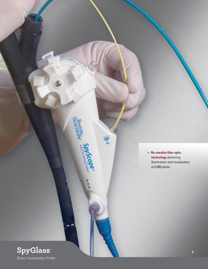
5SpyGlass®
Direct Visualization Probe
➢ Re-useable fiber optic technology delivering illumination and visualization at 6,000 pixels.
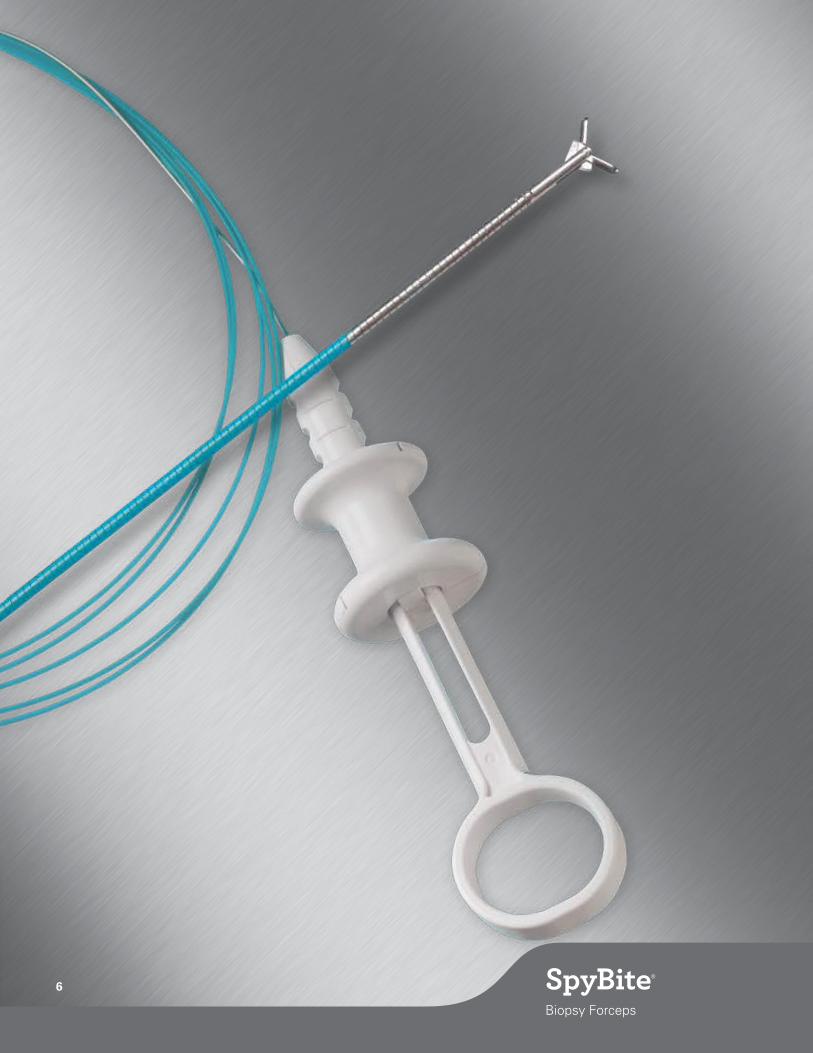
6 SpyBite®
Biopsy Forceps

The SpybiTe biOpSy fOrCepS is designed to be inserted through the accessory channel of the SpyScope® Access and Delivery Catheter to facilitate site-specific specimen acquisition
under direct visualization throughout the pancreatico-biliary system.
• The 1mm outer diameter is designed to fit easily through the SpyScope Catheter working channel, but also allow for 4.1mm jaw opening at 55° for substantial tissue pathology acquisition.
• The central spike in the specimen cup further aids in securing tissue samples in difficult anatomies.
• Has demonstrated to be 88% effective in acquiring sufficient tissue for histologi-cal evaluation, significantly better than cytology brushing alone*.
7SpyBite®
Biopsy Forceps
SpyBite®
Biopsy Forceps
SpyBite Forceps
Tissue acquisition in the common bile duct (CBD) using
SpyBite Forceps
Breaking a large stone in the CBD using EHL Probe
➢ Biopsy Cup: 4.1mm opening at 55° for maximum tissue acquisition
* Meining A, Chen Y, Pleskow D, et al; Direct visualization of indeterminate pancreticobiliary strictures with probe-based confocal laser endomicroscopy: a multicenter experience, Gastrointestinal Endoscopy, November 2011.
SpybiTe biOpSy fOrCepS
Order Number
Description
Cable Diameter
Jaw Outer Diameter
Jaw Opening
Working Length
Required Endoscope Working Channel
M00546270 SpyBite Biopsy Forceps 0.039” (1.0mm)
1.0mm 4.1mm, 55°
270cm 1.2mm

Camera, Camera head and Ocular
The camera utilizes a ¼ CCD color-image sensor that provides three types of video outputs (RGBs, S-video or composite) to easily integrate into any procedure room. The camera head attaches to the camera control unit by a multi-pin cable connector on the proximal end and the ocular on the distal end. The ocular is an opto-mechanic coupler that facilitates focus and transmission of the image from the SpyGlass Probe.
light Source and Cable
The light source utilizes 300W Xenon light technology and features a simple control wheel to adjust the light output. The light guide is composed of a flexible fiber-optic bundle that attaches to the light source via a Wolf connector port and transmits the light to the SpyGlass Probe.
irrigation pump, Tubing and footswitch
The irrigation pump and tubing provide fluid flow to the SpyScope® Access and Delivery Catheter in order to clear the duct of debris and maintain clear visualization. The flow rate is adjustable and continuous flow can be controlled by the footswitch.
Pump Specifications
Power 100 - 240 V~, 50/60 Hz, 25 VA
Flow Rate 0 - 375 ml / min +/- 20% using SpyGlass Irrigation Tube Set M00546451 (without SpyScope® Access and Delivery Catheter)
SpyGlass probe Trays
The SpyGlass Probe re-processing and storage trays come in large and small sizes, and are designed to protect the probe in between uses and during Cidex® Solution or Cidex OPA Solution for disinfecting.
Large Storage Trays are recommended whenever possible to maintain a larger loop radius for the coiled probe, which puts less strain on the probe fibers.
monitor
High-resolution LCD monitor (1280x1024) is designed to attach to the cart and provide bright, clear images under varying lighting conditions.
8
SpyGlass®
Direct Visualization System Capital Components
SpyGlass Direct Visualization System Capital Components

Cart (3-Joint arm and isolation Transformer)
Designed for easy transfer between rooms, the SpyGlass® System cart provides space for all required capitalequipment along with two storage drawers to fit SpyGlass System storage trays, an isolation transformer (optional) to manage all electrical demands of the equipment on the cart, and flexible 3-joint arm to hold the camera in place and out of the way (also available separately with bedside attachment). The cart also features a mounting bracket for the LCD monitor and four locking casters for simple transportation and stable positioning within the endoscopy suite.
Inputs Outputs
120 / 240 VAC – 50/60 Hz 120 or 240 VAC (voltage selector switch)
Input via an IEC 320 Power entry module 1000 VA Maximum
On / off switch 9 Receptacles (IEC 320)
power Cable pack
The power cable pack includes 5 cables of varying lengths to connect the capital units to the isolation transformer. This ensures electrical isolation and enables simple setup after moving from room to room requiring only the connection of the transformer power cable rather than 5 separate connections.
9
1 Cart includes a 3-joint arm with connector for ocular, but no table clamp; 2Camera System includes camera controller (box), camera head, and video cables for connection to monitor. US Power cord is included in NTSC version. PAL version does not contain a power cord. For ordering information for PAL version, please contact your local sales representative; 33-Joint Arm (without clamp) is included with the Travel Cart (M00546160). The 3-Joint Arm with Clamp (M00546070) is useful if the account does not want to purchase the Travel Cart as it allows the account to mount the arm to another cart or table;
4 For ordering information for 240 V transformer, please contact your local salesperson; 5Large Storage Trays are recommended whenever possible to maintain a larger loop radius for the coiled probe, which puts less strain on the probe fibers.
SpyGlaSS SySTem CapiTal COmpOnenTS
Order Number Component Dimensions Weight (lbs) Weight (kg)
M00546160 Travel Cart1 19.5”W x 48.25”H x 21”D 150 68
M00546120 Video Monitor 17.5”W x 15.8”H x 4.75”D 20 9.06
M00546190 Lightsource 12.6”W x 5.6”H x 9.7”D 13 5.89
M00546100 Camera2 – NTSC 12.6”W x 4”H x 14”D 10 4.53
M00546140 Irrigation Pump with Footswitch 8”W x 5.5”H x 6.5”D 5 2.26
M00546070 3-Joint Arm with Clamp3 31.5”L (extended) 1.61 0.73
M00546040 Ocular <0.5 <0.226
M00546210 Light Cable 5’ Long <1 <0.453
M00546260 Isolation Transformer4 (120V) 12”W x 4.5”H x 8.6”D 23 10.432
M00546250 Power Cable Pack Cables included: 2 – 0.5 meter; 1 – 1.0 meter; 2 – 1.5 meter n/a n/a
M00546060 Large Probe Storage Tray5 11”W x 2”H x 8”D 2.5 1.134
M00546050 Small Probe Storage Tray5 7”W x 2”H x 5.5D 1.3 0.589
Note: SpyGlass System, when completely assembled with monitor, has a space footprint of 28”W x 70”H x 26”D (711mm x 1778mm x 660mm). Total weight of the assembled system is 214 lbs (97.01 kg).

SpyGlass, SpyScope and SpyBite are registered trademarks of Boston Scientific Corporation or its affiliates. All other trademarks are the property of their respective owners.
Indications, Contraindications, Warnings and Instructions for Use can be found in the product labeling supplied with each device.CauTION: Federal (uSa) law restricts this device to sale by or on the order of a physician.
Visit the Boston Scientific Endoscopy Channel by
using your Smartphone to scan this code or go to
www.youTube.com/bostonScientif icendo
Boston Scientific CorporationOne Boston Scientific PlaceNatick, MA 01760-1537www.bostonscientific.com/endo-resources
Ordering Information 1.888.272.1001
©2013 Boston Scientific Corporation or its affiliates. All rights reserved.
ENDO-61903-AB April 2013