![Etiopathogenesis of obsessive compulsive disorder [autosaved]](https://static.fdocuments.net/doc/165x107/58ee7c571a28ab2b328b46d7/etiopathogenesis-of-obsessive-compulsive-disorder-autosaved.jpg)
Etiopathogenesis of obsessive compulsive disorder [autosaved]
Continuing mediCAl eduCAtion
Sporotrichosis: an update on epidemiology, etiopathogenesis, laboratory and clinical therapeutics*
RosaneOrofino-Costa1,4 PriscilaMarquesdeMacedo2
AndersonMessiasRodrigues3 AndréaReisBernardes-Engemann1,4
DOI: http://dx.doi.org/10.1590/abd1806-4841.2017279
Abstract: Inthelate90’stherewasachangeinboththerouteoftransmissionandthepeopleatriskforsporotrichosis.Thiszoonoticcat-manalternativetransmissionrouteelicitedchangesinstrategiestocontroltheepidemic.Therewasaprogressiveincrease in the number of cases involving especially children and the elderly. In addition to becoming hyperendemic,uncommonclinicalpictureslikeimmunoreactiveclinicalpresentationsorseveresystemiccaseshaveemerged.Newspecieswereidentifiedandclassifiedthroughmoleculartoolsusingmorevirulentclinicalisolates,likeS. brasiliensis,comparedtotheenvironmentalisolates.Likewise,differentspeciesofSporothrix have been associated with different geographic regions. The serologicalandmoleculartechniquesareusedasanauxiliarytoolforthediagnosisand/orforspeciesidentification,althoughtheisolationandtheidentificationofSporothrix spp. in clinical specimen is still the gold standard. Currently sporotrichosis epidemicsrequirestheknowledgeoftheepidemiological-molecularprofiletocontrolthediseaseandthespecifictreatment.Itraconazole,potassiumiodide,terfinafine,andamphotericinBaretheavailabledrugsinBraziltotreatsporotrichosis.Thedrugofchoice,itsposology,andtreatmentdurationvaryaccordingtotheclinicalpresentation,theSporothrixspecies,andhostimmunestatus.Newtreatmentchoices,includingavaccine,arebeingdeveloped;nevertheless,moreclinicaltrialsarerequiredtoconfirmitsefficacy.Keywords: Diagnosis;Epidemiology;Molecularbiology;Serology;Sporothrix;Therapeutics
s
606
Received on 11.05.2017.ApprovedbytheAdvisoryBoardandacceptedforpublicationon23.07.2017.* WorkperformedattheDermatologyDepartment,FaculdadedeCiênciasMédicas,UniversidadedoEstadodoRiodeJaneiro(FCM-UERJ),RiodeJaneiro,RJ,Brazil. Financial support: None Conflictofinterests:None
1 DermatologyDepartment,FaculdadedeCiênciasMédicas,UniversidadedoEstadodoRiodeJaneiro(FCM-UERJ),RiodeJaneiro,RJ,Brazil.2 InfectiousDermatologyClinicalResearchLaboratory, InstitutoNacionaldeInfectologiaEvandroChagas,FundaçãoOswaldoCruz(INI/Fiocruz),Riode
Janeiro,RJ,Brazil.3 LaboratoryofEmergingFungalPathogen,DepartmentofMicrobiology,ImmunologyandParasitology,UniversidadeFederaldeSãoPaulo(UNIFESP),SP,
Brazil.4 MedicalMycologyLaboratory,DermatologyDepartment,HospitalUniversitárioPedroErnesto,RiodeJaneiro,RJ,Brazil.
©2017byAnaisBrasileirosdeDermatologia
An Bras Dermatol. 2017;92(5):606-20.
INTRODUCTIONSporotrichosisisasubacuteorchronicinfection,causedby
thermodimorphic fungi of the genus Sporothrix. It is a cosmopolitan disease, occurring preferably in tropical and subtropical regions,andisconsideredthemostfrequentsubcutaneousmycosisinLatinAmerica,whereitisendemic.1
Sporotrichosiswasfirstdescribed in1898,by themedicalstudentBenjaminSchenck,at the JohnsHopkinsHospital, inBal-timore,USA.2 The fungus isolated from skin lesions on the rightupperlimbofapatienttreatedbySchenckwasevaluatedbythepa-thologistErwinF.Smith,andidentifiedasaspeciesbelongingtothegenus Sporotrichum.In1900,HektoenandPerkinsreportedanothercaseinChicagoandproposedanewname,Sporothrix schenckii,al-though Sporotrichum schenckii was used for decades.3 Due to the re-productioncharacteristicsofthisgenus,thebinomialwaschanged
to Sporothrix schenckii.4 Currently,thespeciesinvolvedinthehumanoranimaldisease,andotherthatareenvironmental,havebeenrec-ognized.5
In the early 20th century, in France, sporotrichosiswas acommondisease,aswerethereportsoftheextracutaneousclinicalforms.3InBrazil,LutzandSplendorefirstlydescribedinfectionsinratsandhumans,demonstratingthepresenceofasteroidbodiesintissues,whichareusefulforthehistopathologicaldiagnosisofthismycosis.6
EPIDEMIOLOGY AND ETIOPATHOGENESISForalongtime,sporotrichosiswasknownasthe“rosebush
mycosis”,orthe“gardener’smycosis”,giventhattheinfectionusu-ally resulted from the agent’s inoculation on the skin ormucous

An Bras Dermatol. 2017;92(5):606-20.
Sporotrichosis: an update on epidemiology, etiopathogenesis, laboratory and clinical therapeutics 607
membrane,bytraumawithcontaminatedplantmaterial.However,somecasesofzoonotictransmissionhavebeenreported,aswellaslessfrequentcasesofinhaledinfectivefungalpropagules,clinicallypresenting as a systemic mycosis.7,8
Occasionally,theremaybeanenvironmentaltransmissionof sporotrichosis, classicallyassociatedwithsoilmanipulationac-tivities,whether for occupational or leisure reasons; in outbreakssuchasthosethatoccurredintheUSA,especiallyintheMississippiValley,inthe80’s,involvingreforestationworkersinfectedbypinetrees andmoss seedlings; also asmicroepidemics like thatwhichoccuredintheearly90’s,withpeopleinfectedbythecontactwithhay stored in an abandoned house where Halloween parties were held;aswellaslargeepidemics,suchasthatwhichoccurredinthe40’s,whenthreethousandminersinSouthAfricawereinfectedbythecontactwithcontaminatedwoodsupportingbeams,beingcon-sidered the largest epidemic in the 20th century.9-11More recently,457 cases were described between 2007 and 2009 in a province in the northeastChina,wherethediseaseisendemic.12
Thezoonotictransmissionofsporotrichosiswasdescribedsporadically involvingaccidentswithsnakesandbirds,alsomos-quito,rat,horse,squirrel,andfishbites.3,13,14 Epidemics were report-edinUruguayand,morerecently,inBrazilandArgentina,relatedtoarmadillohunting,giventhecloserelationofthearmadillowiththe soil.15,16Theimportanceofthecatinzoonotictransmissionwasfirstnoticedwhenanoutbreakinvolvingfivepeopleexposedtoasickanimalwasreported.17InBrazil,themainzoonoticsporotricho-sisoutbreaks,involvinghumansandasmallgroupofdomesticcats,occurredinthestatesofSãoPauloandRioGrandedoSul,withaneffective epidemiological control.18,19
InSeptember1997,thefirstcasesofthegreatestfelinezoo-notic transmission epidemic ever described were admitted at the Pedro Ernesto University Hospital, Rio de Janeiro. Three peoplefromthesamefamilywereinfectedbyasickcatthatdied(authors’report,unpublished) (Figure1).Then, thefirstpublicationsaboutthis epidemic,which is currently considered to be hyperendemicinthestateofRiodeJaneiro,appeared.20,21 The capital city and the surrounding municipalities known as “Baixada Fluminense” arecurrently the most affected places where poor socioeconomic condi-
tionsareobserved.Theepidemiologicalprofileismainlycharacter-izedbychildren,elderly,andwomen,becausethesegroupsusuallyhavedirectandmorefrequentcontactwiththeseanimals.22 Alongwiththeconsolidationoftheurbanepidemics,vulnerablepatientsalsobecameaworrisomeat-riskpopulation,especiallythoseinfect-edbythehumanimmunodeficiencyvirus.23Since2013,thenotifica-tionismandatoryinthestateofRiodeJaneiro,butnotintheotherBrazilianstates.Therefore,prevalenceandincidencemeasuresareobtainedmainlybasedoncasesreportedintheliterature,certainlyunderestimatingtherealepidemiologicalimportance,especiallyre-gardingoutbreaksandepidemics.
Althoughhumanoranimalsporotrichosiscaseswerepub-lishedinthestatesofAmazonas,Pará,MinasGerais,EspíritoSanto,SãoPaulo,RiodeJaneiro,Paraná,andRioGrandedoSul,mostcas-esoccurintheSouthandSoutheastBrazil.24
Theadvancesinthemicrobiologicalknowledgealongwiththe use of molecular tools led to important advances concerning epidemiologicalstudies,enablingtheidentificationoftheSporothrix speciesin14Brazilianstates,indicatingthatsporotrichosisismorewidespreadthanpreviouslythoughtfortheBrazilianterritory.24
In Brazil, there are two important disease transmissionroutesforhumans,asapronoticrouteinvolvingdirectcontactwiththesoilanddecomposingorganicmatter;andazoonoticroute,inwhich felines participate actively in the disease transmission. The outbreaksfromclassictransmissionroute,inwhichS. schenckii and S. globosaprevail,requiredtheremovaloffungussourcesinnature.Thealternativetransmissionroute,mainlyinvolvinghorizontalani-maltransmission(cat-cat),aswellaszoonotictransmission,requiresdifferent epidemic control strategies. Such measures include the streetanimalsneuteringandthetreatmentofsickcats,aswellastheeducationabout responsibleownershipof animals, knowledgeofthe main aspect main aspects of Sporothrixtransmission,especiallyinhyperendemicareas.Deadinfectedanimalsmustbeincinerated,ratherthanburied,thusavoidingS. brasiliensis dissemination in the soil and the pathogen progression in nature.
Even though sporotrichosis has been described worldwide-ly,thereisacuriousdivergenceregardingthegeographicdistribu-tion and the incidence of the etiological agents.25Indeed,somespe-
FIgure 1: Ulcerated lesions on the hands of three members ofthesamefamily,atthebeginningofthezoonotictransmission sporotricho-sis epidemics in Rio de Janeiro,in1997,treatedat Hospital Universitário Pedro Ernesto
608 Orofino-CostaR,deMacedoPM,RodriguesAM,Bernardes-EngemannAR
An Bras Dermatol. 2017;92(5):606-20.
ciesaremoreubiquitousthanothers. InAsia,especiallyChina,S. globosa is estimated to be the etiological agent in 99.3% of the human sporotrichosis cases.26,27 In other endemic areas, such asAustraliaandSouthAfrica (94%),also inNorthAmericaandpartofSouthAmerica(89%),S. schenckii is the predominant species.26 In the South andSoutheastBrazilianregions,S. brasiliensis(88%)isthemainetio-logical agent of human and animal sporotrichosis.28,29
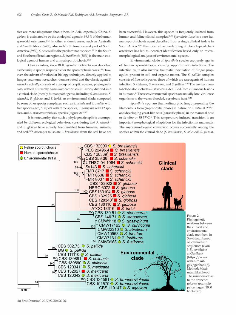
Overacentury,since1898,Sporothrix schenckii was described astheuniquespeciesresponsibleforthesporotrichosiscases.2,30 How-ever,theadventofmolecularbiologytechniques,directlyappliedtofungustaxonomyresearches,demonstratedthattheclassicagentS. schenckii actually consistsofagroupofcrypticspecies,phylogeneti-callyrelated.Currently,Sporothrixcomprises51taxons,dividedintoaclinicalclade(mostlyhumanpathogens),includingS. brasiliensis,S. schenckii,S. globosa,andS. luriei,anenvironmentalclade,composedbysomeotherspeciescomplexes,suchasS. pallida and S. candida with fivespecieseach,S.inflatawiththreespecies,S. gossypina with 12 spe-cies,andS. stenoceraswithsixspecies(Figure2).31,32
It is noteworthy that such a phylogenetic split is accompa-niedbydifferentecologicalbehaviors,consideringthatS. schenckii and S. globosa have already been isolated fromhumans, animals,and soil.33,34AttemptstoisolateS. brasiliensis from the soil have not
beensuccessful.However, this species is frequently isolated fromhuman and feline clinical samples.28,35 Sporothrix luriei is a rare hu-man sporotrichosis agent described from a single clinical isolate in SouthAfrica.36,37Historically,theoverlappingofphenotypicalchar-acteristicshas led to incorrect identificationbasedonlyonmicro-morphological analyses of environmental species.
Environmental clade of Sporothrix species are rarely agents of human sporotrichosis, causing opportunistic infections. Theinfection route also involve traumatic inoculation of fungal prop-agules present in soil and organic matter. The S. pallida complex consistsoffivesoilspecies,threeofwhicharerareagentsofhumaninfection: S. chilensis, S. mexicana, and S. pallida.38-40 The environmen-tal clade also includes S. stenocerasidentifiedfromcutaneouslesionsin humans.41 These environmental species are usually low-virulence organismstothewarm-blooded,vertebratehost.38,42
Sporothrixspp.arethermodimorphicfungi,presentingthefilamentousform(saprophyticphase)innature or in vitroat25°C,anddevelopingyeast-likecells(parasiticphase)inthemammalhostor in vitro at 35-37°C.43 This temperature-induced transition is an important morphological adaptation for the infection in mammals. The mycelium-to-yeast conversion occurs successfully among the species within the clinical clade (S. brasiliensis,S. schenckii,S. globosa,
FIgure 2: Phylogenetic relations between the clinical and environmental clade members in Sporothrix,basedon calmodulin sequences(exon3-5).AvailableatGenBank(https://www.ncbi.nlm.nih.gov/genbank/).Method: Maxi-mumlikelihoodThe numbers close to the branches refer to resample percentages (1000 bootstrap)

Sporotrichosis: an update on epidemiology, etiopathogenesis, laboratory and clinical therapeutics 609
An Bras Dermatol. 2017;92(5):606-20.
and S. luriei),whiletheenvironmentalspecies(S.inflata,S. humicola,S. pallida,S. mexicana,S. chilensis,amongothers)showdeficientmor-phological transition, producing few yeast-like cells. Perhaps, thesuccessful emergence of S. brasiliensisinmammals,aswellasthelowvirulence of environmental species may be related to the thermotoler-anceorthermosensitivityofthesespecies,respectively.44Anotherim-portant virulence factor is the Sporothrix ability to produce melanin. Melanininducesfungalescapefromthehost’sdefenses,andisalsoconsideredaresistancefactoragainstsomeantifungaldrugs,suchasamphotericinB,itraconazoleandterbinafine.45,46
Thevirulenceprofileschangedependingonthepathogencharacteristics and the host defenses. Sporothrix brasiliensis is the mostvirulent species,due to its ability to invade tissue and leadtodeath,whereasS. schenckii has different levels of virulence and S. globosa exhibits little or no virulence in murine models.42,47-49 The environmental species of Sporothrix present low virulence in murine models,withlowinvasivepotential,andthehostisabletocontroltheinfectionafewweeksafterinoculation.38,42
The host’s defense against the Sporothrix species hasnotyetbeenestablished.Thefungus’cellwallcomponents,es-peciallythe70kDaglycoprotein(gp70),hasaprotectiveeffectinthehost,mediatedbyT-helpercells(Th1inhumans),butparadoxically,italsomakestheadherenceofconidiatotheepithelium,increasingfungal invasive potential. The cell-mediated immune response seems toberesponsible foreliminatingorcontrolling infection.However,thehumoralimmuneresponseelicitsspecificantibodiesagainsttheSporothrix’ cell wall.5,50 The dissemination of sporotrichosis is usually relatedtocellularimmunitydeficiencies,suchasAIDS.51
CLINICAL ASPECTSUsually, the clinical manifestations of sporotrichosis are
dividedintocutaneousandextracutaneous,theformerismorefre-quent.22After the zoonotic sporotrichosis epidemic in the state ofRio de Janeiro, new clinical presentations, uncommonuntil then,wereidentified.Forthisreason,anewclassificationwasproposed,to better describe the clinical features of the patients cared by the reference sporotrichosis teams.52 Inthepresentarticle, theauthorsproposeanupdateofthisclinicalclassificationbasedonthegroup’sexpertise(Table1).
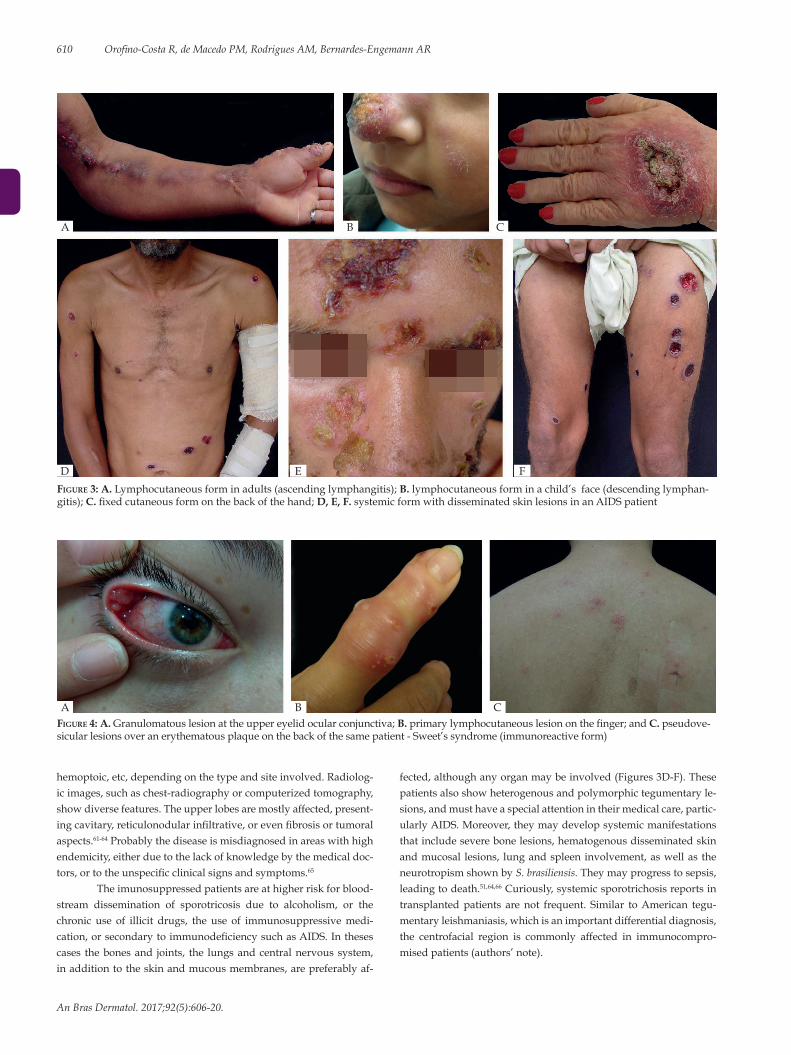
Nearly 80% of the affected patients present the lymphocu-taneous form.52Initially,thelesionhasapapulonodularappearancewherethefunguswasintroducedintotheskin,appearingbetweentwotofourweeksafterthetrauma.Afterwards,thelesionmayul-cerate,andfistulizedrainingapurulentdischarge.Thisisso-calledthe inoculation chancre. The lesions, usually nodules, progressalongtheregionallymphangiticchannels,upwardsordownwardsdepending on the anatomical site, after someweeks. Later, thesenodules,mayulcerate,fistulize,andheal,caractherizingagumma(Figures3Aand3B).Thefixedcutaneousformconsistsofasinglelesion,usually similar to the inoculationchancre,withno region-allymphaticspreading(Figure3C).Insomeoccasions,thediseasemayappearaslargerulcerswithwell-definedandframedborders,or erythematous-scaly, papulopustular, vegetative, infiltrative, orcrusty lesions (Figures3D-F). Somepatients exhibitmultiple skinlesions,disseminatedonthetegument,withnosystemic invasion
andpolymorphicappearance,allofthemarisingatthesametime.Ingeneral, thesepatients are immunocompetent individualswhodescribe having multiple traumas.
Althoughanymucousmembranemaybeaffectedbyspo-rotrichosis, the ocularmucosa ismore commonly involved, caus-ing conjunctivitis, episcleritis,uveitis, choroiditis, and retrobulbarlesions, among others (Figure 4A).53-55 When the lacrimal duct is affected, dacryocystitis may occur as sequela.56,57 The retrobulbar lesions,suchaschorioretinitis,aremorefrequentlyrelatedtohema-togenousspread,andanteriorlesionsareassociatedwiththefungalinnoculation. The simultaneous affection of the ocular mucosa and theregionallymphnodesisnotrare,anditisoneofthecausesofthe Parinaud syndrome.58
Thebonesandjointsmaybeinvolvedbydirecttrauma,bythe invasion through a preexisting overlying cutaneous lesion or secondarytoahematogenousspreading,thelatterathighestriskofsepsis due to the deep site of infection. Osteoarticular sporotrichosis may appear as a monoarthritis associated or not with an overlying cutaneouslesions,aswellasboneresorptionandosteolyticlesionsin the most severe cases.59,60Thesynovialfluidexhibitincreasedcel-lularity mostly consisting of polymorphonuclear leukocytes, lowglucose and high protein levels.
The respiratory transmission through the inhalation of Spo-rothrix propagules is acceptable, characterizing the primary pul-monary systemic form of sporotrichosis. The lungs may also be af-fectedbythehematogenousspread,mainlyinimmunosuppressedpatients presenting with the disseminated systemic form of sporo-trichosis.Thesignsandsymptomsmayincludecoughing,dyspnea,
Table 1: Clinical classification of sporotrichosis
Skin
Lymphocutaneous
Fixed cutaneous
Multiple inoculation
Mucous membrane
Ocular
Nasal
Others
Systemic
Osteoarticular
Cutaneous disseminated
Pulmonary
Neurological
Other locations/sepsis
Immunoreactive
Erythema nodosum
Erythema multiforme
Sweet's syndrome
Reactive arthritis
Spontaneous regression
Modifiedfrom:Lopes-Bezerra,et al. 2006.52
610 Orofino-CostaR,deMacedoPM,RodriguesAM,Bernardes-EngemannAR
An Bras Dermatol. 2017;92(5):606-20.
hemoptoic,etc,dependingonthetypeandsiteinvolved.Radiolog-icimages,suchaschest-radiographyorcomputerizedtomography,showdiversefeatures.Theupperlobesaremostlyaffected,present-ingcavitary,reticulonodularinfiltrative,orevenfibrosisortumoralaspects.61-64 Probably the disease is misdiagnosed in areas with high endemicity,eitherduetothelackofknowledgebythemedicaldoc-tors,ortotheunspecificclinicalsignsandsymptoms.65
Theimunosuppressedpatientsareathigherriskforblood-stream dissemination of sporotricosis due to alcoholism, or thechronic use of illicit drugs, the use of immunosuppressivemedi-cation,orsecondarytoimmunodeficiencysuchasAIDS.Inthesescases thebonesand joints, the lungsandcentralnervoussystem,inadditiontotheskinandmucousmembranes,arepreferablyaf-
fected,althoughanyorganmaybeinvolved(Figures3D-F).Thesepatients also show heterogenous and polymorphic tegumentary le-sions,andmusthaveaspecialattentionintheirmedicalcare,partic-ularlyAIDS.Moreover,theymaydevelopsystemicmanifestationsthatincludeseverebonelesions,hematogenousdisseminatedskinandmucosal lesions, lungandspleen involvement,aswellas theneurotropism shown by S. brasiliensis. Theymayprogresstosepsis,leading to death.51,64,66Curiously,systemicsporotrichosisreportsintransplantedpatients are not frequent. Similar toAmerican tegu-mentaryleishmaniasis,whichisanimportantdifferentialdiagnosis,the centrofacial region is commonly affected in immunocompro-misedpatients(authors’note).
FIgure 3: A.Lymphocutaneousforminadults(ascendinglymphangitis);B. lymphocutaneous form in a child’s face (descending lymphan-gitis);C.fixedcutaneousformonthebackofthehand;D, E, F. systemicformwithdisseminatedskinlesionsinanAIDSpatient
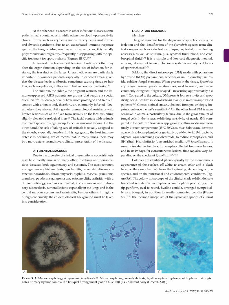
FIgure 4: A. Granulomatouslesionattheuppereyelidocularconjunctiva; B.primarylymphocutaneouslesiononthefinger;andC. pseudove-sicularlesionsoveranerythematousplaqueonthebackofthesamepatient-Sweet’ssyndrome(immunoreactiveform)
A B
D E F
C
A B C
Sporotrichosis: an update on epidemiology, etiopathogenesis, laboratory and clinical therapeutics 611
An Bras Dermatol. 2017;92(5):606-20.
Attheotherend,asoccursinotherinfectiousdiseases,somepatientshealspontaneously,whileothersdevelophypersensitivityclinical forms, such as erythemanodosum, erythemamultiforme,and Sweet’s syndrome due to an exacerbated immune response against the fungus.Also, reactive arthritis canoccur, it is usuallypolyarticularandmigratory,frequentlydisappearingwiththespe-cifictreatmentforsporotrichosis(Figures4B-C).67-69
Ingeneral, the lesionsheal leavingfibroticscars thatmayaltertheorganfunctiondependingonthesiteof infection,for in-stance,thetearductorthelungs.Unaestheticscarsareparticularlyimportant inyoungerpatients, especially in exposedareas, giventhatthediseaseleadstofibrosis,sometimescausingtissueorhairloss,suchaseyelashes,inthecaseofbulbarconjunctivallesion.52
Thechildren,theelderly,thepregnantwomen,andtheim-munosuppressedAIDS patients are groups that require a specialattention.70,71Childrengenerallyhavemoreprolongedandfrequentcontactwithanimalsand, therefore,arecommonly infected.Nev-ertheless,theyalsoexhibitagreaterimmunologicalresistancewithlimitedlesionssuchasthefixedform,usuallyontheface;exhibitingslightly elevated serological titers.71 The facial contact with animals also predisposes this age group to ocular mucosal lesions. On the otherhand,thetaskoftakingcareofanimalsisusuallyassignedtotheelderly,especiallyfemales.Inthisagegroup,thehostimmunedefenseisdeclining,whichmeansthat, inmanytimes, theremaybe a more extensive and severe clinical presentation of the disease.
DIFFERENTIAL DIAGNOSISDuetothediversityofclinicalpresentations,sporotrichosis
may be clinically similar to many other infectious and non-infec-tiousdiseases,bothtegumentaryandsystemic.Themostcommonaretegumentaryleishmaniasis,pyodermitis,cat-scratchdisease,cu-taneousnocardiosis,chromomycosis,syphilis,rosacea,granulomaannulare, pyoderma gangrenosum, osteomyelitis, arthritiswith adifferentetiology,suchasrheumatoid,alsocutaneousandpulmo-narytuberculosis,tumorallesions,especiallyinthelungsandinthecentralnervoussystem,andmeningitis,besidesothers.Inregionsofhighendemicity,theepidemiologicalbackgroundmustbetakeninto consideration.
LABORATORY DIAGNOSISMycology
The gold standard for the diagnosis of sporotrichosis is the isolationandthe identificationof theSporothrix species from clin-ical samples such as skin lesions, biopsy, aspirated from floatingabscesses,aswellassputum,pus,synovialfluid,blood,andcere-brospinal fluid.8,72 It is a simple and low-cost diagnosticmethod,although it may not be useful for some systemic and atypical forms of sporotrichosis.52,73
Seldom, thedirectmicroscopy (DM)madewithpotassiumhydroxide (KOH)preparations,whetherornot indimethylsulfox-ide,exhibitsfungalelements.Whenpresentinthetissue,Sporothrix spp. show several yeast-like structures, oval to round, andmorecommonlyelongated,“cigar-shaped”,measuringapproximately5-8µm.74Comparedtotheculture,DMpresentslowsensitivityandspec-ificity,beingpositiveinsporotrichosismainlyinimmunosuppressedpatients.75,76Giemsa-stainedsmears,obtainedfrompusorbiopsyim-prints, enhance the test’s sensitivity.8 On the other hand DM is more sensitiveinanimals,particularlyfelines,duetothegreatamountoffungalcellsinthetissues,exhibitingsensitivityofnearly85%com-pared to the culture.77 Sporothrix spp. grow in culture media used rou-tinely,atroomtemperature(25ºC-30ºC),suchasSabourauddextroseagarwithchloramphenicolorgentamicin,addedtoinhibitbacteria;Mycoselagarcontainingcycloheximide,toreducesaprophytes,andBHI(BrainHeartInffusion),anenrichedmedium.8,76 Sporothrix spp. is usuallyisolatedin4-6days,forsamplescollectedfromskinlesions,andin10-19days,forextracutaneouslesions;timecanalsovaryde-pending on the species of Sporothrix.73,76,78,79
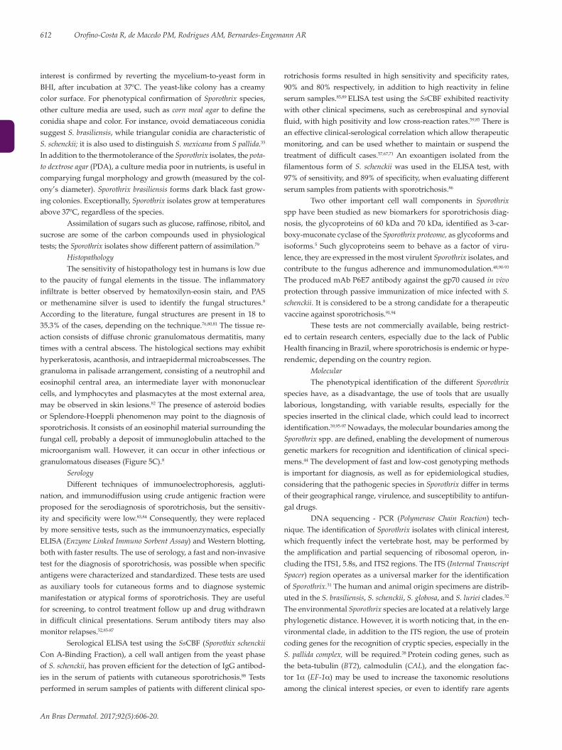
Coloniesareidentifiedphenotypicallybythemembranousappearance of the surface, off-white to cream color and a blackhalo,or theymaybedark from thebeginning,dependingon thespecies,andonthenutritionalandenvironmentalconditions(Fig-ure5A).Thecolonymicroscopyoftheclinicalcladeexhibitdelicatebranchedseptatehyalinehyphae,aconidiophoreproducingatthetippyriform,ovaltoround,hyalineconidia,arrangedsympodial-ly as a bouquet, in addition to sessile pigmented conidia (Figure5B).33,74 The thermodimorphism of the Sporothrix species of clinical
FIgure 5: A. Macromorphology of Sporothrix brasiliensis;B.Micromorphologyrevealsdelicate,hyalineseptatehyphae,conidiophorethatorigi-natesprimaryhyalineconidiainabouquetarrangement(cottonblue,x400);C.Asteroidbody(Grocott,X400)
A B C
interest is confirmed by reverting themycelium-to-yeast form inBHI, after incubationat 37ºC.Theyeast-like colonyhasa creamycolorsurface.ForphenotypicalconfirmationofSporothrix species,otherculturemediaareused, such as corn meal agar todefine theconidiashapeandcolor.Forinstance,ovoiddematiaceousconidiasuggest S. brasiliensis,whiletriangularconidiaarecharacteristicofS. schenckii; it is also used to distinguish S. mexicana from S pallida.33 In addition to the thermotolerance of the Sporothrixisolates,thepota-to dextrose agar(PDA),aculturemediapoorinnutrients,isusefulincomparying fungal morphology and growth (measured by the col-ony’sdiameter).Sporothrix brasiliensis formsdarkblackfastgrow-ingcolonies.Exceptionally,Sporothrix isolates grow at temperatures above37ºC,regardlessofthespecies.
Assimilationofsugarssuchasglucose,raffinose,ribitol,andsucrose are some of the carbon compounds used in physiological tests;theSporothrix isolates show different pattern of assimilation.79
Histopathology The sensitivity of histopathology test in humans is low due
to thepaucityof fungalelements in the tissue.The inflammatoryinfiltrate is better observed by hematoxilyn-eosin stain, and PASor methenamine silver is used to identify the fungal structures.8 According to the literature, fungal structures are present in 18 to35.3%ofthecases,dependingonthetechnique.76,80,81 The tissue re-actionconsistsofdiffusechronicgranulomatousdermatitis,manytimes with a central abscess. The histological sections may exhibit hyperkeratosis,acanthosis,andintraepidermalmicroabscesses.Thegranulomainpalisadearrangement,consistingofaneutrophilandeosinophil central area, an intermediate layer with mononuclearcells,andlymphocytesandplasmacytesatthemostexternalarea,maybeobservedinskinlesions.82 The presence of asteroid bodies or Splendore-Hoeppli phenomenon may point to the diagnosis of sporotrichosis. It consists of an eosinophil material surrounding the fungalcell,probablyadepositofimmunoglobulinattachedtothemicroorganismwall.However, it can occur in other infectious orgranulomatousdiseases(Figure5C).8
Serology Different techniques of immunoelectrophoresis, aggluti-
nation,and immunodiffusionusingcrudeantigenic fractionwereproposed for theserodiagnosisof sporotrichosis,but thesensitiv-ityandspecificitywere low.83,84Consequently, theywere replacedbymoresensitivetests,suchastheimmunoenzymatics,especiallyELISA(EnzymeLinkedImmunoSorbentAssay)andWesternblotting,bothwithfasterresults.Theuseofserology,afastandnon-invasivetestforthediagnosisofsporotrichosis,waspossiblewhenspecificantigenswerecharacterizedandstandardized.Thesetestsareusedas auxiliary tools for cutaneous forms and to diagnose systemic manifestation or atypical forms of sporotrichosis. They are useful forscreening,tocontroltreatmentfollowupanddrugwithdrawnin difficult clinical presentations. Serum antibody titersmay alsomonitor relapses.52,85-87
SerologicalELISAtestusingtheSsCBF (Sporothix schenckii ConA-BindingFraction),acellwallantigenfromtheyeastphaseof S. schenckii,hasprovenefficientforthedetectionofIgGantibod-ies in the serum of patients with cutaneous sporotrichosis.88 Tests performed in serum samples of patients with different clinical spo-
rotrichosis forms resulted inhigh sensitivityandspecificity rates,90% and 80% respectively, in addition to high reactivity in felineserum samples.85,89ELISAtestusingtheSsCBF exhibited reactivity withother clinical specimens, suchas cerebrospinal and synovialfluid,withhighpositivityandlowcross-reactionrates.59,85 There is an effective clinical-serological correlation which allow therapeutic monitoring,andcanbeusedwhether tomaintainorsuspendthetreatment of difficult cases.57,67,71An exoantigen isolated from thefilamentous formofS. schenckiiwasused in theELISA test,with97%ofsensitivity,and89%ofspecificity,whenevaluatingdifferentserum samples from patients with sporotrichosis.86
Two other important cell wall components in Sporothrix spphavebeenstudiedasnewbiomarkersforsporotrichosisdiag-nosis,theglycoproteinsof60kDaand70kDa,identifiedas3-car-boxy-muconate cyclase of the Sporothrix proteome, as glycoforms and isoforms.5 Such glycoproteins seem to behave as a factor of viru-lence,theyareexpressedinthemostvirulentSporothrix isolates,andcontribute to the fungus adherence and immunomodulation.48,90-93 TheproducedmAbP6E7antibodyagainstthegp70causedin vivo protectionthroughpassiveimmunizationofmiceinfectedwithS. schenckii. It is considered to be a strong candidate for a therapeutic vaccine against sporotrichosis.91,94
These tests are not commercially available, being restrict-ed tocertainresearchcenters,especiallydue to the lackofPublicHealthfinancinginBrazil,wheresporotrichosisisendemicorhype-rendemic,dependingonthecountryregion.
MolecularThe phenotypical identification of the differentSporothrix
species have, as a disadvantage, the use of tools that are usuallylaborious, longstanding, with variable results, especially for thespeciesinsertedintheclinicalclade,whichcouldleadtoincorrectidentification.39,95-97Nowadays,themolecularboundariesamongtheSporothrixspp.aredefined,enablingthedevelopmentofnumerousgeneticmarkersforrecognitionandidentificationofclinicalspeci-mens.44 The development of fast and low-cost genotyping methods is important fordiagnosis, aswellas forepidemiological studies,considering that the pathogenic species in Sporothrix differ in terms oftheirgeographicalrange,virulence,andsusceptibilitytoantifun-gal drugs.
DNA sequencing - PCR (Polymerase Chain Reaction) tech-nique.TheidentificationofSporothrixisolateswithclinicalinterest,whichfrequently infect thevertebratehost,maybeperformedbytheamplificationandpartial sequencingof ribosomaloperon, in-cludingtheITS1,5.8s,andITS2regions.TheITS(InternalTranscriptSpacer)regionoperatesasauniversalmarkerfortheidentificationof Sporothrix.31 The human and animal origin specimens are distrib-uted in the S. brasiliensis,S. schenckii,S. globosa,andS. luriei clades.32 The environmental Sporothrix species are located at a relatively large phylogeneticdistance.However,itisworthnoticingthat,intheen-vironmentalclade,inadditiontotheITSregion,theuseofproteincodinggenesfortherecognitionofcrypticspecies,especiallyintheS. pallida complex, willberequired.38 Proteincodinggenes,suchasthe beta-tubulin (BT2), calmodulin (CAL), and theelongation fac-tor 1α (EF-1α)maybeused to increase the taxonomicresolutionsamongtheclinical interestspecies,orevento identifyrareagents
612 Orofino-CostaR,deMacedoPM,RodriguesAM,Bernardes-EngemannAR
An Bras Dermatol. 2017;92(5):606-20.
within the S. pallidacomplex,intheenvironmentalclade,suchasS. pallida,S. mexicana,andS. chilensis. The region between the 3 and 5 exonsofthecalmodulingeneappearsasthemainmarkerforrecog-nition of clinical interest Sporothrix.98-101 In addition to the possibility of identifying the infectiousagent,protein-codinggenesarecom-monly used in studies on genetic diversity, population structure,and molecular epidemiology of sporotrichosis.29
PCR-RFLP - the amplification of a target sequence in thefungusgenomebymeansofPCR,followedbyamplicondigestionwithoneoracombinationofrestrictionenzymes(RFLP),hasbeensuccessfullyusedtodetectinter-andintra-specificvariabilityinsev-eral fungus species. In clinical interest Sporothrixspp.,theidentifi-cation of morphologically similar species becomes possible with the useofPCR-RFLP.Afterthepartialamplificationofthecalmodulingene(exon3-5),theamplicondigestionwiththeHhaIenzymetakesplace,producingfivedifferentrestrictionprofiles(species-specific),representing all species with medical importance.102 Some important advantagesofthistechniqueincludelow-cost,fastandeasyexecu-tion,associatedwiththeabsenceofneedforadvancedinstruments.
Species-specificPCR - the identificationofSporothrix spp. maybeperformedbymeansofPCR,byusingprimers that selective-lyamplifyDNAfromS. brasiliensis,S. schenckii,S. globosa,S. mexi-cana,S. pallida,andS. stenoceras.103Therefore,theprimersequencesarepreservedwithinasingletargetspeciesandinter-specificallydi-vergent.Thistechniqueisalow-cost,fastandrobustmoleculartool,capableofdetectingandidentifyingsmallpathogenDNAbasedonisolatedspecimens,aswellascomplexbiologicalsamples(biopsy,soil,mixedcultures,etc.),withnoneedtoisolatethepathogen.
Sporotrichosis: an update on epidemiology, etiopathogenesis, laboratory and clinical therapeutics 613
An Bras Dermatol. 2017;92(5):606-20.
RollingCircleAmplification (RCA) is amethod that pro-videshighsensitivityandrobustness,whichmaybeappliedfrommonosporiccultures toenvironmentalsamples,withpotential forecology studies.104TheidentificationofSporothrixbasedonRCAhasproven to be a reliable identification tool, alternative toDNA se-quencing,althoughlittleusedinthemycologicaldiagnosis,despitebeing a simple and powerful technique, capable of synthesizinglargeDNAamountsbasedonverylowinitialconcentrations.104,105
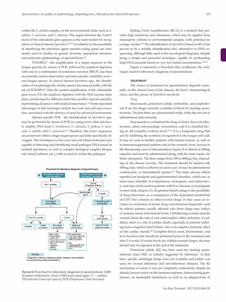
Figure 6 represents a flowchart that synthesizes the tech-niquesusedforlaboratorydiagnosisofsporotrichosis.
TREATMENT
The choice of treatment for sporotrichosis depends essen-tiallyontheclinicalformofthedisease,thehost’simmunologicalstatus,andthespeciesofSporothrix involved.
DrugItraconazole,potassiumiodide,terbinafine,andamphoter-
icinBarethedrugscurrentlyavailableinBrazilfortreatingsporo-trichosis.Thefirstthreeareadministeredorally,whilethelastoneisadministered intravenously.
Itraconazoleisconsideredthedrugofchoiceduetoitseffec-tiveness,safety,andposologicconvenience,anditisclassifiedhav-inganAIIscientificevidencelevel.72,106 It is a fungistatic drug that acts by inhibiting the synthesis of ergosterol in the fungus cell wall. Itmaybeusedinhealthypatientswithlimitedlesions,aswellasinimmunosuppressedpatientsandinthesystemicform,butnotinlife-threatening cases of dissemination/sepsis. It is offered in 100mg capsulesandmustbeadministeredalongwiththemainmeals,forbetterabsorption.Thedoserangesfrom100to400mg/day,depend-ing on the disease severity. The treatment should be started with 100mg/day,whichiseffectiveinmostcases.Itmaybeadministeredcontinuouslyor intermittently (pulse).107 The main adverse effects reportedareheadacheandgastrointestinaldisorders,whichare,inmostcases,tolerable.Itishepatotoxic,teratogenic,andembryotox-ic,andmaynotbeusedinpatientswithliverdiseasesorinpregnantwomen(riskcategoryC).Itsgreatestdisadvantageisthepossibilityofdruginteraction,asaconsequenceofthedependentmetabolismonCYP3A4common toother severaldrugs. Itmaycausean in-creaseorareductionofserumdrugconcentrationsfrequentlyusedby elderlypatientsusually affected, also thesedrugsmay reduceorincreaseserumitraconazolelevels.Childbearingwomenmustbewarnedabouttheriskoforalcontraceptiveeffectreduction.Inad-dition,thereisariskofsuddendeath,especiallyinpatientssuffer-ingfromcongestiveheartfailure,duetoitsnegativeinotropiceffecton the cardiac muscle.108Completebloodcount,biochemistry,andliver function tests should be performed prior to the treatment and after3-4weeks.Ifserumlevelsarewhithinnormalranges,thetestsshould only be repeated at the end of the treatment.
Potassium iodide (KI) has been used for treating sporo-trichosis, since 1903, as initially suggested by Saboraud.7At thattime,specificantifungaldrugswerenotavailableand iodidewasused for several infectious and non-infectious diseases. The KI mechanismofactionisnotyetcompletelyunderstood,despiteitsalreadyknownactionontheimmuneresponse,destructuringgran-ulomas, on neutrophil chemotaxis, aswell as on phagocytosis of
FIgure 6: Flowchart for laboratory diagnosis of sporotrichosis. GMS (Gomori methenamine silver);CMA(cornmealagar);‘C’–carbon;ITS (InternalTranscriptSpacer);PCR(PolymeraseChainReaction)
Sporothrix cells.109,110ThescientificevidencelevelisAII,thesameofitraconazole,anditmaybepreparedassaturatedorconcentratedsolution. In the saturated solution, eachdrop contains 0.07g, andin theconcentratedsolution,0.05g.111 Doses of up to 4-6g/day for adultsarerecommended.However,arecentstudyhasdemonstrat-edthatdosesof1-2g/dayforchildren,and2-4g/dayforadults,ad-ministeredt.i.dwithmilk,juiceoryogurtareeffectivetocuremostpatients.112Thetreatmentstartswithlowerdoses,increasingdailyinbothintakesuntiltheeffectiveandtolerateddoseisreached.Thisisespeciallyusefulfortheelderlyandforchildren,asitisavailableintheliquidform.KIisindicatedforlocalizedsporotrichosiscasesinpatientswhoseimmunityispreserved,butitmayalsobeusedin immunoreactive forms, suchas erythemanodosumor reactivearthritis,duetoitsimmunomodulatoryeffect.Itiscontraindicatedforpatientswiththyroiddysfunction,kidneyfailure,iodineallergy,autoimmunediseases, and in pregnant andnursingwomen (riskcategoryD).Up tonow, it isnot indicated forpatientswhohavedeficiencyof immuneresponse,andextensiveorsystemicclinicalmanifestations. The main adverse events are metallic taste and nau-sea,followedbyacneiformeruption.Inadditiontothelaboratorytestsformonitoringitraconazoleuse,forKIitisimportanttochecktheTSHandT4 serum levelsduring treatment, althougha slightincrease in TSH serum levels is considered to be physiological.111
Terbinafine,afungicideallylaminethatinhibitsthesynthe-sisofergosterolinthefunguscellwall,isanexcellenttherapeuticoptionforpatientswithcontraindicationstoitraconazoleorKIuse,as its effectiveness in the sporotrichosis treatment is well demon-strated.113,114Thismedication ismetabolized through theCYP2D6,which is not involved inmany other drugs, thus it exhibits few-erdruginteractions,anditisespeciallyusefulforelderlypatientswith other comorbidities. It is available in tablets of 125 and 250 mg facilitating pediatric administration. The recommended dose is 250 mg/day,butitmaybeincreasedupto500mg/dayforadults.Thepediatric dose depends on the child’s weight and is the same rec-ommendedfortreatingdermatophytosis.Itmaycauseheadaches,nausea, taste alteration, and neutropenia. Terbinafine is contrain-dicated forpatientswith lupus erythematosus, and is consideredariskcategoryBdrugduringpregnancy.Itsusehasnotyetbeentested for other clinical forms other than the cutaneous. The labora-tory exams are the same as those for monitoring the treatment with itraconazole.
In severe, life-threatening cases, amphotericin B, deoxy-cholate or, preferably, liposomal, is recommended until the clin-ical improvementhasbeen achieved,when it shouldbe replacedby itraconazole.72 Amphotericin B is a polyene that links to theergosterolof fungalmembrane,modifying itspermeability.Whenadministered intravenously, amphotericin B is cardiotoxic andnephrotoxic,therebyrequiringconstantevaluationofkidneyfunc-tion and of the serum potassium levels. The total cumulative dose recommendedranges from1 to3g fordeoxycholatepresentation,or the corresponding liposomal dose. The precautions and types of amphotericin B administration for sporotrichosis treatment are the same as those used in other mycoses for which the drug is indicated. This is the only drug recommended for pregnant women with se-veredisease,giventhatitisnotteratogenic,althoughitmayworsen
metabolic disorders that are already common during pregnancy. 115
Sporotrichosis treatment must be maintained until the clin-icalcureisreached,whichusuallyoccurswithin2to3months.Itisnot necessary to maintain the drug use for 1 to 3 months after the cure,aspreviouslyrecommended.Clinicalcureisconsideredwhenthereisnodisease’sactivity,suchaspus,exsudation,orcrustintheskin lesions, even if adiscrete erythema,fibrosis, ormilia appearduring the healing process. Systemic forms require longer treat-ment,rangingfrom6to12months.72
Miscellaneous
Thelocalheatwasinitiallyusedtotreatchromomycosis,itisbasedonthefungus’thermosensitivity,andmaybeusefulwhenconventional drugs are contraindicated.115,116Cryosurgeryusingliq-uid nitrogen may be used as a therapeutic complement in refractory cases, especiallywhen lesionsare crusty and infiltrate, aswell asinisolatedcasesoflocalizedlesionsinimmunocompetentpatients.Electrosurgery and surgical removal of small lesions constitute oth-er therapeutic options in selected cases.All therapeuticmethodsdescribedmaybeusedasmonotherapyorasadjuvanttreatment.Photodynamic therapy was tested in vivo and in vitro for the treat-mentofskinsporotrichosis.117
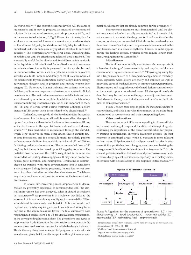
Figure 7 shows basic steps to guide the therapeutic choice in sporotrichosis,andtable2providesthesummaryofthemaindrugsadministered in sporotrichosis and their corresponding doses.
Other considerations
There are important differences regarding in vitro sensitivity to themain antifungal drugs and thedifferent etiological agents,reinforcingtheimportanceofthecorrectidentificationforproper-ly treating sporotrichosis. Sporothrix brasiliensis presents the best response to antifungal drugs, while S. mexicana is more tolerant to drug action.118 Epidemiological analyses reveal that the in vitro susceptibilityprofilehasbeenchangingovertime,emphasizingtheemergence of S. brasiliensisisolatestoleranttoitraconazole.119 In this context,potassiumiodide,terbinafine,andposaconazolemaybeal-ternative drugs against S. brasiliensis,especiallyinrefractorycases,or for those with no satisfactory in vivoresponsetoitraconazole.96,112
614 Orofino-CostaR,deMacedoPM,RodriguesAM,Bernardes-EngemannAR
An Bras Dermatol. 2017;92(5):606-20.
FIgure 7:Algorithm for the treatment of sporotrichosis. LC – lym-phocutaneous;CF–fixedcutaneous;KI–potassium iodide; ITZ–itraconazole;TBF–terbinafine;AmB–amphotericinB
Modified from: Orofino-Costa,etal.201570
1Hyperkeratoticor refractory cutaneous lesions:heat, cryosurgery, electrosurgery, exci-
sion/drainage,Kl+ITZorKl+TBF2Children,elderly,immunoreactiveforms:Kl
Pregnantwomen:Heat,cryosurgery,AmB

KI has been used in association with itraconazole, for refractorymycosisinfelines.However,itsuseinimmunosuppressedhumansneeds to be better evaluated due to the possibility of changes in the immunological reactivity, and worsening of the underlying dis-ease.120 This association has been used in humans with conidiobo-lomycosis.121 The effectiveness of this combination of drugs is prob-ably due to the different mechanisms of action and synergism of both drugs. KI capsules still needs to be tested in humans regarding absorption,pharmacodynamics,andbioavailabilityfortheeffectivedoseadjustmentinthetreatmentofsporotrichosis.
Thedrugs5-fluorocytosine,caspofungin,andfluconazoledonot exhibit in vitro antifungal activity against S. brasiliensis,S. schenckii,S. globosa,orS. mexicana.122,123Invitroanalysesindicatesignificantdif-ferencesamongtheminimumconcentrationsrequiredtoinhibitthegrowth of Sporothrix spp.and therequiredconcentration toreducethenumberof colony-formingunits,demonstrating the fungistaticand non-fungicide effect for the most available antifungal drugs.
PROGNOSISImmunocompetent individualswithskinormucosal clin-
icalformsusuallyheal inashortperiodoftime,althoughfibrousscarsarefrequentcomplaintsleadingtofunctionalorunestheticse-quelae.Inimmunosuppressedpatients,especiallyAIDS,thediseasemay disseminate and cause death. Untreated patients with chronic skinlesionsmaydevelopsevereclinicalforms,withsystemicmani-festations,frequentlyrequiringinpatienttreatment.
PERSPECTIVESForoveracentury,sporotrichosiswasdescribedasanoc-
cupationaldisease,withastrongruralprofile.However,inthelastdecades Sporothrix spp. as a threat to the warm-blooded vertebrate hosts’ health has emerged. The close relation between S. brasiliensis andthe felinehost iscurious, so that it causes largeepizooties inurban areas.124 Indeed, zoonotic pathogens aremore often associ-ated with emerging diseases than the non-zoonotic pathogens.125
This indicates the need to investigate the S. brasiliensis’ transmission dynamics involving cats and humans. Such a transmission route (cat-man)brokeaparadigminadiseaseinwhichepidemiologyhadalreadybeenconsideredtoberesolved,toalargeextent.Ecological
Sporotrichosis: an update on epidemiology, etiopathogenesis, laboratory and clinical therapeutics 615
An Bras Dermatol. 2017;92(5):606-20.
studiesareimportant,giventhattheuncertaintyregardingthefun-gus’reservoirsintheenvironmentandreasonsforthefluctuation of Sporothrix population still exists.
Theknowledgeoftheepidemiological-molecularprofileisessential tounderstand thedynamics of species occurrence, fromthestandpointofboththecharacterizationofadifferentiatedclin-icalstandardandtheresponsetothetreatment,aswellastheim-plementation of strategic Public Health policies intended to control epidemics. The low-cost molecular methods are important because theyprovidefastandaccurateresults,particularlyduringdiseaseoutbreaks. However, for countries with limited health budgets,isolating the Sporothrix in culture media is still the best diagnostic method,concerningbothcostandpositivity.
Investigations on the host-parasite interaction have evolved regardingbothcellandhumoralimmuneresponses.However,re-cent researches involving 3-carboxy-muconate cyclase (gp60-gp70)andthehumoralactivityreconductedtheseekforspecificantigensthat could be used in the diagnosis and antibodies based vaccines.5 Anti-gp70antibodiesarepotentiallyusefulfordisease’stherapybe-causetheystronglyreducethehostfungalburden,thuspreventingSporothrix adhesion to theextracellularmatrixcomponents,or in-ducingtheyeastsopsonizationinthehost-pathogeninteraction.126
The need to develop new antifungal drugs is encouraged by the increasing number of refractory cases resulting from the emer-gence of the resistance phenotype among the etiological agents.127 Currently,researchesonalternativetreatmentsforsporotrichosisre-vealpromisingmolecules,suchasterpinen-4-olandfarnesol,milte-fosine,TCAN26(astructuralanalogousofmiltefosine)andtheH3molecule(aninhibitorofthesterolmethyltransferaseenzyme).127-131 However, it shouldbepointedout that it is stillnecessary touseappropriate animal models and clinical tests to ensure the effective-ness and safety of treatment for such alternatives. q
ACKNOWLEDGMENTS
TheauthorsthankthetechnicalteamattheMycologyLab-oratoryat thePedroErnestoUniversityHospital (UERJ), for their support during all stages of preparation of this article. The authors alsowould like to thankRaphaeldaSilvaRoma forhis technicalcomputersupportandhisassistanceinpreparingthefigures.
Table 2: Main drugs used in sporotrichosis treatment with the corresponding dosages
Drug Itraconazole1 100 mg capsules Terbinafine125and250mgtablets Potassium solution
Continuous Pulse Continuous Pulse 1.42g/mL(0.07g/drop)
Adult 100-400mg/d2 400mg/d 250-500mg/d4 500mg/d 2- 4g/d
7d/month 7d/month
Pediatric 3-5mg/Kg/d3 ------ 62.5–250mg/d5 ---- 1-2 g/d
Dosage 1-2 x/d 2x/d 1-2 x/d 1-2 x/d 2 x/d
Laboratory control6 Bloodcount,biochemistry,LFT Bloodcount,biochemistry,LFTBloodcount,biochemistry,LFT,TSH,T4L
1 Takeatmealtime;2Startat100mg/d;3Maximumof200mg/d;4Startat250mg/d;5Dosevariesaccordingtoweight;6Priortotreatment,at3-4treatmentweeks,attheendofthetreatment.
LFT-liverfunctiontestsAdaptedfrom:Orofino-Costa,et al. 201570
REFERENCES1. Conti Diaz IA. Epidemiology of sporotrichosis in Latin America. Mycopathologia.
1989;108:113-6.2. Schenck BR. On refractory subcutaneous abscess caused by a fungus possibly
related to the Sporotricha. Bull Johns Hopkins Hosp. 1898;93:286-90.3. Kwon-Chung KJ, Bennett JE. Sporotrichosis. In: Kwon-Chung KJ, Bennett JE,
editors. Medical Mycology. Philadelphia: Lea & Febiger; 1992. p.707-29. 4. Carmichael JW. Chrysosporium and some other aleuriosporic hyphomycetes. Can
J Bot. 1962;40:1137-73.5. Rodrigues AM, Kubitschek-Barreira PH, Fernandes GF, de Almeida SR, Lopes-
Bezerra LM, de Camargo ZP. Immunoproteomic analysis reveals a convergent humoral response signature in the Sporothrix schenckii complex. J Proteomics. 2015 Feb 6;115:8-22.
6. Lutz A, Splendore A. Sobre uma micose observada em homens e ratos. Rev Med S. Paulo. 1907; 21:433-50.
7. Rippon JW. Sporotrichosis. In: Rippon JW, editor. Medical Mycology: the pathogenic fungi and the pathogenic actinomycetes. Philadelphia: WB Saunders; 1988. p. 325-52.
8. Morris-Jones R. Sporotrichosis. Clin Exp Dermatol. 2002;27:427-31.9. CDC. Epidemiologic notes and reports multistate outbreak of sporotrichosis in
seedling handlers. MMWR. 1988;37:652-3.10. Dooley DP, Bostic PS, Beckius ML. Spook house sporotrichosis: A point-source
outbreak of sporotrichosis associated with hay bale props in a Halloween haunted-house. Arch Intern Med. 1997 ;157:1885-7.
11. Helm MAF, Berman C. The clinical, therapeutic and epidemiological features of the sporotrichosis infection on the mines. In: Proceedings of the Transvaal Mine Medical Officers’ Association. Sporotrichosis Infection on Mines of the Witwatersrand. Johannesburg: The Transvaal Chamber of Mines; 1947. p. 59-74.
12. Song Y, Li SS, Zhong SX, Liu YY, Yao L, Huo SS. Report of 457 sporotrichosis cases from Jilin province northeast China, a serious endemic region. J Eur Acad Dermatol Venereol. 2013;27:313-8.
13. Saravanakumar PS, Eslami P, Zar FA. Lymphocutaneous sporotrichosis associated with a squirrel bite: case report and review. Clin Infect Dis. 1996;23:647-8.
14. Lacaz CS, Porto E, Martins JEC, Heins-Vaccari EM, Melo NT. Esporotricose e outras micoses gomosas. In: Lacaz CS, Porto E, Martins JEC, Heins-Vaccari EM, Melo NT editores. Tratado de micologia médica. São Paulo: Sarvier; 2002. p. 479-97.
15. Conti-Diaz IA. Sporotrichosis in Uruguay: epidemiologic and clinical aspects. Washington DC: Pan American Health Organization Scientific Publication; 1980. p. 312-21.
16. Alves SH, Boettcher CS, Oliveira DC, Tronco-Alves GR, Sgaria MA, Thadeu P, et al. Sporothrix schenckii associated with armadillo hunting in Southern Brazil: epidemiological and antifungal susceptibility profiles. Rev Soc Bras Med Trop. 2010;43:523-5.
17. Read SI, Sperling LC. Feline sporotrichosis. Transmission to man. Arch Dermatol. 1982;118:429-31.
18. Marques SA, Franco SRVS, Camargo RMP, Dias LDF, Haddad-Júnior V, Fabris VE. Esporotricose do gato doméstico (Felis catus): transmissão humana. Rev Inst Med Trop São Paulo. 1993;35:327-30.
19. Madrid IM, Mattei A, Martins A, Nobre M, Meireles M. Feline sporotrichosis in the southern region of Rio Grande do Sul, Brazil: clinical, zoonotic and therapeutic aspects. Zoonoses Public Health. 2010;57:151-4.
20. de Lima Barros MB, Schubach TM, Galhardo MC, de Oliveira Schubach A, Monteiro PC, Reis RS, et al. Sporotrichosis: an emergent zoonosis in Rio de Janeiro. Mem Inst Oswaldo Cruz. 2001;96:777-9.
21. Barros MB, Schubach A de O, do Valle AC, Gutierrez Galhardo MC, Conceição-Silva F, Schubach TM, et al. Cat-transmited sporotrichosis epidemic in Rio de Janeiro, Brazil: description of a series of cases. Clin Infect Dis. 2004;38:529-35.
22. Barros MB, Schubach AO, Schubach TM, Wanke B, Lambert-Passos SR. An epidemic of sporotrichosis in Rio de Janeiro, Brazil: epidemiological aspects of a series of cases. Epidemiol Infect. 2008;136:1192-6.
23. Freitas DF, Valle AC, da Silva MB, Campos DP, Lyra MR, de Souza RV, et al. Sporotrichosis: an emerging neglected opportunistic infection in HIV-infected patients in Rio de Janeiro, Brazil. PLoS Negl Trop Dis. 2014;8:e3110
24. Gutierrez-Galhardo MC, Freitas DFS, do Valle ACF, Almeida-Paes R, Oliveira MME, Zancope-Oliveira RM et al. Epidemiological aspects of sporotrichosis epidemic in Brazil. Curr Fungal Infect Rep. 2015;9:238-45.
25. Chakrabarti A, Bonifaz A, Gutierrez-Galhardo MC, Mochizuki T, Li S. Global epidemiology of sporotrichosis. Med Mycol. 2015;53:3-14.
26. Zhang Y, Hagen F, Stielow B, Rodrigues AM, Samerpitak K, Zhou X, et al. Phylogeography and evolutionary patterns in Sporothrix spanning more than 14,000 human and animal case reports. Persoonia. 2015 ;35:1-20.
27. Moussa TAA, Kadasa NMS, Al Zahrani HS, Ahmed SA, Feng P, Gerrits van den Ende AHG, et al. Origin and distribution of Sporothrix globosa causing sapronoses in Asia. J Med Microbiol. 2017;66:560-569.
28. Rodrigues AM, de Melo Teixeira M, de Hoog GS, Schubach TM, Pereira SA, Fernandes GF, et al. Phylogenetic analysis reveals a high prevalence of Sporothrix brasiliensis in feline sporotrichosis outbreaks. PLoS Negl Trop Dis. 2013;7:e2281
29. Rodrigues AM, de Hoog G, Zhang Y, de Camargo ZP. Emerging sporotrichosis is driven by clonal and recombinant Sporothrix species. Emerg Microbes Infect. 2014;3:e32.
30. Marimon R, Gené J, Cano J, Trilles L, Dos Santos Lazéra M, Guarro J. Molecular phylogeny of Sporothrix schenkii. J Clin Microbiol. 2006;44:3251-6.
31. Zhou X, Rodrigues AM, Feng P, Hoog GS. Global ITS diversity in the Sporothrix schenckii complex. Fungal Divers. 2014;66:153-65.
32. de Beer ZW, Duong TA, Wingfield MJ. The divorce of Sporothrix and Ophiostoma: solution to a problematic relationship. Stud Mycol. 2016;83:165-91.
33. Marimon R, Cano J, Gené J, Sutton DA, Kawasaki M, Guarro J. Sporothrix brasiliensis, S. globosa, and S. mexicana, three new Sporothrix species of clinical interest. J Clin Microbiol. 2007;45:3198-206.
34. Rodrigues AM, Bagagli E, de Camargo ZP, Bosco S de M. Sporothrix schenckii sensu stricto isolated from soil in an armadillo’s burrow. Mycopathologia. 2014;177:199-206.
35. Montenegro H, Rodrigues AM, Dias MA, da Silva EA, Bernardi F, de Camargo ZP. Feline sporotrichosis due to Sporothrix brasiliensis: an emerging animal infection in São Paulo, Brazil. BMC Vet Res. 2014;10:269.
36. Ajello L, Kaplan W. A new variant of Sporothrix Schenckii. Mykosen. 1969;12:633-44.37. Marimon R, Gené J, Cano J, Guarro J. Sporothrix luriei: a rare fungus from clinical
origin. Med Mycol. 2008;46:621-5. 38. Rodrigues AM, Cruz Choappa R, Fernandes GF, de Hoog GS, de Camargo ZP.
Sporothrix chilensis sp. nov. (Ascomycota: Ophiostomatales), a soil-borne agent of human sporotrichosis with mild-pathogenic potential to mammals. Fungal Biol. 2016;120(2):246-64.
39. Rodrigues AM, de Hoog S, de Camargo ZP. Emergence of pathogenicity in the Sporothrix schenckii complex. Med Mycol. 2013;51:405-12.
40. Morrison AS, Lockhart SR, Bromley JG, Kim JY, Burd EM. An environmental Sporothrix as a cause of corneal ulcer. Med Mycol Case Rep. 2013;2:88-90.
41. Mariat F, Escudié A, Gaxotte P. Isolation of Ceratocystis strains sp. conidial forms Sporotrichum, from human scalp and from tails of rats. Comparison with the pathogenic species Sporotrichum schenckii. C R Acad Sci Hebd Seances Acad Sci D. 1968;267:974-6.
42. Arrillaga-Moncrieff I, Capilla J, Mayayo E, Marimon R, Mariné M, Gené J, et al. Different virulence levels of the species of Sporothrix in a murine model. Clin Microbiol Infect. 2009;15:651-5.
43. Howard DH. Dimorphism of Sporotrichum schenckii. J Bacteriol. 1961;81:464-9.44. Rodrigues AM, de Hoog GS, de Camargo ZP. Sporothrix species causing outbreaks
in animals and humans driven by animal-animal transmission. PLoS Pathog. 2016;12:e1005638.
45. Mario DA, Santos RC, Denardi LB, Vaucher R de A, Santurio JM, Alves SH. Interference of melanin in the susceptibility profile of Sporothrix species to amphotericin B. Rev Iberoam Micol. 2016;33:21-5.
46. Almeida-Paes R, Figueiredo-Carvalho MH, Brito-Santos F, Almeida-Silva F, Oliveira MM, Zancopé-Oliveira RM. Melanins protect Sporothrix brasiliensis and Sporothrix schenckii from the antifungal effects of terbinafine. PLoS One. 2016;11:e0152796.
47. Castro RA, Kubitschek-Barreira PH, Teixeira PA, Sanches GF, Teixeira MM, Quintella LP,et al. Differences in cell morphometry, cell wall topography and Gp70 expression correlate with the virulence of Sporothrix brasiliensis clinical isolates. PLoS One. 2013;8:e75656.
48. Fernandes GF, dos Santos PO, Rodrigues AM, Sasaki AA, Burger E, de Camargo ZP. Characterization of virulence profile, protein secretion and immunogenicity of different Sporothrix schenckii sensu stricto isolates compared with S. globosa and S. brasiliensis species. Virulence. 2013;4:241-9.
49. Almeida-Paes R, de Oliveira LC, Oliveira MM, Gutierrez-Galhardo MC, Nosanchuk JD, Zancopé-Oliveira RM. Phenotypic characteristics associated with virulence of clinical isolates from the Sporothrix complex. Biomed Res Int. 2015;2015:212308.
50. Alba-Fierro CA, Pérez-Torres A, Toriello C, Romo-Lozano Y, López-Romero E, Ruiz-Baca E. Molecular components of the Sporothrix schenckii complex that induce immune response. Curr Microbiol. 2016;73:292-300
51. Freitas DF, De Siqueira Hoagland B, do Valle AC, Fraga BB, de Barros MB, de Oliveira Schubach A et al. Sporotrichosis in HIV-infected patients: report of 21 cases of endemic sporotrichosis in Rio de Janeiro, Brazil. Med Mycol. 2012;50(2):170-8.
52. Lopes-Bezerra LM, Schubach A, Costa RO. Sporothrix schenckii and sporotrichosis. An Acad Bras Cienc. 2006;78:293-308.
616 Orofino-CostaR,deMacedoPM,RodriguesAM,Bernardes-EngemannAR
An Bras Dermatol. 2017;92(5):606-20.
Sporotrichosis: an update on epidemiology, etiopathogenesis, laboratory and clinical therapeutics 617
An Bras Dermatol. 2017;92(5):606-20.
53. Kurosawa A, Pollock SC, Collins MP, Kraff CR, Tso MO. Sporothrix schenckii endophthalmitis in a patient with human immunodeficiency virus infection. Arch Ophthalmol. 1988;106:376-80.
54. Sena CM, Dias D, Oréfice F, Tanuri MA. Uveíte anterior granulomatosa causada por Sporothrix schenckii. Rev Bras Oftalmol. 1995;54:27-30.
55. Curi AL, Félix S, Azevedo KM, Estrela R, Villar EG, Saraça G. Retinal granuloma caused by Sporothrix schenckii. Am J Ophthalmol. 2003;136:205-7.
56. Freitas DF, Lima IA, Curi CL, Jordão L, Zancopé-Oliveira RM, Valle AC, Galhardo MC, et al. Acute dacryocystitis: another clinical manifestation of sporotrichosis. Mem Inst Oswaldo Cruz. 2014;109:262-4.
57. de Macedo PM, Sztajnbok DC, Camargo ZP, Rodrigues AM, Lopes-Bezerra LM, Bernardes-Engemann AR, et al. Dacryocystitis due to Sporothrix brasiliensis: a case report of a successful clinical and serological outcome with low-dose potassium iodide treatment and oculoplastic surgery. Br J Dermatol. 2015;172:1116-9
58. Ferreira CP, Nery JA, de Almeida AC, Ferreira LC, Corte-Real S, Conceição-Silva F. Parinaud’s oculoglandular syndrome associated with Sporothrix schenckii. IDCases. 2014;1:38-9.
59. Costa RO, de Mesquita KC, Damasco PS, Bernardes-Engemann AR, Dias CM, Silva IC, et al. Infectious arthritis as the single manifestation of sporotrichosis: serology from serum and synovial fluid samples as an aid to diagnosis. Rev Iberoam Micol. 2008;25:54-6.
60. Ribeiro BN, Ribeiro RN, Penna CR, Frota AC. Bone involvement by Sporothrix schenckii in an immunocompetent child. Pediatr Radiol. 2015;45:1427-30.
61. England DM, Hochholzer L. Sporothrix infection of the lung without cutaneous disease. Primary pulmonary sporotrichosis. Arch Pathol Lab Med. 1987;111:298-300.
62. Padhye AA, Kaufman L, Durry E, Banerjee CK, Jindal SK, Talwar P, et al. Fatal pulmonary sporotrichosis caused by Sporothrix schenckii var. luriei in India. J Clin Microbiol. 1992;30:2492-4.
63. Singhai M, Rawat V, Verma P, Jha PK, Shree D, Goyal R, et al. Primary pulmonary sporotrichosis in a sub-Himalayan patient. J Lab Physicians. 2012;4:48-9.
64. Orofino-Costa R, Unterstell N, Carlos Gripp A, de Macedo PM, Brota A, Dias E, et al. Pulmonary cavitation and skin lesions mimicking tuberculosis in a HIV negative patient caused by Sporothrix brasiliensis. Med Mycol Case Rep. 2013;2:65-71.
65. Aung AK, Spelman DW, Thompson PJ. Pulmonary Sporotrichosis: An Evolving Clinical Paradigm. Semin Respir Crit Care Med. 2015;36:756-66.
66. Vilela R, Souza GF, Fernandes Cota G, Mendoza L. Cutaneous and meningeal sporotrichosis in a HIV patient. Rev Iberoam Micol. 2007;24:161-3.
67. Orofino-Costa R, Bóia MN, Magalhães GA, Damasco PS, Bernardes-Engemann AR, Benvenuto F, et al. Arthritis as a hypersensitivity reaction in a case of sporotrichosis transmitted by a sick cat: clinical and serological follow up of 13 months. Mycoses. 2010;53:81-3.
68. Freitas DF, Valle AC, Cuzzi T, Brandão LG, Zancope-Oliveira RM, Galhardo MC. Sweet syndrome associated with sporotrichosis. Br J Dermatol. 2012;166:212-3.
69. Zhang Y, Pyla V. Sweet’s syndrome-like sporotrichosis. Int J Dermatol. 2014;53:e324-5.
70. Orofino-Costa R, de Macedo PM, Bernardes-Engemann AR. Hyperendemia of Sporotrichosis in the Brazilian Southeast: Learning From Clinics and Therapeutics. Curr Fungal Infect Rep. 2015;9:220-8.
71. Bernardes-Engemann AR, de Lima Barros M, Zeitune T, Russi DC, Orofino-Costa R, Lopes-Bezerra LM. Validation of a serodiagnostic test for sporotrichosis: a follow-up study of patients related to the Rio de Janeiro zoonotic outbreak. Med Mycol. 2015;53:28-33.
72. Kauffman CA, Bustamante B, Chapman SW, Pappas PG; Infectious Diseases Society of America. Clinical practice guidelines for the management of sporotrichosis: 2007 update by the Infectious Diseases Society of America. Clin Infect Dis. 2007;45:1255-65.
73. Hessler C, Kauffman CA, Chow FC. The Upside of Bias: A Case of Chronic Meningitis Due to Sporothrix schenckii in an Immunocompetent Host. Neurohospitalist. 2017;7:30-4.
74. de Hoog GS, Guarro J. Atlas of Clinical Fungi. Baarn: Centralbureau voor Schimmelcultures; 1995.
75. Mahajan VK. Sporotrichosis: an overview and therapeutic options. Dermatol Res Pract. 2014;2014:272376.
76. Oyarce JA, García C, Alave J, Bustamante B. Epidemiological clinical and laboratory characterization of sporotrichosis in patients of a tertiary care hospital in Lima, Peru, from 1991 to 2014. Rev Chilena Infectol. 2016;33:315-21.
77. Jessica N, Sonia RL, Rodrigo C, Isabella DF, Tânia MP, Jeferson C, et al. Diagnostic accuracy assessment of cytopathological examination of feline sporotrichosis. Med Mycol. 2015;53:880-4.
78. Pappas PG, Tellez I, Deep AE, Nolasco D, Holgado W, Bustamante B. Sporotrichosis in Peru: description of an area of hyperendemicity. Clin Infect Dis. 2000;30:65-70.
79. Zhao MD, Zhou X, Liu TT, Yang ZB. Morphological and physiological comparison of taxa comprising the Sporothrix schenckii complex. J Zhejiang Univ Sci B.
2015;16:940-7.80. Agarwal S, Gopal K, Umesh, Kumar B. Sporotrichosis in Uttarakhand (India): a
report of nine cases. Int J Dermatol. 2008;47:367-71.81. Quintella LP, Passos SR, do Vale AC, Galhardo MC, Barros MB, Cuzzi T, et al.
Histopathology of cutaneous sporotrichosis in Rio de Janeiro: a series of 119 consecutive cases. J Cutan Pathol. 2011;38:25-32.
82. Zhang YQ, Xu XG, Zhang M, Jiang P, Zhou XY, Li ZZ, Zhang MF, et al. Sporotrichosis: clinical and histopathological manifestations. Am J Dermatopathol. 2011;33:296-302.
83. Casserone S, Conti-Díaz IA, Zanetta E, Peña de Pereira ME. Serology of cutaneous sporotrichosis. Sabouraudia. 1983;21:317-21.
84. de Albornoz MB, Villanueva E, de Torres ED.. Application of immunoprecipitation techniques to the diagnosis of cutaneous and extracutaneousforms of sporotrichosis. Mycopathologia. 1984;85:177-83.
85. Bernardes-Engemann AR, Costa RC, Miguens BR, Penha CV, Neves E, Pereira BA, et al. Development of an enzyme-linked immunosorbent assay for the serodiagnosis of several clinical forms of sporotrichosis. Med Mycol. 2005;43:487-93.
86. Almeida-Paes R, Pimenta MA, Pizzini CV, Monteiro PC, Peralta JM, Nosanchuk JD, et al. Use of mycelial-phase Sporothrix schenckii exoantigens in an enzyme-linked immunosorbent assay for diagnosis of sporotrichosis by antibody detection. Clin Vaccine Immunol. 2007;14:244-9.
87. Almeida-Paes R, Bailão AM, Pizzini CV, Reis RS, Soares CM, Peralta JM, et al. Cell-free antigens of Sporothrix brasiliensis: antigenic diversity and application in an immunoblot assay. Mycoses. 2012;55:467-75.
88. Penha CV, Bezerra LM. Concanavalin A-binding cell wall antigens of Sporothrix schenckii: a serological study. Med Mycol. 2000;38:1-7.
89. Fernandes GF, Lopes-Bezerra LM, Bernardes-Engemann AR, Schubach TM, Dias MA, Pereira SA, et al. Serodiagnosis of sporotrichosis infection in cats by enzyme-linked immunosorbent assay using a specific antigen, SsCBF, and crude exoantigens. Vet Microbiol. 2011;147:445-9.
90. Alba-Fierro CA, Pérez-Torres A, Toriello C, Pulido-Camarillo E, López-Romero E, Romo-Lozano Y, et al. Immune response induced by an immunodominant 60 kDa glycoprotein of the cell wall of Sporothrix schenckii in two mice strains with experimental sporotrichosis. J Immunol Res. 2016;2016:6525831
91. Nascimento RC, Espíndola NM, Castro RA, Teixeira PA, Loureiro y Penha CV, Lopes-Bezerra LM, et al. Passive immunization with monoclonal antibody against a 70-kDa putative adhesin of Sporothrix schenckii induces protection in murine sporotrichosis. Eur J Immunol. 2008;38:3080-9.
92. Teixeira PA, de Castro RA, Nascimento RC, Tronchin G, Torres AP, Lazéra M, et al. Cell surface expression of adhesins for fibronectin correlates with virulence in Sporothrix schenckii. Microbiology. 2009;155:3730-8.
93. Alba-Fierro CA, Pérez-Torres A, López-Romero E, Cuéllar-Cruz M, Ruiz-Baca E. Cell wall proteins of Sporothrix schenckii as immunoprotective agents. Rev Iberoam Micol. 2014 Jan-Mar;31(1):86-9.
94. Almeida SR. Therapeutic monoclonal antibody for sporotrichosis. Front Microbiol. 2012;3:409.
95. Oliveira MM, Almeida-Paes R, Muniz MM, Gutierrez-Galhardo MC, Zancope-Oliveira RM. Phenotypic and molecular identification of Sporothrix isolates from an epidemic area of sporotrichosis in Brazil. Mycopathologia. 2011;172:257-67.
96. Ottonelli Stopiglia CD, Magagnin CM, Castrillón MR, Mendes SD, Heidrich D, Valente P,et al. Antifungal susceptibilities and identification of species of the Sporothrix schenckii complex isolated in Brazil. Med Mycol. 2014;52:56-64
97. Camacho E, León-Navarro I, Rodríguez-Brito S, Mendoza M, Niño-Vega GA. Molecular epidemiology of human sporotrichosis in Venezuela reveals high frequency of Sporothrix globosa. BMC Infect Dis. 2015 Feb 25;15:94.
98. Madrid H, Cano J, Gené J, Bonifaz A, Toriello C, Guarro J. Sporothrix globosa, a pathogenic fungus with widespread geographical distribution. Rev Iberoam Micol. 2009;26:218-22.
99. Romeo O, Scordino F, Criseo G. New insight into molecular phylogeny and epidemiology of Sporothrix schenckii species complex based on calmodulin-encoding gene analysis of Italian isolates. Mycopathologia. 2011;172:179-86
100. Yu X, Wan Z, Zhang Z, Li F, Li R, Liu X. Phenotypic and molecular identification of Sporothrix isolates of clinical origin in Northeast China. Mycopathologia. 2013;176:67-74.
101. Kano R, Okubo M, Siew HH, Kamata H, Hasegawa A. Molecular typing of Sporothrix schenckii isolates from cats in Malaysia. Mycoses. 2015;58:220-4.
102. Rodrigues AM, de Hoog GS, de Camargo ZP. Genotyping species of the Sporothrix schenckii complex by PCR-RFLP of calmodulin. Diagn Microbiol Infect Dis. 2014;78:383-7.
103. Rodrigues AM, de Hoog GS, de Camargo ZP. Molecular diagnosis of pathogenic Sporothrix species. PLoS Negl Trop Dis. 2015;9(:e0004190.
104. Rodrigues AM, Najafzadeh MJ, de Hoog GS, de Camargo ZP. Rapid identification of emerging human-pathogenic Sporothrix species with rolling circle amplification. Front Microbiol. 2015 Dec 8;6:1385
Mailing address:RosaneOrofinoCostaAv. 28 de Setembro, 77 - 2º andarVilaIsabel20551-030RiodeJaneiro-RJBrazilE-mail:[email protected]
How to cite this article: Orofino-Costa R, de Macedo PM, Rodrigues AM, Bernardes-Engemann AR. Sporothrichosis: an update onepidemiology,etiopathogenesis,laboratoryandclinicaltherapeutics.AnBrasDermatol.2017;92(5):606-20.
105. Fire A, Xu SQ. Rolling replication of short DNA circles. Proc Natl Acad Sci U S A. 1995;92:4641-5.
106. de Lima Barros MB, Schubach AO, de Vasconcellos Carvalhaes de Oliveira R, Martins EB, Teixeira JL, Wanke B. Treatment of cutaneous sporotrichosis with itraconazol-study of 645 patients. Clin Infect Dis. 2011;52:e200-6.
107. Song Y, Zhong SX, Yao L, Cai Q, Zhou JF, Liu YY, et al. Efficacy and safety of itraconazole pulses vs. continuous regimen in cutaneous sporotrichosis. J Eur Acad Dermatol Venereol. 2011;25:302-5.
108. Paul V, Rawal H. Cardiotoxicity with Itraconazole. BMJ Case Rep. 2017 Apr 10;2017.
109. Sterling JB, Heymann WR. Potassium iodide in dermatology: a 19th century drug for the 21st century-uses, pharmacology, adverse effects, and contraindications. J Am Acad Dermatol. 2000;43:691-7.
110. Xue SL, Li L. Oral potassium iodide for the treatment of sporotrichosis. Mycopathologia. 2009;167:355-6.
111. Costa RO, Macedo PM, Carvalhal A, Bernardes-Engemann AR. Use of potassium iodide in dermatology: updates on an old drug. An Bras Dermatol. 2013 May-Jun;88:396-402.
112. Macedo PM, Lopes-Bezerra LM, Bernardes-Engemann AR, Orofino-Costa R. New posology of potassium iodide for the treatment of cutaneous soporotrichosis: study of efficacy and safety in 102 patients. J Eur Acad Dermatol Venereol. 2015;29:719-24.
113. Antunes Tde A, Nobre M de O, Faria RO, Meinerz AR, Martins AA, Cleff MB, et al. Experimental cutaneous sporotrichosis: In vivo evaluation of itraconazol and terbinafine. Rev Soc Bras Med Trop. 2009;42:706-10.
114. Francesconi G, Valle AC, Passos S, Reis R, Galhardo MC. Terbinafine (250 mg/d): An effective and safe treatment of cutaneous sporotrichosis. J Eur Acad Dermatol Venereol. 2009;23:1273-6.
115. Costa RO, Bernardes-Engemann AR, Azulay-Abulafia L, Benvenuto F, Neves Mde L, Lopes-Bezerra LM. Sporotrichosis in pregnancy: case reports of 5 patients in a zoonotic epidemic in Rio de Janeiro, Brasil. An Bras Dermatol. 2011;86:995-8.
116. Bonifaz A, Vásquez-Gonzáles D. Diagnosis and treatment of cutaneous sporotrichosis: what are the options? Curr Fungal Infect Rep. 2013;7:252-9.
117. Gilaberte Y, Aspiroz C, Alejandre MC, Andres-Ciriano E, Fortuño B, Charlez L, et al. Cutaneous sporotrichosis treated with photodynamic therapy: An in vivo and in vitro study. Photomed Laser Surg. 2014;32:54-7.
118. Oliveira DC, Lopes PG, Spader TB, Mahl CD, Tronco-Alves GR, Lara VM, et al. Antifungal susceptibilities of Sporothrix albicans, S. brasiliensis and S. luriei of the S. schenckii complex identified in Brazil. J Clin Microbiol. 2011;49:3047-9.
119. Borba-Santos LP, Rodrigues AM, Gagini TB, Fernandes GF, Castro R, de Camargo ZP, et al. Susceptibility of Sporothrix brasiliensis isolates to amphotericin B, azoles, and terbinafine. Med Mycol. 2015;53:178-88
120. Reis EG, Gremião ID, Kitada AA, Rocha RF, Castro VS, Barros MB, et al. Potassium iodide capsule treatment of feline sporotrichosis. J Feline Med Surg. 2012;14:399-404.
121. Gupta M, Narang T, Kaur RJ, Manhas A, Saikia UN, Dogra S. A prospective case series evaluating efficacy and safety of combination of itraconazole and potassium iodide in rhinofacial conidiobolomycosis. Int J Dermatol. 2016;55:208-14
122. Marimon R, Serena C, Gené J, Cano J, Guarro J. In vitro antifungal susceptibilities of five species of Sporothrix. Antimicrob Agents Chemother. 2008;52:732-4.
123. Rodrigues AM, de Hoog GS, de Cássia Pires D, Brihante RS, Sidrim JJ, Gadelha MF, et al. Genetic diversity and antifungal susceptibility profiles in causative agents of sporotrichosis. BMC Infect Dis. 2014;14:219.
124. Gremião ID, Miranda LH, Reis EG, Rodrigues AM, Pereira SA. Zoonotic epidemic of sporotrichosis: Cat to human transmission. PLoS Pathog. 2017;13:e1006077.
125. Taylor LH, Latham SM, Woolhouse ME. Risk factors for human disease emergence. Philos Trans R Soc Lond B Biol Sci. 2001;356:983-9.
126. Ruiz-Baca E, Toriello C, Perez-Torres A, Sabanero-Lopez M, Villagomez-Castro JC, Lopez-Romero E. Isolation and some properties of a glycoprotein of 70 kDa (Gp70) from the cell wall of Sporothrix schenckii involved in fungal adherence to dermal extracellular matrix. Med Mycol. 2009;47:185-96.
127. Borba-Santos LP, Gagini T, Ishida K, de Souza W, Rozental S. Miltefosine is active against Sporothrix brasiliensis isolates with in vitro low susceptibility to amphotericin B or itraconazole. J Med Microbiol. 2015;64:415-22.
128. Brilhante RS, Silva NF, Marques FJ, Castelo-Branco Dde S, de Lima RA, Malaquias AD, et al. In vitro inhibitory activity of terpenic derivatives against clinical and environmental strains of the Sporothrix schenkii complex. Med Mycol. 2015;53:93-8.
129. Brilhante RS, Malaquias AD, Caetano EP, Castelo-Branco Dde S, Lima RA, Marques FJ, et al. In vitro inhibitory effect of miltefosine against strains of Histoplasma capsulatum var. capsulatum and Sporothrix spp. Med Mycol. 2014;52:320-5.
130. Borba-Santos LP, Ishida K, Calogeropoulou T, Souza Wd, Rozental S. Adamantylidene-substituted alkylphosphocholine TCAN26 is more active against Sporothrix schenckii than miltefosine. Mem Inst Oswaldo Cruz. 2016;111:523-7.
131. Borba-Santos LP, Visbal G, Gagini T, Rodrigues AM, de Camargo ZP, Lopes-Bezerra LM, et al. ∆24-sterol methyltransferase plays an important role in the growth and development of Sporothrix schenckii and Sporothrix brasiliensis. Front Microbiol. 2016;7:311.
618 Orofino-CostaR,deMacedoPM,RodriguesAM,Bernardes-EngemannAR
An Bras Dermatol. 2017;92(5):606-20.

Questions s
1. The most common clinical presentation of sporotricho-sis in Brazil is: a. lymphocutaneous b. fixedcutaneous c. osteoarticular d. pulmonary
2. Concerning the sporotrichosis transmission, it may be stated that: a. zoonotic transmissiononly takesplacewhentheetio-
logical agent is Sporothrix globosa b. transmission by means of trauma only happens when
the etiological agent is S. schenckii c. respiratory transmission results from inhalation of Spo-
rothrix conidia d.respiratory transmission is restricted to specific geo-
graphic regions
3. The histopathological exam in sporotrichosis shows: a.highsensitivityandlowspecificity b.lowsensitivityandhighspecificity c. highsensitivityandhighspecificity d.lowsensitivityandlowspecificity
4. To isolate Sporothrix spp. the following basic fungus culture media are preferably used: a.Cerealagar(Cornmealagar)andBHI(BrainHeartIn-
fusion) b.Mycoselagarandcerealagar(Cornmealagar) c.SabouraudagarandPDA(PotatoDextroseAgar) d. Sabouraud dextrose agar and Mycosel agar
5. The asteroid body found on the dermis of the patients with sporotrichosis: a. consistsofnon-specificinflammatorycells b.confirmsthediagnosisofsporotrichosis c. represents a probable immunological reaction to the
fungal cell d.suggeststhehost’simmunodeficiency
6. It may be stated that serology for the diagnosis of spo-rotrichosis: a.isonlyindicatedforskinforms b. does not provide a good clinical-laboratory correlation c. is useful in diagnosing systemic and atypical manifesta-
tions d.maynotbeusedinorganicfluidsotherthanserum
7. Regarding infectious arthritis caused by Sporothrix spp., it is correct to state that: a. it is almost always polyarticular and migratory
b. it is almost always monoarticular with phlogistic signs c.adjacentskinlesionsarenotobserved d.itispolyarticular,butdoesnotshowphlogisticsigns
8. Check the true statement regarding the laboratory diag-nosis of sporotrichosis: a.the direct microscopy is more sensitive and specific
than the culture in humans b.thegoldstandardistheisolationandidentificationof
Sporothrix spp. c.thehistopathologicalexam,impregnatedbysilver,gen-
erally exhibits abundance of fungal structures d. the molecular identification of the Sporothrix species
has no diagnostic value.
9. Feline zoonotic sporotrichosis, predominant in the South and Southeast regions of Brazil, is mostly associated with the following Sporothrix species: a. S. schenckii b. S. lurei c. S. brasiliensis d. S. globosa
10. Examples of immunoreactive sporotrichosis clinical forms include: a. erythema multiforme and meningitis b. Sweet’s syndrome and erythema nodosum c. infectious arthritis and erythema multiforme d. Sweet’s syndrome and infectious arthritis
11. The choice treatment for localized cutaneous forms of sporotrichosis in immunocompetent people is: a.terbinafine b.fluconazole c. griseofulvin d.itraconazole
12. Recent advances in Sporothrix taxonomy have led to the identification of new clinical interest agents. The clini-cal clade includes the following species: a. S. chilensis,S. pallida,S. mexicana,andS. globosa b. S. brasiliensis,S. schenckii,S. chilensis,andS. pallida c. S. brasiliensis,S. schenckii,S. globosa,and S. luriei d. S. schenckii,S. globosa,S. mexicana,andS. luriei
13. Check the correct alternative regarding potassium io-dide used in sporotrichosis treatment: a. it is recommended for systemic forms with lung in-
volvement
Sporotrichosis: an update on epidemiology, etiopathogenesis, laboratory and clinical therapeutics 619
An Bras Dermatol. 2017;92(5):606-20.
b. its mechanism of action is well established, inhibiting ergosterol synthesis
c. it is useful in treating immunoreactive forms due to its immunomodulatory effect
d. it is used in systemic forms with meningeal involve-ment
14. Check the alternative that includes the laboratory exams to be requested prior to starting treatment with itraconazole and as a therapeutic control: a. biochemistry, blood count, and liver function tests b. liver function tests, blood count, and TSH c. blood count, liver function tests, and free T4 d. biochemistry, liver function tests, and TSH
15. If required, the drug that may be administered to preg-nant women affected by sporotrichosis is: a. fluconazole b. itraconazole c. amphotericin B d. posaconazole
16. Among the actions below, check the one that is not rec-ommended as a preventive measure against the propaga-tion of zoonotic transmission sporotrichosis cases: a. street animals neutering b. removal of Sporothrix from soil and vegetation c. treatment of sick animals d. incineration of dead infected animals
17. Among the statements below, check the one in which the correlation between the etiological agent and the geo-graphic distribution is appropriate: a. S. brasiliensis in Mexico b. S. globosa in Chile c. S. pallida in the USA d. S. globosa in China
18. The biochemical compound present in S. brasiliensis that may be related to its virulence is: a. quinine b. adrenalin c. melanin d. erythropoietin
19. The disease most commonly present in the differential diagnosis of lymphocutaneous sporotrichosis is: a. tegumentary leishmaniasis b. granuloma annulare c. paracoccidioidomycosis d. epidermoid carcinoma
20. The important biomarker present in the Sporothrix cell wall that can be used for the production of a therapeutic vaccine is the glycoprotein: a. gp40 b. gp80 c. gp70 d. gp100
Answer key
Behçet´s Disease: a review with emphasis on dermatological as-pects. An Bras Dermatol. 2017;92(4):452-64
1. A
2. C
3. D
4. D
5. C
6. C
7. B
8. B
9. C
10. B
11. D
12. C
13. C
14. A
15. C
16. B
17. D
18. C
19. A
20. C
Papers
Information for all members: The EMC-D questionnaire is now available at the homepage of the Brazilian Annals of Dermatology: www.anaisdedermatologia.org.br. The dead-line for completing the questionnaire is 30 days from the date of online publication.
620 Orofino-CostaR,deMacedoPM,RodriguesAM,Bernardes-EngemannAR
AnBrasDermatol.2017;92(5):606-20.