SPOP suppresses tumorigenesis by regulating Hedgehog/Gli2 ...
12
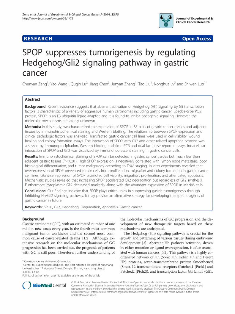
RESEARCH Open Access SPOP suppresses tumorigenesis by regulating Hedgehog/Gli2 signaling pathway in gastric cancer Chunyan Zeng 1 , Yao Wang 1 , Quqin Lu 2 , Jiang Chen 3 , Junyan Zhang 1 , Tao Liu 1 , Nonghua Lv 3 and Shiwen Luo 1* Abstract Background: Recent evidence suggests that aberrant activation of Hedgehog (Hh) signaling by Gli transcription factors is characteristic of a variety of aggressive human carcinomas including gastric cancer. Speckle-type POZ protein, SPOP, is an E3 ubiquitin ligase adaptor, and it is found to inhibit oncogenic signaling. However, the molecular mechanisms are largely unknown. Methods: In this study, we characterized the expression of SPOP in 88 pairs of gastric cancer tissues and adjacent tissues by immunohistochemical staining and Western blotting. The relationship between SPOP expression and clinical pathologic factors was analyzed. Transfected gastric cancer cell lines were used in cell viability, wound healing and colony formation assays. The interaction of SPOP with Gli2 and other related apoptotic proteins was assessed by immunoprecipitation, Western blotting, real-time PCR and dual luciferase reporter assays. Intracellular interaction of SPOP and Gli2 was visualized by immunofluorescent staining in gastric cancer cells. Results: Immunohistochemical staining of SPOP can be detected in gastric cancer tissues but much less than adjacent gastric tissues (P < 0.01). High SPOP expression is negatively correlated with lymph node metastasis, poor histological differentiation, and tumor malignancy according to TNM staging. In vitro experiments revealed that over-expression of SPOP prevented tumor cells from proliferation, migration and colony formation in gastric cancer cell lines. Likewise, repression of SPOP promoted cell viability, migration, proliferation, and attenuated apoptosis. Mechanistic studies revealed that increasing SPOP accelerated Gli2 degradation but regardless of Gli2 synthesis. Furthermore, cytoplasmic Gli2 decreased markedly along with the abundant expression of SPOP in MKN45 cells. Conclusions: Our findings indicate that SPOP plays critical roles in suppressing gastric tumorigenesis through inhibiting Hh/Gli2 signaling pathway. It may provide an alternative strategy for developing therapeutic agents of gastric cancer in future. Keywords: SPOP, Gli2, Hedgehog, Degradation, Apoptosis, Gastric cancer Background Gastric carcinoma (GC), with an estimated number of one million new cases every year, is the fourth most common malignant tumor worldwide and the second most com- mon cause of cancer-related deaths [1,2]. Although ex- tensive research on the molecular mechanisms of GC progression has been carried out, the prognosis of patients with GC is still poor. Therefore, further understanding of the molecular mechanisms of GC progression and the de- velopment of new therapeutic targets based on these mechanisms are anticipated. The Hedgehog (Hh) signaling pathway is crucial for the growth and patterning of various tissues during embryonic development [3]. Aberrant Hh pathway activation, driven by either mutation or ligand overexpression, is often associ- ated with human cancers [4,5]. This pathway is a highly co- ordinated network of Hh (Sonic Hh, Indian Hh and Desert Hh) proteins, seven-transmembrane protein Smoothened (Smo), 12-transmembrane receptors (Patched1 [Ptch1] and Patched2 [Ptch2]), and transcription factor Gli family (Gli1, * Correspondence: [email protected] 1 Center for Experimental Medicine, The First Affiliated Hospital of Nanchang University, No. 17 Yongwai Street, Donghu District, Nanchang, Jiangxi 330006, China Full list of author information is available at the end of the article © 2014 Zeng et al.; licensee BioMed Central Ltd. This is an Open Access article distributed under the terms of the Creative Commons Attribution License (http://creativecommons.org/licenses/by/4.0), which permits unrestricted use, distribution, and reproduction in any medium, provided the original work is properly credited. The Creative Commons Public Domain Dedication waiver (http://creativecommons.org/publicdomain/zero/1.0/) applies to the data made available in this article, unless otherwise stated. Zeng et al. Journal of Experimental & Clinical Cancer Research 2014, 33:75 http://www.jeccr.com/content/33/1/75
Transcript of SPOP suppresses tumorigenesis by regulating Hedgehog/Gli2 ...
Zeng et al. Journal of Experimental & Clinical Cancer Research 2014, 33:75http://www.jeccr.com/content/33/1/75
RESEARCH Open Access
SPOP suppresses tumorigenesis by regulatingHedgehog/Gli2 signaling pathway in gastriccancerChunyan Zeng1, Yao Wang1, Quqin Lu2, Jiang Chen3, Junyan Zhang1, Tao Liu1, Nonghua Lv3 and Shiwen Luo1*
Abstract
Background: Recent evidence suggests that aberrant activation of Hedgehog (Hh) signaling by Gli transcriptionfactors is characteristic of a variety of aggressive human carcinomas including gastric cancer. Speckle-type POZprotein, SPOP, is an E3 ubiquitin ligase adaptor, and it is found to inhibit oncogenic signaling. However, themolecular mechanisms are largely unknown.
Methods: In this study, we characterized the expression of SPOP in 88 pairs of gastric cancer tissues and adjacenttissues by immunohistochemical staining and Western blotting. The relationship between SPOP expression andclinical pathologic factors was analyzed. Transfected gastric cancer cell lines were used in cell viability, woundhealing and colony formation assays. The interaction of SPOP with Gli2 and other related apoptotic proteins wasassessed by immunoprecipitation, Western blotting, real-time PCR and dual luciferase reporter assays. Intracellularinteraction of SPOP and Gli2 was visualized by immunofluorescent staining in gastric cancer cells.
Results: Immunohistochemical staining of SPOP can be detected in gastric cancer tissues but much less thanadjacent gastric tissues (P < 0.01). High SPOP expression is negatively correlated with lymph node metastasis, poorhistological differentiation, and tumor malignancy according to TNM staging. In vitro experiments revealed thatover-expression of SPOP prevented tumor cells from proliferation, migration and colony formation in gastric cancercell lines. Likewise, repression of SPOP promoted cell viability, migration, proliferation, and attenuated apoptosis.Mechanistic studies revealed that increasing SPOP accelerated Gli2 degradation but regardless of Gli2 synthesis.Furthermore, cytoplasmic Gli2 decreased markedly along with the abundant expression of SPOP in MKN45 cells.
Conclusions: Our findings indicate that SPOP plays critical roles in suppressing gastric tumorigenesis throughinhibiting Hh/Gli2 signaling pathway. It may provide an alternative strategy for developing therapeutic agents ofgastric cancer in future.
Keywords: SPOP, Gli2, Hedgehog, Degradation, Apoptosis, Gastric cancer
BackgroundGastric carcinoma (GC), with an estimated number of onemillion new cases every year, is the fourth most commonmalignant tumor worldwide and the second most com-mon cause of cancer-related deaths [1,2]. Although ex-tensive research on the molecular mechanisms of GCprogression has been carried out, the prognosis of patientswith GC is still poor. Therefore, further understanding of
* Correspondence: [email protected] for Experimental Medicine, The First Affiliated Hospital of NanchangUniversity, No. 17 Yongwai Street, Donghu District, Nanchang, Jiangxi330006, ChinaFull list of author information is available at the end of the article
© 2014 Zeng et al.; licensee BioMed Central LCommons Attribution License (http://creativecreproduction in any medium, provided the orDedication waiver (http://creativecommons.orunless otherwise stated.
the molecular mechanisms of GC progression and the de-velopment of new therapeutic targets based on thesemechanisms are anticipated.The Hedgehog (Hh) signaling pathway is crucial for the
growth and patterning of various tissues during embryonicdevelopment [3]. Aberrant Hh pathway activation, drivenby either mutation or ligand overexpression, is often associ-ated with human cancers [4,5]. This pathway is a highly co-ordinated network of Hh (Sonic Hh, Indian Hh and DesertHh) proteins, seven-transmembrane protein Smoothened(Smo), 12-transmembrane receptors (Patched1 [Ptch1] andPatched2 [Ptch2]), and transcription factor Gli family (Gli1,
td. This is an Open Access article distributed under the terms of the Creativeommons.org/licenses/by/4.0), which permits unrestricted use, distribution, andiginal work is properly credited. The Creative Commons Public Domaing/publicdomain/zero/1.0/) applies to the data made available in this article,
Zeng et al. Journal of Experimental & Clinical Cancer Research 2014, 33:75 Page 2 of 12http://www.jeccr.com/content/33/1/75
Gli2, Gli3) [6-8]. Hh exerts its biological function througha signaling cascade that culminates in an alteration of thebalance between activator and repressor forms of the Glitranscription factors. The vertebrate Gli repressor func-tion largely depends on Gli3, while the primary Gli activa-tor function largely depends on Gli2. Gli1 acts as atranscriptional activator and thus a reliable indicator ofHh pathway activity [9,10]. In Drosophila, the Hh signal ismediated by the Cubitus Interruptus (Ci) transcriptionfactor. Yet the Ci function is positively regulated by Fused(Fu) serine/threonine kinase, but suppressed by Suppres-sor of Fused (SuFu) and the kinesin-like molecule Costal2(Cos2) [11]. SuFu is antagonized by HIB (Hh-inducedMATH and BTB domain protein), of which speckle-typePOZ protein, SPOP, is the vertebrate homolog [12].SPOP is demonstrated as an E3 ubiquitin ligase adaptor.
It has 374 residues and contains three domains: an N-terminal MATH domain (residues 28–166) that recruitssubstrates, an internal BTB domain (residues 190–297)that binds Cul3, and a C-terminal nuclear localization se-quence (residues 365–374) [13]. SPOP is mostly studiedin ubiquitin-dependent proteolysis, however, its role intumorigenesis is largely unknown [13,14]. Li C., et al.demonstrated that SPOP is responsible for full length Gli2and Gli3 ubiquitination and proteolysis [15]. Recently,Geng C., et al. revealed that wild type SPOP functions as acritical tumor suppressor in prostate cancer cells by pro-moting the turnover of SRC-3 (p160 steroid receptorcoactivator-3) protein and, thus suppressing androgen re-ceptor transcription activity [16]. Wang C., et al. showedthat SPOP promotes full-length Gli2 and Gli3 degrad-ation, which may indicate a potential tumor suppressorrole of SPOP [17]. The limited studies indicate the import-ance of SPOP in tumorigenesis, which prompts us to findout more evidence of it.In this study, we evaluated SPOP expression in gastric
cancer tissues and adjacent gastric tissues. We then ex-hibited possible correlations between SPOP expressionand the clinical pathologic factors. Based on the resultsof clinical findings, we performed in vitro experimentsand studied the effects of up-regulated or down-regulatedSPOP expression on the proliferation, migration andapoptosis of GC cell lines. Our results indicate that SPOPreduces gastric cancer cell invasion and proliferation byregulating Hh/Gli2 signaling pathway.
MethodsConstructs, antibodies and reagentsHuman SPOP cDNA was amplified by PCR and theninserted into KpnI and NotI sites of Pub6/V5-HisB vec-tor. SPOP - miRNA (miR-SPOP) expressed vectors weregenerated using the BLOCK-iT™ Pol II miR RNAi Expres-sion Vector Kit (K4936-00, Invitrogen, Carlsbad, CA).Clones were confirmed by DNA sequencing of both
strands (Generay, Shanghai, China). The oligonucleotidesequences for miRNA constructs were as follows: for miR-SPOP-607, 5′-AAT GAC TTC ACC CAT TTC CTC-3′;for miR-SPOP-917, 5′-TAA GCT TGT CAT CAG GGAGAA-3′; and for miR-SPOP-1430, 5′-AGT TGA TGAAAT CCA CTG CCT-3′. The numbers in the miRNAconstructs indicate the start of targeted nucleotide se-quence of the gene. All transfectants were selected with7 μg/ml Blasticidin for 3 weeks and selected clones wereidentified by Western blotting. Antibodies were purchasedfrom Abcam (Gli2, ab26056; p16, ab108349; PTEN,ab31392), Cell Signaling (Gli1, L42B10; Caspase-3, 9665S;cleaved Caspase-3, 9664; Ki-67, 9449; Cyclin B1, 4135S;p21, 2947; p27, 3686; Daxx, 4533; p-ERK, 4376S), Novus(SPOP, H00008405-B01P), Protein-Tech (SPOP, 16750-1-AP), Santa Cruz (PCNA, sc-9857; PARP, sc-7150; Actin, sc-1616), Abgent (DUSP7, AP8450a), Millipore (GAPDH,MAB374), Thermo (goat anti-rabbit IgG, 31460). Enhancedchemiluminescence (ECL) Western blot detection re-agents were from Thermo Fisher Scientific (Rockford, IL).MG-132 (Z-Leu-Leu-Leu-CHO, I-130) was from Boston-Biochem. Protease inhibitor cocktail, Lubrol-PX, and3-(4,5-dimethylthiazol-2-yl)-2,5-diphenyltetrazolium brom-ide (MTT) were purchased from Sigma-Aldrich (St. Louis,MO).
Cell culture and tissue specimensHuman gastric epithelial cell line GES-1, human gastriccancer cell lines AGS, MKN28, SGC-7901, MKN45 andhuman embryonic kidney 293 T (HEK293T) cell lineswere obtained from American Type Culture Collection(ATCC, Manassas). Cells were maintained in DMEM(Invitrogen, CA) supplemented with 10% fetal bovine serum(FBS; Invitrogen, CA) and 100 μg/ml streptomycin in hu-midified incubator at 37°C with 5% CO2.
Gastric cancer samples were obtained from 88 patientsdiagnosed gastric carcinoma from March 2007 to April2010 in the Department of Pathology, the First AffiliatedHospital of Nanchang University. All the patients hadnot received chemotherapy or radiotherapy before sur-gery and diagnosed by clinic pathologic staging based onthe 2010 American Joint Committee on Cancer (AJCC)criteria. Detailed clinic pathologic information of all pa-tients and their correlation with SPOP expression weresummarized in Table 1. This study was approved by theinstitutional review board of the First Affiliated Hospitalof Nanchang University. All patients had given writteninformed consent.
Immunohistochemical stainingParaffin sections (5 μm) of formalin-fixed gastric cancertissue and adjacent tissue arrays were de-waxed, rehy-drated and incubated in 3% H2O2 for 10 min to block en-dogenous peroxidase. Then the sections were autoclaved

Table 1 Correlation between SPOP expression and clinic pathological information
Variable Groups SPOP expression scoring Spearman’s R P
Low (<6) High (≥6)
Gender Male 54 10
Female 22 2 −0.095 0.380
Age ≤50 18 1
>50 62 7 0.070 0.518
Lymp node Metastasis 54 4
No metastasis 23 7 0.236 0.027*
Histopathologic grading Poorly differentiated 34 0
Moderately differentiated 36 10
Well differentiated 8 0 −0.232 0.029*
TNM staging I 13 1
II 33 7
III + IV 30 4 −0.375 0.0003*
*Significant difference.
Zeng et al. Journal of Experimental & Clinical Cancer Research 2014, 33:75 Page 3 of 12http://www.jeccr.com/content/33/1/75
in 10 mM sodium citrate buffer (pH 6.0) for 15 min. Nor-mal goat serum (10%) was used to block non-specificstaining and then the tissue sections were exposed to indi-cated antibodies. All of the stainings were observed by atleast 2 independent investigators blinded to the histopath-ologic features and patient data of the samples [18]. Thewidely accepted German semi-quantitative scoring systemwas used for assessing the staining intensity and area ex-tent [19]. Each specimen was assigned a score accordingto the intensity of the nucleic, cytoplasmic, and/or mem-brane staining (no staining, not detected = 0; weak stain-ing, light yellow = 1; moderate staining, yellowish brown =2; strong staining, brown =3) and the extent of stainedcells (no staining = 0, 1-24% = 1, 25-49% = 2, 50-74% = 3,75-100% = 4). The final immunoreactive score was deter-mined by multiplying the intensity score with the extentscore of stained cells, ranging from 0 (the minimum) to12 (the maximum).
Western blottingCells were solubilized in the extraction buffer (0.5%Lubrol-PX, 50 mM KCl, 2 mM CaCl2, 20% glycerol,50 mM Tris–HCl, and inhibitors of proteases and phos-phatases, pH 7.4) at 4°C for 30 min and centrifuged(12,000 rpm, 15 min at 4°C) to maintain the supernatant.Protein concentrations were determined by Bradfordassay (Bio-Rad, CA). Proteins were resolved by SDS-PAGE and transferred to a polyvinylidene difluoridemembrane (Bio-Rad), and then probed with the specifiedprimary antibody followed by the appropriate secondaryantibody. The immunostaining was visualized by usingKodak X-ray films, and quantified by scanning films con-taining nonsaturated signals with an Epson 1680 scannerbefore analyzed with ImageJ software [20].
ImmunoprecipitationSupernatant of the cell lysates was incubated with indi-cated antibodies and protein-G agarose beads at 4°Covernight. The beads were washed with the buffer (0.1%Lubrol-PX, 50 mM KCl, 2 mM CaCl2, 20% glycerol,50 mM Tris–HCl, and inhibitors of proteases and phos-phatases, pH 7.4), then subjected to SDS-PAGE and sub-sequent immunoblotting with antibodies.
Cell viability analysisCell viability assay was performed using 3-(4,5-dimethyl-thiazol-2-yl)-2,5- diphenyltetrazolium bromide (MTT,Sigma) reduction assay [21]. Briefly, cells were seeded ata density of 300 cells per well in a 96-well plate and cul-tured for indicated days. Culture medium was replacedwith fresh media every day. MTT (20 μl) was added for4 h to allow formation of the colored formazan product.Then, the supernatant was replaced with 150 μl dimethylsulfoxide (DMSO). Plates were incubated for a further2 h and then optical density was measured using a mi-croplate ELISA reader (Bio-Rad) at 490 nm. The relativesurvival rates were presented as the percentage of con-trol. Triplicate wells were used for each cell line. Eachexperiment was repeated three times.
Scratching assayCell migration was measured using a scratch assay [22].AGS cells were plated in 6-well plates to create a confluentmonolayer. At a confluence of 90-100%, a 200 μl pipettetip was used to make a “scratch” in the cell monolayer.Then the cells were transfected with SPOP plasmid orempty plasmid as control for 24 h. After removing debrisand adding fresh media containing 2% FBS, cells werephotographed using phase contrast microscopy (Olympus,

Zeng et al. Journal of Experimental & Clinical Cancer Research 2014, 33:75 Page 4 of 12http://www.jeccr.com/content/33/1/75
IX71). The migration distance was assessed using ImageJsoftware. A relative migration rate was calculated by cellrelative migration rate for each treatment.
Colony formation assayOne or two hundred cells were plated into 6 cm dishesand incubated in DMEM with 10% FBS at 37°C. Twoweeks later, the cells were fixed and stained with 0.1%crystal violet. The number of colonies, defined as >50cells/colony, were counted. The experiments were per-formed in triplicate.
Real-time PCRTotal RNA (1 μg) was employed to prepare cDNA viareverse transcription using cDNA synthesis kit (Invitro-gen) according to manufacturer’s instructions and ana-lyzed using an Applied Biosystems 7500 PCR DetectionSystem (Applied Biosystems Inc.). Target gene mRNA ex-pressions were normalized to the expression of GAPDHand quantified using the comparative Ct method. Theprimers used for real-time PCR are as follows: SPOP(forward, 5′-CAA GGC AAA GAC TGG G-3′; Reverse,5′-AAC ACT CAC CTC GCA GA-3′); Gli1 (forward, 5′-TCC TAC CAG AGT CCC AAG TT-3′; reverse, 5′- CCCTAT GTG AAG CCC TAT TT-3′); Gli2 (forward, 5′-CCT GGC ATG ACT ACC ACT ATG AG-3′; reverse,5′-GGC TTG GCT GGC ATG TTG-3′); β-actin (for-ward, 5′-CGG GAA ATC GTG CGT GAC-3′; reverse,5′-ATG CCC AGG AAG GAA GGC T-3′).
Dual luciferase reporter assayWe inserted Gli binding sites (8 × GBS) into plasmidPGL4.2 in HEK293T cells and screened for stable celllines [23]. Myc-SPOP or empty vector were co-transfectedinto the cells for 48 h. Luciferase activity was assayedusing dual luciferase reporter assay system with proceduredescribed by the manufacturer (Promega, Madison, WI).Change of luciferase activity in treated cells was expressedas percentage of the controls.
Immunofluorescence analysisMKN45 cells were transfected with Myc-SPOP, miR-SPOPor empty vector as control. Approximately 48 h after trans-fection, cells were fixed with 4% paraformaldehyde/phosphate-buffered saline (PBS), permeated with 0.1%Triton/PBS, blocked in 3% BSA/PBS for 30 min, andthen incubated with indicated antibodies (1: 200) in2% BSA/PBS at 4°C overnight. Double-labelled immu-nostaining was done with appropriate fluorochrome-conjugated secondary antibodies. Protein localizationwas visualized using a Zeiss-700 (Jena, Germany) con-focal microscope.
Statistical analysisValues were indicated as mean ± SD. The differences be-tween the groups were evaluated by Student’s t test orone-way analysis of variance (ANOVA). For the relation-ship between SPOP expression and clinical pathologicfactors, the Chi-square (χ2) test, Fisher’s exact test andSpearman’s correlation were used. All analyses were carriedout by the use of SPSS17.0 software (SPSS Inc., Chicago,IL). Differences were considered significant if P < 0.05.
ResultsClinical significance of SPOP expression in gastric cancerEighty eight gastric cancer cases were eligible for ourstudy. SPOP protein expression was found in most GCtissues and adjacent gastric tissues. Interestingly, the im-munostaining of SPOP was much less in GC tissues thanin paired adjacent tissues (Figure 1A). In high powerfield, SPOP was mainly expressed in the cytoplasm andnuclear of adjacent gastric mucosa epithelium cells butless or no staining in gastric cancer cells. The immuno-histochemical scoring of SPOP revealed a significant dif-ferential staining between GC group and adjacent gastrictissue group (P < 0.05), indicating a critical role of SPOPin tumorigenesis of GC (Figure 1B,C). Then, we ran-domly selected 4 pairs of tissues to detect SPOP proteinexpression using Western blotting. Consistent with theresults above, SPOP protein was found less in GC thanin adjacent gastric tissues in most cases (Figure 1D).We further analyzed the correlation between SPOP ex-
pression and clinical pathologic characteristics of patients.Since SPOP scoring ranged from 0 to 12, we defined thescore less than 6 as low expression, and equal or morethan 6 as high expression. As shown in Table 1, it is not-able that SPOP was negatively correlated with lymph nodemetastasis, poor histopathologic differentiation and ad-vanced TNM stages, regardless of age and gender. Takentogether, these results indicate that SPOP is a potentialoncogenic suppressor.
SPOP inhibits gastric cancer cell proliferation andmigrationSPOP protein is detected in several human GC cell lines(MKN45, MKN28, AGS, SGC7901) and human gastricmucosal cell line GES-1 by Western blotting assay(Figure 2A). The expression of SPOP protein in differ-ent gastric cell lines is imbalanced. We found much higherSPOP expression in MKN28 and MKN45 cells when com-pared with a relatively low expression in GES-1, AGS andSGC-7901 cells. In order to study effects of SPOP on GC,we generated SPOP transfected AGS cells and two stablecell lines (marked as #1, #2) were screened for furtherstudies (Figure 2B). In MTT cell viability assays, as shownby Figure 2C, cell proliferated in all groups in course oftime. As expected, SPOP slowed down proliferation of

Figure 1 SPOP expression in gastric cancer. (A) The representative photos of SPOP expression in gastric cancer tissues and adjacent gastrictissues by using immunohistochemical staining (DAB staining, scale bar, 40 μm). Magnified local images reflecting detailed information wereshown on the bottom. (B, C) SPOP expression was plotted using the immunochemical scores as described in the Methods. B, plot of SPOPscores in each gastric carcinoma and adjacent tissues. C, scores of SPOP expression are shown as box plots. The horizontal lines represent themedian. The bottom and top edges of the boxes represent the 25th and 75th percentiles, respectively. And the vertical bars represent the rangeof the data. n = 88. (D) Representative immunoblotting of SPOP protein in gastric cancer samples. C refers to gastric cancer tissue and A refers topaired adjacent non-tumor gastric tissue from the same patient.
Zeng et al. Journal of Experimental & Clinical Cancer Research 2014, 33:75 Page 5 of 12http://www.jeccr.com/content/33/1/75
AGS cells significantly when compared with non-transfected AGS cells (Control). In addition, AGS cellmigration was attenuated by over-expressed SPOP inwound healing assay (Figure 2D,E). Colony formationassay revealed that the SPOP transfected AGS cellsformed significantly less colonies than control AGS cells(Figure 2F,G). These in vitro results further confirmed thatSPOP is a potential tumor suppressor in GC.
Repression of SPOP promotes proliferation and migrationof MKN45 cellsWe next selected MKN45 cells to study the effects ofdown-regulation of SPOP on cancer cell proliferationand migration. We screened a stable cell line expressingGFP-tagged miR-SPOP in MKN45 cells by flow cytometryafter 48 h incubation, and a diminished expression of SPOPprotein was detected by Western blotting (Figure 3A). Thecell viability of all GFP-tagged cells was suddenly in-creased from the 5th day of incubation, and the cells withrepressed SPOP showed stronger viability than the control
(Figure 3B). In order to examine the rate of cell prolifera-tion, we performed immunofluorescent staining of Ki-67in miR-SPOP transfected MKN45 cells. We observed in-creased fluorescence intense of Ki-67 in the nucleus ofmiR-SPOP transfected cells (Figure 3C, indicated witharrow heads). Moreover, repression of SPOP effect-ively enhanced AGS cell migration and colony formation(Figure 3D-G). Therefore, these results indicate that down-regulation of SPOP expression promotes proliferation andmigration of human GC cells.
SPOP inhibits Hh/Gli2 signaling activityTo explore the molecular basis that SPOP suppressestumorigenesis, we studied the relationship of SPOP andHh signaling pathway which is generally accepted to beinvolved in tumorigenesis [4,24,25]. Immunoprecipitationshowed the interaction of Gli2 with SPOP in MKN28 cells(Figure 4A). With increasing expression of SPOP, mRNAof Gli1 and Gli2 detected by real-time PCR had no signifi-cant change (Figure 4B). However, the immunoreactive

Figure 2 SPOP inhibits gastric cancer cell proliferation and migration. (A) The expression of SPOP protein in human GC cell lines - MKN45,MKN28, AGS, SGC7901, and human gastric mucosal cell line GES-1 by Western blotting. (B) Detection of SPOP expression in transfected AGS cells.Expression of SPOP was detected in cell lysates of SPOP-V5 transfected AGS cells by Western blotting. Control, AGS cells with empty vector plasmid;#1 and #2 represent different groups of stable AGS cell lines transfected with SPOP. (C) Decreased viability of gastric cancer cells transfected with SPOP.The viability of AGS cells was assessed by using an MTT assay. Data represent the average of three experiments (mean ± SD). **P < 0.01, compared tocontrol cells. (D, E) Reduced migration of AGS cells following treatment with SPOP transfection. Cell migration was examined using a scratch assay.D, images acquired at indicated time points. E, quantification of cell migration rates. Data represent the average of three experiments (mean ± SD).**P< 0.01, compared to DMSO treated cells. (F, G) Colony formation of SPOP over-expressed AGS cells. One or two hundred stable cell lines with SPOPover-expression were seeded into 12-well plates for 2 weeks. Colonies were fixed and stained with crystal violet, then counted if the colonies > 50 cells. G,histograms show the rate of colony formation. Data represent the average of three experiments (mean ± SD). **P< 0.01, compared to DMSO treated cells.
Zeng et al. Journal of Experimental & Clinical Cancer Research 2014, 33:75 Page 6 of 12http://www.jeccr.com/content/33/1/75
Gli2 protein decreased in a dose-dependent manneralong with the increasing SPOP (Figure 4C). These resultsindicate a role that SPOP affects Gli2 on post-transcriptionaldegradation but not on its transcription. A proteasomeinhibitor MG-132 was used to inhibit proteasome-dependent degradation of Gli2 protein (Figure 4D).Gli2 degradation was attenuated by treatment with MG-132. However, Gli2 abundance under the treatment of MG-132 can be reduced when SPOP was repressed in trans-fected cells. These data suggested that SPOP regulates thestability of Gli2 through proteasome-dependent pathway.In addition, dual luciferase reporter assay was performed
and we found that increasing Myc-SPOP led to a dose-dependent decrease of Gli transcription activity inHEK293T cells (Figure 4E). We then generated Myc-SPOPtransfected MKN45 cells to detect SPOP and endogenousGli2 expression. We found that the intensity of Gli2 stain-ing was negatively related to SPOP intensity in cytoplasmand perinuclear region (Figure 4F).
SPOP promotes apoptosis of gastric cancer cellsSeveral studies indicate that SPOP regulates apoptosis intumor cells [12,26]. However, apoptotic function ofSPOP in gastric cancer was seldom studied. Therefore,

Figure 3 Repression of SPOP promotes proliferation and migration of gastric cancer cells. (A) MKN45 cells were transfected with GFP-taggedmiR-SPOP and screened by flow cytometry for the stable cells. Western blotting was performed to detect SPOP expression. (B) Repression of SPOPenhances gastric cancer cell viability. The viability of MKN45 cells was assessed using an MTT assay. Data represent the average of three experiments(mean ± SD). *P < 0.05, compared to control cells. (C) Repression of SPOP promotes Ki-67 expression in nucleus. Immunofluorescent stainings oftransfected miR-SPOP and endogenous Ki-67 were performed in MKN45 cells. MKN45 cells were transfected with GFP-tagged miR-SPOP for 48 h.Ki-67 was detected by incubating cells with mouse anti-Ki-67 and subsequently with Alexa Fluor 594 goat anti-mouse antibodies. Nucleus wasidentified by DAPI staining. (D, E) Repression of SPOP accelerates MKN45 cell migration in a scratch assay. Bars represent the standard deviationof three independent experiments. **P < 0.01, compared to DMSO treated control. (F) Repression of SPOP increases gastric cancer cell colonyformation. Two hundred stable miR-SPOP transfected MKN45 cells were seeded into 12-well plates for 2 weeks. Colonies were fixed and stainedwith crystal violet and then counted if the colonies > 50 cells. (G) Histograms show the colony formation rate. Data represent the average of threeexperiments (mean ± SD). **P < 0.01, compared to DMSO treated cells.
Zeng et al. Journal of Experimental & Clinical Cancer Research 2014, 33:75 Page 7 of 12http://www.jeccr.com/content/33/1/75
we utilize plasmid transfected gastric cancer cells to ob-serve effects of over-expressed or repressed SPOP onapoptosis. As shown in Figure 5A, in Myc-SPOP trans-fected cells, Hh/Gli pathway related apoptotic proteins,such as Caspase-3, cleaved Caspase-3 and PARP are allincreased. Nevertheless, repressed SPOP resulted in de-creased Caspase-3, cleaved Caspase-3, tumor suppressorPTEN, and Cyclin-dependent kinase inhibitors p16, p21and p27 (Figure 5B). In addition, Cyclin B1 which
promotes several events of early mitosis, PCNA (prolif-erating cell nuclear antigen), p-ERK and Daxx are upreg-ulated when SPOP was knockdown. No tendency ofDUSP7 abundance was observed (Figure 5B). Since theactive form of Caspase-3 (cleaved Caspase-3) marks thefrequency of cell apoptosis, we examined the expressionof cleaved Caspase-3 in SPOP over-expressed gastriccancer cells and gastric cancer tissues by immunofluor-escent and immunohistochemical staining methods.

Figure 4 SPOP inhibits Hh/Gli signaling activity. (A) SPOP interacts with Gli2 protein in MKN28 cells by co-immunoprecipitation method. Goatanti-rabbit IgG is used as negative control. (B) Quantification of Gli1 and Gli2 mRNA in Myc-SPOP transfected MKN28 cells. (C) Increasing SPOPreduced Gli2 expression. AGS cells were transfected with different amount of Myc-SPOP for 48 h. Full length of Gli2 is detected in cell lysatesby Western blotting. (D) SPOP promotes Gli2 degradation through proteasome pathway. MKN45 cells were treated with vehicle (DMSO) ormiR-SPOP for 48 h, with or without 10 mM MG-132. In order to limit toxicity, MG-132 was added 4 h before cell harvest. Lysates were subjectedto immunoblotting with indicated antibodies. (E) Dual luciferase reporter assay is performed in HEK293T cells. Myc-SPOP were transfected intothe cells and lysed after 48 h incubation. The percentage of decrease in luciferase activity was calculated. (F) SPOP affects Gli2 abundance incytoplasm. Immunofluorescent stainings of transfected Myc-SPOP and endogenous Gli2 were performed in MKN45 cells. MKN45 cells weretransfected with Myc-SPOP for 48 h. Myc-SPOP was detected by incubating cells with mouse anti-Myc antibody and subsequently Alexa Fluor594 goat anti-mouse antibody. Gli2 was detected by incubating cells with rabbit anti-Gli2 antibody and subsequently Alexa Fluor 488 donkeyanti-rabbit antibody. Nucleus was identified by DAPI staining.
Zeng et al. Journal of Experimental & Clinical Cancer Research 2014, 33:75 Page 8 of 12http://www.jeccr.com/content/33/1/75
Cleaved Caspase-3 was found intensively stained inMyc-SPOP transfected AGS cells (Figure 5C). In humangastric cancer tissues, immunostaining of cleaved Caspase-3 was mainly found in adjacent gastric tissues but muchless in cancer tissues (Figure 5D). Taken together, these re-sults indicate that SPOP plays critical roles in gastric can-cer cell apoptosis.A working model is shown in Figure 6. In brief, Gli2 is
normally kept in the cytoplasm. We propose that SPOPmay directly combine with Gli2 and the complex is rec-ognized by ubiquitin. Then the ubiquitinated Gli2 en-counters a proteasome-dependent degradation. Or SPOPacts to inhibit Gli2-mediated transcriptional activationand thereby block the effect of Gli2 on the activation oftarget genes, thus further impact on initiation of cancercell proliferation, migration, invasion and apoptosis.
DiscussionSPOP is an E3 ubiquitin ligase adaptor and its role intumorigenesis is largely unknown. In this study, we de-scribed the relationship between SPOP expression andclinical pathology parameters of 88 GC cases by immuno-histochemical analysis. Interestingly, immunostaining ofSPOP is differentially detected in GC tissues and adjacentgastric tissues. Dramatically decreased SPOP expression inGC tissues indicates a role of it in gastric cancer process-ing. Further correlation analysis revealed that SPOP ex-pression was negatively related to lymph node metastasis,poor histopathologic differentiation and advanced TNMstages. In vitro experiments provide evidence that SPOPfunctions as a tumor suppressor in different gastric cancercell lines and contributes to post-transcriptional modifica-tion of Gli2. Intracellular studies found that SPOP

Figure 5 SPOP promotes tumor cell apoptosis. (A) Overexpression of SPOP upregulates the expression of apoptotic proteins. MKN45 cellswere transfected with Myc-SPOP and incubated for 48 h. Target proteins in cell lysates were detected by indicated antibodies using Westernblotting. (B) Repression of SPOP inhibits cancer cell apoptosis. MKN45 cells were transfected with miR-SPOP-1430 and incubated for 48 h. Targetproteins in cell lysates were probed with indicated antibodies using Western blotting. (C) Overexpressed SPOP promotes cleaved Caspase-3expression. AGS cells were transfected with Myc-SPOP for 48 h and then fixed. Myc-SPOP was detected by incubating cells with mouse anti-Mycantibody and subsequently Alexa Fluor 488 goat anti-mouse antibody. Cleaved Caspase-3 was detected by incubating cells with rabbit anti-cleavedCaspase-3 and subsequently Alexa Fluor 594 donkey anti-rabbit antibody. Nucleus was identified by DAPI staining. (D) The representative photos ofcleaved Caspase-3 expression in gastric cancer tissues and adjacent gastric tissues by using immunohistochemical staining (DAB staining, scale bar,100 μm). Magnified local images reflecting detailed information were shown on the bottom.
Zeng et al. Journal of Experimental & Clinical Cancer Research 2014, 33:75 Page 9 of 12http://www.jeccr.com/content/33/1/75
inhibited Gli2 expression inside the cytoplasm. In addition,SPOP promotes tumor cell apoptosis through regulatingthe expression of Hh/Gli related apoptotic proteins. To-gether, our study elucidates the tumor protective functionof SPOP through inhibiting Hh/Gli2 pathway, and the pos-sible molecular mechanism of Gli2 stability regulated bySPOP.Aberrant activation of Hh pathway has been reported
in the proliferation of a variety of human cancers, suchas BCCs, gastric cancers, prostate cancers, breast can-cers, pancreatic cancers [27-34]. As the end effectors ofcanonical Hh signaling pathway, Gli family play roles oftranscription factors, and the abnormal regulation of Gliproteins leads to tumorigenesis [35,36]. Their activity istightly regulated through various posttranslational modi-fications, such as ubiquitin-mediated degradation, pro-teolytic processing, and nuclear-cytoplasmic shuttling. In
the present study, Gli mRNA and protein were differen-tially affected by SPOP in human gastric epidermal celllines, suggesting a post-transcription modification ofGli by SPOP. As an E3 ubiquitin ligase adaptor, SPOPis supposed to process in degradation of Gli dependingon ubiquitin/proteasome system. In fact, a recent studyconfirmed our speculation [17]. Wang C., et al. found thattreatment with proteasome inhibitor MG-132 inhibitedSPOP-mediated full-length Gli2 and Gli3 degradation.Co-expression of SPOP and Myc-tagged ubiquitin witheither Gli2 or Gli3 resulted in increased levels of ubi-quitinated forms of both Gli2 and Gli3 proteins, althoughthe extent of ubiquitination between the two proteins wasdifferent. In our study, we demonstrated that increasingexogenous SPOP expression led to decreased endogenousGli2 both in cytoplasm and nucleus, which providedanother direct evidence of Gli2 turnover by SPOP. The

Figure 6 A working model. Gli2 is normally kept in the cytoplasm. We propose that SPOP may directly combine with Gli2 and the complex isrecognized by ubiquitin. Then the ubiquitinated Gli2 encounters a proteasome-dependent degradation. Or SPOP acts to inhibit Gli2-mediatedtranscriptional activation and thereby block the effect of Gli2 on the activation of target genes, thus further impact on initiation of cancer cellproliferation, migration, invasion, and apoptosis.
Zeng et al. Journal of Experimental & Clinical Cancer Research 2014, 33:75 Page 10 of 12http://www.jeccr.com/content/33/1/75
biological function of Gli2 is suggested to translocate to thenucleus, where it acts as a transcriptional activator [37]. Ac-tually, we did find endogenous Gli2 expression within thenucleus by immunofluorescent staining, and diminishedmarkedly when SPOP overexpressed. In other words,SPOP interferes Gli2 nucleus/cytoplasm shuttling thusfurther blocks Gli2 function as a transcription activator intumorigenesis.Our proposed degradation of Gli2 by SPOP is analo-
gous to that of Ci by the HIB/Cul3 complex in Drosoph-ila [38]. Like HIB, exogenous mammalian SPOP mayrecruit Cul3 from the cytoplasm along with degradationsubstrates, likely including Gli2. The molecular basis ofGli2 degradation by SPOP is affected by another inhibi-tory regulator for Gli proteins-SuFu, which is localizedto both the cytoplasm and the nucleus. SuFu sequestersGli proteins in the cytoplasms, and in the nucleus SuFuplays as a co-repressor of Gli proteins [39]. SuFu andSPOP competitively interact with Gli2 and Gli3 proteins,and SPOP is likely to exhibit a lower binding affinitythan SuFu to Gli2 and Gli3 [17]. This might ensure theprompt activation and deactivation of Gli2 and Gli3 pro-teins in response to Hh signaling.Limited studies suggest that SPOP also behaves in
apoptosis. A study revealed that SPOP BTB proteinserves as an adaptor of Daxx, which is a pro-apoptoticprotein under various stress condition [12]. Likewise,our data proved that SPOP knockdown by miR-SPOPtransfection resulted in reduced expression of Caspase-3,cleaved Caspase-3, p16, p27, and p21 which are cellcycle inhibitors. Furthermore, we found that repressedSPOP promotes early mitosis through enhancing the ex-pression of PCNA and Cyclin B1 respectively. These
may indicate a function of SPOP besides E3 ligaseadaptor.Noted that in the control groups of our cultured
AGS cell line and MKN45 cell line (Figure 2D,F andFigure 3C,E), under the same incubatory condition,the baseline cell ability of migration and proliferationwere different from each other. Lower expression ofSPOP may contribute to a more severe malignancy ofAGS cells than MKN45 cells.A recent published study of clear cell renal cell cancer
(ccRCC) raises another question that SPOP acts as mul-tiple regulators of cellular proliferation and apoptosis, in-cluding not only Gli2 but also tumor suppressor - PTEN,ERK phosphatases and pro-apoptotic molecule Daxx [39].Thus the total effect of SPOP on clear cell renal cell car-cinoma is promoting tumorigenesis. However, in our gas-tric cancer cell line MKN45, different from ccRCC study,tumor suppressor PTEN was reduced and p-ERK was acti-vated when SPOP was repressed (Figure 5B). These dis-crepancies indicate multiple roles of SPOP in tumors fromdifferent sources of tissues, and the molecular mecha-nisms are under investigation.
ConclusionsWe report herein that SPOP negatively regulates Hh/Gli2 signaling pathway mediated transcription throughinterfering Gli2 abundance in gastric cell lines, thusresults in decreased tumor cell proliferation, invasion,migration and enhanced cell apoptosis. The identifi-cation of SPOP as a negative regulator of Gli2-mediatedtranscription may provide an alternative strategy fordeveloping therapeutic agents for gastric cancer infuture.

Zeng et al. Journal of Experimental & Clinical Cancer Research 2014, 33:75 Page 11 of 12http://www.jeccr.com/content/33/1/75
Competing interestsThe authors declare that they have no competing interests.
Authors’ contributionsCZ and YW carried out the experiments and drafted the manuscript;QL was involved in the statistical analysis; JC contributed to theimmunohistochemical staining; JZ performed the immunofluorescentstaining, apoptosis related experiments; NL and TL reviewed the manuscriptcritically; SL managed the experimental design, reviewed the manuscript andgave funding support. All authors had read and approved the finalmanuscript.
AcknowledgementsThis work was supported in part by grants from the China National BasicResearch Program (2010CB535001), the National Natural Science Foundationof China (81060095 and 31171359), the Natural Science Foundation ofJiangxi Province (20114BAB205035) and the National Science andTechnology Major Projects program for “Major New Drugs Innovation andDevelopment” of China (2011ZX09302-007-03).
Author details1Center for Experimental Medicine, The First Affiliated Hospital of NanchangUniversity, No. 17 Yongwai Street, Donghu District, Nanchang, Jiangxi330006, China. 2Department of Biostatistics & Epidemiology, School of PublicHealth, Nanchang University, Nanchang, Jiangxi 330006, China. 3Institute ofDigestive Disease, The First Affiliated Hospital of Nanchang University,Nanchang, Jiangxi 330006, China.
Received: 28 April 2014 Accepted: 2 September 2014
References1. Jemal A, Bray F, Center MM, Ferlay J, Ward E, Forman D: Global cancer
statistics. CA Cancer J Clin 2011, 61:69–90.2. Jemal A, Siegel R, Ward E, Hao Y, Xu J, Thun MJ: Cancer statistics, 2009.
CA Cancer J Clin 2009, 59:225–249.3. Cohen MM Jr: The Hedgehog signaling network. Am J Med Genet A 2003,
123A:5–28.4. Evangelista M, Tian H, de Sauvage FJ: The Hedgehog signaling pathway in
cancer. Clin Cancer Res 2006, 12:5924–5928.5. Long B, Zhu H, Zhu C, Liu T, Meng W: Activation of the Hedgehog
pathway in chronic myelogeneous leukemia patients. J Exp Clin CancerRes 2011, 30:8.
6. Ingham PW, McMahon AP: Hedgehog signaling in animal development:paradigms and principles. Genes Dev 2001, 15:3059–3087.
7. Kalderon D: Similarities between the Hedgehog and Wnt signalingpathways. Trends Cell Biol 2002, 12:523–531.
8. Ruel L, Rodriguez R, Gallet A, Lavenant-Staccini L, Therond PP: Stability andassociation of Smoothened, Costal2 and Fused with Cubitus interruptusare regulated by Hedgehog. Nat Cell Biol 2003, 5:907–913.
9. Jiang J, Hui CC: Hedgehog signaling in development and cancer. Dev Cell2008, 15:801–812.
10. Merchant JL: Hedgehog signalling in gut development, physiology andcancer. J Physiol 2012, 590:421–432.
11. Hooper JE, Scott MP: Communicating with Hedgehogs. Nat Rev Mol CellBiol 2005, 6:306–317.
12. Kwon JE, La M, Oh KH, Oh YM, Kim GR, Seol JH, Baek SH, Chiba T, Tanaka K,Bang OS, Joe CO, Chung CH: BTB domain-containing speckle-type POZprotein (SPOP) serves as an adaptor of Daxx for ubiquitination byCul3-based ubiquitin ligase. J Biol Chem 2006, 281:12664–12672.
13. Zhuang M, Calabrese MF, Liu J, Waddell MB, Nourse A, Hammel M, MillerDJ, Walden H, Duda DM, Seyedin SN, Hoggard T, Harper JW, White KP,Schulman BA: Structures of SPOP-substrate complexes: insights intomolecular architectures of BTB-Cul3 ubiquitin ligases. Mol Cell 2009,36:39–50.
14. Bunce MW, Boronenkov IV, Anderson RA: Coordinated activation ofthe nuclear ubiquitin ligase Cul3-SPOP by the generation ofphosphatidylinositol 5-phosphate. J Biol Chem 2008, 283:8678–8686.
15. Li C, Ao J, Fu J, Lee DF, Xu J, Lonard D, O’Malley BW: Tumor-suppressorrole for the SPOP ubiquitin ligase in signal-dependent proteolysis of theoncogenic co-activator SRC-3/AIB1. Oncogene 2011, 30:4350–4364.
16. Geng C, He B, Xu L, Barbieri CE, Eedunuri VK, Chew SA, Zimmermann M,Bond R, Shou J, Li C, Blattner M, Lonard DM, Demichelis F, Coarfa C, RubinMA, Zhou P, O’Malley BW, Mitsiades N: Prostate cancer-associatedmutations in speckle-type POZ protein (SPOP) regulate steroidreceptor coactivator 3 protein turnover. Proc Natl Acad Sci U S A2013, 110:6997–7002.
17. Wang C, Pan Y, Wang B: Suppressor of fused and Spop regulate thestability, processing and function of Gli2 and Gli3 full-length activatorsbut not their repressors. Development 2010, 137:2001–2009.
18. Wang YY, Li L, Zhao ZS, Wang YX, Ye ZY, Tao HQ: L1 and epithelial celladhesion molecules associated with gastric cancer progression andprognosis in examination of specimens from 601 patients. J Exp ClinCancer Res 2013, 32:66.
19. Pan X, Zhou T, Tai YH, Wang C, Zhao J, Cao Y, Chen Y, Zhang PJ, Yu M,Zhen C, Mu R, Bai ZF, Li HY, Li AL, Liang B, Jian Z, Zhang WN, Man JH,Gao YF, Gong WL, Wei LX, Zhang XM: Elevated expression of CUEDC2protein confers endocrine resistance in breast cancer. Nat Med 2011,17:708–714.
20. Luo S, Zhang B, Dong XP, Tao Y, Ting A, Zhou Z, Meixiong J, Luo J, Chiu FC,Xiong WC, Mei L: HSP90 beta regulates rapsyn turnover and subsequentAChR cluster formation and maintenance. Neuron 2008, 60:97–110.
21. Bannazadeh Amirkhiz M, Rashtchizadeh N, Nazemieh H, Abdolalizadeh J,Mohammadnejad L, Baradaran B: Cytotoxic effects of alcoholic extract ofdorema glabrum seed on cancerous cells viability. Adv Pharm Bull 2013,3:403–408.
22. Liang CC, Park AY, Guan JL: In vitro scratch assay: a convenient andinexpensive method for analysis of cell migration in vitro. Nat Protoc2007, 2:329–333.
23. Sasaki H, Hui C, Nakafuku M, Kondoh H: A binding site for Gli proteins isessential for HNF-3beta floor plate enhancer activity in transgenics andcan respond to Shh in vitro. Development 1997, 124:1313–1322.
24. Onishi H, Katano M: Hedgehog signaling pathway as a therapeutic targetin various types of cancer. Cancer Sci 2011, 102:1756–1760.
25. Xie K, Abbruzzese JL: Developmental biology informs cancer: theemerging role of the Hedgehog signaling pathway in uppergastrointestinal cancers. Cancer Cell 2003, 4:245–247.
26. Byun B, Tak H, Joe CO: BTB/POZ domain of speckle-type POZ protein(SPOP) confers proapoptotic function in HeLa cells. Biofactors 2007,31:165–169.
27. Caro I, Low JA: The role of the Hedgehog signaling pathway in thedevelopment of basal cell carcinoma and opportunities for treatment.Clin Cancer Res 2010, 16:3335–3339.
28. Yuan Z, Goetz JA, Singh S, Ogden SK, Petty WJ, Black CC, Memoli VA,Dmitrovsky E, Robbins DJ: Frequent requirement of Hedgehog signalingin non-small cell lung carcinoma. Oncogene 2007, 26:1046–1055.
29. Cui W, Wang LH, Wen YY, Song M, Li BL, Chen XL, Xu M, An SX, Zhao J,Lu YY, Mi XY, Wang EH: Expression and regulation mechanisms of SonicHedgehog in breast cancer. Cancer Sci 2010, 101:927–933.
30. Olive KP, Jacobetz MA, Davidson CJ, Gopinathan A, McIntyre D, Honess D,Madhu B, Goldgraben MA, Caldwell ME, Allard D, Frese KK, Denicola G, FeigC, Combs C, Winter SP, Ireland-Zecchini H, Reichelt S, Howat WJ, Chang A,Dhara M, Wang L, Ruckert F, Grutzmann R, Pilarsky C, Izeradjene K, HingoraniSR, Huang P, Davies SE, Plunkett W, Egorin M, et al: Inhibition of Hedgehogsignaling enhances delivery of chemotherapy in a mouse model ofpancreatic cancer. Science 2009, 324:1457–1461.
31. Sheng T, Li C, Zhang X, Chi S, He N, Chen K, McCormick F, Gatalica Z, Xie J:Activation of the Hedgehog pathway in advanced prostate cancer.Mol Cancer 2004, 3:29.
32. Fan L, Pepicelli CV, Dibble CC, Catbagan W, Zarycki JL, Laciak R, Gipp J,Shaw A, Lamm ML, Munoz A, Lipinski R, Thrasher JB, Bushman W:Hedgehog signaling promotes prostate xenograft tumor growth.Endocrinology 2004, 145:3961–3970.
33. Katoh Y, Katoh M: Hedgehog signaling pathway and gastric cancer.Cancer Biol Ther 2005, 4:1050–1054.
34. Ma X, Chen K, Huang S, Zhang X, Adegboyega PA, Evers BM, Zhang H,Xie J: Frequent activation of the Hedgehog pathway in advanced gastricadenocarcinomas. Carcinogenesis 2005, 26:1698–1705.
35. Snijders AM, Huey B, Connelly ST, Roy R, Jordan RC, Schmidt BL, Albertson DG:Stromal control of oncogenic traits expressed in response to theoverexpression of GLI2, a pleiotropic oncogene. Oncogene 2009,28:625–637.

Zeng et al. Journal of Experimental & Clinical Cancer Research 2014, 33:75 Page 12 of 12http://www.jeccr.com/content/33/1/75
36. Lauth M, Bergstrom A, Shimokawa T, Toftgard R: Inhibition of GLI-mediatedtranscription and tumor cell growth by small-molecule antagonists.Proc Natl Acad Sci U S A 2007, 104:8455–8460.
37. Aza-Blanc P, Lin HY, Ruiz i Altaba A, Kornberg TB: Expression of thevertebrate Gli proteins in Drosophila reveals a distribution of activatorand repressor activities. Development 2000, 127:4293–4301.
38. Zhang Q, Zhang L, Wang B, Ou CY, Chien CT, Jiang J: A Hedgehog-inducedBTB protein modulates Hedgehog signaling by degrading Ci/Gli transcriptionfactor. Dev Cell 2006, 10:719–729.
39. Merchant M, Vajdos FF, Ultsch M, Maun HR, Wendt U, Cannon J, DesmaraisW, Lazarus RA, de Vos AM, de Sauvage FJ: Suppressor of fused regulatesGli activity through a dual binding mechanism. Mol Cell Biol 2004,24:8627–8641.
doi:10.1186/s13046-014-0075-8Cite this article as: Zeng et al.: SPOP suppresses tumorigenesis byregulating Hedgehog/Gli2 signaling pathway in gastric cancer. Journal ofExperimental & Clinical Cancer Research 2014 33:75.
Submit your next manuscript to BioMed Centraland take full advantage of:
• Convenient online submission
• Thorough peer review
• No space constraints or color figure charges
• Immediate publication on acceptance
• Inclusion in PubMed, CAS, Scopus and Google Scholar
• Research which is freely available for redistribution
Submit your manuscript at www.biomedcentral.com/submit


![Procesy wytwarzania oprogramowania - elka.pwelka.pw/obieralne/spop/Wyklady/[SPOP] Procesy wytwarzania... · Wprowadzenie O mnie dr inż. Marcin Szlenk Instytut Automatyki i Informatyki](https://static.fdocuments.net/doc/165x107/5c78fd8c09d3f2d2178be084/procesy-wytwarzania-oprogramowania-elka-spop-procesy-wytwarzania-wprowadzenie.jpg)