Split fovea theory and the role of the two cerebral hemispheres in ...
14
This article appeared in a journal published by Elsevier. The attached copy is furnished to the author for internal non-commercial research and education use, including for instruction at the authors institution and sharing with colleagues. Other uses, including reproduction and distribution, or selling or licensing copies, or posting to personal, institutional or third party websites are prohibited. In most cases authors are permitted to post their version of the article (e.g. in Word or Tex form) to their personal website or institutional repository. Authors requiring further information regarding Elsevier’s archiving and manuscript policies are encouraged to visit: http://www.elsevier.com/copyright
Transcript of Split fovea theory and the role of the two cerebral hemispheres in ...
This article appeared in a journal published by Elsevier. The attachedcopy is furnished to the author for internal non-commercial researchand education use, including for instruction at the authors institution
and sharing with colleagues.
Other uses, including reproduction and distribution, or selling orlicensing copies, or posting to personal, institutional or third party
websites are prohibited.
In most cases authors are permitted to post their version of thearticle (e.g. in Word or Tex form) to their personal website orinstitutional repository. Authors requiring further information
regarding Elsevier’s archiving and manuscript policies areencouraged to visit:
http://www.elsevier.com/copyright
Author's personal copy
Neuropsychologia 48 (2010) 353–365
Contents lists available at ScienceDirect
Neuropsychologia
journa l homepage: www.e lsev ier .com/ locate /neuropsychologia
Reviews and perspectives
Split fovea theory and the role of the two cerebral hemispheres in reading:A review of the evidence
Andrew W. Ellisa,∗,1, Marc Brysbaertb,c,1
a Department of Psychology and York Neuroimaging Centre, University of York, York YO10 5DD, UKb Department of Psychology, Ghent University, Belgiumc Department of Psychology, Royal Holloway, University of London, UK
a r t i c l e i n f o
Article history:Received 19 May 2009Received in revised form 20 August 2009Accepted 25 August 2009Available online 29 August 2009
Keywords:Split foveaHemispheresMacular sparingReadingWord recognitionVisual field
a b s t r a c t
Split fovea theory proposes that when the eyes are fixated within a written word, visual information aboutthe letters falling to the left of fixation is projected initially to the right cerebral hemisphere while visualinformation about the letters falling to the right of fixation is projected to the left cerebral hemisphere. Thetwo parts of the word must be re-united before the word can be recognised. Bilateral projection theoryproposes instead that visual information is projected simultaneously to both hemispheres provided that itfalls within the fovea (defined as the central 2–3◦). On this more traditional account, no interhemispherictransfer would be required in order to read a word presented within the fovea. We review the evidencein support of split fovea theory and consider some of the objections that have been raised. We argue thata split fovea affects the reading of words at fixation, something that must be recognised and accountedfor by cognitive, computational and neural models of reading.
© 2009 Elsevier Ltd. All rights reserved.
Contents
1. Introduction . . . . . . . . . . . . . . . . . . . . . . . . . . . . . . . . . . . . . . . . . . . . . . . . . . . . . . . . . . . . . . . . . . . . . . . . . . . . . . . . . . . . . . . . . . . . . . . . . . . . . . . . . . . . . . . . . . . . . . . . . . . . . . . . . . . . . . . . . . 3532. Evidence from hemianopia . . . . . . . . . . . . . . . . . . . . . . . . . . . . . . . . . . . . . . . . . . . . . . . . . . . . . . . . . . . . . . . . . . . . . . . . . . . . . . . . . . . . . . . . . . . . . . . . . . . . . . . . . . . . . . . . . . . . . . . . . . 3543. Evidence from split-brain patients . . . . . . . . . . . . . . . . . . . . . . . . . . . . . . . . . . . . . . . . . . . . . . . . . . . . . . . . . . . . . . . . . . . . . . . . . . . . . . . . . . . . . . . . . . . . . . . . . . . . . . . . . . . . . . . . . . 3564. Evidence from normal readers . . . . . . . . . . . . . . . . . . . . . . . . . . . . . . . . . . . . . . . . . . . . . . . . . . . . . . . . . . . . . . . . . . . . . . . . . . . . . . . . . . . . . . . . . . . . . . . . . . . . . . . . . . . . . . . . . . . . . . . 357
4.1. Optimal viewing positions for readers with left- and right-hemisphere language dominance. . . . . . . . . . . . . . . . . . . . . . . . . . . . . . . . . . . . . . . . . . . . . 3574.2. EEG responses in Chinese readers . . . . . . . . . . . . . . . . . . . . . . . . . . . . . . . . . . . . . . . . . . . . . . . . . . . . . . . . . . . . . . . . . . . . . . . . . . . . . . . . . . . . . . . . . . . . . . . . . . . . . . . . . . . . 3584.3. Effects of the number of letters in a word that fall to the left or right of fixation . . . . . . . . . . . . . . . . . . . . . . . . . . . . . . . . . . . . . . . . . . . . . . . . . . . . . . . . . . . 3584.4. Effects of the number of orthographic neighbours aroused by that portion of a word that falls to the left or right of fixation. . . . . . . . . . . . 3594.5. Effects of case alternation applied to the portion of a word that falls to the left or right of fixation . . . . . . . . . . . . . . . . . . . . . . . . . . . . . . . . . . . . . . . 360
5. Re-evaluating the criticisms directed against SFT . . . . . . . . . . . . . . . . . . . . . . . . . . . . . . . . . . . . . . . . . . . . . . . . . . . . . . . . . . . . . . . . . . . . . . . . . . . . . . . . . . . . . . . . . . . . . . . . . . . 3615.1. Failures to replicate the differential effects of the number of letters falling to the left or right of fixation in centrally presented words . 3615.2. Further replications of the differential effects of the number of letters falling to the left or right of fixation in centrally presented words 3615.3. Why have different studies obtained different patterns of results? . . . . . . . . . . . . . . . . . . . . . . . . . . . . . . . . . . . . . . . . . . . . . . . . . . . . . . . . . . . . . . . . . . . . . . . . . 362
6. Re-considering the usefulness of the Reicher–Wheeler task in hemispheric and split fovea research . . . . . . . . . . . . . . . . . . . . . . . . . . . . . . . . . . . . . . . . . . . . 3637. Closing remarks . . . . . . . . . . . . . . . . . . . . . . . . . . . . . . . . . . . . . . . . . . . . . . . . . . . . . . . . . . . . . . . . . . . . . . . . . . . . . . . . . . . . . . . . . . . . . . . . . . . . . . . . . . . . . . . . . . . . . . . . . . . . . . . . . . . . . . 364
References . . . . . . . . . . . . . . . . . . . . . . . . . . . . . . . . . . . . . . . . . . . . . . . . . . . . . . . . . . . . . . . . . . . . . . . . . . . . . . . . . . . . . . . . . . . . . . . . . . . . . . . . . . . . . . . . . . . . . . . . . . . . . . . . . . . . . . . . . . . 364
∗ Corresponding author. Tel.: +44 1904 433140; fax: +44 1904 433181.E-mail address: [email protected] (A.W. Ellis).
1 Andrew Ellis and Marc Brysbaert were members of the EU Marie Curie ResearchTraining Network on Language and Brain.
1. Introduction
The essence of split fovea theory (SFT) is that the fovea isanatomically and functionally divided down the middle, with allvisual information that originates to the left of fixation projectinginitially to the right cerebral hemisphere while all visual informa-tion that originates to the right of fixation projects first to the leftcerebral hemisphere. In contrast, the longer-established bilateral
0028-3932/$ – see front matter © 2009 Elsevier Ltd. All rights reserved.doi:10.1016/j.neuropsychologia.2009.08.021
Author's personal copy
354 A.W. Ellis, M. Brysbaert / Neuropsychologia 48 (2010) 353–365
projection theory (BPT) proposes that while information presentedin the left and right visual fields outside the fovea projects to theright and left hemispheres respectively, foveal information (usu-ally taken to be the central 2–3◦) is projected simultaneously toboth hemispheres. That would include all the letters in a centrallyfixated word that falls within the fovea.
When skilled readers are processing connected text, somewords are skipped, but most are fixated. When a word is fixated,the fixation point typically falls about one-third of the way into thewritten word (O’Regan & Jacobs, 1992; Rayner, 1998). If the BPT iscorrect then, under normal reading conditions, the whole of a wordthat is contained within the fovea will be projected to visual areas inboth hemispheres, including the language-dominant hemispherefrom which word recognition can proceed. Word recognition willinclude access to the word’s meaning (semantics) and its spokenform (phonology). If, however, the SFT is correct, then those lettersin a centrally fixated word that fall to the right of fixation will beprojected initially to visual areas in the left hemisphere while thoseletters that fall to the left of fixation will be projected first to visualareas in the right hemisphere. The two parts of the word will needto be brought together for identification, which would presumablyinvolve the transfer of those letters projected to the non-dominanthemisphere across the corpus callosum to the language-dominanthemisphere. In readers with left hemisphere language dominance,which is the majority of readers, that would involve transfer of let-ters that fell to the left of fixation from the right hemisphere tothe left hemisphere, where they could be re-united with the lettersthat fell to the right of the original fixation. If the SFT provides a bet-ter account of foveal processing of words and other visual stimuli,then researchers will need to explore the implications for cognitive,computational and brain-based models of reading, including howand where the left and right parts of a fixated word are re-united,and what problems might arise as a result of deficient callosaltransfer (cf. Brysbaert, 1994, 2004; Ellis, 2004, 2009; Hendersonet al., 2007; Lavidor & Walsh, 2004; Monaghan & Shillcock, 2008;Shillcock et al., 2000; Whitney, 2001; Whitney & Cornelissen, 2008).
There are three main lines of evidence relevant to the issue ofwhether the human fovea is or is not split, and whether any puta-tive split affects visual word recognition. The first line of evidencecomes from research involving patients with loss of vision in one orother visual hemifield (hemianopia). SFT predicts that when visionis lost in one visual hemifield, that loss will extend right up to themidline, even within the fovea. In contrast, because the BPT pro-poses that the fovea projects to both hemispheres, the BPT predictsthat the fovea should be spared in hemianopic patients in whomonly one visual field is damaged. The second line of evidence rele-vant to whether the human fovea is split or not concerns so-called‘split-brain’ patients whose cerebral hemispheres have been sur-gically disconnected, either wholly or partially. SFT predicts thatthe problems such patients experience in transferring visual infor-mation between hemispheres as a result of the severing of callosalfibres should mean that one hemisphere will be unable to accessvisual information falling in the opposite visual field, even whenthat input falls within the fovea. The BPT theory makes the oppos-ing prediction that because the fovea projects in its entirety toboth hemispheres, each hemisphere will retain full access to fovealinformation even when extrafoveal information can no longer betransmitted between hemispheres. The third and final line of evi-dence comes from studies of word recognition in normal, healthyreaders which explore parallels between the processing of wholewords in the left or right visual fields (beyond the fovea) with theprocessing of the left and right halves of centrally fixated words. Wewill review each of these lines of evidence in turn, paying attentionto the various points raised by Jordan and Paterson (2009) in theircritical assessment of research in this area, an assessment whichcomes down in favour of BPT over SFT. Other reviews of the evi-
dence, including more extensive coverage of older research, canbe found in Brysbaert (1994, 2004), Ellis (2004), Leff (2004), andLavidor and Walsh (2004). We should note that while our primaryconcern is with the implications of a split fovea for understandingreading processes, some of the crucial studies in the three differ-ent areas employed simple, nonverbal stimuli such as small dotsor geometric figures. The importance of those studies for discrimi-nating between BPT and SFT is such, however, that we will includethem in this review.
2. Evidence from hemianopia
A basic source of evidence commonly cited in favour of BFTand against SFT was published by Huber (1962) who reasonedthat by examining patients with hemianopia (i.e., loss of vision inone visual field as a consequence of unilateral ablation of primaryvisual cortex), he could determine the amount of central overlap(if any) between the left visual field (LVF) and the right visual field(RVF). Such overlap would be interpreted as indicating the regionfrom which there is bilateral projection to both hemispheres. Afterpresenting dots of light around the vertical meridian and askingpatients if they could see them, Huber argued that there was anoverlap of 0.5–1◦, which widened to 1.5◦ on either side in fovealvision (i.e., to include the full fovea). Huber’s conclusion seemedto receive strong support when Stone et al. (1973) cut the leftor right optic tract in monkeys and observed the resulting reti-nal degeneration, reporting a strip of intermingling ganglion cellsdown the vertical meridian that extended 0.5◦ into each visualfield. Because the fovea does not contain ganglion cells (they aredisplaced towards the parafovea to increase the density of thereceptors) it appeared that the complete fovea was still functioning.The latter finding seemed to receive further corroboration whenBunt and colleagues (Bunt & Minckler, 1977; Bunt et al., 1977) andLeventhal et al. (1988) injected a retrograde labeler into the left orthe right optic tract of monkeys and observed which ganglion cellsin the retinas had been marked. Again, there was an interminglingof ganglion cells 0.5◦ into each hemiretina and a band of stainedganglion cells around the fovea, indirectly suggesting that the fullfovea (3◦ in width) projected to the labelled optic tract, and there-fore that the full fovea projected bilaterally to the primary visualcortices in both hemispheres.
Even though the above-mentioned studies did not present directbehavioural evidence showing hemifield overlap in foveal vision,they have been used repeatedly as the basis for proposing bilat-eral representation of foveal vision, both in ophthalmology and inpsychology. In ophthalmology, the BPT seemed to present an expla-nation of macular sparing – the phenomenon whereby central visionis often preserved in patients with hemianopia. In psychology, BPTprovided researchers with a reason not to incorporate split pro-cessing and interhemispheric communication into their models offoveal word recognition and a reason, more generally, to marginal-ize the literature on differences between the processing of wordspresented beyond the fovea in the LVF or RVF as relevant only toneuropsychology and the study of hemispheric differences. The pic-ture has, however, changed considerably over the last 20 years or so,in terms of both anatomical understanding (Leff, 2004; Tootell et al.,1998)2 and psychological theorizing (Brysbaert, 1994, 2004; Ellis,
2 At first sight, the title of the review by Tootell et al. (1998), “The representa-tion of the ipsilateral visual field in human cerebral cortex”, seems to suggest thatTootell revised his earlier opinions. The Tootell et al. (1998) paper begins, however,by saying that, “in macaque monkeys, input to primary visual cortex (V1) appearscompletely crossed, with little or no measurable activation from the ipsilateral visualfield.” (p. 818). The rest of Tootell et al.’s (1998) article deals with the functioning of‘higher-tier cortical areas’, which have increasing input from the ipsilateral visualfield and for which the authors hypothesize that, “Such electrophysiological varia-

Author's personal copy
A.W. Ellis, M. Brysbaert / Neuropsychologia 48 (2010) 353–365 355
Fig. 1. Image from Trauzettel-Klosinski and Reinhard (1998) showing the retinaof the participant, the fixation stimulus (the cross) and the stimulus presented (3black dots against a red background presented for 120 ms; in the figure the dots areshown at an eccentricity of 4◦). Participants had to indicate how many dots theysaw on each trial (announced by an auditory signal). The inset (B) shows the criteriaTrauzettel-Klosinski and Reinhard used. Scenarios (a) and (b) with far eccentricitieswere used to diagnose macular sparing or splitting. A person with macular splittingsaw no dots (a); a person with sparing saw one dot (b). Scenarios (c) and (d) withclose eccentricities were used to decide about a vertical strip of hemifield overlap:Without overlap, the persons see no dots (c); with overlap the person sees all threedots (d). Source: Trauzettel-Klosinski and Reinhard (1998).
2004; Shillcock et al., 2000; Whitney, 2001; Whitney & Cornelissen,2008).
With respect to macular sparing in humans, the stud-ies of Trauzettel-Klosinski and Reinhard (1998) and Reinhardand Trauzettel-Klosinski (2003) are of particular importance.Trauzettel-Klosinski and Reinhard (1998) examined the phe-nomenon of macular sparing under carefully controlled conditions.They used a scanning laser ophthalmoscope which allowed themto film the retina and to reveal the exact position of stimuli onthe retina (Fig. 1). In this way they were able to make sure thatthe data they obtained were not affected by inadequate fixationcontrol. In addition, their stimuli (small black dots against a redbackground, presented for 120 ms) were specifically designed toensure that the findings could not be explained in terms of lightbeing scattered from one part of the retina to another. Scatter isa problem when bright stimuli are presented against a dark back-ground: even though the source of the light cannot be perceived,light scattered from one part of the retina to another may disclosethe fact that a stimulus has occurred out there in the visual world.Trauzettel-Klosinski and Reinhard (1998) took measurements foreach eye separately and were able to distinguish three types ofhemianopia: (1) hemianopia with large macular sparing (up to 5◦
in the affected hemiretina), (2) hemianopia with moderate macularsparing (up to 2–3◦ in the affected hemiretina), and (3) hemianopiawith little or no macular sparing (no more than 0.5◦ in the affectedhemiretina). Trauzettel-Klosinski and Reinhard presented evidencethat the first two categories of macular sparing were due to sparedfunctioning of parts of the affected visual cortex and were not dueto bilateral representation of the central 4–10◦ of the visual field. Asimilar conclusion about the origins of macular sparing was reachedby Miki et al. (1996) on the basis of brain imaging.
Reinhard and Trauzettel-Klosinski (2003) continued theirresearch by testing the subset of hemianopic patients who showed
tions in the ipsilateral activation may reflect corresponding variations in the densityof callosal input. . .” (p. 818, italics added). There is nothing in Tootell et al. (1998)that contradicts SFT. Another reference often cited in favor of BPT (Gazzaniga, 2000)concludes from an evaluation of research on macular sparing in split-brain patientsthat, “The callosotomy research thus supports other work showing that macularsparing cannot be explained by nasotemporal overlap.” (p. 1297).
little or no macular sparing (i.e., the third type mentioned above).In particular, they asked whether these patients would show fovealsparing; that is, preservation of a smaller region of central vision inline with Huber’s (1962) original claim. Reinhard and Trauzettel-Klosinski (2003) used the same methodology as in their previousstudy and were able to test a total of 34 eyes in 20 patients. In 12eyes they found that the patients could see the dots presented 0.5◦
in the blind hemifield but not beyond. In 22 eyes the patients couldsee the dots at an eccentricity of 0.5◦ in parafoveal vision but not infoveal vision. In no eye did the authors observe the pattern of spar-ing in the foveal area proposed by Huber (see Fig. 2). On the basis ofthese observations, Reinhard and Trauzettel-Klosinski (2003) con-cluded that Huber’s (1962) findings must have been an artefact ofinadequate eye-fixation control and/or light scatter from the stim-uli used. According to Reinhard and Trauzettel-Klosinski’s data, theamount of foveal overlap is at most 0.5◦, and could be 0◦ (given thatthe spatial accuracy of the measurements themselves was 0.5◦).
In sum, Trauzettel-Klosinski and Reinhard (1998) and Reinhardand Trauzettel-Klosinski (2003) showed in two careful studies thatmacular sparing is due to spared functioning of the affected cerebralhemisphere and that apparent foveal sparing can be explained as aresult of either scattering of light across the retina to portions thatlie across the vertical meridian and which project to intact visualcortex, or to preserved functioning of parts of the visual cortex inan otherwise damaged hemisphere. When these two possibilitieswere excluded, their estimate for the bilateral projection of fovealinformation varied across individuals from 0◦ to a maximum of 0.5◦
(about 1.5 letters under normal reading conditions). This evidenceagainst significant bilateral foveal projection, coming from the best-controlled studies of hemianopia in the literature, is the first findingon which the SFT is based.
Fig. 2. Amount of foveal sparing in hemianopia patients without macular sparing.This figure represents three possible scenarios of hemifield overlap. In the first sce-nario (A), there is a constant overlap of 0.5◦ . In the second scenario (B) there is awidening of the overlap in foveal vision, as claimed by Huber (1962). In the last sce-nario (C), there is an overlap of 0.5◦ in peripheral vision but not in central vision. Ofthe 34 eyes tested by Reinhard and Trauzettel-Klosinski (2003), 12 fell in scenario Aand 22 in scenario C. No cases were observed of foveal sparing of the sort suggestedby Huber (1962). Source: Fig. 4 of Reinhard and Trauzettel-Klosinski (2003).

Author's personal copy
356 A.W. Ellis, M. Brysbaert / Neuropsychologia 48 (2010) 353–365
3. Evidence from split-brain patients
This brings us to our second line of evidence for SFT whichcomes from the study of ‘split-brain’ patients. If the fovea is split,so that interhemispheric communication via the corpus callosumis required for foveal word recognition, a straightforward predic-tion is that central word recognition should no longer be possible insplit-brain patients who have had their corpus callosum sectioned(provided that their eyes are not allowed to wander to one side ofa word or the other, allowing whole words to be projected to a sin-gle hemisphere). According to SFT, the severing of callosal fibres insplit-brain patients should mean that they are unable to recombinethe portion of a briefly fixated word that falls to the left of fixation,and is therefore projected to the right hemisphere, with the por-tion that falls to the right of fixation, and is therefore projected tothe left hemisphere. The best that an isolated hemisphere coulddo would be to formulate a guess as to the possible identity of theword based on those letters that are projected directly to it. In con-trast, BPT states that foveal words will project in their entirety toboth hemispheres, so reading foveated words should not present aproblem for split-brain patients.
Corballis and Trudel (1993) reported precisely the difficultieswith central word recognition predicted by SFT in two split-brainpatients, one of whom (LB) had undergone a complete sectioningof the corpus callosum for the treatment of intractable epilepsywhile the other (DK) had undergone a posterior callosotomy, sep-arating the visual areas at the back of the brain. Both patients weregiven a lexical decision task requiring the discrimination of 4-letterwords from nonwords. The stimuli were displayed for 100 ms justto the left of fixation, just to the right of fixation, or across fixa-tion (two letters to the left of the fixation point and two to theright). As one would expect, healthy controls with intact interhemi-spheric communication scored highly in all three conditions. Thetwo patients were close to 100% correct for words and nonwordsdisplayed entirely to the right of fixation, and therefore project-ing in full to their left hemispheres. On the basis of the resultsshown in Fig. 5 of Corballis and Trudel (1993), Brysbaert (1994)estimated that DK scored at around 73% on items displayed to theleft of fixation, projecting to the right hemisphere, while LB scoredaround 82%. These LVF performance levels indicate some capabil-ity for word recognition in the isolated right hemispheres of thesepatients (cf. Sidtis et al., 1981; Zaidel, 1998). Both DK and LB were,however, close to chance (around 57% correct) for items presentedacross fixation, despite the fact that acuity is highest at this loca-tion. An inability to discriminate words from nonwords when thosestimuli are presented at fixation is precisely what SFT would predictin split-brain patients, but is not predicted by BPT.
Corballis and Trudel (1993) also asked LB to read aloud brieflypresented words and nonwords (rather than simply indicatewhether each item was a word or a nonword). LB performed well onRVF words and nonwords (10/10 on both). He managed to read 9/10LVF words, suggesting once again a significant capability for readingfamiliar words presented in their entirety to the right hemisphere.His right hemisphere was much less adept, however, at readingLVF nonwords (2/10). He managed to read 5/10 centrally presentedwords correctly but could read none of the 10 central nonwords.LB’s responses to the 5 central words that he managed to read cor-rectly were very slow and laboured. Corballis and Trudel (1993)suggested that LB may have succeeded in guessing some of the cen-trally presented words on the basis of those letters that appearedin the RVF and were therefore projected directly to his left hemi-sphere. Guessing would not be an option for nonwords, which LBwas completely unable to read when displayed across fixation. LBhad previously been one of a set of patients studied by Sperry et al.(1969). Those authors observed that “If a word like catkin [whichis composed of two words, cat and kin] falls half in one field and
half in the other, the two parts are perceived only as two separatewords; the complete single word is never perceived as such unlessthe gaze is centred to the left or right of the whole word” (p. 277).This is exactly what SFT would predict.
Unfortunately, because the above research was not designedspecifically to address the issue of SFT vs BPT, the stimuli used werenot well controlled on their width. The words used by Corballis andTrudel (1993) extended 2◦ to either side of fixation. This leaves thepossibility that the centrally fixated stimuli could not be recognizedbecause the first letter fell outside the bilaterally projecting fovea.Sperry et al. (1969) did not provide information about the widthof their stimuli, but given that the words were reasonably long, itis quite likely that their words also extended beyond foveal vision.The best-controlled study with a split-brain patient that is relevantto SFT was published by Fendrich and Gazzaniga (1989), thoughtheirs was not a study of word recognition. Fendrich and Gazzanigaasked a split-brain patient (VP) to compare target figures presented1◦ or less from the retinal vertical midline with reference figuresthat were presented further from the midline in either the sameor the opposite visual field. Target and reference stimuli consistedof four different geometric figures constructed from straight linesof equal length (a square, a bisected triangle, an asterisk, and anhourglass-like shape). On each trial, one of the figures served as thereferent. It was presented for 1 s prior to the onset of the target, 2.5◦
to the left or right of the fixation point. The target figure was thenpresented for 200 ms with the referent remaining on the screen.The target was the same figure as the referent on half the trialsand one of the three alternative figures on the remaining trials. Thetarget was displayed in one of five positions – 15′ or 1◦ from thefixation point in the same visual field as the referent or 15′, 30′, or1◦ from the fixation point in the opposite visual field. The patient’stask was to indicate whether target and referent were the sameor different. Viewing was monocular (with the right eye) and eyefixations throughout a trial were tightly controlled with an eye-tracker and a gaze-contingent display technique (i.e., the stimuliwere only visible when the participant was looking at the requiredfixation point). VP had few problems comparing geometric figurespresented in the same visual field (90% correct for targets presentedat 15′ and 91% for target presented at 1◦). However, performancedropped dramatically when the figures appeared on opposite sidesof fixation (to 60% for targets presented 15′ in the opposite visualfield, 57% for targets at 30′, and 55% for targets at 1◦). On the basisof these findings, Fendrich and Gazzaniga (1989, pp. 277–278) con-cluded that “A change from near perfect accuracy to near chanceaccuracy was therefore produced by a one half degree shift in targetposition across the center of the subject’s fovea.” SFT predicts thispattern of results: BPT does not.
Fendrich et al. (1996) ran a similar study on another split-brainpatient, JW. This time they were more interested in the temporalaspect rather than the spatial aspect. In particular, they comparedperformance for target durations of 200 ms (‘fast’) with target dura-tions of 2 s (‘slow’). The stimuli in this study were sine wave gratingsand the patient had to indicate whether the two gratings wereboth horizontal, both vertical, or one horizontal and one vertical.Eye movements were tracked to ensure that JW did not move hiseyes during stimulus presentation. When the gratings were pre-sented well away from fixation (2◦ to the left or right), JW was at ornearly at chance whether the presentation was fast or slow. Whenthe gratings were close to fixation (1◦ to the left or the right), JWremained at chance with the fast presentations (49–56% correctdepending on the spatial frequencies of the gratings). Only withpresentation time of 2 s was JW able to get above chance to accuracylevels between 65 and 80% (again depending on the spatial frequen-cies of the gratings). This suggests that some low-level informationmay be capable of being transferred from one hemisphere to theother in the absence of a corpus callosum, but that the quality of
Author's personal copy
A.W. Ellis, M. Brysbaert / Neuropsychologia 48 (2010) 353–365 357
this information and the speed of transfer are too poor to be ofuse in normal reading (for a review of the type of visual informa-tion that can be transferred between the hemispheres in split-brainpatients using non-callosal routes, see Corballis, 1995).3 JW was notable to match stimuli presented 1◦ to the left and right of fixationfor 200 ms, something he should have been perfectly capable ofdoing if that region enjoys the bilateral projections to both cerebralhemispheres proposed by BPT.
The fact that split-brain patients cannot match briefly presentedstimuli displayed either side of fixation, even when the stimulifall within the fovea, and have great difficulty reading words andnonwords presented at fixation despite being able to read itemspresented in the RVF (and sometimes in the LVF), forms the secondline of evidence in support of SFT.
4. Evidence from normal readers
The evidence from hemianopia and split-brain patients stronglysuggests the need for callosal transfer when processing foveal stim-uli that cross the vertical meridian, including written words. Theevidence is less than optimal, however, for drawing firm con-clusions about the need for interhemispheric communication innormal word recognition. The hemianopia evidence is based onstudies using simple, non-alphabetic stimuli, and only one of thesplit-brain studies used stimuli that were small enough to fitentirely within foveal vision. The studies reviewed in this sectionlooked for evidence that a split fovea impacts on visual word recog-nition in healthy, skilled readers.
4.1. Optimal viewing positions for readers with left- andright-hemisphere language dominance
Studies of the optimal viewing position for recognizing writtenwords present the words briefly in such a way that across the trialsof an experiment, fixation falls at different positions in the words(e.g., on each of the component letters of the word from first tolast). Positioning of the words is randomized so that the participantshave no way of knowing exactly where the word will appear on agiven trial. Participants respond to each word as quickly as possi-ble (e.g., by reading it aloud or making a lexical decision response).Fastest responses are typically observed when fixation occurs tothe left of the centre of the word, while the slowest responsesoccur when fixation falls on the last letter (Brysbaert, 1994; O’Regan& Jacobs, 1992). Optimum viewing positions (OVPs) are probablydetermined by a range of factors including the distribution acrosswords of information indicating the identity of the words (whichtends to be skewed towards the initial letters which vary moreacross words than do final letters), the fact that visual acuity fallsaway with distance from fixation, and perceptual experience andlearning derived from the fact that fixations in reading usually landin the first half of each word (Brysbaert & Nazir, 2005). But if SFT iscorrect, a fourth determinant of OVPs could be that when fixationfalls in the first half of a word, the majority of the letters will be inthe RVF, and will therefore project directly to the left hemisphere,which is the language-dominant hemisphere in most readers. Fix-ating to the left of centre will reduce the number of letters thatfall to the left of fixation, and therefore project initially to the righthemisphere, requiring callosal transfer in order to be re-united with
3 We note that a comparable phenomenon exists in hemianopia. When thesepatients are asked to guess where in the blind hemifield a stimulus has been pre-sented, they are able to do so at an above chance level, a phenomenon calledblindsight (e.g., Cowey, 2004). However, there is no evidence that this residual capac-ity would be of any assistance in the identification of words presented at a normalreading speed.
the other letters (Brysbaert, 1994; Brysbaert & Nazir, 2005). BPT, incontrast, maintains that the whole of the fovea projects to bothhemispheres, so that provided words fall within the fovea, all theletters will project directly to the language-dominant hemisphere.BPT would then explain the OVP data in terms of some combinationof information distribution, visual acuity and perceptual learning.
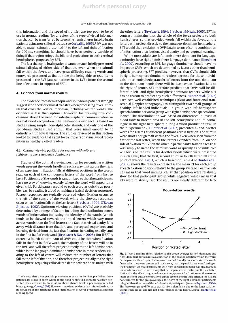
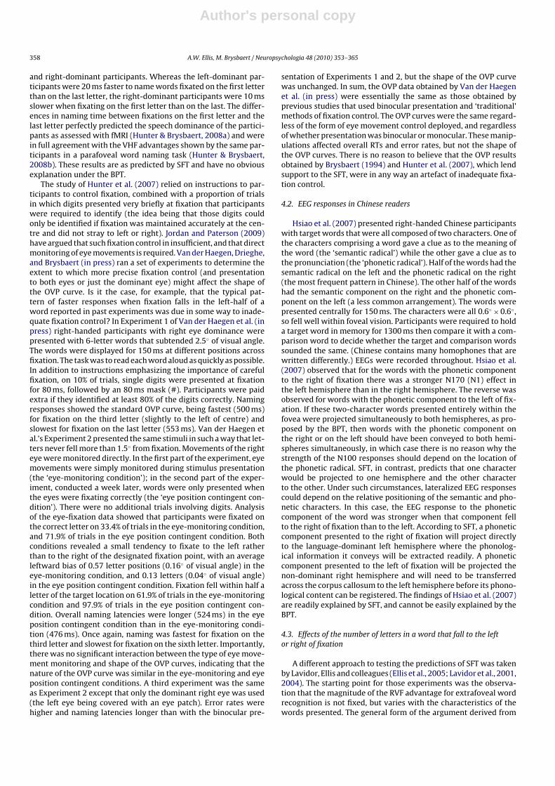
While most adults are left hemisphere dominant for language,a minority have right hemisphere language dominance (Knecht etal., 2000). According to BPT, language dominance should have noimpact on OVPs, which are determined by factors other than hemi-spheric processing. SFT predicts, however, that OVPs should shiftin right hemisphere dominant readers because for those individ-uals, interhemispheric transfer of letters from the non-dominantto the dominant hemisphere will be least when fixation falls tothe right of centre. SFT therefore predicts that OVPs will be dif-ferent in left- and right-hemisphere dominant readers, while BPThas no reason to predict any such differences. Hunter et al. (2007)used two well-established techniques (fMRI and functional tran-scranial Doppler sonography) to distinguish two small groups ofhealthy, left-handed individuals – a group with left hemispherespeech dominance and a group with right hemisphere speech dom-inance. The discrimination was based on differences in levels ofblood flow to Broca’s area in the left hemisphere and its homo-logue in the right hemisphere during a word production task. Intheir Experiment 2, Hunter et al. (2007) presented 4- and 7-letterwords for 180 ms at different positions across fixation. The stimuliwere short enough to fit within the fovea, even when seen from thefirst or the last letter, when the letters extended from 0.2◦ on oneside of fixation to 1.7◦ on the other. A participant’s task on each trialwas simply to name the stimulus word as quickly as possible. Wewill focus on the results for 4-letter words which were presentedin such a way that the first, second, third, or fourth letter fell at thepoint of fixation. Fig. 3, which is based on Table 4 of Hunter et al.(2007), shows the results expressed as the mean RT for each groupat each fixation position relative to the group average. Positive val-ues mean that word naming RTs at that position were relativelyslow for that participant group while negative values mean thatRTs were relatively fast. The results are clearly different for left-
Fig. 3. Word naming times relative to the group average for left dominant andright-dominant participants as a function of the fixation position within the word.Participants with left speech dominance named foveally presented 4-letter wordsfaster when they were presented in such a way that the participants were fixating onthe first letter, whereas participants with right speech dominance had an advantagefor words presented in such a way that participants were fixating on the last letter.Notice that the effect is a gradual one, not only present for fixations on the extremeletter positions but also for fixations on the second and the third letter. If the RTs arenot corrected for the group averages, the curve of the right-dominant participantsis higher than the curve of the left-dominant participants (see also Brysbaert, 1994).This between-group difference was far from significant due to the large variationwithin each group, and has not been retained in the figure. Source: Hunter et al.(2007).
Author's personal copy
358 A.W. Ellis, M. Brysbaert / Neuropsychologia 48 (2010) 353–365
and right-dominant participants. Whereas the left-dominant par-ticipants were 20 ms faster to name words fixated on the first letterthan on the last letter, the right-dominant participants were 10 msslower when fixating on the first letter than on the last. The differ-ences in naming time between fixations on the first letter and thelast letter perfectly predicted the speech dominance of the partici-pants as assessed with fMRI (Hunter & Brysbaert, 2008a) and werein full agreement with the VHF advantages shown by the same par-ticipants in a parafoveal word naming task (Hunter & Brysbaert,2008b). These results are as predicted by SFT and have no obviousexplanation under the BPT.
The study of Hunter et al. (2007) relied on instructions to par-ticipants to control fixation, combined with a proportion of trialsin which digits presented very briefly at fixation that participantswere required to identify (the idea being that those digits couldonly be identified if fixation was maintained accurately at the cen-tre and did not stray to left or right). Jordan and Paterson (2009)have argued that such fixation control in insufficient, and that directmonitoring of eye movements is required. Van der Haegen, Drieghe,and Brysbaert (in press) ran a set of experiments to determine theextent to which more precise fixation control (and presentationto both eyes or just the dominant eye) might affect the shape ofthe OVP curve. Is it the case, for example, that the typical pat-tern of faster responses when fixation falls in the left-half of aword reported in past experiments was due in some way to inade-quate fixation control? In Experiment 1 of Van der Haegen et al. (inpress) right-handed participants with right eye dominance werepresented with 6-letter words that subtended 2.5◦ of visual angle.The words were displayed for 150 ms at different positions acrossfixation. The task was to read each word aloud as quickly as possible.In addition to instructions emphasizing the importance of carefulfixation, on 10% of trials, single digits were presented at fixationfor 80 ms, followed by an 80 ms mask (#). Participants were paidextra if they identified at least 80% of the digits correctly. Namingresponses showed the standard OVP curve, being fastest (500 ms)for fixation on the third letter (slightly to the left of centre) andslowest for fixation on the last letter (553 ms). Van der Haegen etal.’s Experiment 2 presented the same stimuli in such a way that let-ters never fell more than 1.5◦ from fixation. Movements of the righteye were monitored directly. In the first part of the experiment, eyemovements were simply monitored during stimulus presentation(the ‘eye-monitoring condition’); in the second part of the exper-iment, conducted a week later, words were only presented whenthe eyes were fixating correctly (the ‘eye position contingent con-dition’). There were no additional trials involving digits. Analysisof the eye-fixation data showed that participants were fixated onthe correct letter on 33.4% of trials in the eye-monitoring condition,and 71.9% of trials in the eye position contingent condition. Bothconditions revealed a small tendency to fixate to the left ratherthan to the right of the designated fixation point, with an averageleftward bias of 0.57 letter positions (0.16◦ of visual angle) in theeye-monitoring condition, and 0.13 letters (0.04◦ of visual angle)in the eye position contingent condition. Fixation fell within half aletter of the target location on 61.9% of trials in the eye-monitoringcondition and 97.9% of trials in the eye position contingent con-dition. Overall naming latencies were longer (524 ms) in the eyeposition contingent condition than in the eye-monitoring condi-tion (476 ms). Once again, naming was fastest for fixation on thethird letter and slowest for fixation on the sixth letter. Importantly,there was no significant interaction between the type of eye move-ment monitoring and shape of the OVP curves, indicating that thenature of the OVP curve was similar in the eye-monitoring and eyeposition contingent conditions. A third experiment was the sameas Experiment 2 except that only the dominant right eye was used(the left eye being covered with an eye patch). Error rates werehigher and naming latencies longer than with the binocular pre-
sentation of Experiments 1 and 2, but the shape of the OVP curvewas unchanged. In sum, the OVP data obtained by Van der Haegenet al. (in press) were essentially the same as those obtained byprevious studies that used binocular presentation and ‘traditional’methods of fixation control. The OVP curves were the same regard-less of the form of eye movement control deployed, and regardlessof whether presentation was binocular or monocular. These manip-ulations affected overall RTs and error rates, but not the shape ofthe OVP curves. There is no reason to believe that the OVP resultsobtained by Brysbaert (1994) and Hunter et al. (2007), which lendsupport to the SFT, were in any way an artefact of inadequate fixa-tion control.
4.2. EEG responses in Chinese readers
Hsiao et al. (2007) presented right-handed Chinese participantswith target words that were all composed of two characters. One ofthe characters comprising a word gave a clue as to the meaning ofthe word (the ‘semantic radical’) while the other gave a clue as tothe pronunciation (the ‘phonetic radical’). Half of the words had thesemantic radical on the left and the phonetic radical on the right(the most frequent pattern in Chinese). The other half of the wordshad the semantic component on the right and the phonetic com-ponent on the left (a less common arrangement). The words werepresented centrally for 150 ms. The characters were all 0.6◦ × 0.6◦,so fell well within foveal vision. Participants were required to holda target word in memory for 1300 ms then compare it with a com-parison word to decide whether the target and comparison wordssounded the same. (Chinese contains many homophones that arewritten differently.) EEGs were recorded throughout. Hsiao et al.(2007) observed that for the words with the phonetic componentto the right of fixation there was a stronger N170 (N1) effect inthe left hemisphere than in the right hemisphere. The reverse wasobserved for words with the phonetic component to the left of fix-ation. If these two-character words presented entirely within thefovea were projected simultaneously to both hemispheres, as pro-posed by the BPT, then words with the phonetic component onthe right or on the left should have been conveyed to both hemi-spheres simultaneously, in which case there is no reason why thestrength of the N100 responses should depend on the location ofthe phonetic radical. SFT, in contrast, predicts that one characterwould be projected to one hemisphere and the other characterto the other. Under such circumstances, lateralized EEG responsescould depend on the relative positioning of the semantic and pho-netic characters. In this case, the EEG response to the phoneticcomponent of the word was stronger when that component fellto the right of fixation than to the left. According to SFT, a phoneticcomponent presented to the right of fixation will project directlyto the language-dominant left hemisphere where the phonolog-ical information it conveys will be extracted readily. A phoneticcomponent presented to the left of fixation will be projected thenon-dominant right hemisphere and will need to be transferredacross the corpus callosum to the left hemisphere before its phono-logical content can be registered. The findings of Hsiao et al. (2007)are readily explained by SFT, and cannot be easily explained by theBPT.
4.3. Effects of the number of letters in a word that fall to the leftor right of fixation
A different approach to testing the predictions of SFT was takenby Lavidor, Ellis and colleagues (Ellis et al., 2005; Lavidor et al., 2001,2004). The starting point for those experiments was the observa-tion that the magnitude of the RVF advantage for extrafoveal wordrecognition is not fixed, but varies with the characteristics of thewords presented. The general form of the argument derived from
Author's personal copy
A.W. Ellis, M. Brysbaert / Neuropsychologia 48 (2010) 353–365 359
SFT was to propose that if a factor has more of an effect on therecognition of words presented entirely in one visual field than theother (well away from the fovea), then the same factor may havedifferential effects on those portions of centrally presented wordsthat fall to one side or the other of the fixation point. For exam-ple, letter length affects recognition speed for familiar words in theLVF more than in the RVF (Ellis, 2004). Is it the case that recogni-tion times for words presented centrally are more affected by thenumber of letters that fall to the left of the fixation point than bythe number of letters that fall to the right? If they are, that wouldsupport SFT over BPT (which maintains that centrally presentedwords are projected to both hemispheres and so will be processedby the language-dominant (usually left) hemisphere, with no needfor callosal transfer).4
The first attempt to test this general line of reasoning was basedupon the fact that if familiar words in normal formats (lower orupper case) are presented entirely in the LVF or the RVF, the mag-nitude of the RVF advantage varies with the number of letters inthe stimulus words. Longer words generate larger RVF advantagesbecause increasing letter length affects the speed and accuracy ofprocessing LVF words more than RVF words (Bub & Lewine, 1988;Ellis et al., 1988; Lindell et al., 2002; Young & Ellis, 1985). This holdstrue even when the spaces between letters in words are adjusted sothat words containing different numbers of letters have the samephysical length on the screen and on the retina, suggesting thatthe decline in performance with increased length in the LVF has todo with the numbers of letters in a word rather than any effectsof acuity change that may result from longer words projectingfurther into the periphery (Bruyer & Janlin, 1989; Lavidor et al.,2002).
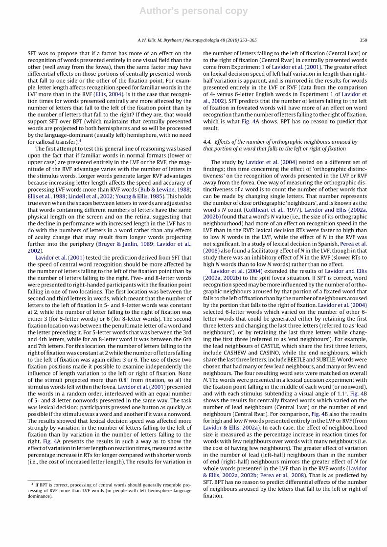
Lavidor et al. (2001) tested the prediction derived from SFT thatthe speed of central word recognition should be more affected bythe number of letters falling to the left of the fixation point than bythe number of letters falling to the right. Five- and 8-letter wordswere presented to right-handed participants with the fixation pointfalling in one of two locations. The first location was between thesecond and third letters in words, which meant that the number ofletters to the left of fixation in 5- and 8-letter words was constantat 2, while the number of letter falling to the right of fixation waseither 3 (for 5-letter words) or 6 (for 8-letter words). The secondfixation location was between the penultimate letter of a word andthe letter preceding it. For 5-letter words that was between the 3rdand 4th letters, while for an 8-letter word it was between the 6thand 7th letters. For this location, the number of letters falling to theright of fixation was constant at 2 while the number of letters fallingto the left of fixation was again either 3 or 6. The use of these twofixation positions made it possible to examine independently theinfluence of length variation to the left or right of fixation. Noneof the stimuli projected more than 0.8◦ from fixation, so all thestimulus words fell within the fovea. Lavidor et al. (2001) presentedthe words in a random order, interleaved with an equal numberof 5- and 8-letter nonwords presented in the same way. The taskwas lexical decision: participants pressed one button as quickly aspossible if the stimulus was a word and another if it was a nonword.The results showed that lexical decision speed was affected morestrongly by variation in the number of letters falling to the left offixation than by variation in the number of letters falling to theright. Fig. 4A presents the results in such a way as to show theeffect of variation in letter length on reaction times, measured as thepercentage increase in RTs for longer compared with shorter words(i.e., the cost of increased letter length). The results for variation in
4 If BPT is correct, processing of central words should generally resemble pro-cessing of RVF more than LVF words (in people with left hemisphere languagedominance).
the number of letters falling to the left of fixation (Central Lvar) orto the right of fixation (Central Rvar) in centrally presented wordscome from Experiment 1 of Lavidor et al. (2001). The greater effecton lexical decision speed of left half variation in length than right-half variation is apparent, and is mirrored in the results for wordspresented entirely in the LVF or RVF (data from the comparisonof 4- versus 6-letter English words in Experiment 1 of Lavidor etal., 2002). SFT predicts that the number of letters falling to the leftof fixation in foveated words will have more of an effect on wordrecognition than the number of letters falling to the right of fixation,which is what Fig. 4A shows. BPT has no reason to predict thatresult.
4.4. Effects of the number of orthographic neighbours aroused bythat portion of a word that falls to the left or right of fixation
The study by Lavidor et al. (2004) rested on a different set offindings; this time concerning the effect of ‘orthographic distinc-tiveness’ on the recognition of words presented in the LVF or RVFaway from the fovea. One way of measuring the orthographic dis-tinctiveness of a word is to count the number of other words thatcan be made by changing single letters. That number representsthe number of close orthographic ‘neighbours’, and is known as theword’s N count (Coltheart et al., 1977). Lavidor and Ellis (2002a,2002b) found that a word’s N value (i.e., the size of its orthographicneighbourhood) had more of an effect on recognition speed in theLVF than in the RVF: lexical decision RTs were faster to high thanto low N words in the LVF, while the effect of N in the RVF wasnot significant. In a study of lexical decision in Spanish, Perea et al.(2008) also found a facilitatory effect of N in the LVF, though in thatstudy there was an inhibitory effect of N in the RVF (slower RTs tohigh N words than to low N words) rather than no effect.
Lavidor et al. (2004) extended the results of Lavidor and Ellis(2002a, 2002b) to the split fovea situation. If SFT is correct, wordrecognition speed may be more influenced by the number of ortho-graphic neighbours aroused by that portion of a fixated word thatfalls to the left of fixation than by the number of neighbours arousedby the portion that falls to the right of fixation. Lavidor et al. (2004)selected 6-letter words which varied on the number of other 6-letter words that could be generated either by retaining the firstthree letters and changing the last three letters (referred to as ‘leadneighbours’), or by retaining the last three letters while chang-ing the first three (referred to as ‘end neighbours’). For example,the lead neighbours of CASTLE, which share the first three letters,include CASHEW and CASINO, while the end neighbours, whichshare the last three letters, include BEETLE and SUBTLE. Words werechosen that had many or few lead neighbours, and many or few endneighbours. The four resulting word sets were matched on overallN. The words were presented in a lexical decision experiment withthe fixation point falling in the middle of each word (or nonword),and with each stimulus subtending a visual angle of 1.1◦. Fig. 4Bshows the results for centrally fixated words which varied on thenumber of lead neighbours (Central Lvar) or the number of endneighbours (Central Rvar). For comparison, Fig. 4B also the resultsfor high and low N words presented entirely in the LVF or RVF (fromLavidor & Ellis, 2002a). In each case, the effect of neighbourhoodsize is measured as the percentage increase in reaction times forwords with few neighbours over words with many neighbours (i.e.the cost of having few neighbours). The greater effect of variationin the number of lead (left-half) neighbours than in the numberof end (right-half) neighbours mirrors the greater effect of N forwhole words presented in the LVF than in the RVF words (Lavidor& Ellis, 2002a, 2002b; Perea et al., 2008). That is as predicted bySFT. BPT has no reason to predict differential effects of the numberof neighbours aroused by the letters that fall to the left or right offixation.
Author's personal copy
360 A.W. Ellis, M. Brysbaert / Neuropsychologia 48 (2010) 353–365
Fig. 4. Illustration of how the effects of letter length (A), neighbourhood size (B) and case alternation (C) in those portions of fixated words that fall to the left or right offixation mirror the effects seen for words presented entirely in the left or right visual field. LVF/RVF = words presented entirely in the left or right visual field, away from thefovea. Central Lvar/Central Rvar = central presentation with variation in length, neighbourhood size or case alternation in that part of the stimulus word that falls to the left(Lvar) or right (Rvar) of fixation. Fig. 1A. Percent RT changes for LVF and RVF words is defined as (((LVF6 − LVF4)/LVF4) × 100) and (((RVF6 − RVF4)/RVF4) × 100) respectively,where LVF4, LVF6, RVF4 and RVF6 are the mean RTs for 4- and 6-letter lower case words presented in the LVF or RVF in Experiment 1 of Lavidor et al. (2002). Percent changeas a function of length variation to the left or right of fixation in centrally presented words is defined as (((Lvar8 − Lvar5)/Lvar5) × 100) and (((Rvar8 − Rvar5)/Rvar5) × 100)respectively, where Lvar5 and Lvar8 are the mean RTs for centrally presented 5- and 8-letter words in Lavidor et al. (2001) when the words were positioned such that thevariation in length occurred to the left of fixation, and Rvar5 and Rvar8 are the mean RTs for centrally presented 5- and 8-letter words when the words were positioned suchthat the variation in length occurred to the right of fixation. Fig. 1B. Percent RT changes as a function of variation in number of orthographic neighbours (N). Mean RTs forwhole words in LVF and RVF come from Experiment 1 of Lavidor and Ellis (2002b) while mean RTs for variation in the number of neighbours for those letters that fall to theleft or right of fixation in centrally presented words come from Experiment 1 of Lavidor et al. (2004). Fig. 1C. Percent RT changes as a function of case alternation. Mean RTsfor whole words in LVF and RVF come from Experiment 1 of Lavidor et al. (2002; 6-letter mixed and lower case) while mean RTs for variation in the number of neighbours forthose letters that fall to the left or right of fixation in centrally presented words come from Experiment 2 of Ellis et al. (2005; words left and right alternated vs lower case).
4.5. Effects of case alternation applied to the portion of a wordthat falls to the left or right of fixation
We noted above that word recognition in right-handed par-ticipants shows a substantial effect of length for words displayedentirely in the LVF but a much smaller (often non-significant) effectfor words displayed entirely in the RVF. The implication is thatthe left hemisphere is better able than the right to process thecomponent letters of words in parallel, reducing the differences inrecognition speed between shorter and longer words (Ellis, 2004).That pattern only applies, however, to familiar words presented ina familiar format (i.e., normal, horizontal, lower or upper case pre-sentation). If familiar words are presented in unfamiliar formats,such as vertically or in MiXeD cAsE, the pattern changes and com-parable length effects are seen in both visual fields. That is becausedistorting the appearance of familiar words induces a sensitivityto word length in the RVF which is like that shown by words inthe LVF under all presentation conditions (Fiset & Arguin, 1999;Lavidor & Ellis, 2001; Lavidor et al., 2002). The implication hereis that parallel processing of the component letters of words bythe left hemisphere depends on those words being presented informats that the left hemisphere is skilled at dealing with. Present-ing words to the left hemisphere in unusual formats causes theleft hemisphere to process the component letters in a more serialfashion, which is how the right hemisphere processes all words,irrespective of format (Ellis, 2004).
SFT predicts that if the left hemisphere is more sensitive to for-mat distortion than the right hemisphere, then distortion appliedto those letters in a centrally presented word that fall to the rightof fixation should affect recognition speed more than distortionapplied to those letters that fall to the left of fixation. Ellis et al.
(2005) created 8-letter words in which case alternation was appliedeither to the first four letters (e.g., eXcHange) or the last four letters(e.g., infiNiTe). The words were presented centrally to right-handedparticipants in a standard lexical decision task with an equal num-ber of similarly manipulated nonwords. Presentation time was150 ms and the fixation point fell in the middle of the word. Thestimuli subtended an angle of 1.8◦, so fell within the fovea. Fig. 4Cshows the impact of format distortion applied to those letters thatfell to the left of fixation (Central Lvar) or to those letters that fellto the right of fixation (Central Rvar). For comparison, Fig. 4C alsoshows the results for case alternation applied to whole words pre-sented entirely in the LVF or RVF (6-letter words from Lavidor et al.,2002, Experiment 1). Effects are again displayed as the percentageincrease in reaction times for distorted over non-distorted words(i.e., the cost of case alternation). As with length and N variation,the effect of case alternation applied to the portion of a word thatfell to the left or right of fixation mirrored the effects seen for wordspresented entirely in the LVF or RVF. This time, however, the effectswere greater in the RVF and in the right halves of centrally fixatedwords than in the LVF and the left halves. So, not all lateralizedmanipulations affecting recognition speed have stronger effects inthe LVF where processing is generally slower. The results of Ellis etal. (2005) are as predicted by SFT and have no ready explanationunder BPT.
The studies on neurologically intact readers by Hunter et al.(2007) and Hsiao et al. (2007), combined with the results concern-ing the effects of length, N and case alternation in the left and righthalves of words (Ellis et al., 2005; Lavidor et al., 2001, 2004), con-stitute the third line of evidence supporting our belief that SFT isworthy of serious consideration, and that a split fovea has conse-quences for normal reading.
Author's personal copy
A.W. Ellis, M. Brysbaert / Neuropsychologia 48 (2010) 353–365 361
5. Re-evaluating the criticisms directed against SFT
For a long time, SFT has been criticized only by reference to theempirical evidence provided by Huber (1962), Stone et al. (1973),Bunt and Minckler (1977), Bunt et al. (1977), and Leventhal etal. (1988). Although the critics usually include more recent arti-cles (e.g., Gazzaniga, 2000; Lindell & Nicholls, 2003) to supporttheir claims, these articles are simply reviews based on the oldermaterial. In section 2 (above) we gave our reasons for believingthat such evidence is not incompatible with SFT, and that other,related evidence (Trauzettel-Klosinski & Reinhard, 1998; Reinhard& Trauzettel-Klosinski, 2003) positively supports SFT. In recentyears, Jordan and colleagues have, however, reported new empir-ical evidence which in their view challenges the notion of a splitfovea, particularly as applied to visual word recognition. In thissection we will consider the extent to which those studies aregenuinely problematic for SFT.
5.1. Failures to replicate the differential effects of the number ofletters falling to the left or right of fixation in centrally presentedwords
Jordan et al. (2009) and Jordan, Paterson, Kurtev, and Xu (inpress) have presented a series of experiments which appear notto replicate the finding reported by Lavidor et al. (2001) that lexi-cal decision performance is more affected by the number of lettersfalling to the left of fixation than by the number falling to theright. Jordan and colleagues claim that the differences in resultsbetween their studies and Lavidor et al. (2001) are due to the factthat their experiments controlled the gaze direction of participantsmore effectively than Lavidor et al. (2001) managed to do throughsimple instructions. At the same time, however, the experimentalconstraints imposed on the participants in the Jordan et al. studiesmeant that the accuracy of lexical decision responses was very lowin some conditions (sometimes approaching chance) while reactiontimes were very long.
Jordan et al.’s (2009) Experiment 1 is presented as a direct repli-cation of Lavidor et al. (2001), using the same materials and, as faras was possible, the same conditions of presentation. The results,however, were very different. Whereas Lavidor et al. (2001) foundmore of an effect of the number of letters falling to the left of fix-ation than to the right (see Fig. 4A), Experiment 1 of Jordan et al.(2009) found no such differential effect. The only detectable effectin that experiment was a tendency for RTs to words to be fasterwhen fixation fell towards the beginnings of the words than whenit fell towards the ends (in line with the OVP effect shown in Fig. 3;Hunter et al., 2007). The accuracy levels in that experiment were,however, much lower than in Lavidor et al. (2001), and the RTswere much longer. Thus, accuracy in Lavidor et al.’s experimentwas 91% when fixation fell towards the beginnings of words and86% when fixation fell towards the end. The corresponding accu-racy levels in Jordan et al.’s (2009) Experiment 1 were 80% and67%. These were lexical decision responses to stimuli that werehalf words and half nonwords, so chance was 50%. The problemwith low performance levels is that many of the ‘correct’ responsesto words that enter into the analysis of reaction times will, in real-ity, be lucky guesses where the participant had little or no ideawhether the stimulus was a word or not and just happened to pressthe right button. The inclusion of a significant proportion of ‘luckyguesses’ will add a great deal of noise to any analysis of RTs forsupposedly ‘correct’ responses. Mean RTs for ‘correct’ responsesto words fixated towards the beginning or the end in Experiment1 of Jordan et al. (2009) were 769 ms and 815 ms respectively.In contrast, the corresponding RTs in Lavidor et al. (2001) were428 ms and 449 ms, a full 350 ms or 80% faster than in Jordan et al.(2009).
Jordan et al.’s (2009) Experiment 2 was very similar to theirExperiment 1 except that the position of eye fixations was mon-itored with an eye tracker. The pattern of results was similar totheir Experiment 1, and therefore different from those of Lavidoret al. (2001). But accuracies were again substantially lower than inLavidor et al., and RTs were again substantially slower (75% slowerfor fixations towards the beginnings of words and 94% slower forfixations towards the ends). Experiment 3 of Jordan et al. (2009)was like their first two experiments, except that this time the eyemovement monitoring apparatus was used to ensure that stimuliwere only presented when participants’ right eyes were fixatingat the desired location. Exposure durations were reduced to 50 msfrom 150 ms (the exposure duration used by Lavidor et al., 2001,and Experiments 1 and 2 of the same study). The resulting accu-racy levels were 85% for words fixated towards the beginning and77% for words fixated towards the end. The pattern of results wassimilar to that in Experiments 1 and 2 of the same study (i.e., dif-ferent from Lavidor et al., 2001). Reaction times in this experimentwere very slow indeed, being 89% slower than Lavidor et al. forwords fixated towards the beginning and 98% slower (i.e., twice aslong) for words fixated towards the end.
Jordan et al. (in press) report two more experiments using theLavidor et al. (2001) stimuli and fixation positions. Presentationtimes were 50 ms (as in Jordan et al., 2009, Experiment 3). Partic-ipants’ non-dominant eyes were occluded using eye patches andstimuli were only presented when the dominant eyes were fix-ating correctly. Experiment 1 of this study used the same sizedtypeface as Jordan et al. (2009) while Experiment 2 used a largertypeface. Accuracy levels were somewhat better than in Jordanet al. (2009), though lexical decision accuracy for centrally pre-sented words fixated towards the ends was still only 69%. RTs werefaster than in Jordan et al. (2009), but still more than 50% slowerthan in Lavidor et al. (2001). Curiously, there is clear evidence fora speed-accuracy trade-off between the two experiments in thisstudy. The use of larger stimuli in Jordan et al.’s (in press) Experi-ment 2 increased the accuracy of responding to words but slowedRTs by 30–40 ms. Participants who were shown the larger stimuliseem to have taken the opportunity to respond more accuratelythan participants who saw the smaller stimuli, but they sacrificedspeed in order to achieve those higher accuracy levels. In our expe-rience, such speed-accuracy trade-offs are rare in studies of eithercentral or lateralized word recognition. Importantly, the patternof results in both of Jordan et al.’s (in press) experiments were thesame as the pattern in the three experiments of Jordan et al. (2009),and therefore different from the pattern obtained by Lavidor et al.(2001).
5.2. Further replications of the differential effects of the numberof letters falling to the left or right of fixation in centrallypresented words
We are reassured in our belief that the results of Lavidor et al.(2001) are not entirely spurious because we know that similar pat-terns can be discerned in other studies (i.e., more of an impacton central word recognition of the number of letters falling to theleft of fixation than the number of letters falling to the right). Ellis(2004) reanalysed data from Brysbaert (1994) who collected nam-ing latencies from participants whose performance on three testsof lateralized perception indicated that they were left hemispheredominant for language. The subset of naming RTs analysed by Ellis(2004) were for trials in which participants fixated on the first orlast letters of Dutch words containing 3, 4, 5, 7 and 9 letters. Whenfixation was on the first letter, different numbers of letters fell in theRVF: when fixation was on the final letter, different numbers of let-ters fell in the LVF. Although the data in Brysbaert (1994) were notcollected with such an analysis in mind, Ellis (2004) showed that
Author's personal copy
362 A.W. Ellis, M. Brysbaert / Neuropsychologia 48 (2010) 353–365
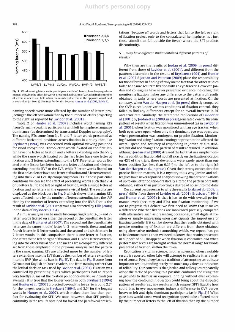
Fig. 5. Word naming latencies for participants with left hemisphere language dom-inance, showing the effect for words presented at fixation of variation in the numberof letters in one visual field when the number of letters in the opposite visual fieldis controlled (at 0 or 1). See text for details. Source: Hunter et al. (2007, Table 2).
naming speeds were more affected by the number of letters pro-jecting to the left of fixation than by the number of letters projectingto the right, as reported by Lavidor et al. (2001).
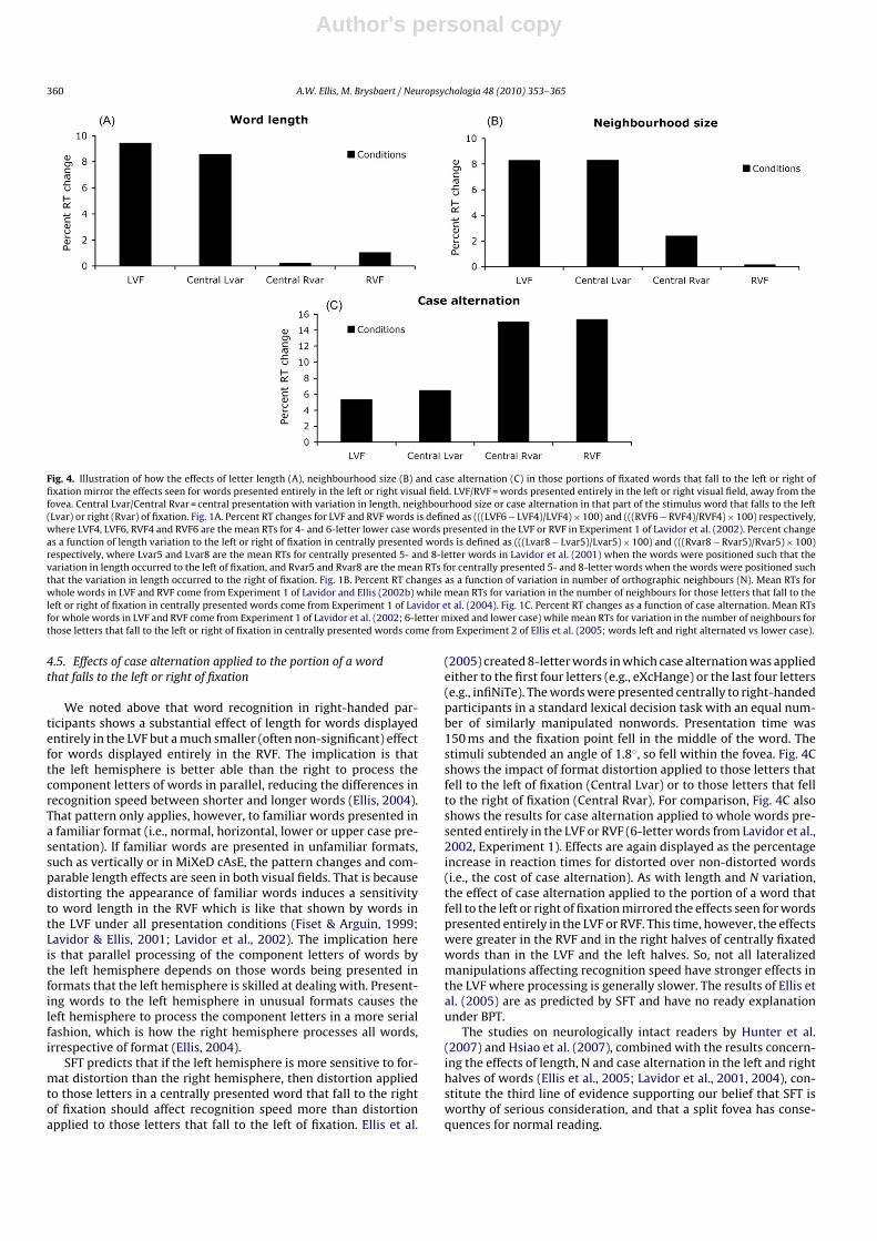
Table 2 of Hunter et al. (2007) includes word naming RTsfrom German-speaking participants with left hemisphere languagedominance (as determined by transcranial Doppler sonography).The naming RTs come from 3-, 5- and 7-letter words presented atdifferent horizontal positions across fixation in a study that, likeBrysbaert (1994), was concerned with optimal viewing positionsfor word recognition. Three-letter words fixated on the first let-ter have one letter at fixation and 2 letters extending into the RVF,while the same words fixated on the last letter have one letter atfixation and 2 letters extending into the LVF. Five-letter words fix-ated on the first or last letter have one letter at fixation and 4 lettersextending into the RVF or the LVF, while 7-letter words fixated onthe first or last letter have one letter at fixation and 6 letters extend-ing into the RVF or LVF. By comparing mean RTs in those particularconditions we can see the effect of presenting words such that 2, 4or 6 letters fall to the left or right of fixation, with a single letter atfixation and no letters in the opposite visual field. The results aredisplayed as the black bars in Fig. 5 which shows that naming RTswere affected more by the number of letters extending into the LVFthan by the number of letters extending into the RVF. That is theresult of Lavidor et al. (2001) that was also detected by Ellis (2004)in the data of Brysbaert (1994).
A similar analysis can be made by comparing RTs to 3-, 5- and 7-letter words fixated on either the second or the penultimate letterin the data of Hunter et al. (2007). The second and the penultimateletter are the same (middle) letter for 3-letter words, the second andfourth letters in 5-letter words, and the second and sixth letters in7-letter words. In this comparison there is one letter at fixation,one letter to the left or right of fixation, and 1, 3 or 5 letters extend-ing into the other visual field. The means are a completely differentset from those employed in the previous analysis, yet the patternis the same: naming RTs are affected more by the number of let-ters extending into the LVF than by the number of letters extendinginto the RVF (the white bars in Fig. 5). The data in Fig. 5 come fromGerman not English or Dutch readers, and from naming rather thanthe lexical decision task used by Lavidor et al. (2001). Fixation wascontrolled by presenting digits which participants had to reportvery briefly (80 ms) at the fixation point once every 6 or 7 trials (onaverage). It is true that the longer words in both Brysbaert (1994)and Hunter et al. (2007) projected beyond the fovea (to around 2.7◦
for the longest words in Brysbaert (1994), and 3.5◦ for the longestwords in Hunter et al., 2007), which makes them less than per-fect for evaluating the SFT. We note, however, that SFT predictscontinuity in the results obtained for foveal and parafoveal presen-
tations (because all words and letters that fall to the left or rightof fixation project only to the contralateral hemisphere, not justthose stimuli that fall outside the fovea) where the BPT predictsdiscontinuity.
5.3. Why have different studies obtained different patterns ofresults?
Why then are the results of Jordan et al. (2009, in press) dif-ferent from those of Lavidor et al. (2001), and different from thepatterns discernible in the results of Brysbaert (1994) and Hunteret al. (2007)? Jordan and Paterson (2009) place the responsibilityfor the difference in findings firmly on the fact that the other studiesfailed to ensure accurate fixation with an eye tracker. However, Jor-dan and colleagues have never presented evidence indicating thatmonitoring fixation makes any difference to the pattern of resultsobtained in tasks where words are presented at fixation. On thecontrary, when Van der Haegen et al. (in press) directly comparedthe OVP-curve under various conditions of fixation control, theyfailed to find any differences except for an overall increase in RTand error rate. Similarly, the attempted replications of Lavidor etal. (2001) by Jordan et al. (2009, in press) generated exactly the samepattern of results when fixation was unmonitored (as in Lavidor etal., 2001), when fixation was monitored with an eye tracker, whenboth eyes were open, when only the dominant eye was open, andwhen presentation was contingent on precise fixation. Monitor-ing fixation and using fixation-contingent presentation affected theoverall speed and accuracy of responding in Jordan et al.’s stud-ied, but did not change the pattern of results obtained. In addition,although Jordan et al. (2009) stressed the fact that in a simple moni-toring condition fixation did not fall exactly on the fixation locationon 42% of the trials, these deviations were rarely more than oneletter position (i.e., less than 0.25◦) to the left or to the right (cf.Van der Haegen et al., in press). Given their strong contention thatprecise fixation matters, it is a mystery to us why Jordan and col-leagues have never reported analyses showing that errant fixationsof up to one letter position drastically altered the pattern of resultsobtained, rather than just injecting a degree of noise into the data.
Our current best guess as to why the results Jordan et al. (2009, inpress) differ from those of Lavidor et al. (2001), Brysbaert (1994),and Hunter et al. (2007) is that it has to do with overall perfor-mance levels (accuracy and RTs), not fixation monitoring. If weare to progress this debate, we first need to know that it makesa difference whether fixations are monitored precisely comparedwith alternative such as presenting occasional, small digits at fix-ation or simply impressing upon participants the importance offixating carefully. If it can be shown that the results obtained withprecise monitoring of fixation are different from those obtainedusing alternative methods (something which, we repeat, has yetto be demonstrated), then we need to know that results presentedin support of SFT disappear when fixation is controlled and whenperformance levels are brought within the normal range for wordspresented at fixation, within the fovea.
Replication is vital in science. In other sciences, when a notableresult is reported, other labs will attempt to replicate it as a mat-ter of course. Psychology lacks a tradition of attempting to replicateimportant results, tending to rely too much on p values as indicatorsof reliability. Our concern is that Jordan and colleagues repeatedlyadopt the tactic of pointing to a possible confound and using thatas grounds to dismiss an empirical finding without ever explain-ing how the confound in question could bring about the disputedpattern of results (i.e., any results which support SFT). Exactly howcould bias in eye movements induce a difference in OVP curvesbetween left and right-dominant participants (as in Fig. 3)? Whatgaze bias would cause word recognition speed to be affected moreby the number of letters to the left of fixation than by the number
Author's personal copy
A.W. Ellis, M. Brysbaert / Neuropsychologia 48 (2010) 353–365 363
of letters to the right (Fig. 4A)? What gaze bias would cause thenumber of ‘lead neighbours’ to affect recognition speed more thanthe number of ‘end neighbours’, or cause case mixing towards theends of words to affect recognition more than case mixing towardsthe beginnings (Fig. 4B and C)? How would a gaze bias explain whyChinese words with the phonetic component to the right of fixationinduce a stronger EEG response in the left hemisphere, whereasChinese words with the phonetic component to the left of fixa-tion induce a stronger ERP effect in the right hemisphere underconditions where the two forms of Chinese words were randomlyintermixed (Hsiao et al., 2007)? These questions are never evenposed by Jordan and colleagues, let alone answered. At the time ofwriting, Jordan and colleagues have failed to identify a single sys-tematic bias in gaze direction that could explain any of the effectswe have summarized above.
6. Re-considering the usefulness of the Reicher–Wheelertask in hemispheric and split fovea research
Many different tasks have been used to investigate central wordrecognition (Grigorenko and Naples, 2008; Lupker, 2008). Mostof those tasks have then been borrowed by researchers inter-ested in the word recognition capabilities of the two cerebralhemispheres (see Banich, 2004; Ellis, 2004). Jordan and colleagueshave championed the use of the Reicher–Wheeler task in hemi-spheric research, including split-fovea research (e.g., Jordan et al.,1998, 2000, 2003, 2008, 2009). We believe, however, that theReicher–Wheeler task has a number of shortcomings which limitits usefulness for addressing the questions of interest here.
The Reicher–Wheeler task is built around pairs of words thatdiffer on a single letter (e.g., beak, bean). In a typical experimentaltrial, one of the two words is presented briefly to a participant. Thatsingle target word is followed by a pair of choice alternatives cre-ated by presenting the pair of letters which differentiate the twowords, with the remaining letters replaced by dashes. For example,following the brief presentation of the target word beak, a partic-ipant would see three dashes then the letters k and n displayedone above the other. The participant’s task is to choose which ofthe letters k or n occurred in that particular position in the tar-get word. Presentation time of the targets is usually adjusted sothat the overall performance level is around 66% across all condi-tions. Reicher (1969), Wheeler (1970) and subsequent researchersshowed that performance on the task is better if the stimuli arefamiliar words (e.g., beak, bean) than if they are letter strings whichare unwordlike and hard to pronounce (e.g., bqfk, bqfn). This wordsuperiority effect has been taken to imply the presence of top-downeffects in which word-level representations influence the percep-tion of the component letters of words. Explaining the presenceof word superiority effects was considered an important require-ment for the first generation of computational models of wordrecognition (e.g., McClelland and Rumelhart, 1981; Paap et al.,1982).
Jordan et al. (2008) questioned SFT on the basis of a study inwhich they presented four-letter words to the left or right of fixa-tion, either in foveal vision (with their ‘medial edge’ at 0.15◦, 0.25◦
or 0.35◦ from fixation) or in parafoveal vision (medial edge at 2.00◦,2.10◦, or 2.20◦ from the fixation point). In an effort to equate fovealand extrafoveal performance levels, the words presented at thefovea were smaller (subtending a visual angle of 0.55◦) than theparafoveal words (subtending a visual angle of 1.10◦). In a variantof the archetypal Reicher–Wheeler task, the briefly presented tar-get words were followed by a choice between two whole wordspresented centrally (not two letters accompanied by dashes as inthe conventional form of the task). Jordan et al. (2008) found bet-ter performance on this task in the RVF than the LVF for parafovealpresentations but not foveal presentations. Performance within the
fovea was symmetrical between presentations left and right of fix-ation. This, Jordan et al. (2008) argued, constituted evidence for abilaterally projecting foveal region of at least 1–2◦.
One major problem with the Reicher–Wheeler task is that it isperformed off-line. Responses are not made to the target stimulithemselves in a way that would, for example, allow reaction timesto be gathered that reflect processing as it happens (as in wordnaming or lexical decision). Instead, scores are based on the accu-racy of choosing between two alternatives presented subsequentto the target. Typically, participants are under no time pressure tomake their choice decisions: they can ponder as long as they likebefore deciding which of the two alternatives matches the brieflypresented target. The off-line, deliberative nature of responses inthe Reicher–Wheeler task matters when it comes to interpretingthe null findings of Jordan et al. (2008). SFT does not claim thatinterhemispheric transfer in healthy participants inevitably resultsin the incomplete or deficient transfer of information that couldcause left foveal performance to be worse than right foveal perfor-mance. That should happen only in a participant with a split or badlyfunctioning corpus callosum. Fig. 3 suggests that interhemispherictransfer in healthy participants introduces a time cost of only 20 ms.This is unlikely to be reflected in a decrement in performance accu-racy in an off-line task such as the Reicher–Wheeler task. Hence,until the findings of Jordan et al. (2008) are repeated using on-lineword processing tasks involving the measurement of reaction timesto target stimuli, we do not regard them as compelling evidenceagainst SFT.
There are other differences between performance on LVF andRVF words in the Reicher–Wheeler task compared with other,on-line processing tasks that may be important. For example,Jordan et al. (2000, Experiment 4) compared performance in theReicher–Wheeler task for 4-letter words and unpronounceablenonwords (letter strings). The accuracy with which participantscould indicate which of two letters had occurred in a specifiedposition in the target stimulus was better for words than for let-ter strings when the target stimuli were presented in the RVF,but there was no significant difference between words and letterstrings when they were presented as targets in the LVF. Jordan etal. (2000, p. 1204) concluded that, “The absence of even a slightword-nonword effect for LVF stimuli in our study suggests thatwords presented briefly to the right hemisphere were unable tomake effective contact with any higher-order representations (i.e.,for words or letter groups).” A similar insensitivity of the LVF tothe normal superiority of words over unpronounceable nonwordscan be discerned in the results of Jordan et al. (2003) and Jordanet al. (2008). In contrast to this evidence that LVF words may failto engage lexical representations during the performance of theReicher–Wheeler task, numerous studies involving naming or lex-ical decision have found lexical influences on the processing of LVFwords. Thus, word frequency and imageability have been shownto affect the speed and accuracy of responses to LVF as well asto RVF words in naming and/or lexical decision (e.g., Boles, 1983;Chiarello et al., 2000; Coney, 2005; Iacoboni & Zaidel, 1996; Scott &Hellige, 1998). This implies that the cognitive processes recruitedin the performance of the Reicher–Wheeler task are not the sameas those recruited in on-line word recognition tasks, particularlywhen words are presented in the left visual field beyond the fovea.These responses may be based on more low-level perceptual judg-ments which do not engage higher level representations and whichresults in performance on familiar words that is indistinguishablefrom performance on unpronounceable letter strings.5
5 Note that changing the case of letters between targets and probes does not pre-vent the use of low-level perceptual strategies in the Reicher–Wheeler task. Imagine,for example, that the target word was beak and that the participant was then offered

Author's personal copy
364 A.W. Ellis, M. Brysbaert / Neuropsychologia 48 (2010) 353–365
In sum, the off-line nature of the Reicher–Wheeler task meansthat it is incapable of detecting effects that need to be measuredin terms of costs to the speed of processing target stimuli. This, wesuggest, may be why the study by Jordan et al. (2008) was insensi-tive to differences in responding to words that fell within the foveabut to the left or right of fixation. Brysbaert has argued in a numberof papers that word naming is the simplest and most informativetask for investigating issues of laterality and interhemispheric com-munication (e.g., Brysbaert & Nazir, 2005; Brysbaert, 1994; Hunter& Brysbaert, 2008a, 2008b; Hunter et al., 2007). Naming is on-line,is close to natural reading, and vocal responses can be timed withmillisecond accuracy. Naming involves the mapping of orthogra-phy onto speech production processes which are clearly lateralized(Banich, 2004; Zaidel, 1998) and can be easily and reliably assessedwith brain imaging (cf. Knecht et al., 2000).
7. Closing remarks
SFT is a relatively new arrival on the theoretical and empiricalstage. There is much to be done before we will be in a position toknow whether SFT, BPT or some third alternative provides the bestexplanation of the mapping between the retinae and the brain, andof the processing of stimuli that fall entirely or partially to the leftor right of fixation. SFT has generated a range of novel and coun-terintuitive findings in relation to word recognition – observationsthat would not have been made without the prompting of the the-ory, but which now need to be accounted for by any satisfactorytheory of visual word recognition. SFT has also stimulated signifi-cant theoretical developments; for example in the computationalmodelling work of Shillcock et al. (2000), Monaghan and Shillcock(2008) and Whitney (2001). We might take issue with aspects ofthose computational models, but that is not the point: the point isthat they have inspired new thinking in an area of both theoreticaland practical importance.
In contrast, nearly a decade of studies by Jordan and colleagues– from Jordan et al. (2000) to Jordan and Paterson (2009) – hasfocussed on questioning SFT data almost exclusively on the basis ofinadequate fixation control. The resulting papers have been persis-tently negative in tone, content to point towards real or imaginaryfaults in other studies and to publish null results dressed up asfailures to replicate effects under properly controlled conditions.Jordan and colleagues have never attempted to explain how thealleged faults of SFT experiments could generate the patterns ofresults upon which the claims of SFT are based, or how smallbiases in fixation location could generate those effects artefactu-ally. Instead, they have been content to point out that the SFT is aminority position (as if that matters) and to imply that any apparentfailure to replicate its predictions is enough to discredit the theoryin its entirety.
SFT will not be the last word on the involvement of the hemi-spheres in perception and reading, but neither will BPT. The taskright now is to decide which theory is leading us in the right direc-tion. We thank Jordan and Paterson (2009) for stimulating us toconduct our own re-evaluation of the evidence for and against SFT.We look forward to the day when Jordan and colleagues employ BPTcreatively to generate novel and original hypotheses which predictpositive results (rather than null effects) that help discriminate BPTfrom SFT, and which the research community can sink its collective
the choice between –K and –N, or between BEAK and BEAN. In a situation whereparticipants can take as long as they like to make a response, they only have tothink whether or not the target word included an ascending letter (k) that projectedabove the rest of the word. If it did, the answer is –K/BEAK: if it did not, the answer is–N/BEAN. The familiarity or otherwise of the word would not be expected to affectperformance when such a perceptual strategy is employed.
teeth into. In the meantime, we hope that readers of this articlewill be inspired to read the original articles reviewed by us and byJordan and Paterson (2009), and to take their own unprejudiced,accurately fixated look at whether the fovea is split and whetherthis has consequences for the way we recognise words in reading.
References
Banich, M. T. (2004). Interaction between the hemispheres and its implications forthe processing capacity of the brain. In K. Hugdahl, & R. J. Davidson (Eds.), Theasymmetrical brain (pp. 261–302). Cambridge, MA: MIT Press.
Boles, D. B. (1983). Dissociated imageability, concreteness, and familiarity in later-alized word recognition. Memory & Cognition, 11, 511–519.
Bruyer, R., & Janlin, D. (1989). Lateral differences in lexical access: Word length vsstimulus length. Brain and Language, 37, 258–265.
Brysbaert, M. (1994). Interhemispheric transfer and the processing of foveally pre-sented stimuli. Behavioral Brain Research, 64, 151–161.
Brysbaert, M. (2004). The importance of interhemispheric transfer for foveal vision:A factor that has been overlooked in theories of visual word recognition andobject perception. Brain and Language, 88, 259–267.
Brysbaert, M., & Nazir, T. A. (2005). Visual constraints on written word recognition:Evidence from the optimal viewing position effect. Journal of Research in Reading,28, 216–228.
Bub, D., & Lewine, J. (1988). Different modes of word recognition in the left and rightvisual fields. Brain and Language, 33, 161–188.
Bunt, A. H., & Minckler, D. S. (1977). Foveal sparing: New anatomical evidencefor bilateral representation of the central retina. Archives of Ophthalmology, 95,1445–1447.
Bunt, A. H., Minckler, D. S., & Johanson, G. W. (1977). Demonstration of bilateralprojection of the central retina of the monkey with horseradish peroxidaseneuronography. Journal of Comparative Neurology, 171, 619–630.
Chiarello, C., Liu, S., Quan, N., & Shears, C. (2000). Imageability and word recognitionin the left and right visual fields: A signal detection analysis. Brain and Cognition,43, 90–94.
Coney, J. (2005). Word frequency and the lateralisation of mental processes. Neu-ropsychologia, 43, 142–148.
Coltheart, M., Davelaar, E., Jonasson, J. T., & Besner, D. (1977). Access to the internallexicon. In S. Dornic (Ed.), Attention and performance (pp. 535–555). New York:Academic Press.
Corballis, M. C. (1995). Visual integration in the split brain. Neuropsychologia, 33,937–959.
Corballis, M. C., & Trudel, A. I. (1993). Role of forebrain commissures in interhemi-spheric integration. Neuropsychology, 7, 306–324.
Cowey, A. (2004). The 30th Sir Frederick Bartlett lecture: Fact, artefact, and mythabout blindsight. Quarterly Journal of Experimental Psychology, 57A, 577–609.
Ellis, A. W. (2004). Length, formats, neighbors, and the processing of words presentedlaterally or at fixation. Brain and Language, 88, 355–366.
Ellis, A. W. (2009). Communication between the cerebral hemispheres in dyslexicand skilled adult readers. Revista Espanola de Logopedia, Fonotria y Audiologia,29, 85–96.
Ellis, A. W., Brooks, J., & Lavidor, M. (2005). Evaluating a split fovea model of visualword recognition: Effects of case alternation in the two visual fields and inthe left and right halves of words presented at the fovea. Neuropsychologia, 43,1128–1137.
Ellis, A. W., Young, A. W., & Anderson, C. (1988). Modes of visual word recognitionin the left and right cerebral hemispheres. Brain and Language, 35, 254–273.
Fendrich, R., & Gazzaniga, M. S. (1989). Evidence of foveal splitting in a commissuro-tomy patient. Neuropsychologia, 27, 273–281.
Fendrich, R., Wessinger, C. M., & Gazzaniga, M. S. (1996). Nasotemporal overlap atthe retinal vertical meridian: Investigations with a callosotomy patient. Neu-ropsychologia, 34, 637–646.
Fiset, S., & Arguin, M. (1999). Case alternation and orthographic neighborhoodsize effects in the left and right cerebral hemispheres. Brain and Cognition, 40,116–118.
Gazzaniga, M. S. (2000). Cerebral specialization and interhemispheric communi-cation: Does the corpus callosum enable the human condition? Brain, 123,1293–1326.
Grigorenko, E. L., & Naples, A. J. (2008). Single-word reading: behavioral and biologicalperspectives. New York: Lawrence Erlbaum.
Henderson, L., Barca, L., & Ellis, A. W. (2007). Interhemispheric cooperation and non-cooperation during word recognition: Evidence for callosal transfer dysfunctionin dyslexic adults. Brain and Language, 103, 276–291.
Hsiao, J. H. W., Shillcock, R., & Lee, C. Y. (2007). Neural correlates of foveal split-ting in reading: Evidence from an ERP study of Chinese character recognition.Neuropsychologia, 45, 1280–1292.
Huber, A. (1962). Homonymous hemianopia after occipital lobectomy. AmericanJournal of Ophthalmology, 54, 623–629.
Hunter, Z. R., & Brysbaert, M. (2008a). Theoretical analysis of interhemispherictransfer costs in visual word recognition. Language and Cognitive Processes, 23,165–182.
Hunter, Z. R., & Brysbaert, M. (2008b). Visual half field experiments are a goodmeasure of cerebral language dominance if used properly. Neuropsychologia,46, 316–325.
Author's personal copy
A.W. Ellis, M. Brysbaert / Neuropsychologia 48 (2010) 353–365 365
Hunter, Z. R., Brysbaert, M., & Knecht, S. (2007). Foveal reading requires interhemi-spheric communication. Journal of Cognitive Neuroscience, 19, 1373–1387.
Iacoboni, M., & Zaidel, E. (1996). Hemispheric independence in word recognition:Evidence from unilateral and bilateral presentations. Brain and Language, 53,121–140.
Jordan, T. R., Patching, G. R., & Milner, A. D. (1998). Central fixations are inadequatelycontrolled by instructions alone: Implications for studying cerebral asymmetry.Quarterly Journal of Experimental Psychology, 51A, 371–391.
Jordan, T. R., Patching, G. R., & Milner, A. D. (2000). Lateralized word recognition:Assessing the role of hemispheric specialization, modes of lexical access, andperceptual asymmetry. Journal of Experimental Psychology: Human Perceptionand Performance, 26, 1192–1208.
Jordan, T. R., Patching, G. R., & Thomas, S. M. (2003). Asymmetries and eccentric-ities in studies of lateralized word recognition: A response to Nazir. CognitiveNeuropsychology, 20, 81–89.
Jordan, T. R., & Paterson, K. (2009). Re-evaluating split-fovea processing in visualword recognition: A critical assessment of recent research. Neuropsychologia,47, 2341–2353.
Jordan, T. R., Paterson, K., Kurtev, S., & Xu, M. (in press). Re-evaluating split-fovea pro-cessing in word recognition: Effects of word length during monocular viewing.Cortex. doi:10.1016/j.cortex.2008.10.008.
Jordan, T. R., Paterson, K., & Stachurski, M. (2008). Re-evaluating split-fovea pro-cessing in word recognition: Effects of retinal eccentricity on hemisphericdominance. Neuropsychology, 22, 738–745.
Jordan, T. R., Paterson, K., & Stachurski, M. (2009). Re-evaluating split-fovea process-ing in word recognition: Effects of word length. Cortex, 45, 495–505.
Knecht, S., Drager, B., Deppe, M., Bobe, L., Lohmann, H., Floel, A., Ringelstein, E. B.,& Henningsen, H. (2000). Handedness and hemispheric language dominance inhealthy humans. Brain, 123, 2512–2518.
Lavidor, M., & Ellis, A. W. (2001). Mixed-case effects in lateralized word recognition.Brain and Cognition, 46, 192–195.
Lavidor, M., & Ellis, A. W. (2002a). Word length and orthographic neighborhood sizeeffects in the left and right cerebral hemispheres. Brain and Language, 80, 45–62.
Lavidor, M., & Ellis, A. W. (2002b). Orthographic neighborhood effects in the rightbut not in the left cerebral hemisphere. Brain and Language, 80, 63–76.
Lavidor, M., Ellis, A. W., & Pansky, A. (2002). Case alternation and length effects in thetwo cerebral hemispheres: A study of English and Hebrew. Brain and Cognition,50, 257–271.
Lavidor, M., Ellis, A. W., Shillcock, R., & Bland, T. (2001). Evaluating a split process-ing model of visual word recognition: Effects of word length. Cognitive BrainResearch, 12, 265–272.
Lavidor, M., Hayes, A., Shillcock, R., & Ellis, A. W. (2004). Evaluating a split processingmodel of visual word recognition: Effects of orthographic neighborhood size.Brain and Language, 88, 312–320.
Lavidor, M., & Walsh, V. (2004). The nature of foveal representation. Nature ReviewsNeuroscience, 5, 729–735.
Leff, A. (2004). A historical review of the representation of the visual field in pri-mary visual cortex with special reference to the neural mechanisms underlyingmacular sparing. Brain and Language, 88, 268–278.
Leventhal, A. G., Ault, S. J., & Vitek, D. J. (1988). The nasotemporal division of primateretina: The neural bases of macular sparing and splitting. Science, 240, 66–67.
Lindell, A. K., & Nicholls, M. E. R. (2003). Cortical representation of the fovea: Impli-cations for visual half-field research. Cortex, 39, 111–117.
Lindell, A. K., Nicholls, M. E. R., & Castles, A. (2002). The effect of word length on hemi-spheric word recognition: Evidence from unilateral and bilateral-redundantpresentations. Brain and Cognition, 48, 447–452.
Lupker, S. J. (2008). Visual word recognition: theories and findings. In M. J. Snowling,& C. Hulme (Eds.), The science of reading: A handbook.. Malden, MA: Blackwell,pp. 39–60
McClelland, J. L., & Rumelhart, D. E. (1981). An interactive activation model of contexteffects in letter perception. Part 1. An account of basic findings. PsychologicalReview, 88, 375–407.
Miki, A., Nakajima, T., Fujita, M., Takagi, M., & Abe, H. (1996). Functional magneticresonance imaging in homonymous hemianopsia. American Journal of Ophthal-mology, 121, 258–266.
Monaghan, P., & Shillcock, R. (2008). Hemispheric dissociation and dyslexia in acomputational model of reading. Brain and Language, 107, 185–193.
O’Regan, J. K., & Jacobs, A. M. (1992). Optimal viewing position effect in word recog-nition: A challenge to current theory. Journal of Experimental Psychology: HumanPerception and Performance, 18, 185–197.
Paap, K. R., Newsome, S. L., McDonald, J. E., & Schvaneveldt, R. W. (1982). Anactivation-verification model for letter and word recognition. PsychologicalReview, 89, 573–594.
Perea, M., Acha, J., & Fraga, I. (2008). Lexical competition is enhanced in the lefthemisphere: Evidence from different types of orthographic neighbors. Brain andLanguage, 105, 199–210.
Rayner, K. (1998). Eye movements in reading and information processing: 20 yearsof research. Psychological Bulletin, 124, 372–422.
Reicher, G. M. (1969). Perceptual recognition as a function of meaningfulness ofstimulus material. Journal of Experimental Psychology, 81, 274–280.
Reinhard, J., & Trauzettel-Klosinski, S. (2003). Nasotemporal overlap of retinal gan-glion cells in humans: A functional study. Investigative Ophthalmology & VisualScience, 44, 1568–1572.
Scott, G. B., & Hellige, J. B. (1998). Hemispheric asymmetry for word naming: Effectsof frequency and regularity of pronunciation. Laterality, 3, 343–371.
Shillcock, R., Ellison, T. M., & Monaghan, P. (2000). Eye-fixation behavior, lexicalstorage and visual word recognition in a split processing model. PsychologicalReview, 107, 824–851.
Sidtis, J. J., Volpe, B. T., Wilson, D. H., Rayport, M., & Gazzaniga, M. S. (1981). Variabil-ity in right hemisphere language function after callosal section: Evidence for acontinuum of generative capacity. Journal of Neuroscience, 1, 323–331.
Sperry, R. W., Gazzaniga, M. S., & Bogen, J. E. (1969). Interhemispheric relations: Theneocortical commissures; syndromes of disconnection. In P. J. Vinken, & G. W.Bruhn (Eds.), Handbook of clinical neurology. Vol. 4. Disorders of speech perception,and symbolic behavior (pp. 273–280). Amsterdam: North-Holland.
Stone, J., Leicester, L., & Sherman, S. M. (1973). The naso-temporal division of themonkey’s retina. Journal of Comparative Neurology, 150, 333–348.
Tootell, R. B. H., Mendola, J. D., Hadjikhani, N. K., Liu, A. K., & Dale, A. M. (1998). Therepresentation of the ipsilateral visual field in human cerebral cortex. Proceed-ings of the National Academy of Sciences of the USA, 95, 818–824.
Trauzettel-Klosinski, S., & Reinhard, J. (1998). The vertical field border in hemianopiaand its significance for fixation and reading. Investigative Ophthalmology & VisualScience, 39, 2177–2186.
Van der Haegen, L., Drieghe, D., & Brysbaert, M. (in press). Split fovea theory and theLeicester critique: What do the data say? Neuropsychologia.
Wheeler, D. D. (1970). Processes in word recognition. Cognitive Psychology, 1,59–85.
Whitney, C. (2001). How the brain encodes the order of letters in a printed word: TheSERIOL model and selective literature review. Psychonomic Bulletin and Review,8, 221–243.
Whitney, C., & Cornelissen, P. (2008). SERIOL reading. Language and Cognitive Pro-cesses, 23, 143–164.
Young, A. W., & Ellis, A. W. (1985). Different methods of lexical access for wordspresented in the left and right visual fields. Brain and Language, 24, 326–358.
Zaidel, E. (1998). Language in the right hemisphere following callosal disconnec-tion. In B. Stemmer, & H. A. Whitaker (Eds.), Handbook of neurolinguistics (pp.369–383). New York: Academic Press.