
Spinal Sensorimotor Systems Part III - uidaho.edurwells/techdocs/Spinal Sensorimotor Systems... ·...
26
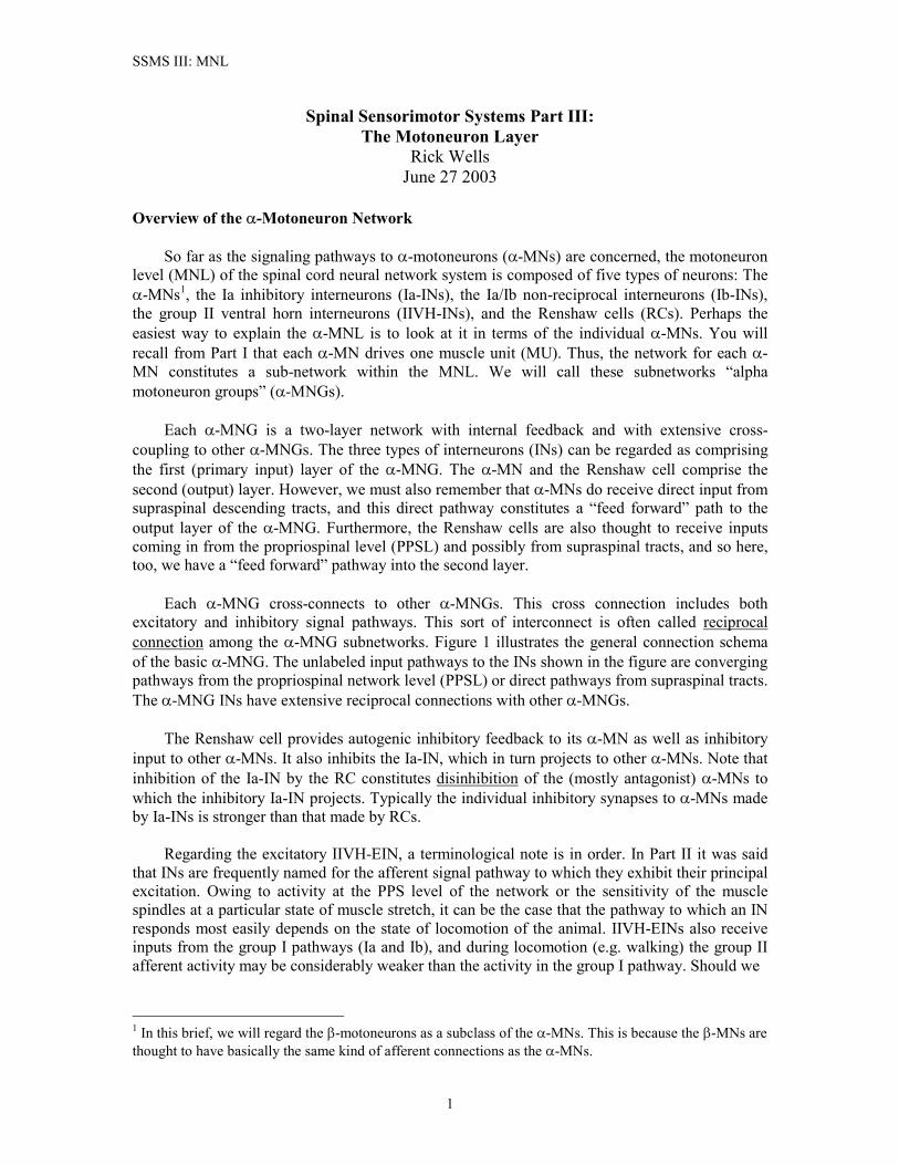
SSMS III: MNL Spinal Sensorimotor Systems Part III: The Motoneuron Layer Rick Wells June 27 2003 Overview of the -Motoneuron Network So far as the signaling pathways to -motoneurons (-MNs) are concerned, the motoneuron level (MNL) of the spinal cord neural network system is composed of five types of neurons: The -MNs 1 , the Ia inhibitory interneurons (Ia-INs), the Ia/Ib non-reciprocal interneurons (Ib-INs), the group II ventral horn interneurons (IIVH-INs), and the Renshaw cells (RCs). Perhaps the easiest way to explain the -MNL is to look at it in terms of the individual -MNs. You will recall from Part I that each -MN drives one muscle unit (MU). Thus, the network for each - MN constitutes a sub-network within the MNL. We will call these subnetworks “alpha motoneuron groups” (-MNGs). Each -MNG is a two-layer network with internal feedback and with extensive cross- coupling to other -MNGs. The three types of interneurons (INs) can be regarded as comprising the first (primary input) layer of the -MNG. The -MN and the Renshaw cell comprise the second (output) layer. However, we must also remember that -MNs do receive direct input from supraspinal descending tracts, and this direct pathway constitutes a “feed forward” path to the output layer of the -MNG. Furthermore, the Renshaw cells are also thought to receive inputs coming in from the propriospinal level (PPSL) and possibly from supraspinal tracts, and so here, too, we have a “feed forward” pathway into the second layer. Each -MNG cross-connects to other -MNGs. This cross connection includes both excitatory and inhibitory signal pathways. This sort of interconnect is often called reciprocal connection among the -MNG subnetworks. Figure 1 illustrates the general connection schema of the basic -MNG. The unlabeled input pathways to the INs shown in the figure are converging pathways from the propriospinal network level (PPSL) or direct pathways from supraspinal tracts. The -MNG INs have extensive reciprocal connections with other -MNGs. The Renshaw cell provides autogenic inhibitory feedback to its -MN as well as inhibitory input to other -MNs. It also inhibits the Ia-IN, which in turn projects to other -MNs. Note that inhibition of the Ia-IN by the RC constitutes disinhibition of the (mostly antagonist) -MNs to which the inhibitory Ia-IN projects. Typically the individual inhibitory synapses to -MNs made by Ia-INs is stronger than that made by RCs. Regarding the excitatory IIVH-EIN, a terminological note is in order. In Part II it was said that INs are frequently named for the afferent signal pathway to which they exhibit their principal excitation. Owing to activity at the PPS level of the network or the sensitivity of the muscle spindles at a particular state of muscle stretch, it can be the case that the pathway to which an IN responds most easily depends on the state of locomotion of the animal. IIVH-EINs also receive inputs from the group I pathways (Ia and Ib), and during locomotion (e.g. walking) the group II afferent activity may be considerably weaker than the activity in the group I pathway. Should we 1 In this brief, we will regard the -motoneurons as a subclass of the -MNs. This is because the -MNs are thought to have basically the same kind of afferent connections as the -MNs. 1
Transcript of Spinal Sensorimotor Systems Part III - uidaho.edurwells/techdocs/Spinal Sensorimotor Systems... ·...
SSMS III: MNL
Spinal Sensorimotor Systems Part III: The Motoneuron Layer
Rick Wells June 27 2003
Overview of the �-Motoneuron Network So far as the signaling pathways to �-motoneurons (�-MNs) are concerned, the motoneuron level (MNL) of the spinal cord neural network system is composed of five types of neurons: The �-MNs1, the Ia inhibitory interneurons (Ia-INs), the Ia/Ib non-reciprocal interneurons (Ib-INs), the group II ventral horn interneurons (IIVH-INs), and the Renshaw cells (RCs). Perhaps the easiest way to explain the �-MNL is to look at it in terms of the individual �-MNs. You will recall from Part I that each �-MN drives one muscle unit (MU). Thus, the network for each �-MN constitutes a sub-network within the MNL. We will call these subnetworks “alpha motoneuron groups” (�-MNGs). Each �-MNG is a two-layer network with internal feedback and with extensive cross-coupling to other �-MNGs. The three types of interneurons (INs) can be regarded as comprising the first (primary input) layer of the �-MNG. The �-MN and the Renshaw cell comprise the second (output) layer. However, we must also remember that �-MNs do receive direct input from supraspinal descending tracts, and this direct pathway constitutes a “feed forward” path to the output layer of the �-MNG. Furthermore, the Renshaw cells are also thought to receive inputs coming in from the propriospinal level (PPSL) and possibly from supraspinal tracts, and so here, too, we have a “feed forward” pathway into the second layer. Each �-MNG cross-connects to other �-MNGs. This cross connection includes both excitatory and inhibitory signal pathways. This sort of interconnect is often called reciprocal connection among the �-MNG subnetworks. Figure 1 illustrates the general connection schema of the basic �-MNG. The unlabeled input pathways to the INs shown in the figure are converging pathways from the propriospinal network level (PPSL) or direct pathways from supraspinal tracts. The �-MNG INs have extensive reciprocal connections with other �-MNGs. The Renshaw cell provides autogenic inhibitory feedback to its �-MN as well as inhibitory input to other �-MNs. It also inhibits the Ia-IN, which in turn projects to other �-MNs. Note that inhibition of the Ia-IN by the RC constitutes disinhibition of the (mostly antagonist) �-MNs to which the inhibitory Ia-IN projects. Typically the individual inhibitory synapses to �-MNs made by Ia-INs is stronger than that made by RCs. Regarding the excitatory IIVH-EIN, a terminological note is in order. In Part II it was said that INs are frequently named for the afferent signal pathway to which they exhibit their principal excitation. Owing to activity at the PPS level of the network or the sensitivity of the muscle spindles at a particular state of muscle stretch, it can be the case that the pathway to which an IN responds most easily depends on the state of locomotion of the animal. IIVH-EINs also receive inputs from the group I pathways (Ia and Ib), and during locomotion (e.g. walking) the group II afferent activity may be considerably weaker than the activity in the group I pathway. Should we
1 In this brief, we will regard the �-motoneurons as a subclass of the �-MNs. This is because the �-MNs are thought to have basically the same kind of afferent connections as the �-MNs.
1
SSMS III: MNL
Figure 1: Simplified diagram of an �-MNG subnetwork. The INs identified in the figure have been
defined previously in Part II of the SSMS tech brief. Excitatory and inhibitory synapses are as noted in the figure key. DTs = descending supraspinal tract pathways. PPS = propriospinal IN pathways. Mono = monosynaptic pathway. The Renshaw cell receives excitatory MN input only from nearby MNs, but
projects its output over a large fraction of the motor nuclei in the ventral horn. Incoming signal pathways generally denote multiple axons. Also, the IN symbols projecting to the �-MN can denote multiple parallel INs of these particular classes, each IN integrating signals from different pathways. Note that the Ia-IN in the �-MNG projects to other �-MNGs, and these other groups reciprocally project back to the MN in this
group. The reason I include the Ia-IN within this �-MNG in this fashion is because the Ia-IN gets a disynaptic inhibiting input (via the RC) from the MN in this group. The IIVH-INs also make lateral
connections with other �-MNGs. Input pathways to the INs are discussed in the text. then call that IN an “excitatory Ib-IN”? The difficulty with IN naming conventions is obvious in such a case. Jankowska2 attempts to distinguish between excitatory group II and an “excitatory group Ib” IN, but notes that the “excitatory Ib interneuron” has “not yet been identified.” Most other authors do not attempt to distinguish between the IIVH-EIN and a “Ib-EIN” as she does.
2 E. Jankowska, “Interneuronal relay in spinal pathways from proprioceptors,” Prog. in Neurobiol. (1992), 38: 335-378.
2
SSMS III: MNL
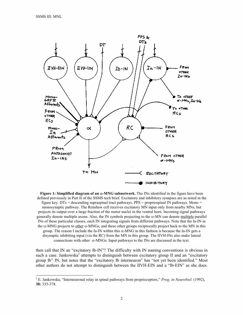
Rather, they simply refer to a generic “excitatory interneuron.” Jankowska’s distinction between the two cases seems to rest primarily on the targets to which IIVH-EIN and “Ib-EIN” project. The Ib-EIN “activate extensors more effectively than flexors”, whereas the IIVH-EIN projects mainly to flexors (but also “have access to” extensors). Both, however, seem to occupy about the same region of the ventral horn, and both receive the same types of afferent pathway inputs at similar synaptic levels (i.e. monosynaptic, disynaptic, etc.). Most probably Jankowska’s distinction between two different class of INs is correct, but for our purposes it is enough to recognize that some neurons of the “IIVH-EIN” class correspond to Jankowska’s “excitatory Ib interneuron” and others correspond to Jankowska’s excitatory group II ventral horn interneuron. Since the IIVH-EIN symbol in figure 1 denotes multiple, parallel INs, we can have whatever mix of “generic excitatory INs” we require. Overview of the �-Motoneuron Network A motor unit is defined by its motoneuron and the muscle fibers that MN excites. Thus, intrafusal fiber excitation by �-MNs must be regarded as a MNL network separate from the �-MNG. We will call these subnetworks �-MNGs. The circuit details of �-MNGs are much less well understood than those of the �-MNGs. The �-MNG lacks the Renshaw cell, the IIVH-IIN, and the Ia-IN. �-MNs do receive monosynaptic inputs from the inhibitory Ib-IN and the IIVH-EIN classes of interneurons.2 What is considerably less clear is whether or not any of the INs feeding the �-MN are the very same INs that feed a related �-MN. If this is the case, we would have co-activation of �-MNs via a direct coupling between the �-MNG and related �-MNGs. Indeed, this question is at the root of a long-standing controversy in neurobiology over the “�-� linkage” question. It does seem to be the case that �-MNs receive their own oligosynaptic descending tract supraspinal inputs, and these pathways in some cases might converge on a group II IN that projects only to the �-MN. It therefore it seems like a safe, conservative approach to assume that at least some of the Ib-INs and IIVH-EINs converging on a �-MN are dedicated to this motor unit. Figure 2 illustrates a putative �-MNG network. The symbolic conventions used in this figure are the same as those in figure 1. A Note on Synaptic Connections to Neurons We are about to examine the plurality of different pathways converging on various INs. As we do so, it is worthwhile to take a precautionary note of the roles the synaptic connections of these pathways play, both for INs as well as for MNs. It is easy to fall into the habit of thinking of all excitatory synaptic inputs as representing motoneuron “data inputs”. However, in a great many cases it is more correct to view excitatory synaptic inputs as providing a biasing “control input” to the neuron they contact. In other words, their role is not so much one of firing the postsynaptic cell but, rather, of making it easier for other pathways, “data pathways”, to fire that cell. This is termed “increasing the excitability” of the postsynaptic neuron. Pathways that primarily serve this function can in a sense be thought of as “gating” signals. The IN can then be regarded as performing a kind of distributed “multiplexer” function in the neural network. The desired muscle response to a spindle, cutaneous, or joint afferent pathway during a simple reflex is usually quite different from the desired response to these same inputs during locomotion. In control system terminology, the SSMS seems to use a kind of “gain scheduling” strategy, with different pathways taking on “primary” control loop responsibility during different states of body position. Gain scheduling is a common strategy in man-made control systems, and it would appear that nature has hit on the same scheme.
3
SSMS III: MNL
Figure 2: Simplified diagram of an �-MNG subnetwork. There are differences in the type and
distribution of input pathways between static and dynamic �-MNs, as discussed in Part II of this tech brief. Group Ia afferents occur in less than 4% of all �-MNGs. It is not known whether or not cross-coupling from �-MNGs occurs in �-MNs. The descending tract (DT) pathways are also different for static and dynamic �-MNs. The �d-MNs receive monosynaptic DT inputs from the rubrospinal tract (which originates in the red nucleus of the midbrain). DT inputs to the �s-MNs are oligosynaptic and it is unclear whether or not any of these pathways are monosynaptic connections to the �-MN. The unlabeled inputs to the INs are from PPSL
interneurons and/ or descending tracts. The INs shown here represent possible parallel INs of each particular class of interneuron. Ib-INs projecting to �-MNs integrate homonymous muscles afferents.
Monosynaptic group II afferents come from both homonymous and heteronymous muscles. It is noteworthy that some �-MNs appear to be predominant recipients of excitatory pathways while others
appear to be predominantly recipients of inhibitory pathways.3 It isn’t entirely clear what sense it makes for a �-MN to be predominantly receptive to inhibitory inputs, but these presumably would counteract tonic
firing invoked from other INs and I would guess that this could be a posture control mechanism. A second control theory analogy that seems to fit the SSMS is the type of control known as “variable structure switching control” (VSSC) systems.4 If some pathways functionally act as “gates” for other pathways, we could say that the SSMS neural network at the MN level exerts control over the MNs by switching among different feedback pathways. This is precisely what is done in man-made VSSC systems, and the benefit obtained in these systems by this method is a
3 B. Appelberg, M. Hulliger, H. Johansson, and P. Sojka, “Actions on �-motoneurones elicited by electrical stimulation of group III muscle afferent fibres in the hind limb of the cat”, J. Physiol. (1983), 335: 275-292. 4 Deterministic Control of Uncertain Systems, A.S.I. Zinober (ed.), London: IEE Press, 1990.
4
SSMS III: MNL
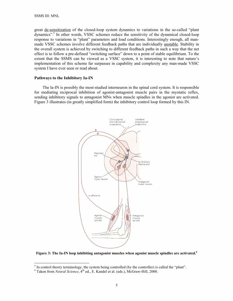
great de-sensitization of the closed-loop system dynamics to variations in the so-called “plant dynamics”.5 In other words, VSSC schemes reduce the sensitivity of the dynamical closed-loop response to variations in “plant” parameters and load conditions. Interestingly enough, all man-made VSSC schemes involve different feedback paths that are individually unstable. Stability in the overall system is achieved by switching to different feedback paths in such a way that the net effect is to follow a pre-defined “switching surface” down to a point of stable equilibrium. To the extent that the SSMS can be viewed as a VSSC system, it is interesting to note that nature’s implementation of this scheme far surpasses in capability and complexity any man-made VSSC system I have ever seen or read about. Pathways to the Inhibitory Ia-IN The Ia-IN is possibly the most-studied interneuron in the spinal cord system. It is responsible for mediating reciprocal inhibition of agonist-antagonist muscle pairs in the myotatic reflex, sending inhibitory signals to antagonist MNs when muscle spindles in the agonist are activated. Figure 3 illustrates (in greatly simplified form) the inhibitory control loop formed by this IN.
Figure 3: The Ia-IN loop inhibiting antagonist muscles when agonist muscle spindles are activated.6
5 In control theory terminology, the system being controlled (by the controller) is called the “plant”. 6 Taken from Neural Science, 4th ed., E. Kandel et al. (eds.), McGraw-Hill, 2000.
5
SSMS III: MNL
The Ia-IN allows higher centers to coordinate opposing muscles at a joint through a single command. Corticospinal descending axons connecting to the Ia-IN that activate one set of muscles automatically leads to relaxation of the antagonists. Inhibitory inputs to the Ia-IN that reduce its activity decrease reciprocal inhibition and allow co-contraction of opposing muscles. Ia-INs project to several, but not all possible, target antagonist motor nuclei. The Ia-IN has a medium-sized soma (about 51 � 27 �m) and long (but not extensive) dendritic branches. Its axon reaches both nearby and fairly distant MNs. Each Ia-IN axon invades several motor nuclei, where it terminates on about 20% of the MNs in those nuclei.7 Besides �-MNs, the only cells inhibited by Ia-INs are other Ia-INs and Renshaw cells. Subpopulations of Ia-INs that have opposing actions (e.g. flexor vs. extensor inhibition) inhibit each other. Strong excitation of one population of Ia-INs favors selective activation of synergists, while weaker excitation favors co-activation of flexors and extensors. (Co-contraction is needed for such things as balance, precise limb positioning, and the like). The inhibitory synaptic weight connecting MNs and Ia-INs is larger than is the inhibitory synaptic weight connecting MNs and Renshaw cells. At least 10, and on the average about 70, Ia-INs converge on each motoneuron. Ia-INs exhibit tonic background firing that hyperpolarizes the target �-MNs, but this hyperpolarization is abolished when the MN fires. Its inhibitory neurotransmitter (NTX) is thought to be glycine. Excitatory synapses on Ia-INs appear to be glutaminergic (that is, the NTX is glutamate, Glu). Ia-INs do not seem to respond to acetylcholine (ACh), and therefore are not directly driven by MN axon collaterals. Ia-INs make synaptic connection to �-MNs primarily at the soma and the proximal dendrites. They are capable of firing high rate bursts (300-400 APs per sec.), but a typical volley of synchronous Ia afferent inputs usually evokes only a single AP. Ia Afferent Pathways. The Ia-IN is the only interneuron for which the dominant peripheral input is from group Ia (velocity-sensing) muscle spindles. Ia afferents entering the ventral horn make monosynaptic excitatory contact8 with the Ia-INs. Group I afferents also participate in polysynaptic excitatory pathways to the Ia-IN, but these pathways come down from the PPSL network and are heavily influenced by group Ib, ipsilateral FRA, and contralateral FRA signals.9 Convergence of group I afferents is limited to one or at most a few muscles.10 The role of the Ia afferents in the polysynaptic pathways is thought to be indirect at best, i.e. the Ia afferent appears to play a gating function role in this pathway. It is thought that this polysynaptic pathway involves spinal circuitry for central pattern generators (CPGs).11 The Ia-IN also terminates a disynaptic inhibitory Ia pathway that comes in via Ia-INs from antagonist muscles or from Ia-INs in synergists. Thus for Ia convergence on the Ia-IN, we have a total of one type of monosynaptic excitatory Ia pathways, one type of inhibitory disynaptic pathways via antagonist Ia-INs, one type of disynaptic inhibitory pathways from synergist Ia-INs, and two or more types of polysynaptic excitatory pathways through a CPG network and other interneurons at the PPS level.
7 E. Jankowska and W.J. Roberts, “Synaptic action of single interneurons mediating reciprocal Ia inhibition of motoneurones,” J. Physiol. (1972) 223: 623-642. 8 When we say monosynaptic here, we mean the Ia afferent synapses directly to the Ia-IN without any intermediate interneuron between them. 9 Ispsilateral means “from the same lateral side”. Contralateral means “from the opposite lateral side”. 10 H. Hultborn, E. Jankowska, and S. Lindström, “Recurrent inhibition of interneurones monosynaptically activated from group Ia afferents,” J. Physiol. (1971), 215: 613-636. 11 C.A. Pratt and L.M. Jordan, “Ia inhibitory interneurons and Renshaw cells as contributors to spinal mechanisms of fictive locomotion,” J. Neurophysiol. (1987), 57(1): 56-71.
6
SSMS III: MNL
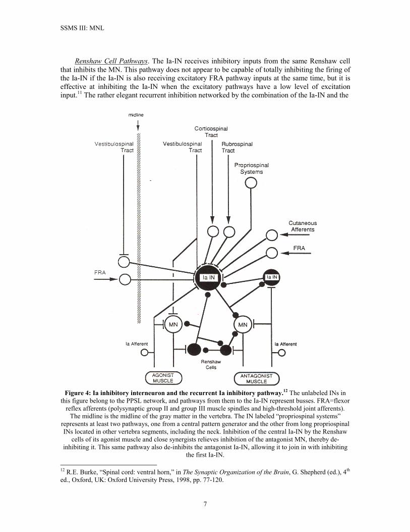
Renshaw Cell Pathways. The Ia-IN receives inhibitory inputs from the same Renshaw cell that inhibits the MN. This pathway does not appear to be capable of totally inhibiting the firing of the Ia-IN if the Ia-IN is also receiving excitatory FRA pathway inputs at the same time, but it is effective at inhibiting the Ia-IN when the excitatory pathways have a low level of excitation input.11 The rather elegant recurrent inhibition networked by the combination of the Ia-IN and the
Figure 4: Ia inhibitory interneuron and the recurrent Ia inhibitory pathway.12 The unlabeled INs in
this figure belong to the PPSL network, and pathways from them to the Ia-IN represent busses. FRA=flexor reflex afferents (polysynaptic group II and group III muscle spindles and high-threshold joint afferents).
The midline is the midline of the gray matter in the vertebra. The IN labeled “propriospinal systems” represents at least two pathways, one from a central pattern generator and the other from long propriospinal INs located in other vertebra segments, including the neck. Inhibition of the central Ia-IN by the Renshaw
cells of its agonist muscle and close synergists relieves inhibition of the antagonist MN, thereby de-inhibiting it. This same pathway also de-inhibits the antagonist Ia-IN, allowing it to join in with inhibiting
the first Ia-IN. 12 R.E. Burke, “Spinal cord: ventral horn,” in The Synaptic Organization of the Brain, G. Shepherd (ed.), 4th ed., Oxford, UK: Oxford University Press, 1998, pp. 77-120.
7
SSMS III: MNL
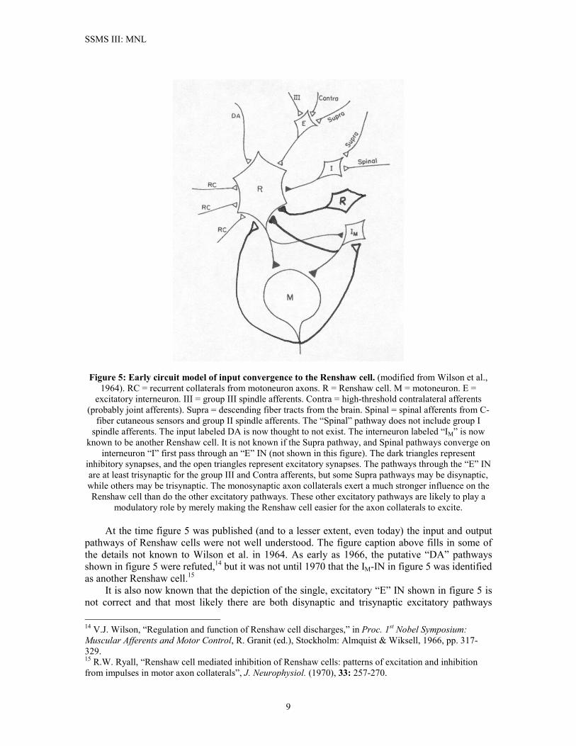
Renshaw cells is illustrated in the simplified circuit in Figure 4. The figure also illustrates the Ia-IN’s role as a primary integrating node in a number of spinal circuit pathways. Although figure 4 only illustrates one Renshaw cell pathway to the Ia-IN, in fact multiple Renshaw cells, primarily from other MNs in the same muscle and from a few close synergists, make disynaptic inhibitory connection to the Ia-IN. Lateral Inhibition Between Ia-INs. Figure 4 also illustrates the pattern of mutual lateral inhibition between Ia-INs of agonist-antagonist muscle pairs. The Ia-IN receives significant excitatory inputs from descending tracts and PPSL networks, and it is intimately linked to the PPSL central pattern generator. These higher-level excitatory inputs, which do not contact the Renshaw cells, can selectively excite Ia-INs, which then inhibit firing of their antagonist Ia-INs. Thus, the mutual inhibition between Ia-INs serves a modulatory role in locomotion. Excitatory FRA and Cutaneous Pathways. The Ia-IN receives polysynaptic excitatory pathways from flexor reflex afferents (FRAs) via subnetworks at the PPS level. There are at least two different kinds of FRA pathways involved. One is a relatively fast-acting pathway that probably serves the flexor reflex. The other is a slower pathway involving the spinal CPG, and it is used in locomotion. Signals descending on the Ia-IN tend to come in volleys in relatively high frequency bursts. Also, as illustrated in figure 4, there are separate pathways for ipsilateral FRAs (i-FRAs) and contralateral FRAs (co-FRAs). The co-FRA is the FRA pathway shown on the left side of figure 4. The FRA pathways shown in figure 4 involve group II and group III spindle afferents and high-threshold joint afferents. The Ia-IN also receives disynaptic excitatory input from low-threshold cutaneous nerves.10 Group Ib Afferent Pathways. Although not shown in figure 4, group Ib spindle afferents have a polysynaptic pathway to the Ia-IN. This pathway most likely descends through the IN marked “propriospinal systems” in figure 4. Only ipsilateral group Ib afferents show a pathway to the Ia-IN.12 Descending Tract Pathways. As illustrated in figure 4, the Ia-IN receives polysynaptic excitatory inputs from the corticospinal and rubrospinal tracts, and from the contralateral tracts of the vestibulospinal tract. Like the �-MN, it also receives a monosynaptic input from the ipsilateral vestibulospinal tract. The Ia-IN exhibits short-term post-tetanic facilitation from volleys coming in through these pathways. Except for the ipsilateral vestibulospinal tract input, the descending supraspinal inputs contact first-order INs in the PPS level, and they appear to be involved in initiating the action of the CPG during locomotion, and in controlling the FRA pathways to the Ia-IN. Renshaw Cells So far as we know, the RC is unique among interneurons in that it is the only one that appears to receive an input from the axons of �-MNs. They receive excitatory input from nearby MNs, but they project their inhibitory outputs to a much larger group of closely-related (synergist) MNs.12 It is currently thought that RCs project outputs only to �-MNs, Ia-INs, and other RCs. The principal excitatory inputs to RCs are the monosynaptic axon collaterals, although it is known that modulatory polysynaptic excitatory pathways to the RC do exist. Figure 5 illustrates an early model of the “minimum circuit” for converging signal pathways to the Renshaw cell.13 13 V.J. Wilson, W.H. Talbot, and M. Kato, “Inhibitory convergence upon Renshaw cells,” J. Neurophysiol. (1964) 27: 1063-1079.
8
SSMS III: MNL
Figure 5: Early circuit model of input convergence to the Renshaw cell. (modified from Wilson et al., 1964). RC = recurrent collaterals from motoneuron axons. R = Renshaw cell. M = motoneuron. E =
excitatory interneuron. III = group III spindle afferents. Contra = high-threshold contralateral afferents (probably joint afferents). Supra = descending fiber tracts from the brain. Spinal = spinal afferents from C-
fiber cutaneous sensors and group II spindle afferents. The “Spinal” pathway does not include group I spindle afferents. The input labeled DA is now thought to not exist. The interneuron labeled “IM” is now
known to be another Renshaw cell. It is not known if the Supra pathway, and Spinal pathways converge on interneuron “I” first pass through an “E” IN (not shown in this figure). The dark triangles represent
inhibitory synapses, and the open triangles represent excitatory synapses. The pathways through the “E” IN are at least trisynaptic for the group III and Contra afferents, but some Supra pathways may be disynaptic, while others may be trisynaptic. The monosynaptic axon collaterals exert a much stronger influence on the Renshaw cell than do the other excitatory pathways. These other excitatory pathways are likely to play a
modulatory role by merely making the Renshaw cell easier for the axon collaterals to excite. At the time figure 5 was published (and to a lesser extent, even today) the input and output pathways of Renshaw cells were not well understood. The figure caption above fills in some of the details not known to Wilson et al. in 1964. As early as 1966, the putative “DA” pathways shown in figure 5 were refuted,14 but it was not until 1970 that the IM-IN in figure 5 was identified as another Renshaw cell.15 It is also now known that the depiction of the single, excitatory “E” IN shown in figure 5 is not correct and that most likely there are both disynaptic and trisynaptic excitatory pathways 14 V.J. Wilson, “Regulation and function of Renshaw cell discharges,” in Proc. 1st Nobel Symposium: Muscular Afferents and Motor Control, R. Granit (ed.), Stockholm: Almquist & Wiksell, 1966, pp. 317-329. 15 R.W. Ryall, “Renshaw cell mediated inhibition of Renshaw cells: patterns of excitation and inhibition from impulses in motor axon collaterals”, J. Neurophysiol. (1970), 33: 257-270.
9
SSMS III: MNL
converging on RCs. Group III spindle afferents and contralateral joint afferents appear to follow a trisynaptic pathway, while group II spindle afferents, descending tract inputs, and C-fiber cutaneous afferents may have a disynaptic pathway. I have seen no evidence that would suggest that the IN mediating this disynaptic pathway is the IIVH-EIN in figure 1. Accordingly, we place the “E” IN within the PPSL network. The inhibitory pathways to the RC have a wide convergence. Cutaneous afferents are part of this pathway system, and it is thought that all classes of myelinated fibers except group I afferents converge on RCs via inhibitory pathways.16 Strong inhibition of RC discharge is evoked by cutaneous and by muscle afferents. This inhibition is very long-lasting, typically between 100 msec. and 200 msec. in duration.13
The inhibitory IN “I” in figure 5 has not been identified, but it is apparently not the IIVH-IIN of figure 1. Inhibition is more frequent than excitation insofar as the “E” and “I” pathways of figure 5 are concerned. One thing that is clear is that supraspinal controls (both excitatory and inhibitory) modulate the firing activity of RCs during locomotion. The role of the FRAs (“III”, “Contra”, and “Spinal” in figure 5) appears to be that of modulating the descending tract’s effects on RCs during locomotion. However, there is unanimous agreement that the principal excitatory pathway to the RCs is from the MN axon collaterals. Unexcited RCs fire spontaneously at a rate of about 15 APs/sec. RC discharges evoked by MNs can consist of as many as 30 action potentials (APs) in a burst lasting as long as 60 msec. RCs are known to be capable of firing in bursts at an unusually high rate (1 kHz). Also, these synapses express both nicotinic and muscarinic ACh receptors. The latter is a metabotropic receptor, and it causes the RC to fire, pause, then resume background firing after about 100-200 msec. Motoneurons of different types excite RCs with different strengths and, in turn, are inhibited by RCs with different synaptic strengths. The strongest exciters of RCs are the FF-type MNs, followed by FR-type MNs, and finally the S-type MNs. The strength of inhibition of MNs by RCs follows just the opposite characteristic, with S-type MNs being the most easily inhibited and FF-type MNs being the least easily inhibited.11 Whereas the Ia-IN receives inputs directly from the CPG in the PPSL network, the RC apparently has no direct connection whatever with the CPG network. Furthermore, it has been established that during locomotion the RC (and, for that matter, of the Ia-IN) play no more than a merely modulatory role. This was established by inhibiting them pharmacologically and observing that locomotion was not substantially interfered with. This is one piece of evidence that strongly suggests that locomotion is driven by excitatory rather than inhibitory inputs to the MNL network. Finally, there has been a long-running debate over whether there was only one type of RC or if there might be two types. The issue was raised by the observation that although the principal inhibitory neurotransmitter used by RCs is glycine, there was also evidence of the presence of the inhibitory NTX GABA. It was thought that a particular RC could only employ one type of NTX, an often-misunderstood hypothesis known as Dale’s law. It is now known that neurons can co-
16 See Part I of this tech brief to review the types of nerves employing myelinated and unmyelinated fibers.
10
SSMS III: MNL
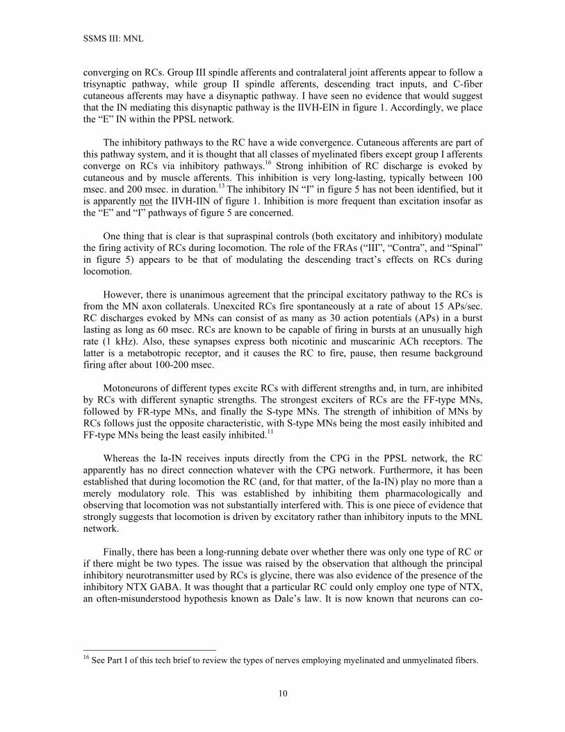
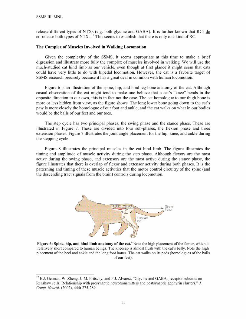
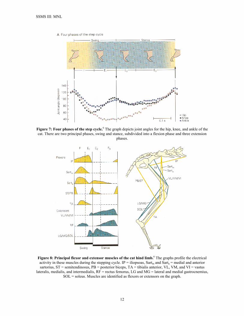
release different types of NTXs (e.g. both glycine and GABA). It is further known that RCs do co-release both types of NTXs.17 This seems to establish that there is only one kind of RC. The Complex of Muscles Involved in Walking Locomotion Given the complexity of the SSMS, it seems appropriate at this time to make a brief digression and illustrate more fully the complex of muscles involved in walking. We will use the much-studied cat hind limb as our vehicle, even though at first glance it might seem that cats could have very little to do with bipedal locomotion. However, the cat is a favorite target of SSMS research precisely because it has a great deal in common with human locomotion. Figure 6 is an illustration of the spine, hip, and hind leg-bone anatomy of the cat. Although casual observation of the cat might tend to make one believe that a cat’s “knee” bends in the opposite direction to our own, this is in fact not the case. The cat homologue to our thigh bone is more or less hidden from view, as the figure shows. The long lower bone going down to the cat’s paw is more closely the homologue of our foot and ankle, and the cat walks on what in our bodies would be the balls of our feet and our toes. The step cycle has two principal phases, the swing phase and the stance phase. These are illustrated in Figure 7. These are divided into four sub-phases, the flexion phase and three extension phases. Figure 7 illustrates the joint angle placement for the hip, knee, and ankle during the stepping cycle. Figure 8 illustrates the principal muscles in the cat hind limb. The figure illustrates the timing and amplitude of muscle activity during the step phase. Although flexors are the most active during the swing phase, and extensors are the most active during the stance phase, the figure illustrates that there is overlap of flexor and extensor activity during both phases. It is the patterning and timing of these muscle activities that the motor control circuitry of the spine (and the descending tract signals from the brain) controls during locomotion.
Figure 6: Spine, hip, and hind limb anatomy of the cat.6 Note the high placement of the femur, which is relatively short compared to human beings. The kneecap is almost flush with the cat’s belly. Note the high placement of the heel and ankle and the long foot bones. The cat walks on its pads (homologues of the balls
of our feet).
17 E.J. Geiman, W. Zheng, J.-M. Fritschy, and F.J. Alvarez, “Glycine and GABAA receptor subunits on Renshaw cells: Relationship with presynaptic neurotransmitters and postsynaptic gephyrin clusters,” J. Comp. Neurol. (2002), 444: 275-289.
11
SSMS III: MNL
Figure 7: Four phases of the step cycle.6 The graph depicts joint angles for the hip, knee, and ankle of the cat. There are two principal phases, swing and stance, subdivided into a flexion phase and three extension
phases.
Figure 8: Principal flexor and extensor muscles of the cat hind limb.6 The graphs profile the electrical activity in these muscles during the stepping cycle. IP = iliopsoas, Sartm and Sarta = medial and anterior sartorius, ST = semitendinosus, PB = posterior biceps, TA = tibialis anterior, VL, VM, and VI = vastus
lateralis, medialis, and intermedialis, RF = rectus femorus, LG and MG = lateral and medial gastrocnemius, SOL = soleus. Muscles are identified as flexors or extensors on the graph.
12
SSMS III: MNL
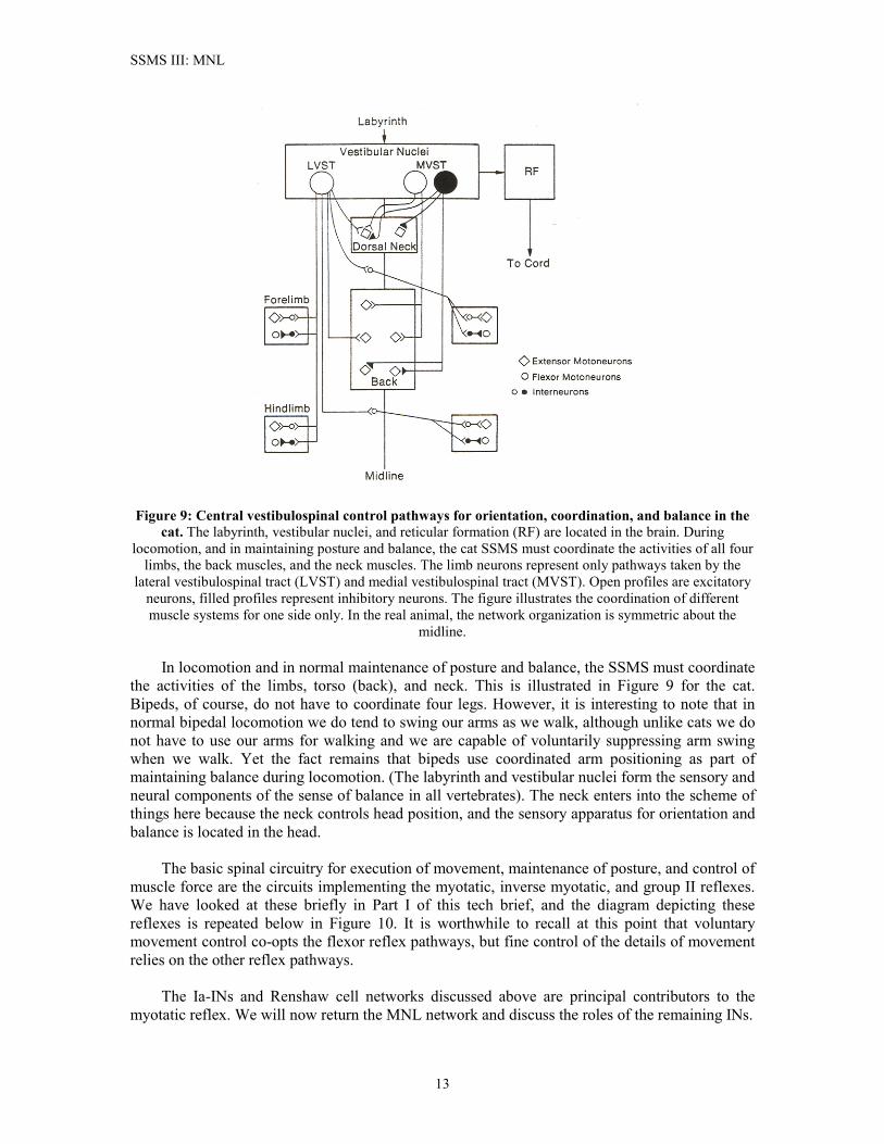
Figure 9: Central vestibulospinal control pathways for orientation, coordination, and balance in the cat. The labyrinth, vestibular nuclei, and reticular formation (RF) are located in the brain. During
locomotion, and in maintaining posture and balance, the cat SSMS must coordinate the activities of all four limbs, the back muscles, and the neck muscles. The limb neurons represent only pathways taken by the
lateral vestibulospinal tract (LVST) and medial vestibulospinal tract (MVST). Open profiles are excitatory neurons, filled profiles represent inhibitory neurons. The figure illustrates the coordination of different muscle systems for one side only. In the real animal, the network organization is symmetric about the
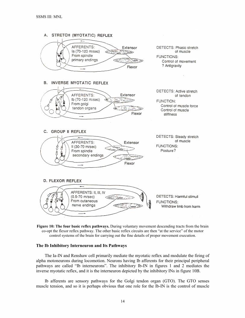
midline. In locomotion and in normal maintenance of posture and balance, the SSMS must coordinate the activities of the limbs, torso (back), and neck. This is illustrated in Figure 9 for the cat. Bipeds, of course, do not have to coordinate four legs. However, it is interesting to note that in normal bipedal locomotion we do tend to swing our arms as we walk, although unlike cats we do not have to use our arms for walking and we are capable of voluntarily suppressing arm swing when we walk. Yet the fact remains that bipeds use coordinated arm positioning as part of maintaining balance during locomotion. (The labyrinth and vestibular nuclei form the sensory and neural components of the sense of balance in all vertebrates). The neck enters into the scheme of things here because the neck controls head position, and the sensory apparatus for orientation and balance is located in the head. The basic spinal circuitry for execution of movement, maintenance of posture, and control of muscle force are the circuits implementing the myotatic, inverse myotatic, and group II reflexes. We have looked at these briefly in Part I of this tech brief, and the diagram depicting these reflexes is repeated below in Figure 10. It is worthwhile to recall at this point that voluntary movement control co-opts the flexor reflex pathways, but fine control of the details of movement relies on the other reflex pathways. The Ia-INs and Renshaw cell networks discussed above are principal contributors to the myotatic reflex. We will now return the MNL network and discuss the roles of the remaining INs.
13
SSMS III: MNL
Figure 10: The four basic reflex pathways. During voluntary movement descending tracts from the brain co-opt the flexor reflex pathway. The other basic reflex circuits are then “at the service” of the motor
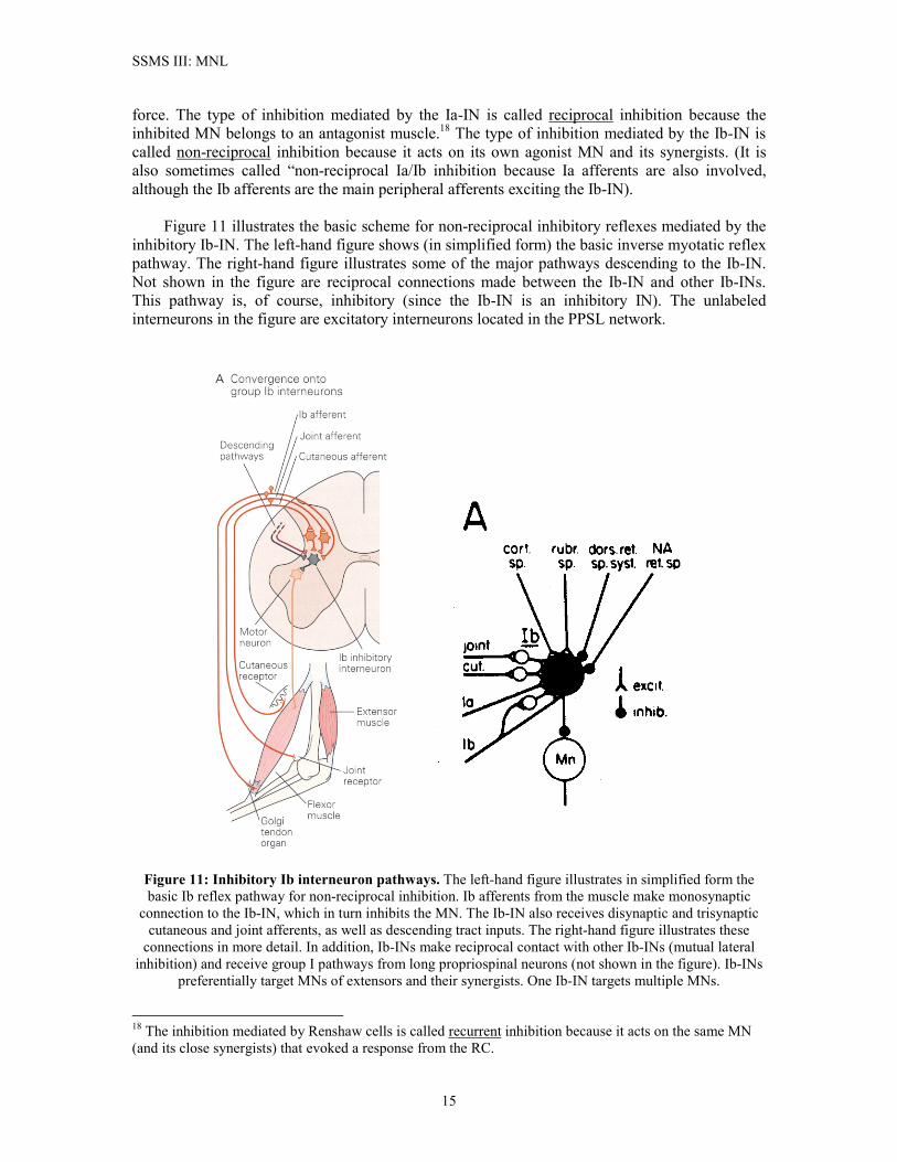
control systems of the brain for carrying out the fine details of proper movement execution. The Ib Inhibitory Interneuron and Its Pathways The Ia-IN and Renshaw cell primarily mediate the myotatic reflex and modulate the firing of alpha motoneurons during locomotion. Neurons having Ib afferents for their principal peripheral pathways are called “Ib interneurons”. The inhibitory Ib-IN in figures 1 and 2 mediates the inverse myotatic reflex, and it is the interneuron depicted by the inhibitory INs in figure 10B. Ib afferents are sensory pathways for the Golgi tendon organ (GTO). The GTO senses muscle tension, and so it is perhaps obvious that one role for the Ib-IN is the control of muscle
14
SSMS III: MNL
force. The type of inhibition mediated by the Ia-IN is called reciprocal inhibition because the inhibited MN belongs to an antagonist muscle.18 The type of inhibition mediated by the Ib-IN is called non-reciprocal inhibition because it acts on its own agonist MN and its synergists. (It is also sometimes called “non-reciprocal Ia/Ib inhibition because Ia afferents are also involved, although the Ib afferents are the main peripheral afferents exciting the Ib-IN). Figure 11 illustrates the basic scheme for non-reciprocal inhibitory reflexes mediated by the inhibitory Ib-IN. The left-hand figure shows (in simplified form) the basic inverse myotatic reflex pathway. The right-hand figure illustrates some of the major pathways descending to the Ib-IN. Not shown in the figure are reciprocal connections made between the Ib-IN and other Ib-INs. This pathway is, of course, inhibitory (since the Ib-IN is an inhibitory IN). The unlabeled interneurons in the figure are excitatory interneurons located in the PPSL network.
Figure 11: Inhibitory Ib interneuron pathways. The left-hand figure illustrates in simplified form the basic Ib reflex pathway for non-reciprocal inhibition. Ib afferents from the muscle make monosynaptic
connection to the Ib-IN, which in turn inhibits the MN. The Ib-IN also receives disynaptic and trisynaptic cutaneous and joint afferents, as well as descending tract inputs. The right-hand figure illustrates these
connections in more detail. In addition, Ib-INs make reciprocal contact with other Ib-INs (mutual lateral inhibition) and receive group I pathways from long propriospinal neurons (not shown in the figure). Ib-INs
preferentially target MNs of extensors and their synergists. One Ib-IN targets multiple MNs.
18 The inhibition mediated by Renshaw cells is called recurrent inhibition because it acts on the same MN (and its close synergists) that evoked a response from the RC.
15

SSMS III: MNL
Figure 12: Proportions of Ib-INs with excitatory input from various sources.22 I = total group I input; Ib = Ib afferents; Ia = Ia afferents; II = group II afferents; cut. = cutaneous; J = joint; i.o. = interosseous
nerve (cutaneous Pacinian afferents mediating touch and vibration; see Part I); f.r.a. = flexor reflex afferents; n.r. = red nucleus (rubrospinal descending tract); pyr. = pyramids (corticospinal tracts).
The Group I Afferent Excitatory Pathways. Afferents from the GTO and the Ia spindle afferents are the principal “data pathways” to the Ib-IN. The GTO generally requires a stronger stimulus to fire its sensory neuron than is the case for the Ia spindle neuron. At one time it was thought that Ib afferents were primarily protective signals, i.e. their activation was indicative of excessive strain on the muscles and tendons. However, it is now pretty well established that the GTO sensors (which are tension sensors) are much more sensitive than once thought, as befits a role for them in controlling muscle force and stiffness.19 It was also thought at one time that group Ib afferents were the only group I afferents that converged on the Ib-IN (hence its name). However, through a series of experiments it was demonstrated that this was not so, and that Ia afferents also converged on the Ib-IN.20,21,22 This does not mean that every Ib-IN necessarily receives Ia afferent inputs. Figure 12 illustrates measured percentages of Ib-INs receiving mono- and disynaptic inputs from various afferent sources. As the figure shows, 100% of the Ib-INs receive input from Ib afferents. In slightly over half of these, the connection from Ib afferent to the IN is monosynaptic. Almost 40% of these connections are disynaptic, which means the afferent pathway passes through the PPSL network at a last-order IN within that network. A minority (roughly 5%) of the Ib-INs made both monosynaptic and disynaptic connections to the Ib afferents. Ia afferents, on the other hand, are found in less than 50% of the Ib-INs. About 25% of the Ib-INs receive monosynaptic Ia afferent input, and about 20% of them receive disynaptic or both
19 K. Pearson and J. Gordon, “Spinal reflexes,” in Neural Science, E. Kandel, J. Schwartz, and T. Jessell (eds.), 4th ed., NY: McGraw-Hill, 2000, pp. 713-736. 20 E.E. Fetz, E. Jankowska, T. Johannisson, and J. Lipski, “Autogenetic inhibition of motoneurones by impulses in group Ia muscle spindle afferents,” J. Physiol. (1979), 293: 173-195. 21 E. Jankowska, D. McCrea, and R. Mackel, “Pattern of ‘non-reciprocal’ inhibition of motoneurones by impulses in group Ia muscle spindle afferents in the cat”, J. Physiol. (1981), 316: 393-409. 22 P.J. Harrison and E. Jankowska, “Sources of input to interneurones mediating group I non-reciprocal inhibition of motoneurones in the cat”, J. Physiol. (1985), 361: 379-401.
16
SSMS III: MNL
monosynaptic and disynaptic Ia afferent input. One rather interesting fact about the Ia afferent connections is that they seem to follow no particular pattern insofar as where they come from relative to the Ib-IN to which they connect. Instead, the distribution of Ia afferent inputs to Ib-INs is consistent with the hypothesis that these connections occur at random. (The same appears to be true for Ib afferents that do not come from MNs it targets). It has been suggested that this randomizing of group I afferent inputs might serve to smooth out the differences in input to individual Ib-INs and thereby produce a statistically uniform input to the MNs they inhibit.23 It is known that Ia afferents are not particularly effective at stimulating inhibition of MNs, and this implies that their main role is to support Ib-evoked inhibition of MNs.20 To me this makes a certain amount of sense because Ia spindle afferents are velocity sensors, whereas Ib sensors provide information about muscle tension. If a moving limb encounters an obstacle, it is reasonable that the fastest-moving muscles should be the ones inhibited when the cutaneous nerves “deliver the message” that the limb’s motion has been intercepted by an obstacle, and that such inhibition should be able to override supraspinal locomotion command signals. Think of what happens when you are walking and you trip over something. Therefore, one possible strategy for determining relative synaptic weights of Ia afferents and cutaneous inputs to the Ib-IN could be to train the EC algorithm to respond properly to trip situations. I deem it likely that disynaptic Ia connections to the Ib-IN (which are not shown in figure 11) share the same PPSL interneuron that mediates the cutaneous inputs shown in figure 11. (I know of no experimental results that prove this, however). Lateral Inhibition from Other Ib-INs. Harrison and Jankowska22 also reported that about 60% of Ib-INs exhibited oligosynaptic inhibition from group I afferents. Usually these IPSPs invoked in the Ib-IN followed upon the occurrence of EPSPs. Because group I afferents use excitatory neurotransmitters, one putative explanation for this was that there might be inhibitory INs in the PPSL networks that received group I inputs and generated an inhibitory signal to the Ib-INs. However, in Jankowska’s 1992 review of spinal interneurons2 she no longer mentioned group I inhibitory pathways to the Ib-IN. Instead, current opinion holds that Ib-INs target other Ib-INs (in addition to their MN targets). Lateral inhibition between Ib-INs could, of course, account for the group I related IPSPs reported in the earlier work. Note that the right-hand diagram in figure 11 does not illustrate this lateral connection between Ib-INs. This figure, taken from Brooks24, was drawn in 1986, before the presence of lateral inhibition between Ib-INs had been established. Group II Excitatory Pathways. A tiny fraction of the Ib-INs, fewer than 10%, received oligosynaptic input from group II afferent pathways. (Group II spindles measure muscle stretch). By themselves, these afferents had little effect on their Ib-INs targets. Because of the way in which the experiment was conducted, it is possible that Harrison and Jankowska might have undercounted the number of such occurrences. Although monosynaptic group II connections to Ib-INs have been reported2, it seems likely that the main pathways descend from the PPSL neural network, where they are integrated with other signals before converging on Ib-INs. Note that this excitatory pathway is also not shown in figure 11, most likely for the same reason that lateral inhibition between Ib-INs does not appear there. Excitatory Low-Threshold Cutaneous Afferents. Cutaneous afferents are the second most potent excitatory pathways to the Ib-IN (after the Ib afferents). 60% of the Ib-INs receive
23 P.J. Harrison and E. Jankowska, “Organization of input to the interneurones mediating group I non-reciprocal inhibition of motoneurons in the cat,” J. Physiol. (1985), 361: 403-418. 24 V.B. Brooks, The Neural Basis of Motor Control, NY: Oxford University Press, 1986.
17
SSMS III: MNL
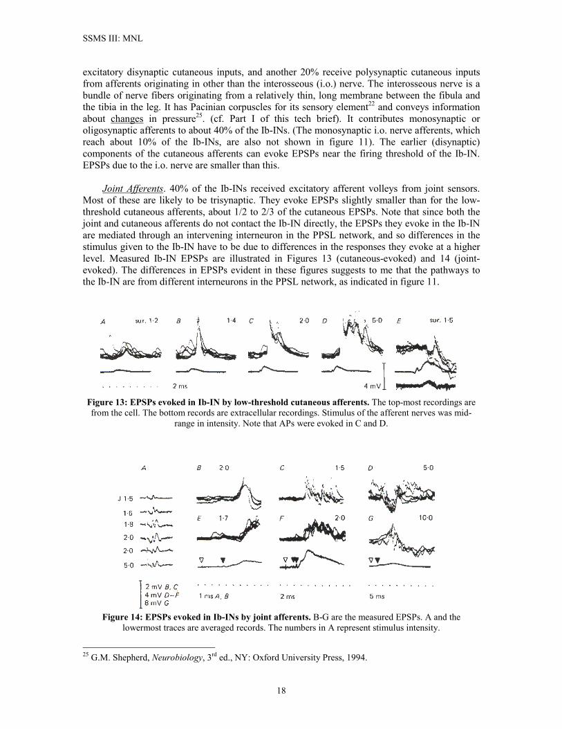
excitatory disynaptic cutaneous inputs, and another 20% receive polysynaptic cutaneous inputs from afferents originating in other than the interosseous (i.o.) nerve. The interosseous nerve is a bundle of nerve fibers originating from a relatively thin, long membrane between the fibula and the tibia in the leg. It has Pacinian corpuscles for its sensory element22 and conveys information about changes in pressure25. (cf. Part I of this tech brief). It contributes monosynaptic or oligosynaptic afferents to about 40% of the Ib-INs. (The monosynaptic i.o. nerve afferents, which reach about 10% of the Ib-INs, are also not shown in figure 11). The earlier (disynaptic) components of the cutaneous afferents can evoke EPSPs near the firing threshold of the Ib-IN. EPSPs due to the i.o. nerve are smaller than this. Joint Afferents. 40% of the Ib-INs received excitatory afferent volleys from joint sensors. Most of these are likely to be trisynaptic. They evoke EPSPs slightly smaller than for the low-threshold cutaneous afferents, about 1/2 to 2/3 of the cutaneous EPSPs. Note that since both the joint and cutaneous afferents do not contact the Ib-IN directly, the EPSPs they evoke in the Ib-IN are mediated through an intervening interneuron in the PPSL network, and so differences in the stimulus given to the Ib-IN have to be due to differences in the responses they evoke at a higher level. Measured Ib-IN EPSPs are illustrated in Figures 13 (cutaneous-evoked) and 14 (joint-evoked). The differences in EPSPs evident in these figures suggests to me that the pathways to the Ib-IN are from different interneurons in the PPSL network, as indicated in figure 11.
Figure 13: EPSPs evoked in Ib-IN by low-threshold cutaneous afferents. The top-most recordings are from the cell. The bottom records are extracellular recordings. Stimulus of the afferent nerves was mid-
range in intensity. Note that APs were evoked in C and D.
Figure 14: EPSPs evoked in Ib-INs by joint afferents. B-G are the measured EPSPs. A and the
lowermost traces are averaged records. The numbers in A represent stimulus intensity.
25 G.M. Shepherd, Neurobiology, 3rd ed., NY: Oxford University Press, 1994.
18
SSMS III: MNL
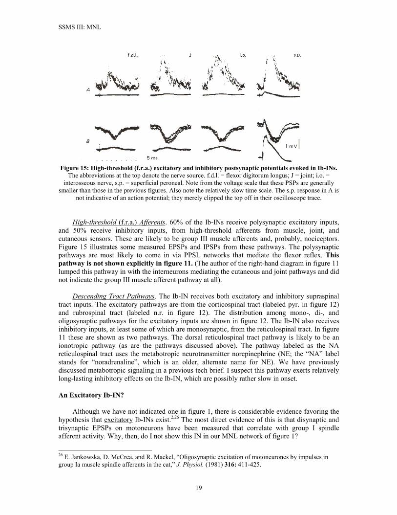
Figure 15: High-threshold (f.r.a.) excitatory and inhibitory postsynaptic potentials evoked in Ib-INs.
The abbreviations at the top denote the nerve source. f.d.l. = flexor digitorum longus; J = joint; i.o. = interosseous nerve, s.p. = superficial peroneal. Note from the voltage scale that these PSPs are generally
smaller than those in the previous figures. Also note the relatively slow time scale. The s.p. response in A is not indicative of an action potential; they merely clipped the top off in their oscilloscope trace.
High-threshold (f.r.a.) Afferents. 60% of the Ib-INs receive polysynaptic excitatory inputs, and 50% receive inhibitory inputs, from high-threshold afferents from muscle, joint, and cutaneous sensors. These are likely to be group III muscle afferents and, probably, nociceptors. Figure 15 illustrates some measured EPSPs and IPSPs from these pathways. The polysynaptic pathways are most likely to come in via PPSL networks that mediate the flexor reflex. This pathway is not shown explicitly in figure 11. (The author of the right-hand diagram in figure 11 lumped this pathway in with the interneurons mediating the cutaneous and joint pathways and did not indicate the group III muscle afferent pathway at all). Descending Tract Pathways. The Ib-IN receives both excitatory and inhibitory supraspinal tract inputs. The excitatory pathways are from the corticospinal tract (labeled pyr. in figure 12) and rubrospinal tract (labeled n.r. in figure 12). The distribution among mono-, di-, and oligosynaptic pathways for the excitatory inputs are shown in figure 12. The Ib-IN also receives inhibitory inputs, at least some of which are monosynaptic, from the reticulospinal tract. In figure 11 these are shown as two pathways. The dorsal reticulospinal tract pathway is likely to be an ionotropic pathway (as are the pathways discussed above). The pathway labeled as the NA reticulospinal tract uses the metabotropic neurotransmitter norepinephrine (NE; the “NA” label stands for “noradrenaline”, which is an older, alternate name for NE). We have previously discussed metabotropic signaling in a previous tech brief. I suspect this pathway exerts relatively long-lasting inhibitory effects on the Ib-IN, which are possibly rather slow in onset. An Excitatory Ib-IN? Although we have not indicated one in figure 1, there is considerable evidence favoring the hypothesis that excitatory Ib-INs exist.2,26 The most direct evidence of this is that disynaptic and trisynaptic EPSPs on motoneurons have been measured that correlate with group I spindle afferent activity. Why, then, do I not show this IN in our MNL network of figure 1?
26 E. Jankowska, D. McCrea, and R. Mackel, “Oligosynaptic excitation of motoneurones by impulses in group Ia muscle spindle afferents in the cat,” J. Physiol. (1981) 316: 411-425.
19
SSMS III: MNL
There are two reasons I omit this putative IN from our network. First, the IN in question has not actually been identified (so far as I know; if someone has found it, they have done so more recently than 1992). The second, and principal, reason is that there is some evidence that the “Ib-EIN” might in fact be the same type of neuron as the group II ventral horn neuron. I discuss this evidence in the next section. This is why I have left it out of our MNL network. Since there is evidence that this “Ib-EIN” does exist and since it is possible that it might be a unique type of neuron, the Ib-EIN is an option available to our EC algorithms should we find one to be necessary.27 The action I call “Ib-EIN” appears to project preferentially to MNs that are antagonists of flexor muscles2. There is some evidence that they may also project from flexors to extensors, but this would be to a lesser extent. I have seen no evidence that suggests this pathway plays any significant role in pure reflexes, and so if it has any significant role at all, it would have to be in locomotion – suggesting that the higher-level network of the PPSL must “enable” this pathway. The Group II Interneurons IIVH-EIN, IIVH-IIN In contrast to the putative character of the unidentified “Ib-EIN”, neurons that Jankowska2 termed “group II ventral horn interneurons” have been identified and to some extent characterized. The name “group II” applied to an IN is meant to indicate that group II spindle afferents constitute the principal data signal pathways into the IN. However, as we are about to see, some of these ventral horn INs in fact respond more strongly to group I afferents than to group II afferents.28 These neurons are found in laminae VII and VIII of the ventral horn in mid- and lower-lumbar spinal segments. Those in lumbar segments L3, L4, and L5 are known to project directly to �-MNs in lumbar segment L7 and the first sacral segments of the spinal cord.29 These INs should technically be regarded as propriospinal interneurons (P-INs) because of their long projections spanning two or more spinal segments. (This terminology is not to be confused with our “PPSL” terminology; the IIVH-INs functionally belong to the MN level of our neural network scheme). Other IIVH-INs that appear to be of the same type as these “P-IN IIVH-INs” have been found in lumbar segments where their projection is non-propriospinal (i.e. they project to more local motor nuclei). Both excitatory and inhibitory IIVH-INs have been identified.2 Except for their effect on motoneurons, there appears to be little difference between IIVH-EINs and IIVH-IINs insofar as their input distributions, EPSP and IPSP responses, and other network-level characteristics are concerned. In one important respect IIVH-EINs do appear to differ from IIVH-IINs, namely that IIVH-EINs are known to project to �-MNs, whereas IIVH-IINs do not. If there is one “theme” that most aptly describes the role of IIVH-INs, it is “convergence.” These INs take in signal pathways from very wide-ranging sources: different muscle groups, cutaneous and joint afferents, and descending tract pathways involving multiple spinal segments.
27 If we do find one to be necessary, this would make something of a splash in the neurocomputing journals. 28 S.A. Edgley and E. Jankowska, “An interneuronal relay for group I and II muscle afferents in the midlumbar segments of the cat spinal cord”, J. Physiol. (1987), 389: 647-674. 29 The cat does not have the same number of spinal segments, nor the same groupings of spinal segments, as the human being. That’s why we can talk about segment L7 in the cat, whereas humans have only 5 lumbar segments.
20
SSMS III: MNL
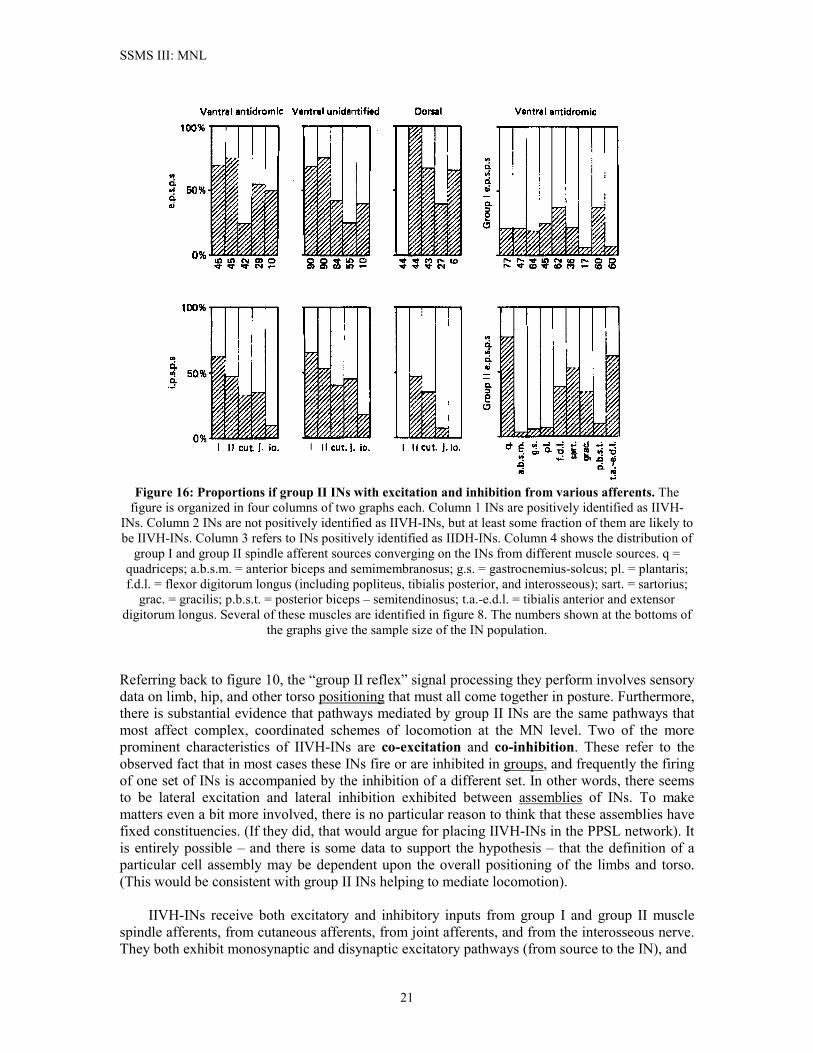
Figure 16: Proportions if group II INs with excitation and inhibition from various afferents. The
figure is organized in four columns of two graphs each. Column 1 INs are positively identified as IIVH-INs. Column 2 INs are not positively identified as IIVH-INs, but at least some fraction of them are likely to be IIVH-INs. Column 3 refers to INs positively identified as IIDH-INs. Column 4 shows the distribution of
group I and group II spindle afferent sources converging on the INs from different muscle sources. q = quadriceps; a.b.s.m. = anterior biceps and semimembranosus; g.s. = gastrocnemius-solcus; pl. = plantaris; f.d.l. = flexor digitorum longus (including popliteus, tibialis posterior, and interosseous); sart. = sartorius;
grac. = gracilis; p.b.s.t. = posterior biceps – semitendinosus; t.a.-e.d.l. = tibialis anterior and extensor digitorum longus. Several of these muscles are identified in figure 8. The numbers shown at the bottoms of
the graphs give the sample size of the IN population. Referring back to figure 10, the “group II reflex” signal processing they perform involves sensory data on limb, hip, and other torso positioning that must all come together in posture. Furthermore, there is substantial evidence that pathways mediated by group II INs are the same pathways that most affect complex, coordinated schemes of locomotion at the MN level. Two of the more prominent characteristics of IIVH-INs are co-excitation and co-inhibition. These refer to the observed fact that in most cases these INs fire or are inhibited in groups, and frequently the firing of one set of INs is accompanied by the inhibition of a different set. In other words, there seems to be lateral excitation and lateral inhibition exhibited between assemblies of INs. To make matters even a bit more involved, there is no particular reason to think that these assemblies have fixed constituencies. (If they did, that would argue for placing IIVH-INs in the PPSL network). It is entirely possible – and there is some data to support the hypothesis – that the definition of a particular cell assembly may be dependent upon the overall positioning of the limbs and torso. (This would be consistent with group II INs helping to mediate locomotion). IIVH-INs receive both excitatory and inhibitory inputs from group I and group II muscle spindle afferents, from cutaneous afferents, from joint afferents, and from the interosseous nerve. They both exhibit monosynaptic and disynaptic excitatory pathways (from source to the IN), and
21
SSMS III: MNL
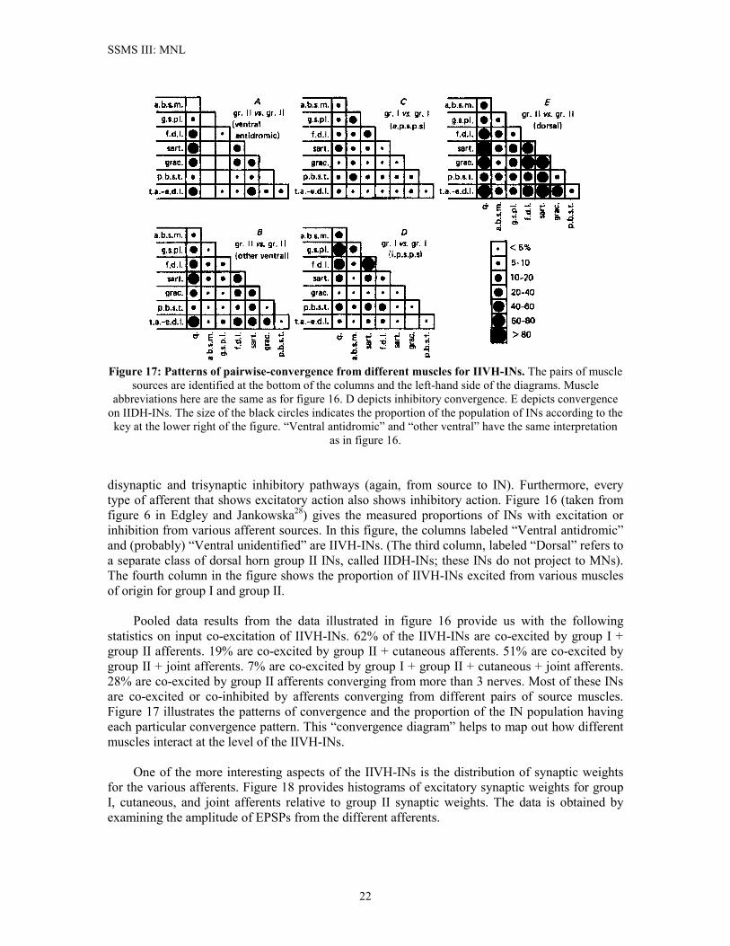
Figure 17: Patterns of pairwise-convergence from different muscles for IIVH-INs. The pairs of muscle
sources are identified at the bottom of the columns and the left-hand side of the diagrams. Muscle abbreviations here are the same as for figure 16. D depicts inhibitory convergence. E depicts convergence
on IIDH-INs. The size of the black circles indicates the proportion of the population of INs according to the key at the lower right of the figure. “Ventral antidromic” and “other ventral” have the same interpretation
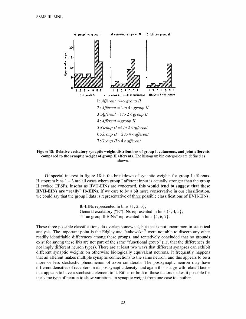
as in figure 16. disynaptic and trisynaptic inhibitory pathways (again, from source to IN). Furthermore, every type of afferent that shows excitatory action also shows inhibitory action. Figure 16 (taken from figure 6 in Edgley and Jankowska28) gives the measured proportions of INs with excitation or inhibition from various afferent sources. In this figure, the columns labeled “Ventral antidromic” and (probably) “Ventral unidentified” are IIVH-INs. (The third column, labeled “Dorsal” refers to a separate class of dorsal horn group II INs, called IIDH-INs; these INs do not project to MNs). The fourth column in the figure shows the proportion of IIVH-INs excited from various muscles of origin for group I and group II. Pooled data results from the data illustrated in figure 16 provide us with the following statistics on input co-excitation of IIVH-INs. 62% of the IIVH-INs are co-excited by group I + group II afferents. 19% are co-excited by group II + cutaneous afferents. 51% are co-excited by group II + joint afferents. 7% are co-excited by group I + group II + cutaneous + joint afferents. 28% are co-excited by group II afferents converging from more than 3 nerves. Most of these INs are co-excited or co-inhibited by afferents converging from different pairs of source muscles. Figure 17 illustrates the patterns of convergence and the proportion of the IN population having each particular convergence pattern. This “convergence diagram” helps to map out how different muscles interact at the level of the IIVH-INs. One of the more interesting aspects of the IIVH-INs is the distribution of synaptic weights for the various afferents. Figure 18 provides histograms of excitatory synaptic weights for group I, cutaneous, and joint afferents relative to group II synaptic weights. The data is obtained by examining the amplitude of EPSPs from the different afferents.
22
SSMS III: MNL
afferentIIGroupafferenttoIIGroupafferenttoIIGroup
IIgroupAfferentIIgrouptoAfferentIIgrouptoAfferent
IIgroupAfferent
��
��
��
�
��
��
��
4:742:621:5
:421:342:2
4:1
Figure 18: Relative excitatory synaptic weight distributions of group I, cutaneous, and joint afferents
compared to the synaptic weight of group II afferents. The histogram bin categories are defined as shown.
Of special interest in figure 18 is the breakdown of synaptic weights for group I afferents. Histogram bins 1 – 3 are all cases where group I afferent input is actually stronger than the group II evoked EPSPs. Insofar as IIVH-EINs are concerned, this would tend to suggest that these IIVH-EINs are “really” Ib-EINs. If we care to be a bit more conservative in our classification, we could say that the group I data is representative of three possible classifications of IIVH-EINs: Ib-EINs represented in bins {1, 2, 3}; General excitatory (“E”) INs represented in bins {3, 4, 5}; “True group II EINs” represented in bins {5, 6, 7}. These three possible classifications do overlap somewhat, but that is not uncommon in statistical analysis. The important point is the Edgley and Jankowska28 were not able to discern any other readily identifiable differences among these groups, and tentatively concluded that no grounds exist for saying these INs are not part of the same “functional group” (i.e. that the differences do not imply different neuron types). There are at least two ways that different synapses can exhibit different synaptic weights on otherwise biologically equivalent neurons. It frequently happens that an afferent makes multiple synaptic connections to the same neuron, and this appears to be a more or less stochastic phenomenon of axon collaterals. The postsynaptic neuron may have different densities of receptors in its postsynaptic density, and again this is a growth-related factor that appears to have a stochastic element to it. Either or both of these factors makes it possible for the same type of neuron to show variations in synaptic weight from one case to another.
23
SSMS III: MNL
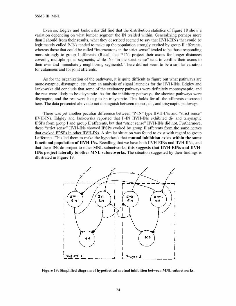
Even so, Edgley and Jankowska did find that the distribution statistics of figure 18 show a variation depending on what lumbar segment the IN resided within. Generalizing perhaps more than I should from their results, what they described seemed to say that IIVH-EINs that could be legitimately called P-INs tended to make up the population strongly excited by group II afferents, whereas those that could be called “interneurons in the strict sense” tended to be those responding more strongly to group I afferents. (Recall that P-INs project their axons for longer distances covering multiple spinal segments, while INs “in the strict sense” tend to confine their axons to their own and immediately neighboring segments). There did not seem to be a similar variation for cutaneous and for joint afferents. As for the organization of the pathways, it is quite difficult to figure out what pathways are monosynaptic, disynaptic, etc. from an analysis of signal latencies for the IIVH-INs. Edgley and Jankowska did conclude that some of the excitatory pathways were definitely monosynaptic, and the rest were likely to be disynaptic. As for the inhibitory pathways, the shortest pathways were disynaptic, and the rest were likely to be trisynaptic. This holds for all the afferents discussed here. The data presented above do not distinguish between mono-, di-, and trisynaptic pathways. There was yet another peculiar difference between “P-IN” type IIVH-INs and “strict sense” IIVH-INs. Edgley and Jankowska reported that P-IN IIVH-INs exhibited di- and trisynaptic IPSPs from group I and group II afferents, but that “strict sense” IIVH-INs did not. Furthermore, these “strict sense” IIVH-INs showed IPSPs evoked by group II afferents from the same nerves that evoked EPSPs in other IIVH-INs. A similar situation was found to exist with regard to group I afferents. This led them to make the hypothesis that mutual inhibition exists within the same functional population of IIVH-INs. Recalling that we have both IIVH-EINs and IIVH-IINs, and that these INs do project to other MNL subnetworks, this suggests that IIVH-EINs and IIVH-IINs project laterally to other MNL subnetworks. The situation suggested by their findings is illustrated in Figure 19.
Figure 19: Simplified diagram of hypothetical mutual inhibition between MNL subnetworks.
24
SSMS III: MNL
In neural network terminology, a cell assembly is a group of tightly-interconnected neurons such that when one neuron in the assembly fires it tends to induce the other neurons in the assembly to also fire. Cell assemblies can be embedded in the midst of many other cells that are not themselves members of the assembly. Cell assemblies are one putative topological network organization that signals “associations” of input signal vectors by having all the cells in the assembly begin to fire when the “associated” pattern of inputs is presented.30 There is some reason to think that MNL subnetworks might be organized in this fashion (for example, through cross-connections to other MNL subnetworks; figures 4 and 19 could be viewed as examples of “inhibitory cell assemblies”). Edgley and Jankowska reported that stimulation of only one or two locations within a motor nucleus were most likely to produce activation of only a small proportion of the IIVH-INs projecting into the MN pools. MN action thus stimulated was “weak” in the sense that the MN did not receive sufficient stimulus to excite it into a state of high-activity in the form of higher-frequency action potential trains. The obvious corollary of this is that simultaneous excitation by multiple afferents is likely to produce widespread activation of populations of INs. Edgley and Jankowska did not investigate this in their paper28, but the presence of widespread co-excitation of different IIVH-INs by the same afferent would seem to make this sort of behavior probable. “Association” by cell assemblies is one theoretically possible means by which the SSMS would be able to “know” how to initiate the appropriate MN response required under conditions of a particular postural or locomotive-positional state of the body. This would be in keeping with the putative role of group II INs as the major integration sites for body position information. Summary Discussion Part III has presented the facts regarding the types of neurons present in MNL subnetworks, and provided some experimental data on their interconnections, afferents, and input distributions. The statistical data especially might prove to be useful in coming up with EC-based algorithms for our PCNNs insofar as the bipedal locomotion application is concerned. Hopefully the review presented here begins to shed some light on the functional advantage of having separate excitatory and inhibitory classes of interneurons. In conventional artificial neural networks, any given artificial neuron can be excitatory for one of its targets and at the same time inhibitory for another of its targets. This means it cannot excite the one without also inhibiting the other and vice versa. In the SSMS, where we have many signal pathways acting as “gates” or “controls”, having separate excitatory and inhibitory INs allows a given subnetwork to inhibit neurons in a different subnetwork without necessarily having to also excite other targets. This gives the subnetworks greater flexibility and a finer degree of “tune-ability” in carrying out complicated muscle coordination tasks. It is my guess that this is the property of SSMS networks that permits them to act as VSSC systems. This still leaves us with some important and unanswered topological questions, many of which are likely to be laid at the doorstep of our EC techniques. I have talked about lateral connections between MNL subnetworks and about co-excitation/ co-inhibition of cell groups. The specifics of these lateral connections are not presently known in detail. We can make use of the interconnection statistics given in this brief to form some kind of handle on this, and we can probably come up with somewhat close first guesses for which subnetworks are interconnected by examining the muscle organization (e.g. figure 8). I think this is certainly worth doing. But, in
30 J.A. Anderson, An Introduction to Neural Networks, Cambridge, MA: The MIT Press, 1996, pp. 281-292.
25
SSMS III: MNL
26
the end, I think it is likely that we’ll have to rely on our EC methods (and perhaps develop some new ones) to come up with all the details of what lateral connections it will take to produce bipedal locomotion. One other point is probably worth mentioning. In the biological system, the neurons tend to be converged upon by great numbers of inputs, yet each input taken individually rarely has enough synaptic strength to fire the postsynaptic cell without the cooperation of many, many other inputs. Is this in any way advantageous? Here I’m not talking about the biological system. (The cat has survived down through the ages, so we can presume its SSMS scheme possesses some kind of survival advantage). Instead I’m reflecting here in more general terms. When we consider the kind of dynamical path-switching that seems to characterize the operation of the various networks in the system, where sometimes an afferent pathway is gated-in and sometimes it is inhibited by other control signals, then it seems to me that the functional advantage that might be gained from the “many relatively-weak-input” scheme that nature has hit upon is this: Extraordinarily fine control and discrimination in how the system will react in different movement and posture situations. Much of this pathway control has been assigned to the higher PPSL network. While I think it may be possible and useful to carry out network design at the MNL aimed at producing the basic reflex responses, as a first approximation of this level of the system, it is nonetheless the case that we require some information about the PPSL network in order to understand locomotion control. That is the topic of Part IV.


