SPINAL CORD ANATOMY AND FUNCTION
86
SPINAL CORD ANATOMY AND SPINAL CORD ANATOMY AND FUNCTION FUNCTION DANIL HAMMOUDI.MD
Transcript of SPINAL CORD ANATOMY AND FUNCTION

SPINAL CORD ANATOMY AND SPINAL CORD ANATOMY AND FUNCTIONFUNCTIONDANIL HAMMOUDI.MD


SPINAL CORD ANATOMYSPINAL CORD ANATOMY

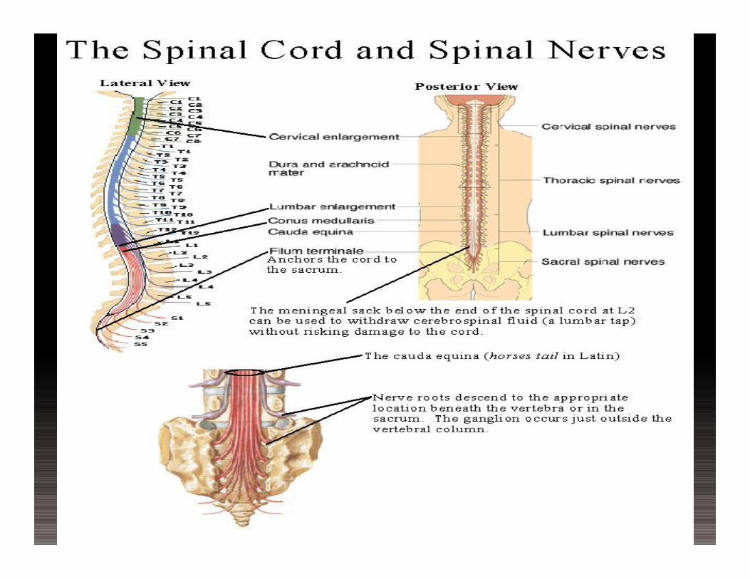
Lumbar TapLumbar Tap
Figure 12.30

Spinal CordSpinal Cord
Figure 12.29a

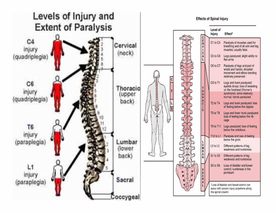
There are 31 spinal cord segments:•8 cervical segments•12 thoracic segments•5 lumbar segments•5 sacral segments•1 coccygeal segment
There are two regions where the spinal cord enlarges:•Cervical enlargement - corresponds roughly to the brachial plexus nerves, which innervate the upper limb. It includes spinal cord segments from about C4 to T1. The vertebral levels of the enlargement are roughly the same (C4 to T1).
•Lumbosacral enlargement - corresponds to the lumbosacral plexus nerves, which innervate the lower limb. It comprises the spinal cord segments from L2 to S3, and is found about the vertebral levels of T9 to T12.

Spinal CordSpinal Cord
Conus medullaris – terminal portion of the spinal cord
Filum terminale – fibrous extension of the pia mater; anchors the spinal cord to the coccyx
Denticulate ligaments – delicate shelves of pia mater; attach the spinal cord to the vertebrae

The dermatomes are somatic or musculocutaneous areas served by fibers from specific spinal nerves.
Referred pain is caused when the sensory fibers from an internal organ enter the spinal cord in the same root as fibers from a dermatome. The brain is poor at interpreting visceral pain and instead interprets it as pain from the somatic area of the dermatome.

Cervical Plexus - the phrenic nerve travels through the thorax to innervate the diaphragm.
Brachial Plexus -Axillary nerve - innervates the deltoid muscle and shoulder, along with the posterior aspect of the upper arm.Musculocutaneous nerve - innervates anterior skin of upper arm and elbow flexors.Radial nerve - innervates dorsal aspect of the arm and extensors of the elbow, wrist, and fingers, abduction of thumb. Median nerve - innervates the middle elbow, wrist and finger flexors, adducts the thumb.Ulnar nerve - innervates the medial aspect wrist and finger flexors.
Lumbar Plexusgenitofemoral - to the external genitaliaobturator - to the adductor musclesfemoral - innervates the skin and muscles of upper thigh, including the quadriceps.
Sacral Plexus gluteal nerves (superior and inferior) - superior innervates the gluteus medius and minimus, inferior innervates the gluteus maximus.sciatic nerve - the body's largest nerve, consisting of two major branches, the tibial and common peroneal. Together they innervate most all of leg including the flexors of the knee, part of adductor magnus, muscles for plantar flexion, dorsiflexion, and other movements of the foot and toes.

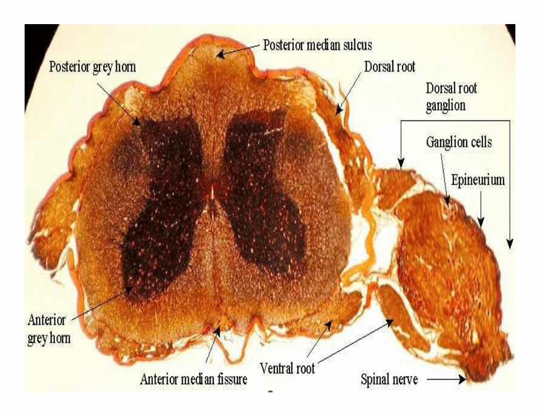
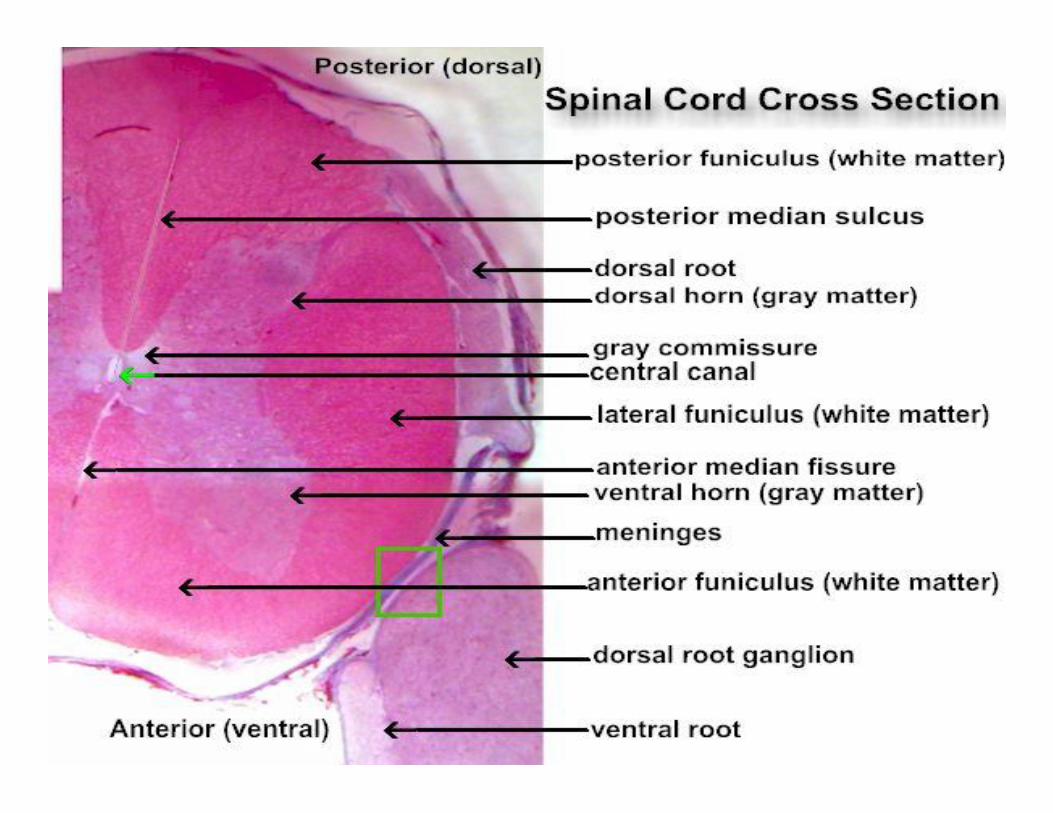
ganglion - a collection of cell bodies located outside the Central Nervous System. The spinal ganglia or dorsal root ganglia contain the cell bodies of sensory neurons entering the cord at that region.
nerve - a group of fibers (axons) outside the CNS. The spinal nerves contain the fibers of the sensory and motor neurons. A nerve does not contain cell bodies. They are located in the ganglion (sensory) or in the gray matter (motor).
tract - a group of fibers inside the CNS. The spinal tracts carry information up or down the spinal cord, to or from the brain. Tracts within the brain carry information from one place to another within the brain. Tracts are always part of white matter.
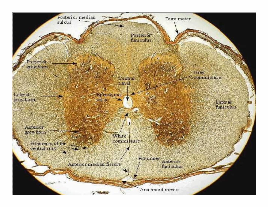
gray matter - an area of unmyelinated neurons where cell bodies and synapses occur. In the spinal cord the synapses between sensory and motor and interneurons occurs in the gray matter. The cell bodies of the interneurons and motor neurons also are found in the gray matter.
white matter - an area of myelinated fiber tracts. Myelination in the CNS differs from that in nerves.

The spinal cord proper begins at the level of the foramen magnum of the skull and ends at the level of the L1ÐL2 intervertebral joint


MyelinatedMyelinatedfibersfibers


a pia mater b subarachnoid space c dura mater d myelinated axon e unipolar neuron of the dorsal root ganglion surrounded by satellite cells (neuroglia).

a Pia mater b Subarachnoid space filled with cerebral spinal fluid, wastes and various cells. c Fibrocyte mixed in the blue collagen fibers of the dura mater. d Nucleus & nucleolus of unipolar neuron e Nucleus of one of many tiny satellite cells surrounding the large unipolar neuron. f Myelinated axon g Node of Ranvier h Nucleus of white Schwann cell

a Synaptic bulbs over the motor end plate - neuromuscular junction b Neuron axon terminal - black fibers

The central canal is the cerebrospinal fluid-filled space that runs longitudinally through the length of the entire spinal cord. The central canal is contiguous with the ventricular system of the brain.

PNS in the Nervous SystemPNS in the Nervous SystemFigure 13.1


Scheuermann kyphosisIn some rare cases, the bones of the back (vertebrae) do not grow correctly. In Scheuermann kyphosis, the front part of the vertebrae does not grow as well as the back part.

Structure of a NerveStructure of a Nerve
Nerve – cordlike organ of the PNS consisting of peripheral axons enclosed by connective tissue
Connective tissue coverings include: Endoneurium – loose connective tissue that
surrounds axons Perineurium – coarse connective tissue that
bundles fibers into fascicles Epineurium – tough fibrous sheath around a nerve


Structure of a NerveStructure of a Nerve
Figure 13.3b

Classification of NervesClassification of Nerves
Sensory and motor divisions Sensory (afferent) – carry impulse to the CNS Motor (efferent) – carry impulses from CNS Mixed – sensory and motor fibers carry
impulses to and from CNS; most common type of nerve

Peripheral NervesPeripheral Nerves
Mixed nerves – carry somatic and autonomic (visceral) impulses
The four types of mixed nerves are: Somatic afferent and somatic efferent Visceral afferent and visceral efferent
Peripheral nerves originate from the brain or spinal column
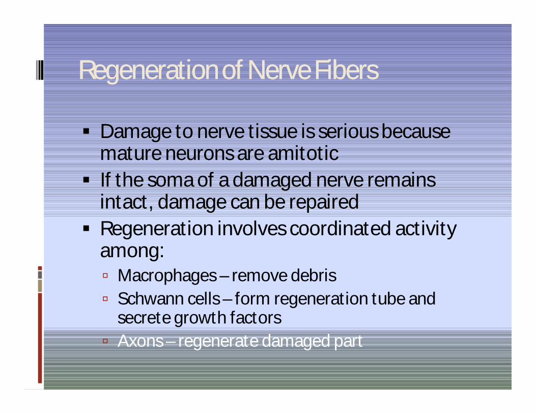
Regeneration of Nerve FibersRegeneration of Nerve Fibers
Damage to nerve tissue is serious because mature neurons are amitotic
If the soma of a damaged nerve remains intact, damage can be repaired
Regeneration involves coordinated activity among: Macrophages – remove debris Schwann cells – form regeneration tube and
secrete growth factors Axons – regenerate damaged part
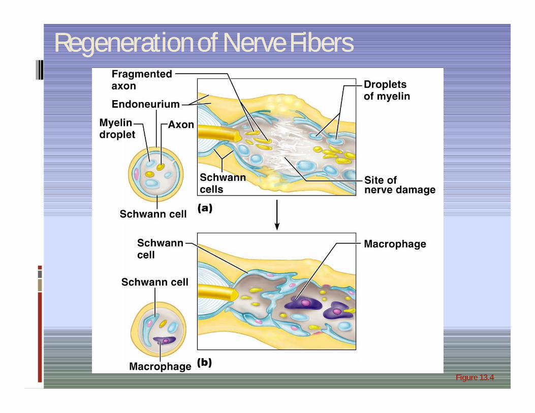
Regeneration of Nerve FibersRegeneration of Nerve Fibers
Figure 13.4

Regeneration of Nerve FibersRegeneration of Nerve Fibers
Figure 13.4

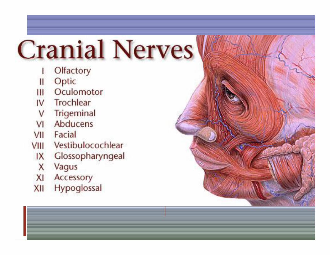
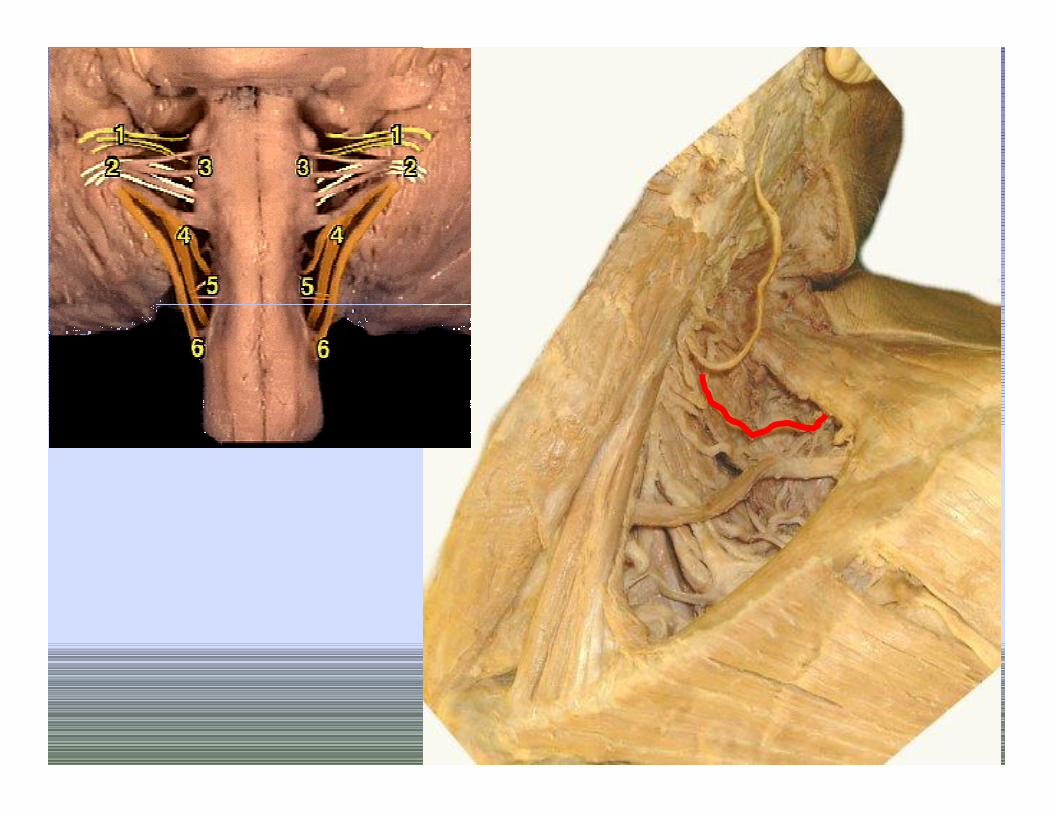
Cranial NervesCranial Nerves
Twelve pairs of cranial nerves arise from the brain
They have sensory, motor, or both sensory and motor functions
Each nerve is identified by a number (I through XII) and a name
Four cranial nerves carry parasympathetic fibers that serve muscles and glands

Peripheral Nervous SystemPeripheral Nervous System
31 spinal nerves We’ve already discussed
their structure
12 cranial nerves How do they differ from
spinal nerves? We need to learn their: Names Locations Functions

Cranial NervesCranial Nerves
Figure 13.5a

Summary of Function of Cranial NervesSummary of Function of Cranial Nerves
Figure 13.5b

12 Cranial Nerves12 Cranial Nerves
How do you remember which nerve is which number? Here is a G-rated mnemonic devices:
Old Opie occasionally tries trigonometry and feels very gloomy, vague, and hypoactive.
There are also several R-rated ones
Some cranial nerves are sensory, some motor, and some are both (mixed)? Some say marry money but my
brother says big butts matter more.


Cranial Nerve I: OlfactoryCranial Nerve I: Olfactory
Arises from the olfactory epithelium Passes through the cribriform plate of the
ethmoid bone Fibers run through the olfactory bulb and
terminate in the primary olfactory cortex Functions solely by carrying afferent impulses
for the sense of smell

Cranial Nerve I: OlfactoryCranial Nerve I: Olfactory
Figure I from Table 13.2
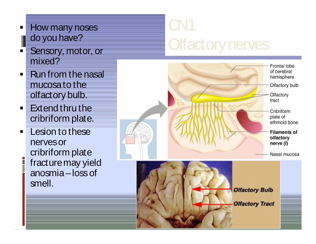
CN1 CN1 Olfactory nervesOlfactory nerves
How many noses do you have?
Sensory, motor, or mixed?
Run from the nasal mucosa to the olfactory bulb.
Extend thru the cribriform plate.
Lesion to these nerves or cribriform plate fracture may yield anosmia – loss of smell.

Cranial Nerve II: OpticCranial Nerve II: Optic
Figure II from Table 13.2

CN2 CN2 Optic NervesOptic Nerves
How many eyes do you have?
Sensory, motor, or mixed?
Begin at the retina, run to the optic chiasm, cross over, continue as the optic tract and synapse in the thalamus.
Optic nerve damage yields blindness in the eye served by the nerve. Optic tract damage yields partial visual loss.
Visual defects = anopsias


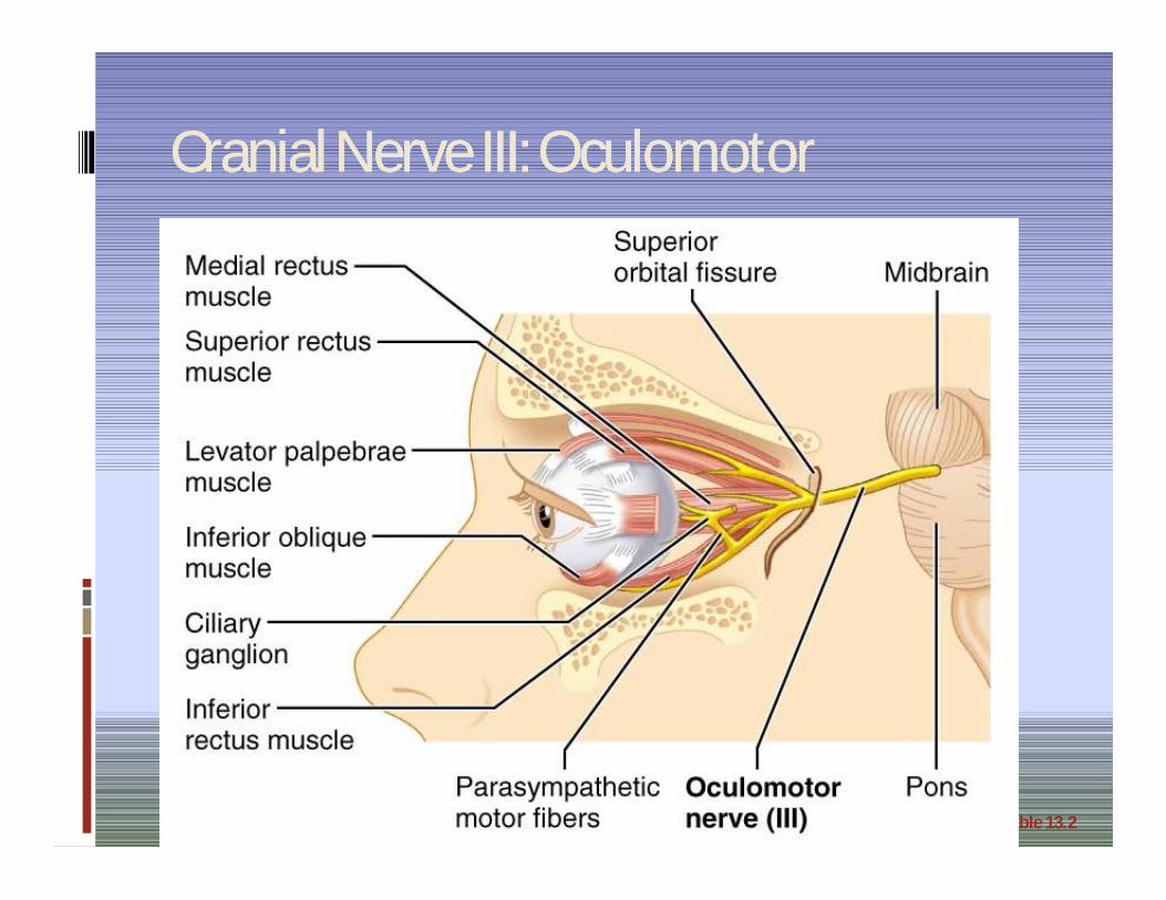
Cranial Nerve III: OculomotorCranial Nerve III: Oculomotor
Fibers extend from the ventral midbrain, pass through the superior orbital fissure, and go to the extrinsic eye muscles
Functions in raising the eyelid, directing the eyeball, constricting the iris, and controlling lens shape
Parasympathetic cell bodies are in the ciliary ganglia
Cranial Nerve III: OculomotorCranial Nerve III: Oculomotor
Figure III from Table 13.2
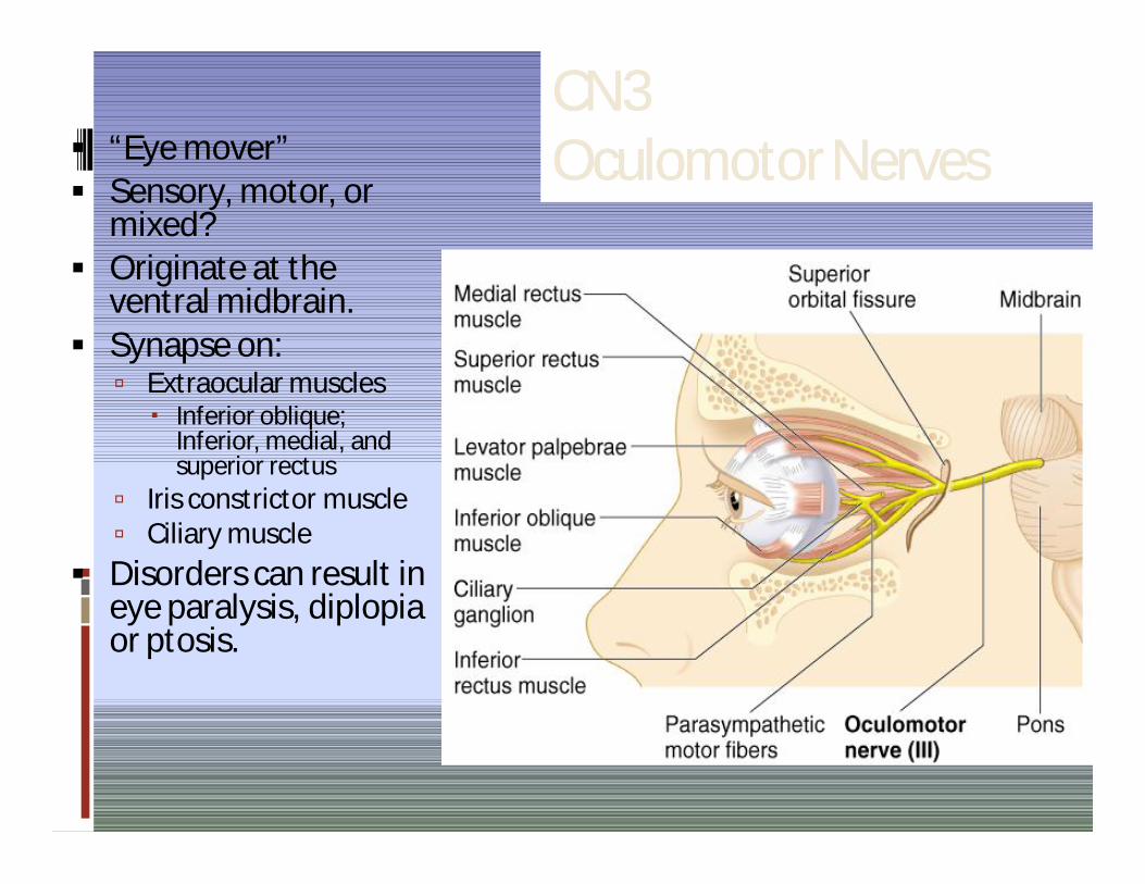
CN3 CN3 Oculomotor NervesOculomotor Nerves “Eye mover”
Sensory, motor, or mixed?
Originate at the ventral midbrain.
Synapse on: Extraocular muscles
Inferior oblique; Inferior, medial, and superior rectus
Iris constrictor muscle Ciliary muscle
Disorders can result in eye paralysis, diplopiaor ptosis.

Cranial Nerve IV: TrochlearCranial Nerve IV: Trochlear
Fibers emerge from the dorsal midbrain and enter the orbits via the superior orbital fissures; innervate the superior oblique muscle
Primarily a motor nerve that directs the eyeball

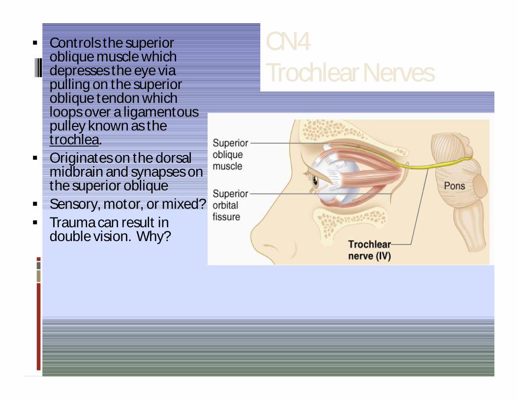
CN4 CN4 Trochlear NervesTrochlear Nerves
Controls the superior oblique muscle which depresses the eye via pulling on the superior oblique tendon which loops over a ligamentouspulley known as the trochlea.
Originates on the dorsal midbrain and synapses on the superior oblique
Sensory, motor, or mixed? Trauma can result in
double vision. Why?

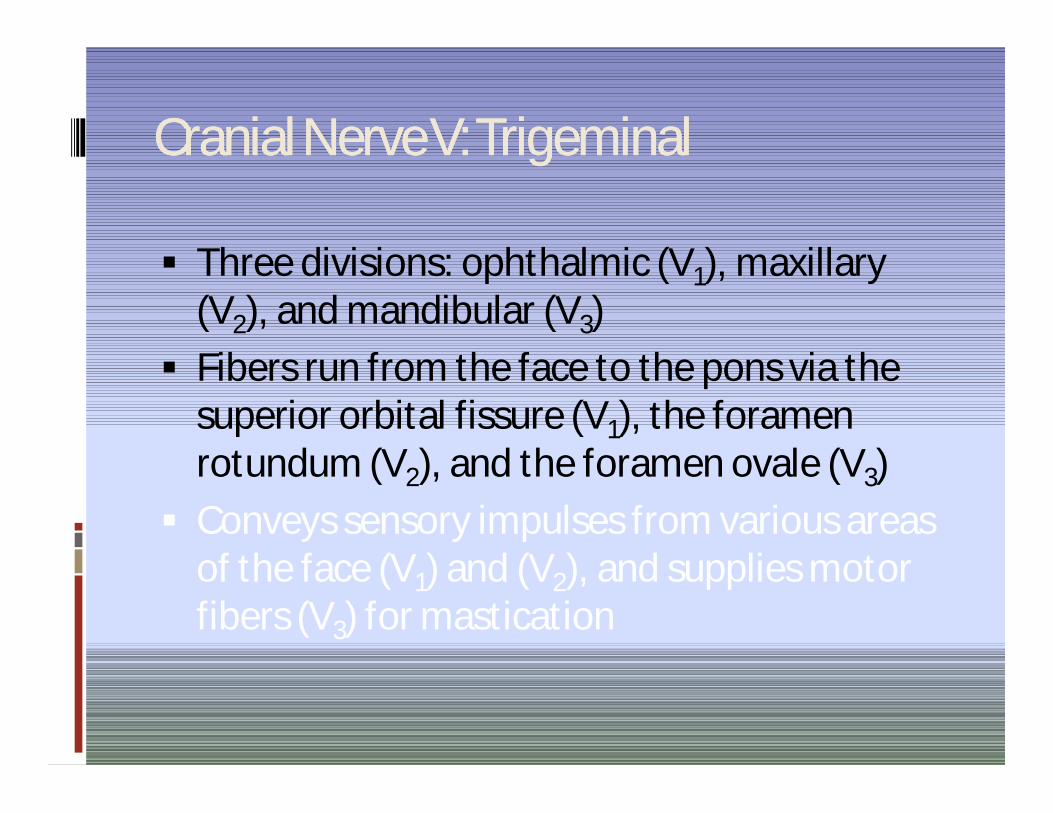
Cranial Nerve V: TrigeminalCranial Nerve V: Trigeminal
Three divisions: ophthalmic (V1), maxillary (V2), and mandibular (V3)
Fibers run from the face to the pons via the superior orbital fissure (V1), the foramen rotundum (V2), and the foramen ovale (V3)
Conveys sensory impulses from various areas of the face (V1) and (V2), and supplies motor fibers (V3) for mastication

Cranial Nerve V: TrigeminalCranial Nerve V: Trigeminal
Figure V from Table 13.2

Sensory, motor, or mixed? Biggest cranial nerve Originates in the pons and
eventually splits into 3 divisions: Ophthalmic (V1), Maxillary
(V2), & Mandibular (V3).
Sensory info (touch, temp., and pain) from face.
Motor info to muscles of mastication
Damage?
CN5 Trigeminal Nerves


The spinal trigeminal nucleus represents pain/temperature sensation from the face. Pain/temperature fibers from peripheral nociceptors are carried in cranial nerves V, VII, IX and X. On entering the brainstem, sensory fibers are grouped together and sent to the spinal trigeminal nucleus. This bundle of incoming fibers can be identified in cross sections of the pons and medulla as the spinal tract of the trigeminal nucleus, which parallels the spinal trigeminal nucleus itself. The spinal tract of V is analogous to, and continuous with, Lissauer’s tract in the spinal cord.

Cranial Nerve VI: AbdcuensCranial Nerve VI: Abdcuens
Fibers leave the inferior pons and enter the orbit via the superior orbital fissure
Primarily a motor nerve innervating the lateral rectus muscle
Figure VI from Table 13.2

Sensory, motor, or mixed?
Runs between inferior pons and lateral rectus.
CN6 Abducens Nerves


Cranial Nerve VII: FacialCranial Nerve VII: Facial Fibers leave the pons, travel through the internal
acoustic meatus, and emerge through the stylomastoid foramen to the lateral aspect of the face
Mixed nerve with five major branches Motor functions include facial expression, and
the transmittal of autonomic impulses to lacrimal and salivary glands
Sensory function is taste from the anterior two-thirds of the tongue

The facial nerve has four components with distinct functions:
Branchial motor(special visceral efferent)
Supplies the muscles of facial expression; posterior belly of digastric muscle; stylohyoid, and stapedius.
Visceral motor(general visceral efferent)
Parasympathetic innervation of the lcrimal, submandibular, and sublingual glands, as well as mucous membranes of nasopharynx, hard and soft palate.
Special sensory(special afferent)
Taste sensation from the anterior 2/3 of tongue; hard and soft palates.
General sensory(general somatic afferent)
General sensation from the skin of the concha of the auricle and from a small area behind the ear.


Branches of facial nerve in the face


Cranial Nerve Cranial Nerve VII: FacialVII: Facial
Figure VII from Table 13.2

CN7 CN7 Facial NervesFacial Nerves
Sensory, motor, or mixed? Originates at the pons Convey motor impulses to facial
skeletal muscles – except for chewing muscles.
Convey parasympathetic motor impulses to tear, nasal, and some salivary glands.
Convey sensory info from taste buds on anterior 2/3 of the tongue.
Facial nerve damage may yield Bell’s palsy, total ipsilateralhemifacial paralysis

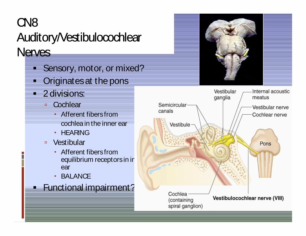
CN8 CN8 Auditory/Auditory/VestibulocochlearVestibulocochlearNervesNerves
Sensory, motor, or mixed? Originates at the pons 2 divisions: Cochlear
Afferent fibers from cochlea in the inner ear
HEARING Vestibular
Afferent fibers from equilibrium receptors in inner ear
BALANCE
Functional impairment?

CN9 CN9 GlossopharyngealGlossopharyngeal NervesNerves Sensory, motor, or mixed? Fibers run emerge from
medulla and run to the throat.
Motor Functions: Motor fibers to some
swallowing muscles Parasympathetic fibers to
some salivary glands
Sensory Functions: Taste, touch, heat from pharynx
and posterior tongue. Info from chemoreceptors on the
level of O2 and CO2 in the blood. Info from baroreceptors on BP. Chemoreceptors and
baroreceptors are located in the carotid sinus – a dilation in the internal carotid artery.

CN10 CN10 VagusVagus NervesNerves Sensory, motor, or mixed? Only cranial nerves to extend
beyond head and neck. Fibers emerge from medulla, leave
the skull, and course downwards into the thorax and abdomen.
Motor Functions: Parasympathetic efferents to the
heart, lungs, and abdominal organs.
Sensory Functions: Input from thoracic and abdominal
viscera; from baro- and chemoreceptors in the carotid sinus; from taste buds in posterior tongue and pharynx


CN11 CN11 Accessory NervesAccessory Nerves Sensory, motor, or mixed?
Formed by the union of a cranial root and a spinal root. CR arises from medulla while SR
arises from superior spinal cord. SR passes thru the FM and joins with CR to form the accessory nerve. They then leave the skull via the jugular foramen.
Cranial division then joins vagusand innervates larynx, pharynx, and soft palate.
Spinal division innervates sternocleidomastoids and trapezius.

CN12 CN12 Hypoglossal NervesHypoglossal Nerves Sensory, motor, or mixed?
Arise from the medulla and exit the skull via the hypoglossal canal and innervate the tongue.
Innervate the intrinsic & extrinsic muscles of the tongue. Swallowing, speech, food
manipulation.
Damage?
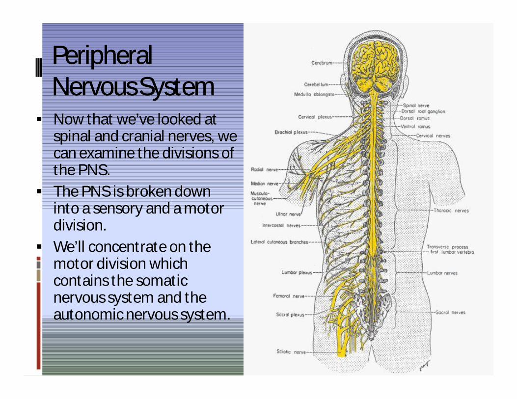
Peripheral Peripheral Nervous SystemNervous System
Now that we’ve looked at spinal and cranial nerves, we can examine the divisions of the PNS.
The PNS is broken down into a sensory and a motor division.
We’ll concentrate on the motor division which contains the somatic nervous system and the autonomic nervous system.