Spinal Bone Marrow Diseases - spinwarp.ucsd.eduspinwarp.ucsd.edu/NeuroWeb/PPT/Spine marrow.pdf ·...
87
Department of Radiology University of California San Diego John R. Hesselink, M.D. Spinal Bone Marrow Diseases
Transcript of Spinal Bone Marrow Diseases - spinwarp.ucsd.eduspinwarp.ucsd.edu/NeuroWeb/PPT/Spine marrow.pdf ·...
Department of Radiology
University of California
San Diego
John R. Hesselink, M.D.
Spinal Bone Marrow Diseases

Bone Marrow
Mineralized osseous matrix. Contributes little to marrow signal on MRI.
Hematopoietic (red) marrow. Most abundant at birth.
Children 60%
Adults 30%
Fatty marrow
MRI appearance of marrow is mostly dependant on the
relative amount of red and fatty marrow
Evelyne Fliszar, UC San Diego

Bone Marrow Composition
Red marrow (Adult): Stem cells, red blood cells, white
blood cells & platelets
40% water
20% protein
40% Fat
Fatty marrow: Fat cells
15% water
5% protein
80% Fat
The different proportions will influence the MR appearance of the marrow.
Cellularity of red marrow varies with age, is highest in infants,
and decreases with conversion to fatty marrow.
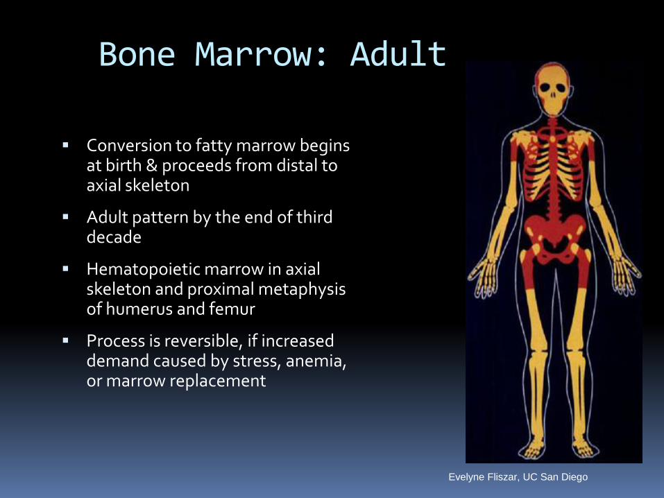
Bone Marrow: Adult
Conversion to fatty marrow begins at birth & proceeds from distal to axial skeleton
Adult pattern by the end of third decade
Hematopoietic marrow in axial skeleton and proximal metaphysis of humerus and femur
Process is reversible, if increased demand caused by stress, anemia, or marrow replacement
Evelyne Fliszar, UC San Diego

History: Newborn male with a
mass on the lower back
624
T2 SE
T1 FSE

27 month old Girl
T2 FSE
T1 FSE
12 month old Girl
T2 FSE
T1 FSE

23 month old Boy
T2 FSE
T1 FSE T1-Gd-FS
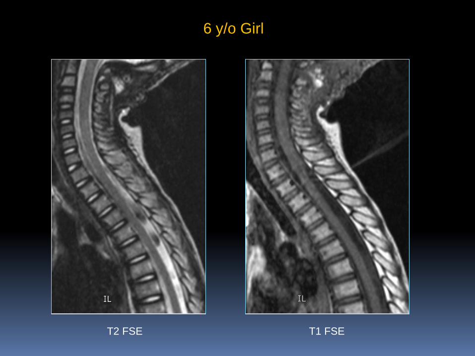
6 y/o Girl
T2 FSE
T1 FSE
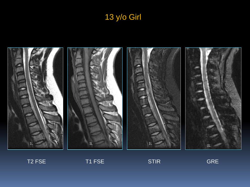
13 y/o Girl
T2 FSE
T1 FSE STIR GRE
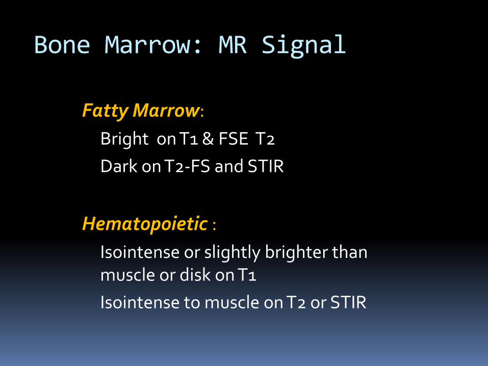
Bone Marrow: MR Signal
Fatty Marrow:
Bright on T1 & FSE T2
Dark on T2-FS and STIR
Hematopoietic :
Isointense or slightly brighter than muscle or disk on T1
Isointense to muscle on T2 or STIR
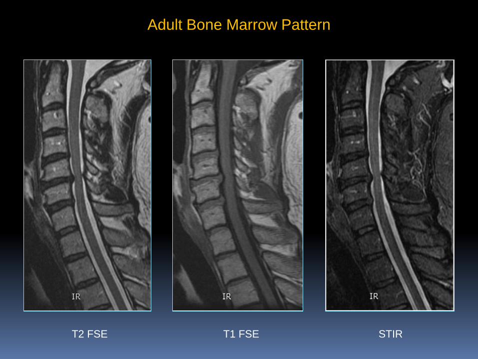
Adult Bone Marrow Pattern
T2 FSE
T1 FSE STIR

Adult Bone Marrow Pattern
T2 FSE
T1 FSE STIR

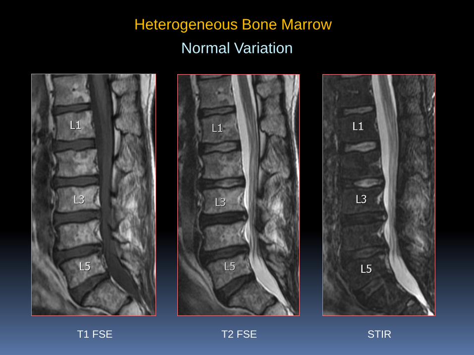
Heterogeneous Bone Marrow
Normal Variation
T1 FSE STIR T2 FSE
Heterogeneous Bone Marrow
Normal Variation
T1 FSE STIR T2 FSE

Degenerative Disk Disease
T1 FSE
Heterogeneous Marrow
T2 FSE

MR Pulse Sequences
T1 FSE Good contrast between fatty marrow and lesion
PD and T2 Sequences Without FS, lack contrast & have low sensitivity
STIR Good fat suppression & high sensitivity
DWI Helpful to assess cellularity
T1 -Gd-FS
Helps characterize T1 & STIR abnormalities

Bone Marrow Disorders
Increased red marrow: failure of conversion / reconversion
Decreased red marrow: marrow depletion
Iron storage disorders
Infiltration by abnormal cells, benign or malignant
Marrow edema
Bone infarcts
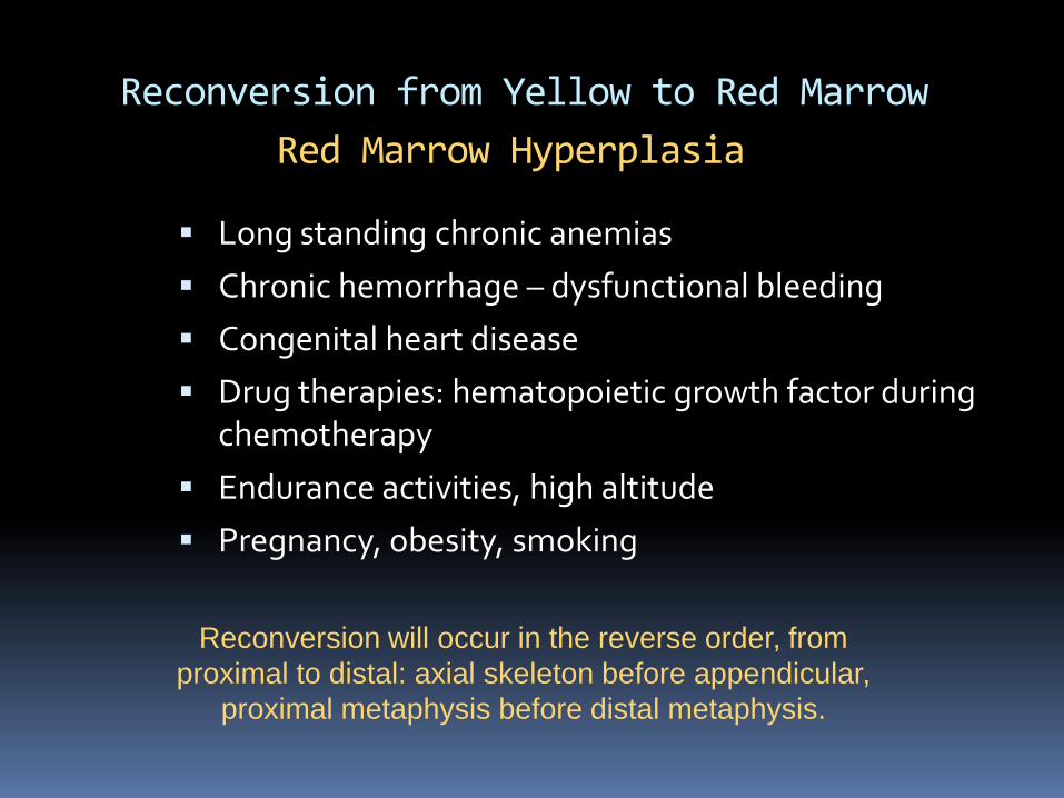
Reconversion from Yellow to Red Marrow
Long standing chronic anemias
Chronic hemorrhage – dysfunctional bleeding
Congenital heart disease
Drug therapies: hematopoietic growth factor during chemotherapy
Endurance activities, high altitude
Pregnancy, obesity, smoking
Red Marrow Hyperplasia
Reconversion will occur in the reverse order, from
proximal to distal: axial skeleton before appendicular,
proximal metaphysis before distal metaphysis.
Thalassemia
Congenital disorder of defect in synthesis of one or more of the subunits of hemoglobin
Gene expression can be homozygous, intermediate, or heterozygous, resulting in various degrees of anemia
Erythropoiesis is ineffective, expansion of red marrow
Massive splenomegaly causes splenic sequestration, increasing the anemia
Iron overload deposited in bone marrow, liver & spleen

Thalassemia
Red marrow hyperplasia
Expansion of marrow cavities
Marrow can have a lower signal on T1w and T2w if iron overload
Extramedullary hematopoiesis
Splenomegaly
777 - 10592921

Sickle Cell Disease
0.15% of African-American children in US are homozygous
Abnormal hemoglobin causes the erythrocyte to become rigid, causing vascular occlusions and bone infarcts
Abnormal shape causes hemolytic anemia
Increased risk for osteomyelitis
Imaging: Abnormal marrow, bone infarcts, H-shaped vertebrae

Sickle Cell Disease Evelyne Fliszar, UC San Diego
Combination of hematopoietic marrow and bone infarcts. Abnormal in distribution and signal
Difficult to differentiate from acute osteomyelitis

BM-2 - 18672071
History: 41 y/o HIV+ male with weakness,
muscle pain & cramping

{Page 2}
DDx: Opportunistic infxn, treatment-related reactive marrow change, lymphoma

BM-2 - 14140982
History: 52 y/o HIV+ male with
fevers for 6 months & back pain
BM Bx 2 months earlier: hypocellular – no lymphoma

Red Marrow Re-conversion
66 y/o obese woman
T1 FSE T1-Gd-FS T2 FSE

T1 FSE STIR
Red Marrow Re-conversion
66 y/o obese man
T2 FSE

Marrow Depleting Disorders
Aplastic anemia ( idiopathic, toxins, viral
infection, drugs)
Chemotherapy
1st week edema, then increasing fat
Partly reversible
Radiation therapy
1st month edema
2 – 12 months: fatty replacement
< 30 Gy – marrow regenerates after 1 year
> 30 Gy – irreversible
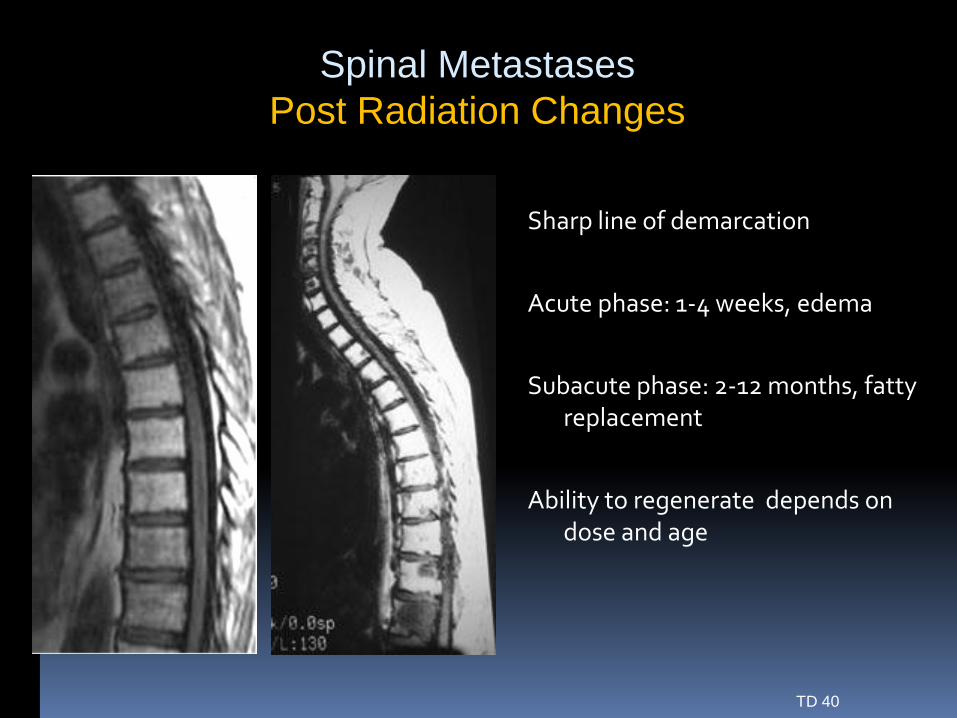
Spinal Metastases
Post Radiation Changes
TD 40
Sharp line of demarcation
Acute phase: 1-4 weeks, edema
Subacute phase: 2-12 months, fatty replacement
Ability to regenerate depends on dose and age

Post Radiation Marrow Recovery
Red marrow is more sensitive
Marrow regeneration starts at the end plates of the vertebral body, producing a band-like pattern
Evelyne Fliszar, UC San Diego

Post Radiation complications
Insufficiency fractures can occur within a few months
Osteonecrosis, usually diagnosed years after RT
Radiation-induced neoplasms: osteochondromas, osteosarcomas, malignant fibrous sarcomas, meningiomas

Insufficiency Fracture: 77 y.o. man
Evelyne Fliszar, UC San Diego

Infiltration by Abnormal Cells
Benign:
Eosinophilic Granuloma (solitary or diffuse)
Gaucher (multifocal or diffuse)
Mucopolysaccharidoses
Mastocytosis
Sarcoidosis
Evelyne Fliszar, UC San Diego

Langerhans Cell Histiocytosis
Hervey Segall, Milwaukee Children’s Hospital
Eosinophilic
Granuloma
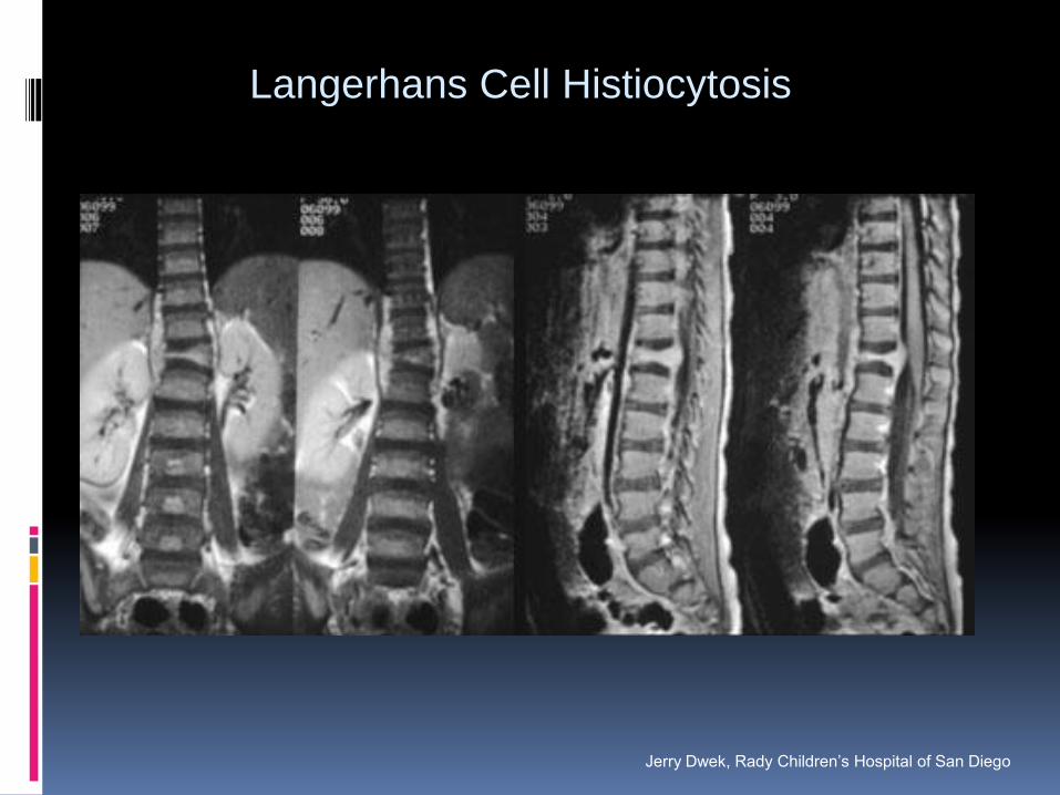
Langerhans Cell Histiocytosis
Jerry Dwek, Rady Children’s Hospital of San Diego

Gaucher’s Disease
Metabolic storage disorder caused by a deficient enzyme
Accumulation of glycolipids in the marrow
Decreased T1 and T2w marrow signal
Preferential involvement of the distal femurs causes Erlenmeyer flask deformity
Marrow infarcts
Splenomegaly
Evelyne Fliszar, UC San Diego

Sarcoidosis
Cor T2FS
Evelyne Fliszar, UC San Diego

Marrow Proliferative Disorders
Polycythemia vera
Myeloid metaplasia with myelofibrosis
Multiple myeloma
Mastocytosis
Leukemia
Lymphoma
Evelyne Fliszar, UC San Diego
Myelofibrosis
Can be primary or secondary (toxins, irradiation, chemotherapy, leukemia)
Bone marrow biopsy: increased megacaryocytes, fibrosis, and decreased fat
Xrays: sclerotic bones
MRI: Low T1 and T2 signal of the marrow
Splenomegaly
Evelyne Fliszar, UC San Diego

Myelofibrosis
Sag T1
Evelyne Fliszar, UC San Diego
Multiple Myeloma
Malignant plasma cells derived from B-lymphocytes
Rare before the age of 40
Monoclonal gammopathies (benign to malignant)
Plasmacytoma - solitary form
May be diffuse, simulating leukemia
Low T1w, variable T2w signal, variable enhancement
20% of MRIs can be normal despite diffuse marrow involvement
Evelyne Fliszar, UC San Diego

History: 49 y/o woman with pelvic pain
Bm-1 - 22747034
Dx: Plasmacytoma

History: 65 y/o woman with back pain & progressive leg
weakness & gait problem for 2 months
656

{Page 2}
Dx: Plasmacytoma – Multiple
myeloma

Multiple Myeloma
Sag T1 Sag T2FS
Diffuse involvment is
not as obvious as
nodular involvment
and can be easily
overlooked.
Evelyne Fliszar, UC San Diego

Multiple Myeloma – s/p Rx
Bm-3
3 months earlier
Sag T1WI
T1–Gd–FS

Diffuse Multiple Myeloma
Bhatia et al, Chapter 74, in Clinical MRI, 2006

History: 53 y/o woman with back pain &
bilateral foot numbness
692

Dx: Plasmacytoma &
Multiple myeloma
{Page 2}
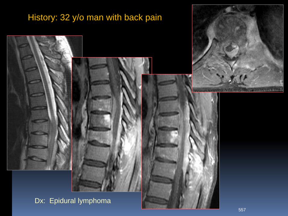
Dx: Epidural lymphoma
History: 32 y/o man with back pain
557
13 - 1662333
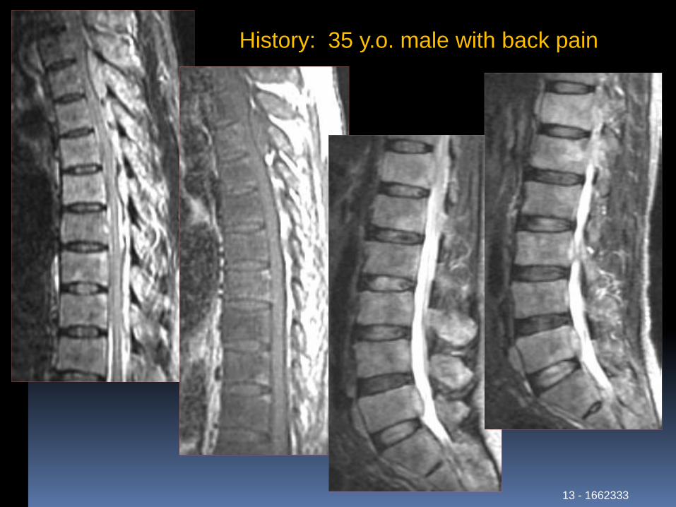
History: 35 y.o. male with back pain

Back pain {Page 2}

Dx: Multiple myeloma
{Page 3}

Multiple Myeloma
Tanenbaum LN, Diffusion imaging in the spine,
Applied Rad iolgy, April 2011
DWI T1- FLAIR STIR
Leukemia / Lymphoma
Lymphoma tends to cause focal marrow tumors, but may be diffuse.
Leukemia is a diffuse process, but can be more focal and irregular in relapse.
Diagnosis by MRI more difficult in children, who have predominantly red marrow.
Chronic leukemia has a more indolent course, with prolonged survival.
Evelyne Fliszar, UC San Diego
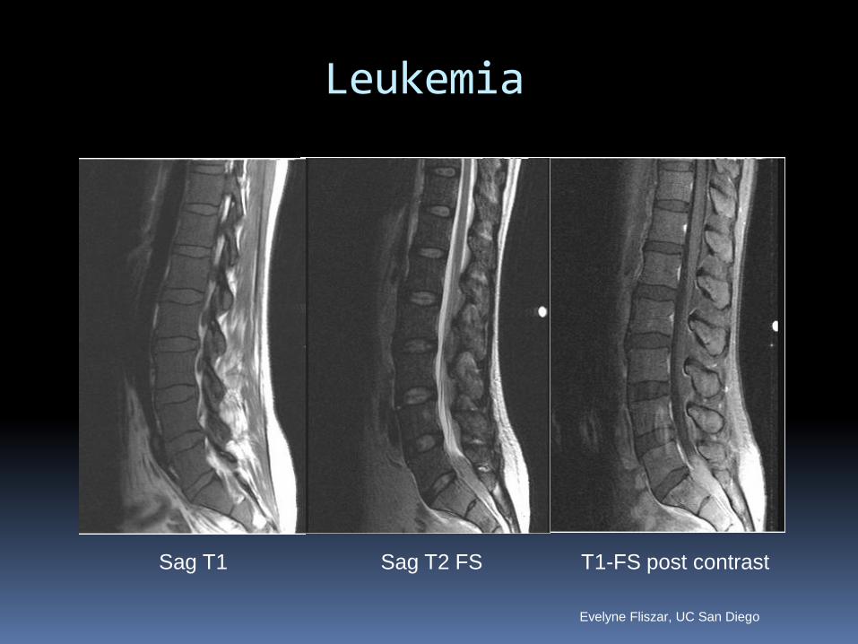
Leukemia
Sag T1 Sag T2 FS T1-FS post contrast
Evelyne Fliszar, UC San Diego

History: 54 y/o woman with CLL & right foot numbess
773 - 19113240
Sag T2
Sag T1
T1-FS post contrast

{Page 2}
Sag T1
T1FS post contrast
Dx: Chronic lymphocytic leukemia

Lymphoma
25415928
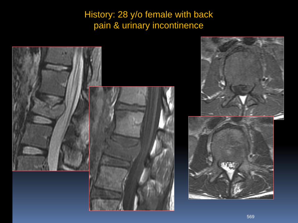
History: 28 y/o female with back
pain & urinary incontinence
569
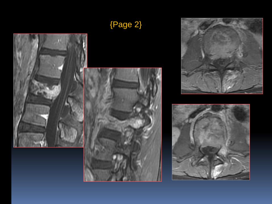
{Page 2}

Dx: Lymphoma
{Page 3}
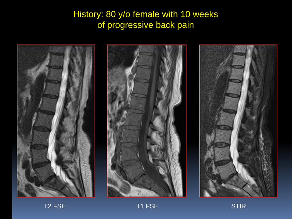
History: 80 y/o female with 10 weeks
of progressive back pain
T2 FSE
T1 FSE STIR

Dx: Lymphoplasmacytic
Lymphoma (Waldenström's
macroglobulinemia)
{Page 2}

• Diagnosed with Lymphoplasmacytic Lymphoma (Waldenström's macroglobulinemia) in 2011
– Low grade lymphoma
– mature plasmacytoid lymphocytes produce monoclonal IgM
– Abnormal lymphoplasmacytoid cells in bone marrow or lymph nodes
– Can be clinically indolent
• Or fatigue, hyperviscosity syndrome, hepatosplenomegaly

Metastatic Disease
Low T1w signal
T2w signal variable depending on cell density.
Lytic metastases are bright on T2w.
Sclerotic metastases may be dark, intermediate or bright on T2w.
“Halo” sign: rim of high T2w signal or rim enhancement on T1w images is very specific.
Evelyne Fliszar, UC San Diego

Metastatic Squamous Cell CA
25415928
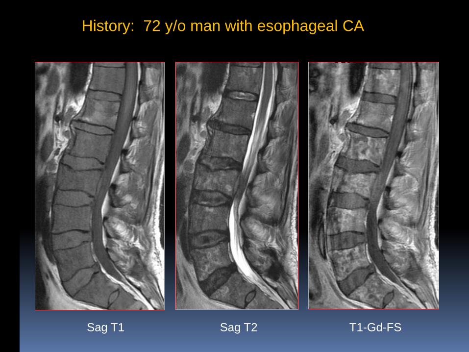
History: 72 y/o man with esophageal CA
Sag T1 Sag T2 T1-Gd-FS

History: 70 y/o woman with back pain
545

{Page 2}
Dx: Renal cell CA

Breast Metastases
T1 FSE STIR T2 FSE

Dx: Diffuse Breast metastases
History: 64 y/o woman with back pain
547

Dx: Diffuse Prostate metastases
History: 62 y/o man with back pain
548
Iron Storage Disorders
Hemochromatosis A genetic disorder
Increased intestinal absorption of iron despite normal dietary intake
Increased iron stores in hepatocytes of liver only
Hemosiderosis Hemolytic anemia
Multiple blood transfusions
Increased iron stores in RE systems of marrow, liver & spleen
Can mask signal from other marrow components
Decreased signal on all MR sequences
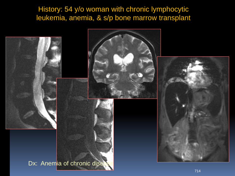
History: 54 y/o woman with chronic lymphocytic
leukemia, anemia, & s/p bone marrow transplant
714
Dx: Anemia of chronic disease
Bone Marrow Edema
Degenerative disk disease (Modic Type I)
Fracture
Infection / inflammation
Tumor
Reflex sympathetic dystrophy

T1 FSE STIR
Degenerative Disk Disease
Bone Marrow Edema
T2 FSE

Benign Vertebral Collapse
T1 FSE STIR
Osteoporosis
T2 FSE

Acute Vertebral Fracture
T2 FSE
T1 FSE STIR

Vertebral Body Fracture
T2 FSE
T1 FSE
STIR
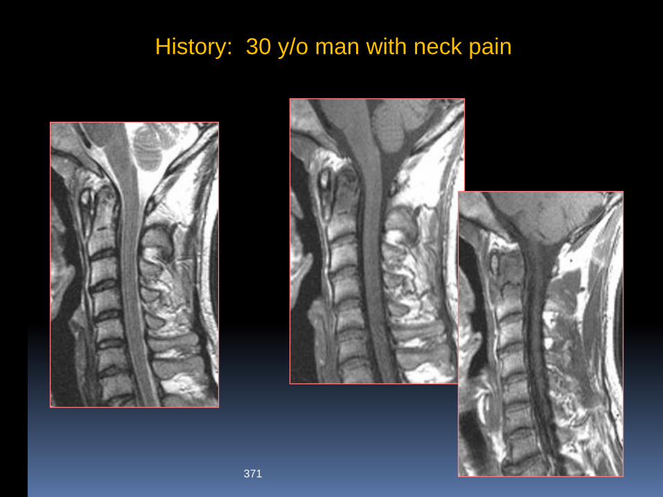
History: 30 y/o man with neck pain
371
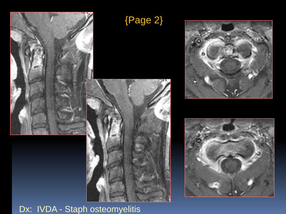
Dx: IVDA - Staph osteomyelitis
{Page 2}

Bone Marrow Transplantation
For leukemia, lymphoma, metabolic & immune disorders, other malignancies
High dose chemo & whole body radiation
Infusion of stem cells to re-populate marrow
T1WIs: central fat sandwiched between bands of hypointensity
Re-populating hematopoietic cells
Progresses to more homogeneous marrow
Can simulate recurrent disease

History: 50 y/o man with weakness & numbness of extremities
794 - 26644146
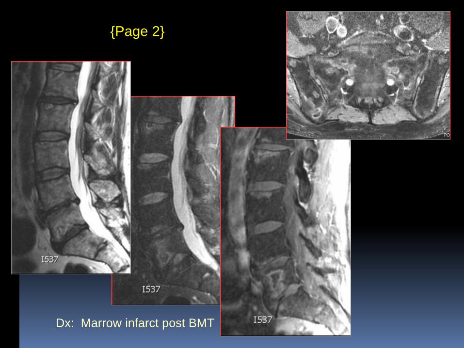
Dx: Marrow infarct post BMT
{Page 2}

In Conclusion…
Be aware of normal variations of marrow patterns
T1w and STIR sequences are most useful
Diffuse marrow processes can be more difficult to detect
Benign and malignant processes can look very similar