Sperm defects in primary ciliary dyskinesia and related ... · Sperm defects in primary ciliary...
20
Vol.:(0123456789) 1 3 Cellular and Molecular Life Sciences (2020) 77:2029–2048 https://doi.org/10.1007/s00018-019-03389-7 REVIEW Sperm defects in primary ciliary dyskinesia and related causes of male infertility Anu Sironen 1 · Amelia Shoemark 2,3 · Mitali Patel 1 · Michael R. Loebinger 4,5 · Hannah M. Mitchison 1 Received: 16 July 2019 / Revised: 12 November 2019 / Accepted: 19 November 2019 / Published online: 28 November 2019 © The Author(s) 2019 Abstract The core axoneme structure of both the motile cilium and sperm tail has the same ultrastructural 9 + 2 microtubular arrange- ment. Thus, it can be expected that genetic defects in motile cilia also have an effect on sperm tail formation. However, recent studies in human patients, animal models and model organisms have indicated that there are differences in compo- nents of specific structures within the cilia and sperm tail axonemes. Primary ciliary dyskinesia (PCD) is a genetic disease with symptoms caused by malfunction of motile cilia such as chronic nasal discharge, ear, nose and chest infections and pulmonary disease (bronchiectasis). Half of the patients also have situs inversus and in many cases male infertility has been reported. PCD genes have a role in motile cilia biogenesis, structure and function. To date mutations in over 40 genes have been identified cause PCD, but the exact effect of these mutations on spermatogenesis is poorly understood. Furthermore, mutations in several additional axonemal genes have recently been identified to cause a sperm-specific phenotype, termed multiple morphological abnormalities of the sperm flagella (MMAF). In this review, we discuss the association of PCD genes and other axonemal genes with male infertility, drawing particular attention to possible differences between their functions in motile cilia and sperm tails. Keywords PCD · MMAF · Infertility · Cilia · Axoneme · Sperm tail · Motility · Dynein Introduction Motile cilia and flagella have a conserved axonemal struc- ture consisting of a ring of nine microtubular doublets and a central pair of microtubules, giving the classical 9 + 2 micro- tubular arrangement (Fig. 1a). Central pair microtubules C1 and C2 are connected by a bridge-like structure and several projections are docked to C1 and C2 forming the central pair complex (CPC). Each outer doublet is composed of type A and B microtubules and connected by radial spokes (RS) to CPC (Fig. 1a). The force for motility is produced by inner and outer dynein arms (IDA and ODA, respectively), which are carried by the A-type tubule and project toward the B-tubule of the adjacent doublet. IDA and ODA are part of a specific protein complex of 96 nm repeat units, which contains four identical ODAs, one two-headed IDA (f/I1), six single-headed IDAs (a–e and g), three RS and a single nexin–dynein regulatory complex (N-DRC) (Fig. 1c) The N-DRC regulates and coordinates the activity of the dynein arms [43]. The current knowledge of motile cilia ultrastruc- ture and protein content has been previously reviewed by Osinka et al. [96]. The sperm flagellum has an ultrastruc- turally comparable axonemal structure, but in addition the sperm tail contains accessory structures: the mitochondrial sheath (MS), fibrous sheath (FS) and outer dense fibres (ODFs) (Fig. 1b). These structures are specific to the sperm tail and are required for fertile sperm production providing, e.g. additional rigidity and energy for the movement of the sperm in the female reproductive tract [58]. Sperm tail is connected to the head by the head tail coupling apparatus (HTCA) and annulus forms a diffusion barrier between the Cellular and Molecular Life Sciences * Anu Sironen [email protected] 1 Genetics and Genomic Medicine, UCL Great Ormond Street Institute of Child Health, University College London, 30 Guilford Street, London WC1N 1EH, UK 2 Department of Paediatrics, Royal Brompton Hospital, London, UK 3 School of Medicine, University of Dundee, Dundee, UK 4 Host Defence Unit, Royal Brompton and Harefield NHS Foundation Trust, London, UK 5 National Heart and Lung Institute, Imperial College London, London, UK
Transcript of Sperm defects in primary ciliary dyskinesia and related ... · Sperm defects in primary ciliary...
Vol.:(0123456789)1 3
Cellular and Molecular Life Sciences (2020) 77:2029–2048 https://doi.org/10.1007/s00018-019-03389-7
REVIEW
Sperm defects in primary ciliary dyskinesia and related causes of male infertility
Anu Sironen1 · Amelia Shoemark2,3 · Mitali Patel1 · Michael R. Loebinger4,5 · Hannah M. Mitchison1
Received: 16 July 2019 / Revised: 12 November 2019 / Accepted: 19 November 2019 / Published online: 28 November 2019 © The Author(s) 2019
AbstractThe core axoneme structure of both the motile cilium and sperm tail has the same ultrastructural 9 + 2 microtubular arrange-ment. Thus, it can be expected that genetic defects in motile cilia also have an effect on sperm tail formation. However, recent studies in human patients, animal models and model organisms have indicated that there are differences in compo-nents of specific structures within the cilia and sperm tail axonemes. Primary ciliary dyskinesia (PCD) is a genetic disease with symptoms caused by malfunction of motile cilia such as chronic nasal discharge, ear, nose and chest infections and pulmonary disease (bronchiectasis). Half of the patients also have situs inversus and in many cases male infertility has been reported. PCD genes have a role in motile cilia biogenesis, structure and function. To date mutations in over 40 genes have been identified cause PCD, but the exact effect of these mutations on spermatogenesis is poorly understood. Furthermore, mutations in several additional axonemal genes have recently been identified to cause a sperm-specific phenotype, termed multiple morphological abnormalities of the sperm flagella (MMAF). In this review, we discuss the association of PCD genes and other axonemal genes with male infertility, drawing particular attention to possible differences between their functions in motile cilia and sperm tails.
Keywords PCD · MMAF · Infertility · Cilia · Axoneme · Sperm tail · Motility · Dynein
Introduction
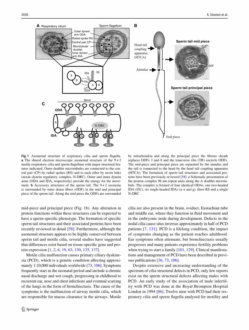
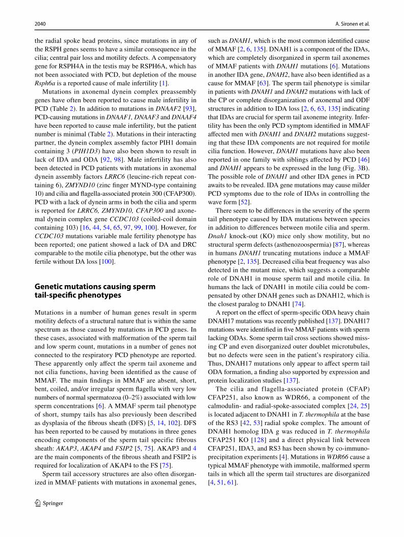
Motile cilia and flagella have a conserved axonemal struc-ture consisting of a ring of nine microtubular doublets and a central pair of microtubules, giving the classical 9 + 2 micro-tubular arrangement (Fig. 1a). Central pair microtubules C1 and C2 are connected by a bridge-like structure and several projections are docked to C1 and C2 forming the central pair complex (CPC). Each outer doublet is composed of
type A and B microtubules and connected by radial spokes (RS) to CPC (Fig. 1a). The force for motility is produced by inner and outer dynein arms (IDA and ODA, respectively), which are carried by the A-type tubule and project toward the B-tubule of the adjacent doublet. IDA and ODA are part of a specific protein complex of 96 nm repeat units, which contains four identical ODAs, one two-headed IDA (f/I1), six single-headed IDAs (a–e and g), three RS and a single nexin–dynein regulatory complex (N-DRC) (Fig. 1c) The N-DRC regulates and coordinates the activity of the dynein arms [43]. The current knowledge of motile cilia ultrastruc-ture and protein content has been previously reviewed by Osinka et al. [96]. The sperm flagellum has an ultrastruc-turally comparable axonemal structure, but in addition the sperm tail contains accessory structures: the mitochondrial sheath (MS), fibrous sheath (FS) and outer dense fibres (ODFs) (Fig. 1b). These structures are specific to the sperm tail and are required for fertile sperm production providing, e.g. additional rigidity and energy for the movement of the sperm in the female reproductive tract [58]. Sperm tail is connected to the head by the head tail coupling apparatus (HTCA) and annulus forms a diffusion barrier between the
Cellular and Molecular Life Sciences
* Anu Sironen [email protected]
1 Genetics and Genomic Medicine, UCL Great Ormond Street Institute of Child Health, University College London, 30 Guilford Street, London WC1N 1EH, UK
2 Department of Paediatrics, Royal Brompton Hospital, London, UK
3 School of Medicine, University of Dundee, Dundee, UK4 Host Defence Unit, Royal Brompton and Harefield NHS
Foundation Trust, London, UK5 National Heart and Lung Institute, Imperial College London,
London, UK
2030 A. Sironen et al.
1 3
mid-piece and principal piece (Fig. 1b). Any alteration in protein functions within these structures can be expected to have a sperm-specific phenotype. The formation of specific sperm tail structures and their associated proteins have been recently reviewed in detail [58]. Furthermore, although the axonemal structure appears to be highly conserved between sperm tail and motile cilia, several studies have suggested that differences exist based on tissue-specific gene and pro-tein expression [1, 2, 6, 19, 63, 130, 135, 137].
Motile cilia malfunction causes primary ciliary dyskine-sia (PCD), which is a genetic condition affecting approxi-mately 1:10,000 individuals worldwide [73, 106]. Symptoms frequently start in the neonatal period and include a chronic nasal discharge and wet cough, progressing in childhood to recurrent ear, nose and chest infections and eventual scarring of the lungs in the form of bronchiectasis. The cause of the symptoms is the malfunction of airway motile cilia, which are responsible for mucus clearance in the airways. Motile
cilia are also present in the brain, oviduct, Eustachian tube and middle ear, where they function in fluid movement and in the embryonic node during development. Defects in the nodal cilia cause situs inversus approximately in half of PCD patients [7, 131]. PCD is a lifelong condition, the impact of symptoms changing as the patient reaches adulthood. Ear symptoms often attenuate, but bronchiectasis usually progresses and many patients experience fertility problems when trying to start a family [101, 129]. Clinical manifesta-tions and management of PCD have been described in previ-ous publications [36, 73, 106].
Despite extensive and increasing understanding of the spectrum of cilia structural defects in PCD, only few reports exist on the sperm structural defects affecting males with PCD. An early study of the association of male infertil-ity with PCD was done at the Royal Brompton Hospital London in 1994 [86]. Twelve men with PCD had their res-piratory cilia and sperm flagella analysed for motility and
Respiratory cilium Sperm flagellum
Outer dynein arm ODA
Inner dynein arm IDA
Central pair CPMicrotubulardoublet
Radial spoke RS
A
RS
ODA
IDA
Sperm tail mid piece
Sperm tail principal piece
ODF
ODF
End piece
Head tailcouplingapparatus(HTCA)
Annulus
B
AB
BA
a b c e g d
f/I1N-DRC
RS1 RS2 RS3
ODA
IDA
96nmC
A
Fig. 1 Axonemal structure of respiratory cilia and sperm flagella. a The shared electron microscopic axonemal structure of the 9 + 2 motile respiratory cilia and sperm flagellum with major structural fea-tures indicated. Outer doublet microtubules are connected to the cen-tral pair (CP) by radial spokes (RS) and to each other by nexin links (nexin–dynein regulatory complex, N-DRC). Outer and inner dynein arms (ODA and IDA, respectively) provide the energy for the move-ment. b Accessory structures of the sperm tail. The 9 + 2 axoneme is surrounded by outer dense fibres (ODF) in the mid and principal piece of the sperm tail. Along the mid piece the ODFs are surrounded
by mitochondria and along the principal piece the fibrous sheath replaces ODFs 3 and 8 and the transverse ribs (TR) encircle ODFs. The mid-piece and principal piece are separated by the annulus and the tail is connected to the head by the head tail coupling apparatus (HTCA). The formation of sperm tail structures and associated pro-teins have been previously reviewed [58]. c Schematic presentation of the protein complex 96 nm repeat units along the A doublet microtu-bule. The complex is formed of four identical ODAs, one two-headed IDA (f/I1), six single-headed IDAs (a–e and g), three RS and a single N-DRC
2031Sperm defects in primary ciliary dyskinesia and related causes of male infertility
1 3
ultrastructure. Two patients had immotile sperm and one had normal motility with oligospermia (low sperm num-bers; Table 1 explains terminology of sperm phenotypic defects). Three of these patients had normal sperm motility and ultrastructure and one had fathered a child. The twelfth patient had slowed sperm with ultrastructural dynein defi-ciency, but had also reportedly fathered a child. Five had azoospermia (no sperm), which the authors attributed at the time to defects in the ciliated portion of the vas def-erens preventing normal transport. However, there are no cilia in the vas deferens, but motile cilia are present in the efferent ductules [20, 142]. Recent studies have shown that these cilia are not required for transport of sperm towards the cauda epididymis, but they agitate the seminal fluid to prevent blockage and to enable reabsorption of the fluid by a rotational swirling motion rather than forward ciliary beat [142]. Thus, the lack of sperm is unlikely to be caused by defects in sperm transport, but may arise from blockage of the efferent ductules. The role of motile cilia in the efferent ductules has not been studied in PCD patients, but mutant mouse models have shown that male infertility can be caused by reduced numbers of motile cilia in the efferent ductules [127, 142]. Furthermore, animal models have also shown that severe spermatid malformations can lead to very low sperm counts in the epididymis, due to sloughing of imma-ture spermatids.
More recently, a 2017 study reported phenotype–geno-type correlations for fertility in 46 male PCD patients, how-ever neither the fertility status of the partner nor the sperm ultrastructure or motility were reported [130]. This study included the cilia structural defects and fertility data avail-able for cases with mutations in 17 different PCD genes, which indicated that the PCD patients more likely to be infertile had cilia showing a loss of inner dynein arms with microtubular disorganization, or a lack of the outer and inner dynein arms. Recent advances in PCD genetics will increas-ingly enable investigations of the effect of specific mutations on male fertility and prediction of their effect based on the genetic test. However, prior to implementing fertility coun-selling in PCD clinics, the association of specific mutations
with sperm phenotype needs to be elucidated. More com-prehensive counselling for PCD would take into account different genetic effects on cilia versus sperm motilities. Therefore, in this review we present the current knowledge of PCD-associated male infertility and sperm tail pheno-types caused by mutations in axonemal genes.
Current PCD diagnostics
Primary ciliary dyskinesia is a genetically heterogeneous disorder of variable clinical impact and thus far there is no single or even combination of tests accurate enough for mak-ing a diagnosis under all circumstances [55, 72, 106, 112]. After suspecting a possible PCD phenotype from clinical history (neonatal respiratory distress, respiratory pheno-types, laterality defects), a series of tests can be used to con-firm the diagnosis. Nasal nitric oxide gas is reduced in most patients with PCD [10]. Analysis of the respiratory epithe-lium by nasal brushing can be used for identification of cili-ary structural defects by transmission electron microscopy (TEM) [113, 118] and defects of the ciliary beat pattern and frequency can be identified by high-speed video microscopy analysis (HSVMA) [107]. HSVMA has excellent sensitivity and specificity for PCD and particular beat patterns have been linked to specific ultrastructural defects [15]. Assist-ing analysis includes immunofluorescence to look at cilia protein defects [119]. This is becoming more routinely used, which in addition to more specialized electron tomography [116] can be used in difficult to diagnose cases.
Primary ciliary dyskinesia is caused by genetic variants in genes coding for proteins, which have a role in motile cilia structure, formation and function. Thus, genetics has become a more prominent component of the diagnostic path-way for PCD over recent years, with confirmation of PCD diagnosis now defined as being made through identification of an ultrastructural defect by transmission electron micros-copy or the identification of bi-allelic mutations in a known PCD gene [72, 112]. Several ultrastructural defects have been identified in the respiratory cilia of patients with PCD,
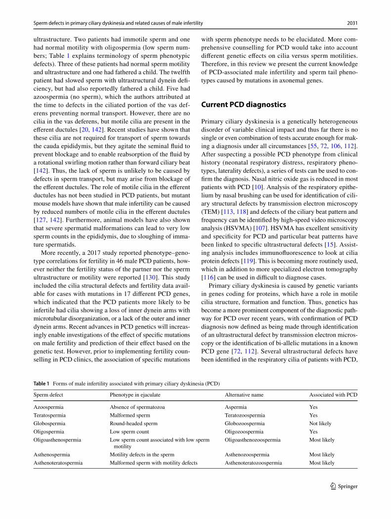
Table 1 Forms of male infertility associated with primary ciliary dyskinesia (PCD)
Sperm defect Phenotype in ejaculate Alternative name Associated with PCD
Azoospermia Absence of spermatozoa Aspermia YesTeratospermia Malformed sperm Teratozoospermia YesGlobospermia Round-headed sperm Globozoospermia Not likelyOligospermia Low sperm count Oligozoospermia YesOligoasthenospermia Low sperm count associated with low sperm
motilityOligoasthenozoospermia Most likely
Asthenospermia Motility defects in the sperm Asthenozoospermia Most likelyAsthenoteratospermia Malformed sperm with motility defects Asthenoteratozoospermia Most likely

2032 A. Sironen et al.
1 3
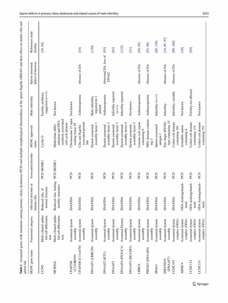
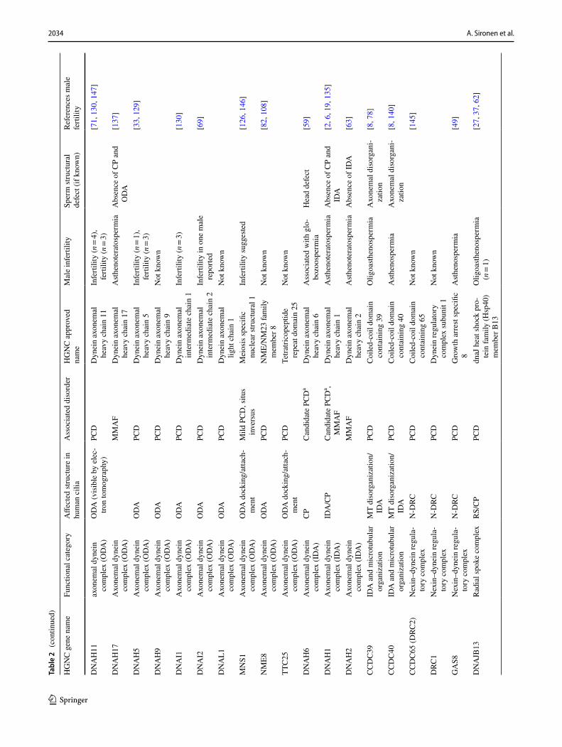
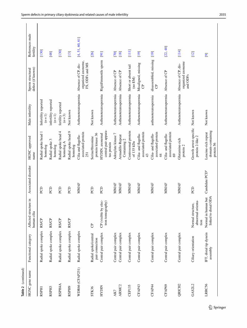
the majority involving absence, partial absence or shorten-ing of the outer or both inner and outer dynein arms. Other defects include complete or partial absence of the central microtubular pair often accompanied by transposition of an outer microtubular doublet [13, 48, 91, 95], a variety of microtubular disarrangements associated with defects of the nexin–dynein regulator complex and inner dynein arm loss [3, 78, 138] or reduced numbers of cilia [11, 132]. A sizeable proportion of patients have no detectable ultras-tructural defect, some explained by subtle defects visual-ized only at high resolution [23, 91, 117], or effects on the axoneme not directly affecting internal structures [9, 12]. To date, more than 40 genes with causative mutations for PCD have been identified (Table 2) and gene panels are available for genetic testing [84, 92, 136]. It is estimated that in about 70% of patients with a clinical PCD phenotype, a bi-allelic mutation in a known PCD gene can be found [105, 112]. Increased awareness of PCD and better testing mean that the average age of diagnosis is now in childhood [35] and therefore fertility assessment is rarely part of frontline PCD diagnostics, although it should be taken into account in adult PCD patients.
How do motile cilia and sperm differ?
The gross axonemal structure of the motile cilia and sperm tail may appear to be identical, but cell type-specific differ-ences in axonemal proteins such as dynein arm components and in assembly of the axoneme exist [32]. This conclu-sion is supported by the fact that basic motility and aspects of morphology differ between cilia and sperm and also since not all mutations in PCD genes cause male infertil-ity (Table 2). Differences exist in the length of the sperm flagellum and in the accessory structures surrounding the axoneme compared to cilia. This is linked to the distinct motility pattern and functions of motile cilia versus sperm tails. Cilia motility includes a forward power stroke to move the mucus in airway epithelia and a recovery return stroke to then produce another power stroke [123], but in sperm the bending waves of the tail produce a constant forward push-ing motion [47]. The respiratory tract cilia are attached to the apical surfaces of the airway epithelium moving the overly-ing fluid, whereas the flagellum of spermatozoa has evolved to power movement of the gamete freely through fluid.
Because of the role of motile cilia in mucus removal in the airways, multiciliogenesis is required for production of multiple cilia in the epithelial cells [17]. This differentia-tion programme is not necessary for sperm tail formation [58] or laterality determination [39] and thus mutations in genes encoding proteins with a role in multiciliogenesis (e.g. CCNO and MCIDAS) are not expected to affect male germ cell development. However, recent studies indicate
that multiciliogenesis is required for correct function of the efferent ductules [20, 142]. In mice it has been shown that mutations in multiciliogenesis genes, Gemc1, Mcidas and Ccno, cause male infertility and complete lack of sperm in the epididymis [127].
Conversely, defects in sperm tail-specific structures can cause infertility with no airway phenotype. The long axo-neme within the sperm flagellum is supported by the ODFs and FS and therefore malformations in these structures may also cause instability of the axonemal structure, as has been seen in patients with mutations in the FS protein, FSIP2 (fibrous sheath interacting protein 2) [28]. Depletion of another FS protein, AKAP4 (A-kinase anchoring protein 4), gives rise to lowered sperm numbers and short sperm tails with disruptions to the principal piece formation of the sperm tail (asthenoteratospermia), but the axoneme appears intact [5, 79]. Male infertility has also been reported to be caused by mutations in outer dense fibre gene ODF1, where the head tail connection was weakened in addition to dis-organization of the ODFs and MS [41, 139]. Sperm tail-specific proteins may also be involved in attachment of the axoneme to the ODFs and FS [19] and unidentified or poorly characterized differences affecting the structural components may also contribute to the specific requirements of the sperm tail. This has been demonstrated by the identification of the tail axoneme intra-lumenal spiral (TAILS), a structure which binds directly to 11 protofilaments on the internal microtu-bule wall, along the end piece of the sperm tail [70, 143].
Differences in tissue distribution of dynein arm genes and the effect of known mutations
There is evidence that different motility patterns of cilia and sperm may require cell specific dynein arm complexes. In respiratory cilia mutations in ODA genes affect the ciliary beating and ODA ultrastructure, but no mutations in IDA genes have been identified in PCD patients to date. ODAs and IDAs are required for motility in both cilia and sperm, but differences in protein content of these complexes may contribute to the specialized motility-specific patterns required for forward swimming sperm, cilia mucus clear-ance in airways and the rotational movement of nodal cilia. In 9 + 0 nodal cilia, their lack of central pair microtubules and radial spokes seems to be the key for their unidirec-tional rotational movement [115], but the structural differ-ences explaining the differences in motility between 9 + 2 cilia and sperm are not immediately obvious.
Recent expression studies in Drosophila indicate that specific ODA and IDA components are required for dif-ferent waveforms in sperm and neuronal cilia [148]. Pre-viously published transcriptome profiles during the first
2033Sperm defects in primary ciliary dyskinesia and related causes of male infertility
1 3
Tabl
e 2
Axo
nem
al g
enes
with
mut
atio
ns c
ausi
ng p
rimar
y ci
liary
dys
kine
sia
(PC
D) a
nd m
ultip
le m
orph
olog
ical
abn
orm
aliti
es o
f the
spe
rm fl
agel
la (M
MA
F) a
nd th
eir e
ffect
on
mot
ile c
ilia
and
sper
m ta
il
HG
NC
gen
e na
me
Func
tiona
l cat
egor
yA
ffect
ed st
ruct
ure
in
hum
an c
ilia
Ass
ocia
ted
diso
rder
HG
NC
app
rove
d na
me
Mal
e in
ferti
lity
Sper
m st
ruct
ural
de
fect
(if k
now
n)Re
fere
nces
mal
e fe
rtilit
y
CC
NO
Mul
ti-ci
liate
d ep
ithe-
lial c
ell d
iffer
entia
-tio
n
Redu
ced
cilia
, of
norm
al st
ruct
ure
PCD
(RG
MC
)C
yclin
OFe
rtilit
y pr
oble
ms
sugg
este
d (n
= 1)
[38,
89]
MC
IDA
SM
ulti-
cilia
ted
epith
e-lia
l cel
l diff
eren
tia-
tion
Redu
ced
cilia
, lac
king
m
otili
ty st
ruct
ures
PCD
(RG
MC
)M
ultic
iliat
e di
ffer-
entia
tion
and
DN
A
synt
hesi
s ass
ocia
ted
cell
cycl
e pr
otei
n
Not
kno
wn
CFA
P298
(C
21O
RF5
9)A
xone
mal
dyn
ein
asse
mbl
yID
A/O
DA
PCD
Chr
omos
ome
21 o
pen
read
ing
fram
e 59
Not
kno
wn
CFA
P300
(C11
orf7
0)A
xone
mal
dyn
ein
asse
mbl
yID
A/O
DA
PCD
Cili
a an
d fla
gella
-as
soci
ated
pro
tein
30
0
Ast
heno
sper
mia
Abs
ence
of D
A[4
4]
DN
AA
F1 (L
RRC
50)
Axo
nem
al d
ynei
n as
sem
bly
IDA
/OD
APC
DD
ynei
n ax
onem
al
asse
mbl
y fa
ctor
1M
ale
infe
rtilit
y re
porte
d fo
r 1
patie
nt
[130
]
DN
AA
F2 (K
TU)
Axo
nem
al d
ynei
n as
sem
bly
IDA
/OD
APC
DD
ynei
n ax
onem
al
asse
mbl
y fa
ctor
2A
sthe
nosp
erm
iaA
bnor
mal
DA
, los
s of
DN
AI2
[93]
DN
AA
F3A
xone
mal
dyn
ein
asse
mbl
yID
A/O
DA
PCD
Dyn
ein
axon
emal
as
sem
bly
fact
or 3
Infe
rtilit
y re
porte
d (n
= 1)
[83]
DN
AA
F4 (D
YX
1C1)
Axo
nem
al D
ynei
n as
sem
bly
IDA
/OD
APC
DD
ynei
n ax
onem
al
asse
mbl
y fa
ctor
4In
ferti
lity
repo
rted
[125
]
DN
AA
F5 (H
EATR
2)A
xone
mal
dyn
ein
asse
mbl
yID
A/O
DA
PCD
Dyn
ein
axon
emal
as
sem
bly
fact
or 5
Not
kno
wn
[21]
LRRC
6A
xone
mal
dyn
ein
asse
mbl
yID
A/O
DA
PCD
Leuc
ine-
rich
repe
at
cont
aini
ng 6
Ast
heno
sper
mia
Abs
ence
of D
A[5
4, 6
5]
PIH
1D3
(DN
AA
F6)
Axo
nem
al d
ynei
n as
sem
bly
IDA
/OD
APC
DPI
H1
dom
ain
cont
ain-
ing
3A
sthe
nosp
erm
iaA
bsen
ce o
f DA
[92,
98]
SPA
G1
Axo
nem
al d
ynei
n as
sem
bly
IDA
/OD
APC
DSp
erm
-ass
ocia
ted
antig
en 1
Infe
rtilit
y (n
= 1)
[88,
130
]
ZMY
ND
10
(DN
AA
F7)
Axo
nem
al d
ynei
n as
sem
bly
IDA
/OD
APC
DZi
nc fi
nger
MY
ND
-ty
pe c
onta
inin
g 10
Infe
rtilit
yA
bsen
ce o
f DA
[16,
85,
97]
CC
DC
103
Axo
nem
al d
ynei
n co
mpl
ex (O
DA
)ID
A/O
DA
PCD
Coi
led-
coil
dom
ain
cont
aini
ng 1
03In
ferti
lity,
var
iabl
eA
bsen
ce o
f DA
[99,
100
]
AR
MC
4A
xone
mal
dyn
ein
com
plex
(OD
A)
OD
A d
ocki
ng/a
ttach
-m
ent
PCD
Arm
adill
o re
peat
co
ntai
ning
4N
ot k
now
n[1
8]
CC
DC
114
Axo
nem
al d
ynei
n co
mpl
ex (O
DA
)O
DA
doc
king
/atta
ch-
men
tPC
DC
oile
d-co
il do
mai
n co
ntai
ning
114
Ferti
lity
not a
ffect
ed[9
4]
CC
DC
151
Axo
nem
al d
ynei
n co
mpl
ex (O
DA
)O
DA
doc
king
/atta
ch-
men
tPC
DC
oile
d-co
il do
mai
n co
ntai
ning
151
Not
kno
wn
2034 A. Sironen et al.
1 3
Tabl
e 2
(con
tinue
d)
HG
NC
gen
e na
me
Func
tiona
l cat
egor
yA
ffect
ed st
ruct
ure
in
hum
an c
ilia
Ass
ocia
ted
diso
rder
HG
NC
app
rove
d na
me
Mal
e in
ferti
lity
Sper
m st
ruct
ural
de
fect
(if k
now
n)Re
fere
nces
mal
e fe
rtilit
y
DN
AH
11ax
onem
al d
ynei
n co
mpl
ex (O
DA
)O
DA
(vis
ible
by
elec
-tro
n to
mog
raph
y)PC
DD
ynei
n ax
onem
al
heav
y ch
ain
11In
ferti
lity
(n =
4),
ferti
lity
(n =
3)[7
1, 1
30, 1
47]
DN
AH
17A
xone
mal
dyn
ein
com
plex
(OD
A)
MM
AF
Dyn
ein
axon
emal
he
avy
chai
n 17
Ast
heno
tera
tosp
erm
iaA
bsen
ce o
f CP
and
OD
A[1
37]
DN
AH
5A
xone
mal
dyn
ein
com
plex
(OD
A)
OD
APC
DD
ynei
n ax
onem
al
heav
y ch
ain
5In
ferti
lity
(n =
1),
ferti
lity
(n =
3)[3
3, 1
29]
DN
AH
9A
xone
mal
dyn
ein
com
plex
(OD
A)
OD
APC
DD
ynei
n ax
onem
al
heav
y ch
ain
9N
ot k
now
n
DN
AI1
Axo
nem
al d
ynei
n co
mpl
ex (O
DA
)O
DA
PCD
Dyn
ein
axon
emal
in
term
edia
te c
hain
1In
ferti
lity
(n =
3)[1
30]
DN
AI2
Axo
nem
al d
ynei
n co
mpl
ex (O
DA
)O
DA
PCD
Dyn
ein
axon
emal
in
term
edia
te c
hain
2In
ferti
lity
in o
ne m
ale
repo
rted
[69]
DN
AL1
Axo
nem
al d
ynei
n co
mpl
ex (O
DA
)O
DA
PCD
Dyn
ein
axon
emal
lig
ht c
hain
1N
ot k
now
n
MN
S1A
xone
mal
dyn
ein
com
plex
(OD
A)
OD
A d
ocki
ng/a
ttach
-m
ent
Mild
PC
D, s
itus
inve
rsus
Mei
osis
spec
ific
nucl
ear s
truct
ural
1In
ferti
lity
sugg
este
d[1
26, 1
46]
NM
E8A
xone
mal
dyn
ein
com
plex
(OD
A)
OD
APC
DN
ME/
NM
23 fa
mily
m
embe
r 8N
ot k
now
n[8
2, 1
08]
TTC
25A
xone
mal
dyn
ein
com
plex
(OD
A)
OD
A d
ocki
ng/a
ttach
-m
ent
PCD
Tetra
trico
pept
ide
repe
at d
omai
n 25
Not
kno
wn
DN
AH
6A
xone
mal
dyn
ein
com
plex
(ID
A)
CP
Can
dida
te P
CD
aD
ynei
n ax
onem
al
heav
y ch
ain
6A
ssoc
iate
d w
ith g
lo-
bozo
ospe
rmia
Hea
d de
fect
[59]
DN
AH
1A
xone
mal
dyn
ein
com
plex
(ID
A)
IDA
/CP
Can
dida
te P
CD
a , M
MA
FD
ynei
n ax
onem
al
heav
y ch
ain
1A
sthe
note
rato
sper
mia
Abs
ence
of C
P an
d ID
A[2
, 6, 1
9, 1
35]
DN
AH
2A
xone
mal
dyn
ein
com
plex
(ID
A)
MM
AF
Dyn
ein
axon
emal
he
avy
chai
n 2
Ast
heno
tera
tosp
erm
iaA
bsen
ce o
f ID
A[6
3]
CC
DC
39ID
A a
nd m
icro
tubu
lar
orga
niza
tion
MT
diso
rgan
izat
ion/
IDA
PCD
Coi
led-
coil
dom
ain
cont
aini
ng 3
9O
ligoa
sthe
nosp
erm
iaA
xone
mal
dis
orga
ni-
zatio
n[8
, 78]
CC
DC
40ID
A a
nd m
icro
tubu
lar
orga
niza
tion
MT
diso
rgan
izat
ion/
IDA
PCD
Coi
led-
coil
dom
ain
cont
aini
ng 4
0A
sthe
nosp
erm
iaA
xone
mal
dis
orga
ni-
zatio
n[8
, 140
]
CC
DC
65 (D
RC2)
Nex
in–d
ynei
n re
gula
-to
ry c
ompl
exN
-DRC
PCD
Coi
led-
coil
dom
ain
cont
aini
ng 6
5N
ot k
now
n[1
45]
DRC
1N
exin
–dyn
ein
regu
la-
tory
com
plex
N-D
RCPC
DD
ynei
n re
gula
tory
co
mpl
ex su
buni
t 1N
ot k
now
n
GA
S8N
exin
–dyn
ein
regu
la-
tory
com
plex
N-D
RCPC
DG
row
th a
rres
t spe
cific
8
Ast
heno
sper
mia
[49]
DN
AJB
13R
adia
l spo
ke c
ompl
exR
S/C
PPC
Ddn
aJ h
eat s
hock
pro
-te
in fa
mily
(Hsp
40)
mem
ber B
13
Olig
oast
heno
sper
mia
(n
= 1)
[27,
37,
62]
2035Sperm defects in primary ciliary dyskinesia and related causes of male infertility
1 3
Tabl
e 2
(con
tinue
d)
HG
NC
gen
e na
me
Func
tiona
l cat
egor
yA
ffect
ed st
ruct
ure
in
hum
an c
ilia
Ass
ocia
ted
diso
rder
HG
NC
app
rove
d na
me
Mal
e in
ferti
lity
Sper
m st
ruct
ural
de
fect
(if k
now
n)Re
fere
nces
mal
e fe
rtilit
y
RSP
H1
Rad
ial s
poke
com
plex
RS/
CP
PCD
Rad
ial s
poke
hea
d 1
hom
olog
Infe
rtilit
y re
porte
d (n
= 1)
[130
]
RSP
H3
Rad
ial s
poke
com
plex
RS/
CP
PCD
Rad
ial s
poke
3
hom
olog
infe
rtilit
y re
porte
d (n
= 1)
[48]
RSP
H4A
Rad
ial s
poke
com
plex
RS/
CP
PCD
Rad
ial s
poke
hea
d 4
hom
olog
Afe
rtilit
y re
porte
d (n
= 3)
[130
]
RSP
H9
Rad
ial s
poke
com
plex
RS/
CP
PCD
Rad
ial s
poke
hea
d 9
hom
olog
Not
kno
wn
[13]
WD
R66
(CFA
P251
)R
adia
l spo
ke c
ompl
exM
MA
FC
ilia
and
flage
lla-
asso
ciat
ed p
rote
in
251
Ast
heno
tera
tosp
erm
iaA
bsen
ce o
f CP,
dis
-or
gani
zed
axon
eme,
FS
, OD
Fs a
nd M
S
[4, 5
1, 6
0, 6
1]
STK
36R
adia
l spo
ke/c
entra
l pa
ir co
nnec
tion
CP
PCD
Serin
e/th
reon
ine
prot
ein
kina
se 3
6N
ot k
now
n[2
6]
HY
DIN
Cen
tral p
air c
ompl
exC
P (v
isib
le b
y el
ec-
tron
tom
ogra
phy)
PCD
HY
DIN
, axo
nem
al
cent
ral p
air a
ppar
a-tu
s pro
tein
Ast
heno
tera
tosp
erm
iaR
igid
/imm
otile
sper
m[9
1]
AK
7C
entra
l pai
r com
plex
MM
AF
Ade
nyla
te k
inas
e 7
Ast
heno
tera
tosp
erm
iaA
bsen
ce o
f CP
[70]
AR
MC
2C
entra
l pai
r com
plex
MM
AF
Arm
adill
o Re
peat
C
onta
inin
g 2
Ast
heno
tera
tosp
erm
iaA
bsen
ce o
f CP
[18]
CEP
135
Cen
tral p
air c
ompl
exM
MA
FC
entro
som
al p
rote
in
of 1
35 k
Da
Ast
heno
tera
tosp
erm
iaSh
ort o
r abs
ent t
ail
(no
EM)
[111
]
CFA
P43
Cen
tral p
air c
ompl
exM
MA
FC
ilia-
and
flag
ella
-as
soci
ated
pro
tein
43
Ast
heno
tera
tosp
erm
iaM
isal
igne
d, m
issi
ng
CP
[19]
CFA
P44
Cen
tral p
air c
ompl
exM
MA
FC
ilia-
and
flag
ella
-as
soci
ated
pro
tein
44
asth
enot
erat
ospe
rmia
disa
ssem
bled
, mis
sing
C
P[1
9]
CFA
P69
Cen
tral p
air c
ompl
exM
MA
FC
ilia-
and
flag
ella
-as
soci
ated
pro
tein
69
Ast
heno
tera
tosp
erm
iaA
bsen
ce o
f CP
[22,
40]
QR
ICH
2C
entra
l pai
r com
plex
MM
AF
Glu
tam
ine-
rich
prot
ein
2A
sthe
note
rato
sper
mia
Abs
ence
of C
P, d
is-
orga
nize
d ax
onem
e an
d O
DFs
[114
]
GA
S2L2
Cili
ary
orie
ntat
ion
Nor
mal
stru
ctur
e,
abno
rmal
orie
nta-
tion
PCD
Gro
wth
arr
est-s
peci
fic
prot
ein
2-lik
e 2
Not
kno
wn
[12]
LRRC
56IF
T, d
istal
tip
dyne
in
asse
mbl
yN
orm
al in
hum
an b
ut
linke
d to
dist
al O
DA
Can
dida
te P
CD
aLe
ucin
e-ric
h re
peat
do
mai
n-co
ntai
ning
pr
otei
n 56
Not
kno
wn
[9]
2036 A. Sironen et al.
1 3
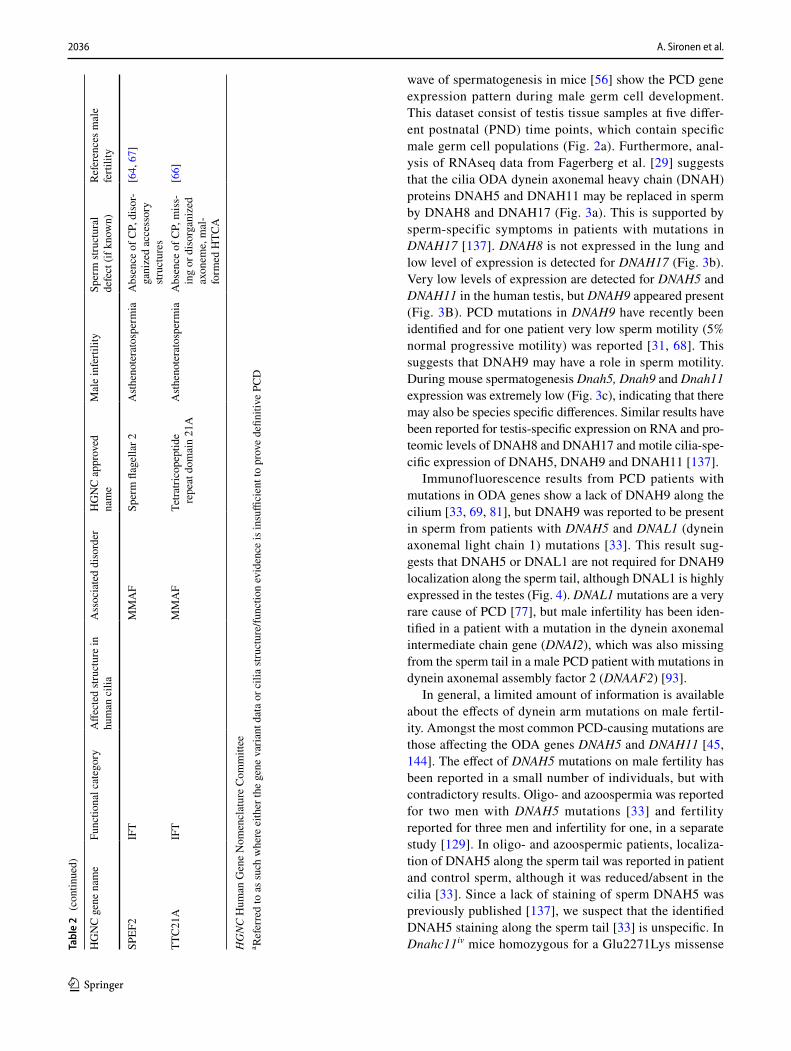
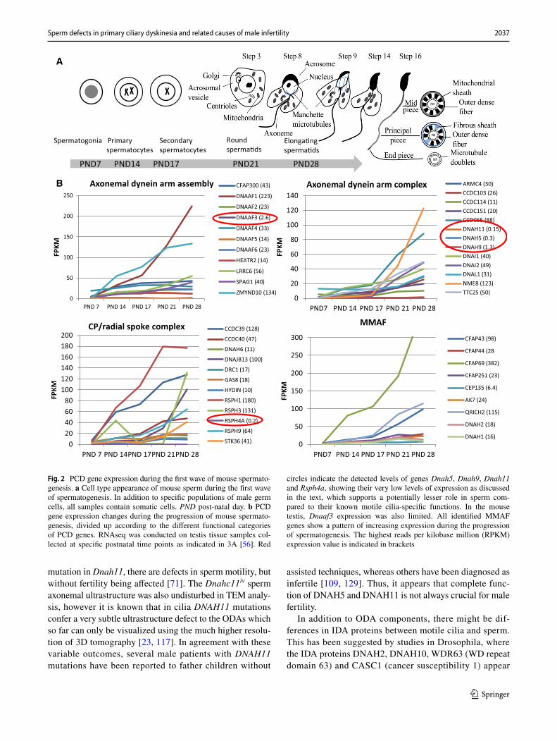
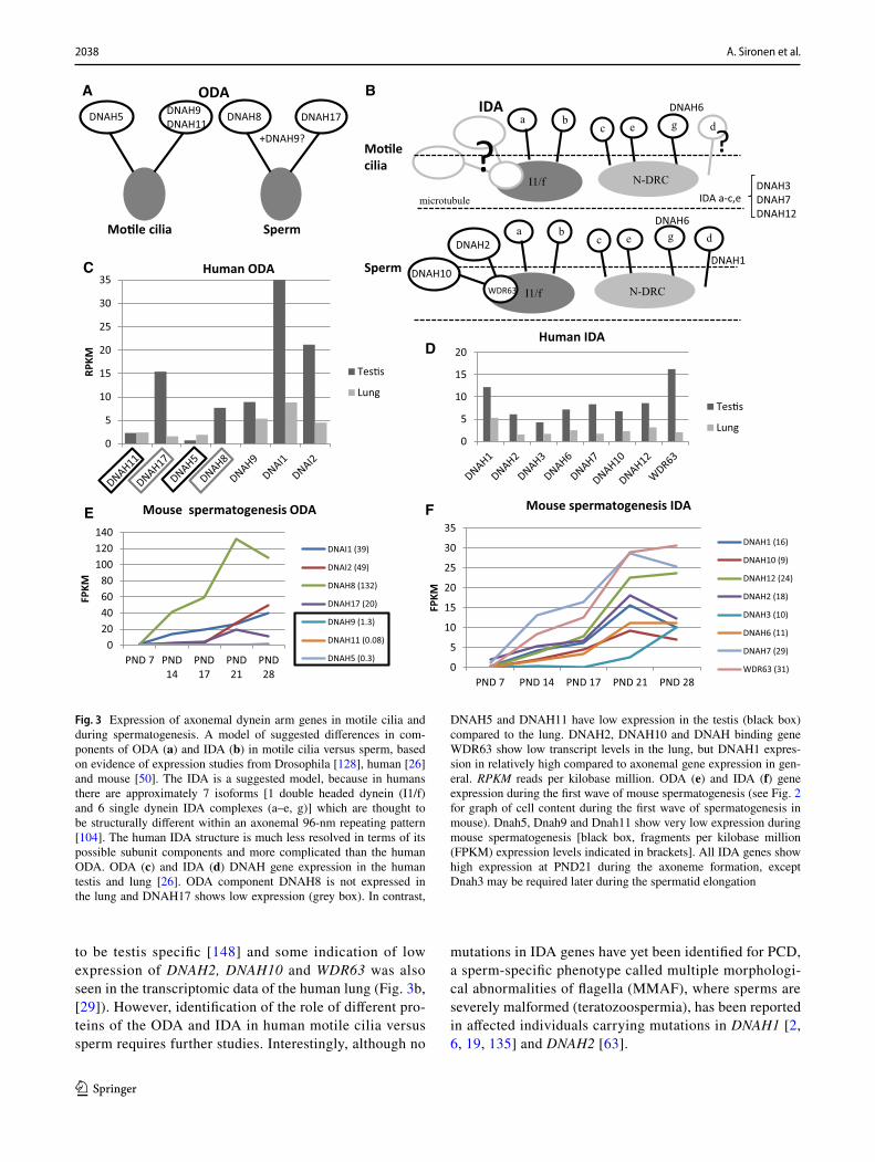
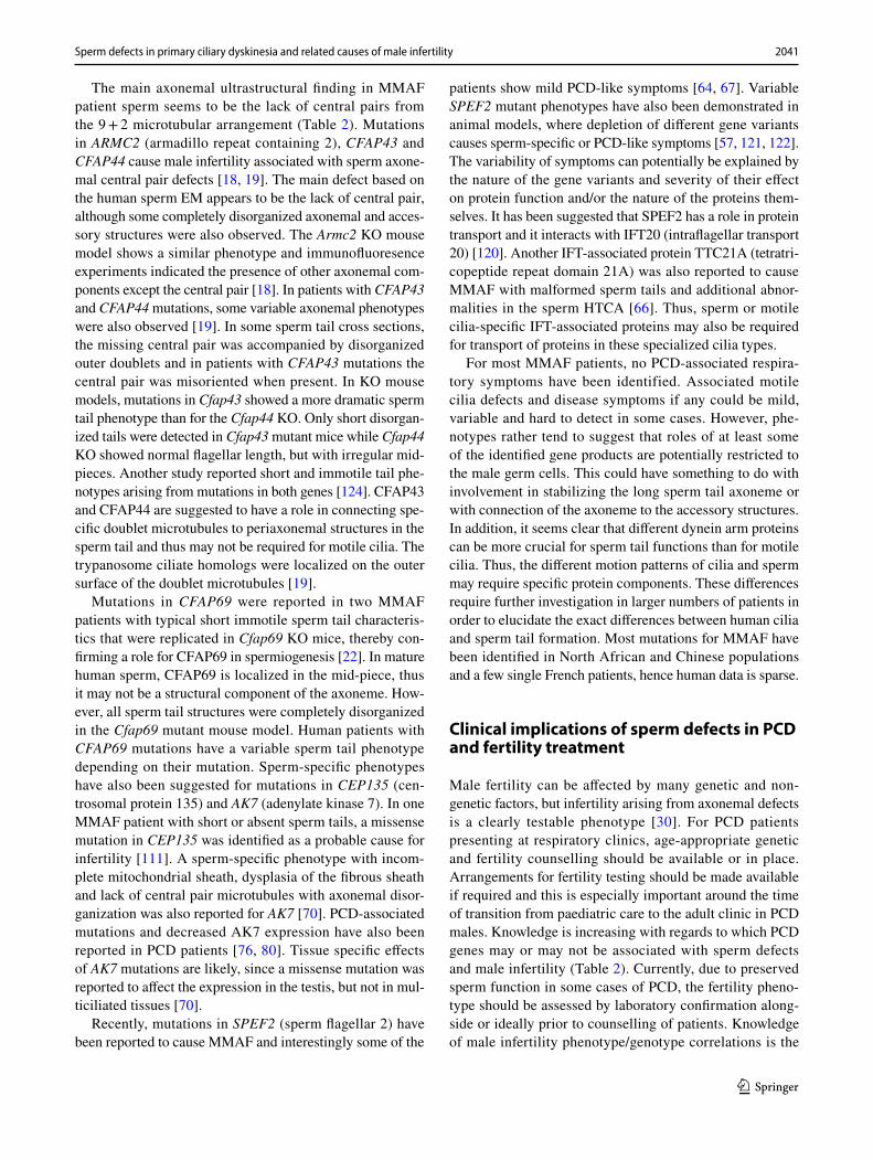
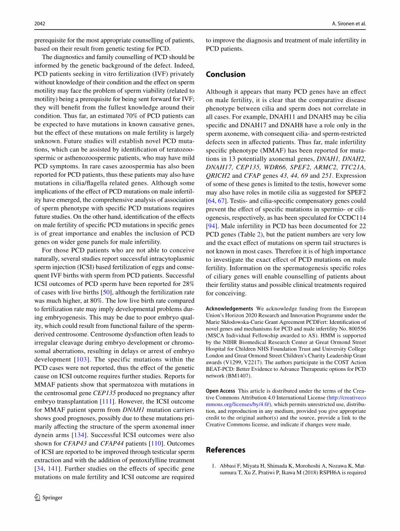
wave of spermatogenesis in mice [56] show the PCD gene expression pattern during male germ cell development. This dataset consist of testis tissue samples at five differ-ent postnatal (PND) time points, which contain specific male germ cell populations (Fig. 2a). Furthermore, anal-ysis of RNAseq data from Fagerberg et al. [29] suggests that the cilia ODA dynein axonemal heavy chain (DNAH) proteins DNAH5 and DNAH11 may be replaced in sperm by DNAH8 and DNAH17 (Fig. 3a). This is supported by sperm-specific symptoms in patients with mutations in DNAH17 [137]. DNAH8 is not expressed in the lung and low level of expression is detected for DNAH17 (Fig. 3b). Very low levels of expression are detected for DNAH5 and DNAH11 in the human testis, but DNAH9 appeared present (Fig. 3B). PCD mutations in DNAH9 have recently been identified and for one patient very low sperm motility (5% normal progressive motility) was reported [31, 68]. This suggests that DNAH9 may have a role in sperm motility. During mouse spermatogenesis Dnah5, Dnah9 and Dnah11 expression was extremely low (Fig. 3c), indicating that there may also be species specific differences. Similar results have been reported for testis-specific expression on RNA and pro-teomic levels of DNAH8 and DNAH17 and motile cilia-spe-cific expression of DNAH5, DNAH9 and DNAH11 [137].
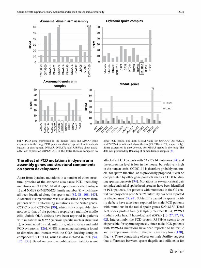
Immunofluorescence results from PCD patients with mutations in ODA genes show a lack of DNAH9 along the cilium [33, 69, 81], but DNAH9 was reported to be present in sperm from patients with DNAH5 and DNAL1 (dynein axonemal light chain 1) mutations [33]. This result sug-gests that DNAH5 or DNAL1 are not required for DNAH9 localization along the sperm tail, although DNAL1 is highly expressed in the testes (Fig. 4). DNAL1 mutations are a very rare cause of PCD [77], but male infertility has been iden-tified in a patient with a mutation in the dynein axonemal intermediate chain gene (DNAI2), which was also missing from the sperm tail in a male PCD patient with mutations in dynein axonemal assembly factor 2 (DNAAF2) [93].
In general, a limited amount of information is available about the effects of dynein arm mutations on male fertil-ity. Amongst the most common PCD-causing mutations are those affecting the ODA genes DNAH5 and DNAH11 [45, 144]. The effect of DNAH5 mutations on male fertility has been reported in a small number of individuals, but with contradictory results. Oligo- and azoospermia was reported for two men with DNAH5 mutations [33] and fertility reported for three men and infertility for one, in a separate study [129]. In oligo- and azoospermic patients, localiza-tion of DNAH5 along the sperm tail was reported in patient and control sperm, although it was reduced/absent in the cilia [33]. Since a lack of staining of sperm DNAH5 was previously published [137], we suspect that the identified DNAH5 staining along the sperm tail [33] is unspecific. In Dnahc11iv mice homozygous for a Glu2271Lys missense Ta
ble
2 (c
ontin
ued)
HG
NC
gen
e na
me
Func
tiona
l cat
egor
yA
ffect
ed st
ruct
ure
in
hum
an c
ilia
Ass
ocia
ted
diso
rder
HG
NC
app
rove
d na
me
Mal
e in
ferti
lity
Sper
m st
ruct
ural
de
fect
(if k
now
n)Re
fere
nces
mal
e fe
rtilit
y
SPEF
2IF
TM
MA
FSp
erm
flag
ella
r 2A
sthe
note
rato
sper
mia
Abs
ence
of C
P, d
isor
-ga
nize
d ac
cess
ory
struc
ture
s
[64,
67]
TTC
21A
IFT
MM
AF
Tetra
trico
pept
ide
repe
at d
omai
n 21
AA
sthe
note
rato
sper
mia
Abs
ence
of C
P, m
iss-
ing
or d
isor
gani
zed
axon
eme,
mal
-fo
rmed
HTC
A
[66]
HG
NC
Hum
an G
ene
Nom
encl
atur
e C
omm
ittee
a Ref
erre
d to
as s
uch
whe
re e
ither
the
gene
var
iant
dat
a or
cili
a str
uctu
re/fu
nctio
n ev
iden
ce is
insu
ffici
ent t
o pr
ove
defin
itive
PC
D
2037Sperm defects in primary ciliary dyskinesia and related causes of male infertility
1 3
mutation in Dnah11, there are defects in sperm motility, but without fertility being affected [71]. The Dnahc11iv sperm axonemal ultrastructure was also undisturbed in TEM analy-sis, however it is known that in cilia DNAH11 mutations confer a very subtle ultrastructure defect to the ODAs which so far can only be visualized using the much higher resolu-tion of 3D tomography [23, 117]. In agreement with these variable outcomes, several male patients with DNAH11 mutations have been reported to father children without
assisted techniques, whereas others have been diagnosed as infertile [109, 129]. Thus, it appears that complete func-tion of DNAH5 and DNAH11 is not always crucial for male fertility.
In addition to ODA components, there might be dif-ferences in IDA proteins between motile cilia and sperm. This has been suggested by studies in Drosophila, where the IDA proteins DNAH2, DNAH10, WDR63 (WD repeat domain 63) and CASC1 (cancer susceptibility 1) appear
0
50
100
150
200
250
PND 7 PND 14 PND 17 PND 21 PND 28
FPKM
Axonemal dynein arm assembly CFAP300 (43)
DNAAF1 (223)
DNAAF2 (23)
DNAAF3 (2.6)
DNAAF4 (33)
DNAAF5 (14)
DNAAF6 (23)
HEATR2 (14)
LRRC6 (56)
SPAG1 (40)
ZMYND10 (134)
PND7 PND14 PND17 PND21 PND28
0
20
40
60
80
100
120
140
PND7 PND 14 PND 17 PND 21 PND 28
FPKM
Axonemal dynein arm complex ARMC4 (30)CCDC103 (26)CCDC114 (11)CCDC151 (20)CCDC65 (88)DNAH11 (0.15)DNAH5 (0.3)DNAH9 (1.3)DNAI1 (40)DNAI2 (49)DNAL1 (31)NME8 (123)TTC25 (50)
020406080
100120140160180200
PND 7 PND 14PND 17PND 21PND 28
FPKM
CP/radial spoke complex CCDC39 (128)
CCDC40 (47)
DNAH6 (11)
DNAJB13 (100)
DRC1 (17)
GAS8 (18)
HYDIN (10)
RSPH1 (180)
RSPH3 (131)
RSPH4A (0.2)
RSPH9 (64)
STK36 (41)
Spermatogonia Primaryspermatocytes
Secondaryspermatocytes
Roundsperma�ds
Elonga�ngsperma�ds
A
B
0
50
100
150
200
250
300
PND7 PND 14 PND 17 PND 21 PND 28
FPKM
MMAF
CFAP43 (98)
CFAP44 (28
CFAP69 (382)
CFAP251 (23)
CEP135 (6.4)
AK7 (24)
QRICH2 (115)
DNAH2 (18)
DNAH1 (16)
Fig. 2 PCD gene expression during the first wave of mouse spermato-genesis. a Cell type appearance of mouse sperm during the first wave of spermatogenesis. In addition to specific populations of male germ cells, all samples contain somatic cells. PND post-natal day. b PCD gene expression changes during the progression of mouse spermato-genesis, divided up according to the different functional categories of PCD genes. RNAseq was conducted on testis tissue samples col-lected at specific postnatal time points as indicated in 3A [56]. Red
circles indicate the detected levels of genes Dnah5, Dnah9, Dnah11 and Rsph4a, showing their very low levels of expression as discussed in the text, which supports a potentially lesser role in sperm com-pared to their known motile cilia-specific functions. In the mouse testis, Dnaaf3 expression was also limited. All identified MMAF genes show a pattern of increasing expression during the progression of spermatogenesis. The highest reads per kilobase million (RPKM) expression value is indicated in brackets
2038 A. Sironen et al.
1 3
to be testis specific [148] and some indication of low expression of DNAH2, DNAH10 and WDR63 was also seen in the transcriptomic data of the human lung (Fig. 3b, [29]). However, identification of the role of different pro-teins of the ODA and IDA in human motile cilia versus sperm requires further studies. Interestingly, although no
mutations in IDA genes have yet been identified for PCD, a sperm-specific phenotype called multiple morphologi-cal abnormalities of flagella (MMAF), where sperms are severely malformed (teratozoospermia), has been reported in affected individuals carrying mutations in DNAH1 [2, 6, 19, 135] and DNAH2 [63].
DNAH5 DNAH9DNAH11
DNAH8 DNAH17
Mo�le cilia Sperm
ODAA B
020406080
100120140
PND 7 PND14
PND17
PND21
PND28
FPKM
Mouse spermatogenesis ODA
DNAI1 (39)
DNAI2 (49)
DNAH8 (132)
DNAH17 (20)
DNAH9 (1.3)
DNAH11 (0.08)
DNAH5 (0.3)
IDA
C
Mo�lecilia
Sperm
0
5
10
15
20
25
30
35
RPKM
Tes�s
Lung
Human ODA
+DNAH9?
DNAH3DNAH7DNAH12
IDA a-c,e
?a b
c e g d
I1/f N-DRC
DNAH6
0
5
10
15
20Human IDA
Tes�s
Lung
DNAH2
DNAH10WDR63
DNAH1
a bc e g d
I1/f N-DRC
DNAH6
?
0
5
10
15
20
25
30
35
PND 7 PND 14 PND 17 PND 21 PND 28
FPKM
Mouse spermatogenesis IDA
DNAH1 (16)
DNAH10 (9)
DNAH12 (24)
DNAH2 (18)
DNAH3 (10)
DNAH6 (11)
DNAH7 (29)
WDR63 (31)
D
E F
microtubule
Fig. 3 Expression of axonemal dynein arm genes in motile cilia and during spermatogenesis. A model of suggested differences in com-ponents of ODA (a) and IDA (b) in motile cilia versus sperm, based on evidence of expression studies from Drosophila [128], human [26] and mouse [50]. The IDA is a suggested model, because in humans there are approximately 7 isoforms [1 double headed dynein (I1/f) and 6 single dynein IDA complexes (a–e, g)] which are thought to be structurally different within an axonemal 96-nm repeating pattern [104]. The human IDA structure is much less resolved in terms of its possible subunit components and more complicated than the human ODA. ODA (c) and IDA (d) DNAH gene expression in the human testis and lung [26]. ODA component DNAH8 is not expressed in the lung and DNAH17 shows low expression (grey box). In contrast,
DNAH5 and DNAH11 have low expression in the testis (black box) compared to the lung. DNAH2, DNAH10 and DNAH binding gene WDR63 show low transcript levels in the lung, but DNAH1 expres-sion in relatively high compared to axonemal gene expression in gen-eral. RPKM reads per kilobase million. ODA (e) and IDA (f) gene expression during the first wave of mouse spermatogenesis (see Fig. 2 for graph of cell content during the first wave of spermatogenesis in mouse). Dnah5, Dnah9 and Dnah11 show very low expression during mouse spermatogenesis [black box, fragments per kilobase million (FPKM) expression levels indicated in brackets]. All IDA genes show high expression at PND21 during the axoneme formation, except Dnah3 may be required later during the spermatid elongation
2039Sperm defects in primary ciliary dyskinesia and related causes of male infertility
1 3
The effect of PCD mutations in dynein arm assembly genes and structural components on sperm development
Apart from dyneins, mutations in a number of other struc-tural proteins of the axoneme also cause PCD, including mutations in CCDC65, SPAG1 (sperm-associated antigen 1) and NME8 (NME/NM23 family member 8) which have all been localized along the sperm tail [82, 88, 108, 145]. Axonemal disorganization was also described in sperm from patients with PCD-causing mutations in the ‘ruler genes’ CCDC39 and CCDC40 [90], which is a comparable phe-notype to that of the patient’s respiratory multiple motile cilia. Subtle ODA defects have been reported in patients with mutations in MNS1 (meiosis specific nuclear structural 1), accompanied by male infertility, situs inversus and mild PCD symptoms [126]. MNS1 is an axonemal protein found to dimerize and interact with the ODA docking complex component CCDC114, which is also mutated in PCD [94, 126, 133]. Based on previous publications, fertility is not
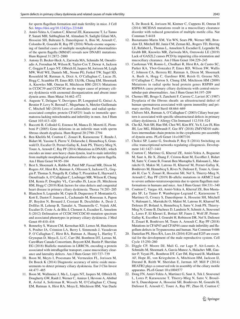
affected in PCD patients with CCDC114 mutations [94] and the expression level is low in the mouse, but relatively high in the human testis. CCDC114 is therefore probably not cru-cial for sperm function, or as previously proposed, it can be compensated by other gene products such as CCDC63 dur-ing spermatogenesis [94]. Mutations in several central pair complex and radial spoke head proteins have been identified in PCD patients. For patients with mutations in the C2 cen-tral pair projection gene HYDIN, infertility has been reported in affected men [59, 91]. Subfertility caused by sperm motil-ity defects have also been reported for male PCD patients with mutations in the radial spoke genes DNAJB13 (DnaJ heat shock protein family (Hsp40) member B13), RSPH3 (radial spoke head 3 homolog) and RSPH9 [13, 27, 37, 48, 62]. Interestingly, the PCD protein RSPH4A seems to be dispensable for spermatogenesis, since male PCD patients with RSPH4A mutations have been reported to be fertile and its expression levels in the testis are very low ([130], Fig. 4). These contrasting effects on male fertility suggest that differences between sperm flagella and cilia exist for
0
10
20
30
40
50
60
C21O
RF59
CFAP
300
DNAA
F1DN
AAF2
DNAA
F3DN
AAF4
DNAA
F5DN
AAF6
HEAT
R2LR
RC6
SPAG
1ZM
YND1
0
RPKM
Axonemal dynein arm assembly
0
10
20
30
40
50
60
ARM
C4CC
DC10
3CC
DC11
4CC
DC15
1CC
DC65
DNAH
11DN
AH5
DNAH
9DN
AI1
DNAI
2DN
AL1
NM
E8
RPKM
Axonemal dynein arm complex
0
10
20
30
40
50
60
CCDC
39CC
DC40
DNAH
6DN
AJB1
3DR
C1GA
S8HY
DIN
RSPH
1RS
PH3
RSPH
4ARS
PH9
STK3
6
RPKM
CP/radial spoke complex21073
0
10
20
30
40
50
60RP
KMMMAF genes
Tes�s
Lung
71
Fig. 4 PCD gene expression in the human testis and MMAF gene expression in the lung. PCD genes are divided up into functional cat-egories in each graph. DNAH5, DNAH11 and RSPH4A show mark-edly low expression (RPKM < 3) in the testis (boxes) compared to
other PCD genes. The high RPKM value for DNAAF3, ZMYND10 and TTC21A is indicated above the bar (73, 210 and 71, respectively). Some expression is also detected for MMAF genes in the lung. The data was produced by RNAseq of human tissues samples [29]
2040 A. Sironen et al.
1 3
the radial spoke head proteins, since mutations in any of the RSPH genes seems to have a similar consequence in the cilia; central pair loss and motility defects. A compensatory gene for RSPH4A in the testis may be RSPH6A, which has not been associated with PCD, but depletion of the mouse Rsph6a is a reported cause of male infertility [1].
Mutations in axonemal dynein complex preassembly genes have often been reported to cause male infertility in PCD (Table 2). In addition to mutations in DNAAF2 [93], PCD-causing mutations in DNAAF1, DNAAF3 and DNAAF4 have been reported to cause male infertility, but the patient number is minimal (Table 2). Mutations in their interacting partner, the dynein complex assembly factor PIH1 domain containing 3 (PIH1D3) have also been shown to result in lack of IDA and ODA [92, 98]. Male infertility has also been detected in PCD patients with mutations in axonemal dynein assembly factors LRRC6 (leucine-rich repeat con-taining 6), ZMYND10 (zinc finger MYND-type containing 10) and cilia and flagella-associated protein 300 (CFAP300). PCD with a lack of dynein arms in both the cilia and sperm is reported for LRRC6, ZMYND10, CFAP300 and axone-mal dynein complex gene CCDC103 (coiled-coil domain containing 103) [16, 44, 54, 65, 97, 99, 100]. However, for CCDC103 mutations variable male fertility phenotype has been reported; one patient showed a lack of DA and DRC comparable to the motile cilia phenotype, but the other was fertile without DA loss [100].
Genetic mutations causing sperm tail‑specific phenotypes
Mutations in a number of human genes result in sperm motility defects of a structural nature that is within the same spectrum as those caused by mutations in PCD genes. In these cases, associated with malformation of the sperm tail and low sperm count, mutations in a number of genes not connected to the respiratory PCD phenotype are reported. These apparently only affect the sperm tail axoneme and not cilia functions, having been identified as the cause of MMAF. The main findings in MMAF are absent, short, bent, coiled, and/or irregular sperm flagella with very low numbers of normal spermatozoa (0–2%) associated with low sperm concentrations [6]. A MMAF sperm tail phenotype of short, stumpy tails has also previously been described as dysplasia of the fibrous sheath (DFS) [5, 14, 102]. DFS has been reported to be caused by mutations in three genes encoding components of the sperm tail specific fibrous sheath: AKAP3, AKAP4 and FSIP2 [5, 75]. AKAP3 and 4 are the main components of the fibrous sheath and FSIP2 is required for localization of AKAP4 to the FS [75].
Sperm tail accessory structures are also often disorgan-ized in MMAF patients with mutations in axonemal genes,
such as DNAH1, which is the most common identified cause of MMAF [2, 6, 135]. DNAH1 is a component of the IDAs, which are completely disorganized in sperm tail axonemes of MMAF patients with DNAH1 mutations [6]. Mutations in another IDA gene, DNAH2, have also been identified as a cause for MMAF [63]. The sperm tail phenotype is similar in patients with DNAH1 and DNAH2 mutations with lack of the CP or complete disorganization of axonemal and ODF structures in addition to IDA loss [2, 6, 63, 135] indicating that IDAs are crucial for sperm tail axoneme integrity. Infer-tility has been the only PCD symptom identified in MMAF affected men with DNAH1 and DNAH2 mutations suggest-ing that these IDA components are not required for motile cilia function. However, DNAH1 mutations have also been reported in one family with siblings affected by PCD [46] and DNAH1 appears to be expressed in the lung (Fig. 3B). The possible role of DNAH1 and other IDA genes in PCD awaits to be revealed. IDA gene mutations may cause milder PCD symptoms due to the role of IDAs in controlling the wave form [52].
There seem to be differences in the severity of the sperm tail phenotype caused by IDA mutations between species in addition to differences between motile cilia and sperm. Dnah1 knock-out (KO) mice only show motility, but no structural sperm defects (asthenozoospermia) [87], whereas in humans DNAH1 truncating mutations induce a MMAF phenotype [2, 135]. Decreased cilia beat frequency was also detected in the mutant mice, which suggests a comparable role of DNAH1 in mouse sperm tail and motile cilia. In humans the lack of DNAH1 in motile cilia could be com-pensated by other DNAH genes such as DNAH12, which is the closest paralog to DNAH1 [74].
A report on the effect of sperm-specific ODA heavy chain DNAH17 mutations was recently published [137]. DNAH17 mutations were identified in five MMAF patients with sperm lacking ODAs. Some sperm tail cross sections showed miss-ing CP and even disorganized outer doublet microtubules, but no defects were seen in the patient’s respiratory cilia. Thus, DNAH17 mutations only appear to affect sperm tail ODA formation, a finding also supported by expression and protein localization studies [137].
The cilia and flagella-associated protein (CFAP) CFAP251, also known as WDR66, a component of the calmodulin- and radial-spoke-associated complex [24, 25] is located adjacent to DNAH1 in T. thermophila at the base of the RS3 [42, 53] radial spoke complex. The amount of DNAH1 homolog IDA g was reduced in T. thermophila CFAP251 KO [128] and a direct physical link between CFAP251, IDA3, and RS3 has been shown by co-immuno-precipitation experiments [4]. Mutations in WDR66 cause a typical MMAF phenotype with immotile, malformed sperm tails in which all the sperm tail structures are disorganized [4, 51, 61].
2041Sperm defects in primary ciliary dyskinesia and related causes of male infertility
1 3
The main axonemal ultrastructural finding in MMAF patient sperm seems to be the lack of central pairs from the 9 + 2 microtubular arrangement (Table 2). Mutations in ARMC2 (armadillo repeat containing 2), CFAP43 and CFAP44 cause male infertility associated with sperm axone-mal central pair defects [18, 19]. The main defect based on the human sperm EM appears to be the lack of central pair, although some completely disorganized axonemal and acces-sory structures were also observed. The Armc2 KO mouse model shows a similar phenotype and immunofluoresence experiments indicated the presence of other axonemal com-ponents except the central pair [18]. In patients with CFAP43 and CFAP44 mutations, some variable axonemal phenotypes were also observed [19]. In some sperm tail cross sections, the missing central pair was accompanied by disorganized outer doublets and in patients with CFAP43 mutations the central pair was misoriented when present. In KO mouse models, mutations in Cfap43 showed a more dramatic sperm tail phenotype than for the Cfap44 KO. Only short disorgan-ized tails were detected in Cfap43 mutant mice while Cfap44 KO showed normal flagellar length, but with irregular mid-pieces. Another study reported short and immotile tail phe-notypes arising from mutations in both genes [124]. CFAP43 and CFAP44 are suggested to have a role in connecting spe-cific doublet microtubules to periaxonemal structures in the sperm tail and thus may not be required for motile cilia. The trypanosome ciliate homologs were localized on the outer surface of the doublet microtubules [19].
Mutations in CFAP69 were reported in two MMAF patients with typical short immotile sperm tail characteris-tics that were replicated in Cfap69 KO mice, thereby con-firming a role for CFAP69 in spermiogenesis [22]. In mature human sperm, CFAP69 is localized in the mid-piece, thus it may not be a structural component of the axoneme. How-ever, all sperm tail structures were completely disorganized in the Cfap69 mutant mouse model. Human patients with CFAP69 mutations have a variable sperm tail phenotype depending on their mutation. Sperm-specific phenotypes have also been suggested for mutations in CEP135 (cen-trosomal protein 135) and AK7 (adenylate kinase 7). In one MMAF patient with short or absent sperm tails, a missense mutation in CEP135 was identified as a probable cause for infertility [111]. A sperm-specific phenotype with incom-plete mitochondrial sheath, dysplasia of the fibrous sheath and lack of central pair microtubules with axonemal disor-ganization was also reported for AK7 [70]. PCD-associated mutations and decreased AK7 expression have also been reported in PCD patients [76, 80]. Tissue specific effects of AK7 mutations are likely, since a missense mutation was reported to affect the expression in the testis, but not in mul-ticiliated tissues [70].
Recently, mutations in SPEF2 (sperm flagellar 2) have been reported to cause MMAF and interestingly some of the
patients show mild PCD-like symptoms [64, 67]. Variable SPEF2 mutant phenotypes have also been demonstrated in animal models, where depletion of different gene variants causes sperm-specific or PCD-like symptoms [57, 121, 122]. The variability of symptoms can potentially be explained by the nature of the gene variants and severity of their effect on protein function and/or the nature of the proteins them-selves. It has been suggested that SPEF2 has a role in protein transport and it interacts with IFT20 (intraflagellar transport 20) [120]. Another IFT-associated protein TTC21A (tetratri-copeptide repeat domain 21A) was also reported to cause MMAF with malformed sperm tails and additional abnor-malities in the sperm HTCA [66]. Thus, sperm or motile cilia-specific IFT-associated proteins may also be required for transport of proteins in these specialized cilia types.
For most MMAF patients, no PCD-associated respira-tory symptoms have been identified. Associated motile cilia defects and disease symptoms if any could be mild, variable and hard to detect in some cases. However, phe-notypes rather tend to suggest that roles of at least some of the identified gene products are potentially restricted to the male germ cells. This could have something to do with involvement in stabilizing the long sperm tail axoneme or with connection of the axoneme to the accessory structures. In addition, it seems clear that different dynein arm proteins can be more crucial for sperm tail functions than for motile cilia. Thus, the different motion patterns of cilia and sperm may require specific protein components. These differences require further investigation in larger numbers of patients in order to elucidate the exact differences between human cilia and sperm tail formation. Most mutations for MMAF have been identified in North African and Chinese populations and a few single French patients, hence human data is sparse.
Clinical implications of sperm defects in PCD and fertility treatment
Male fertility can be affected by many genetic and non-genetic factors, but infertility arising from axonemal defects is a clearly testable phenotype [30]. For PCD patients presenting at respiratory clinics, age-appropriate genetic and fertility counselling should be available or in place. Arrangements for fertility testing should be made available if required and this is especially important around the time of transition from paediatric care to the adult clinic in PCD males. Knowledge is increasing with regards to which PCD genes may or may not be associated with sperm defects and male infertility (Table 2). Currently, due to preserved sperm function in some cases of PCD, the fertility pheno-type should be assessed by laboratory confirmation along-side or ideally prior to counselling of patients. Knowledge of male infertility phenotype/genotype correlations is the
2042 A. Sironen et al.
1 3
prerequisite for the most appropriate counselling of patients, based on their result from genetic testing for PCD.
The diagnostics and family counselling of PCD should be informed by the genetic background of the defect. Indeed, PCD patients seeking in vitro fertilization (IVF) privately without knowledge of their condition and the effect on sperm motility may face the problem of sperm viability (related to motility) being a prerequisite for being sent forward for IVF; they will benefit from the fullest knowledge around their condition. Thus far, an estimated 70% of PCD patients can be expected to have mutations in known causative genes, but the effect of these mutations on male fertility is largely unknown. Future studies will establish novel PCD muta-tions, which can be assisted by identification of teratozoo-spermic or asthenozoospermic patients, who may have mild PCD symptoms. In rare cases azoospermia has also been reported for PCD patients, thus these patients may also have mutations in cilia/flagella related genes. Although some implications of the effect of PCD mutations on male infertil-ity have emerged, the comprehensive analysis of association of sperm phenotype with specific PCD mutations requires future studies. On the other hand, identification of the effects on male fertility of specific PCD mutations in specific genes is of great importance and enables the inclusion of PCD genes on wider gene panels for male infertility.
For those PCD patients who are not able to conceive naturally, several studies report successful intracytoplasmic sperm injection (ICSI) based fertilization of eggs and conse-quent IVF births with sperm from PCD patients. Successful ICSI outcomes of PCD sperm have been reported for 28% of cases with live births [50], although the fertilization rate was much higher, at 80%. The low live birth rate compared to fertilization rate may imply developmental problems dur-ing embryogenesis. This may be due to poor embryo qual-ity, which could result from functional failure of the sperm-derived centrosome. Centrosome dysfunction often leads to irregular cleavage during embryo development or chromo-somal aberrations, resulting in delays or arrest of embryo development [103]. The specific mutations within the PCD cases were not reported, thus the effect of the genetic cause on ICSI outcome requires further studies. Reports for MMAF patients show that spermatozoa with mutations in the centrosomal gene CEP135 produced no pregnancy after embryo transplantation [111]. However, the ICSI outcome for MMAF patient sperm from DNAH1 mutation carriers shows good prognoses, possibly due to these mutations pri-marily affecting the structure of the sperm axonemal inner dynein arms [134]. Successful ICSI outcomes were also shown for CFAP43 and CFAP44 patients [110]. Outcomes of ICSI are reported to be improved through testicular sperm extraction and with the addition of pentoxifylline treatment [34, 141]. Further studies on the effects of specific gene mutations on male fertility and ICSI outcome are required
to improve the diagnosis and treatment of male infertility in PCD patients.
Conclusion
Although it appears that many PCD genes have an effect on male fertility, it is clear that the comparative disease phenotype between cilia and sperm does not correlate in all cases. For example, DNAH11 and DNAH5 may be cilia specific and DNAH17 and DNAH8 have a role only in the sperm axoneme, with consequent cilia- and sperm-restricted defects seen in affected patients. Thus far, male infertility specific phenotype (MMAF) has been reported for muta-tions in 13 potentially axonemal genes, DNAH1, DNAH2, DNAH17, CEP135, WDR66, SPEF2, ARMC2, TTC21A, QRICH2 and CFAP genes 43, 44, 69 and 251. Expression of some of these genes is limited to the testis, however some may also have roles in motile cilia as suggested for SPEF2 [64, 67]. Testis- and cilia-specific compensatory genes could prevent the effect of specific mutations in spermio- or cili-ogenesis, respectively, as has been speculated for CCDC114 [94]. Male infertility in PCD has been documented for 22 PCD genes (Table 2), but the patient numbers are very low and the exact effect of mutations on sperm tail structures is not known in most cases. Therefore it is of high importance to investigate the exact effect of PCD mutations on male fertility. Information on the spermatogenesis specific roles of ciliary genes will enable counselling of patients about their fertility status and possible clinical treatments required for conceiving.
Acknowledgements We acknowledge funding from the European Union’s Horizon 2020 Research and Innovation Programme under the Marie Skłodowska-Curie Grant Agreement PCDFert: Identification of novel genes and mechanisms for PCD and male infertility No. 800556 (MSCA Individual Fellowship awarded to AS). HMM is supported by the NIHR Biomedical Research Center at Great Ormond Street Hospital for Children NHS Foundation Trust and University College London and Great Ormond Street Children’s Charity Leadership Grant awards (V1299, V2217). The authors participate in the COST Action BEAT-PCD: Better Evidence to Advance Therapeutic options for PCD network (BM1407).
Open Access This article is distributed under the terms of the Crea-tive Commons Attribution 4.0 International License (http://creat iveco mmons .org/licen ses/by/4.0/), which permits unrestricted use, distribu-tion, and reproduction in any medium, provided you give appropriate credit to the original author(s) and the source, provide a link to the Creative Commons license, and indicate if changes were made.
References
1. Abbasi F, Miyata H, Shimada K, Morohoshi A, Nozawa K, Mat-sumura T, Xu Z, Pratiwi P, Ikawa M (2018) RSPH6A is required
2043Sperm defects in primary ciliary dyskinesia and related causes of male infertility
1 3
for sperm flagellum formation and male fertility in mice. J Cell Sci. https ://doi.org/10.1242/jcs.22164 8
2. Amiri-Yekta A, Coutton C, Kherraf ZE, Karaouzene T, Le Tanno P, Sanati MH, Sabbaghian M, Almadani N, Sadighi Gilani MA, Hosseini SH, Bahrami S, Daneshipour A, Bini M, Arnoult C, Colombo R, Gourabi H, Ray PF (2016) Whole-exome sequenc-ing of familial cases of multiple morphological abnormalities of the sperm flagella (MMAF) reveals new DNAH1 mutations. Hum Reprod 31:2872–2880
3. Antony D, Becker-Heck A, Zariwala MA, Schmidts M, Onoufri-adis A, Forouhan M, Wilson R, Taylor-Cox T, Dewar A, Jackson C, Goggin P, Loges NT, Olbrich H, Jaspers M, Jorissen M, Leigh MW, Wolf WE, Daniels ML, Noone PG, Ferkol TW, Sagel SD, Rosenfeld M, Rutman A, Dixit A, O’Callaghan C, Lucas JS, Hogg C, Scambler PJ, Emes RD, Uk10k, Chung EM, Shoemark A, Knowles MR, Omran H, Mitchison HM (2013) Mutations in CCDC39 and CCDC40 are the major cause of primary cili-ary dyskinesia with axonemal disorganization and absent inner dynein arms. Hum Mutat 34:462–472
4. Auguste Y, Delague V, Desvignes JP, Longepied G, Gnisci A, Besnier P, Levy N, Beroud C, Megarbane A, Metzler-Guillemain C, Mitchell MJ (2018) Loss of calmodulin- and radial-spoke-associated complex protein CFAP251 leads to immotile sper-matozoa lacking mitochondria and infertility in men. Am J Hum Genet 103:413–420
5. Baccetti B, Collodel G, Estenoz M, Manca D, Moretti E, Piom-boni P (2005) Gene deletions in an infertile man with sperm fibrous sheath dysplasia. Hum Reprod 20:2790–2794
6. Ben Khelifa M, Coutton C, Zouari R, Karaouzene T, Rendu J, Bidart M, Yassine S, Pierre V, Delaroche J, Hennebicq S, Grun-wald D, Escalier D, Pernet-Gallay K, Jouk PS, Thierry-Mieg N, Toure A, Arnoult C, Ray PF (2014) Mutations in DNAH1, which encodes an inner arm heavy chain dynein, lead to male infertility from multiple morphological abnormalities of the sperm flagella. Am J Hum Genet 94:95–104
7. Best S, Shoemark A, Rubbo B, Patel MP, Fassad MR, Dixon M, Rogers AV, Hirst RA, Rutman A, Ollosson S, Jackson CL, Gog-gin P, Thomas S, Pengelly R, Cullup T, Pissaridou E, Hayward J, Onoufriadis A, O’Callaghan C, Loebinger MR, Wilson R, Chung EM, Kenia P, Doughty VL, Carvalho JS, Lucas JS, Mitchison HM, Hogg C (2019) Risk factors for situs defects and congenital heart disease in primary ciliary dyskinesia. Thorax 74:203–205
8. Blanchon S, Legendre M, Copin B, Duquesnoy P, Montantin G, Kott E, Dastot F, Jeanson L, Cachanado M, Rousseau A, Papon JF, Beydon N, Brouard J, Crestani B, Deschildre A, Desir J, Dollfus H, Leheup B, Tamalet A, Thumerelle C, Vojtek AM, Escalier D, Coste A, de Blic J, Clement A, Escudier E, Amselem S (2012) Delineation of CCDC39/CCDC40 mutation spectrum and associated phenotypes in primary ciliary dyskinesia. J Med Genet 49:410–416
9. Bonnefoy S, Watson CM, Kernohan KD, Lemos M, Hutchinson S, Poulter JA, Crinnion LA, Berry I, Simmonds J, Vasudevan P, O’Callaghan C, Hirst RA, Rutman A, Huang L, Hartley T, Grynspan D, Moya E, Li C, Carr IM, Bonthron DT, Leroux M, Care4Rare Canada Consortium, Boycott KM, Bastin P, Sheridan EG (2018) Biallelic mutations in LRRC56, encoding a protein associated with intraflagellar transport, cause mucociliary clear-ance and laterality defects. Am J Hum Genet 103:727–739
10. Boon M, Meyts I, Proesmans M, Vermeulen FL, Jorissen M, De Boeck K (2014) Diagnostic accuracy of nitric oxide meas-urements to detect primary ciliary dyskinesia. Eur J Clin Invest 44:477–485
11. Boon M, Wallmeier J, Ma L, Loges NT, Jaspers M, Olbrich H, Dougherty GW, Raidt J, Werner C, Amirav I, Hevroni A, Abitbul R, Avital A, Soferman R, Wessels M, O’Callaghan C, Chung EM, Rutman A, Hirst RA, Moya E, Mitchison HM, Van Daele
S, De Boeck K, Jorissen M, Kintner C, Cuppens H, Omran H (2014) MCIDAS mutations result in a mucociliary clearance disorder with reduced generation of multiple motile cilia. Nat Commun 5:4418
12. Bustamante-Marin XM, Yin WN, Sears PR, Werner ME, Brot-slaw EJ, Mitchell BJ, Jania CM, Zeman KL, Rogers TD, Herring LE, Refabert L, Thomas L, Amselem S, Escudier E, Legendre M, Grubb BR, Knowles MR, Zariwala MA, Ostrowski LE (2019) Lack of GAS2L2 causes PCD by impairing cilia orientation and mucociliary clearance. Am J Hum Genet 104:229–245
13. Castleman VH, Romio L, Chodhari R, Hirst RA, de Castro SC, Parker KA, Ybot-Gonzalez P, Emes RD, Wilson SW, Wallis C, Johnson CA, Herrera RJ, Rutman A, Dixon M, Shoemark A, Bush A, Hogg C, Gardiner RM, Reish O, Greene ND, O’Callaghan C, Purton S, Chung EM, Mitchison HM (2009) Mutations in radial spoke head protein genes RSPH9 and RSPH4A cause primary ciliary dyskinesia with central-micro-tubular-pair abnormalities. Am J Hum Genet 84:197–209
14. Chemes HE, Brugo S, Zanchetti F, Carrere C, Lavieri JC (1987) Dysplasia of the fibrous sheath: an ultrastructural defect of human spermatozoa associated with sperm immotility and pri-mary sterility. Fertil Steril 48:664–669
15. Chilvers MA, Rutman A, O’Callaghan C (2003) Ciliary beat pat-tern is associated with specific ultrastructural defects in primary ciliary dyskinesia. J Allergy Clin Immunol 112:518–524
16. Cho KJ, Noh SH, Han SM, Choi WI, Kim HY, Yu S, Lee JS, Rim JH, Lee MG, Hildebrandt F, Gee HY (2018) ZMYND10 stabi-lizes intermediate chain proteins in the cytoplasmic pre-assembly of dynein arms. PLoS Genet 14:e1007316
17. Choksi SP, Lauter G, Swoboda P, Roy S (2014) Switching on cilia: transcriptional networks regulating ciliogenesis. Develop-ment 141:1427–1441
18. Coutton C, Martinez G, Kherraf ZE, Amiri-Yekta A, Boguenet M, Saut A, He X, Zhang F, Cristou-Kent M, Escoffier J, Bidart M, Satre V, Conne B, Fourati Ben Mustapha S, Halouani L, Mar-rakchi O, Makni M, Latrous H, Kharouf M, Pernet-Gallay K, Bonhivers M, Hennebicq S, Rives N, Dulioust E, Toure A, Gour-abi H, Cao Y, Zouari R, Hosseini SH, Nef S, Thierry-Mieg N, Arnoult C, Ray PF (2019) Bi-allelic mutations in ARMC2 lead to severe astheno-teratozoospermia due to sperm flagellum mal-formations in humans and mice. Am J Hum Genet 104:331–340
19. Coutton C, Vargas AS, Amiri-Yekta A, Kherraf ZE, Ben Musta-pha SF, Le Tanno P, Wambergue-Legrand C, Karaouzene T, Martinez G, Crouzy S, Daneshipour A, Hosseini SH, Mitchell V, Halouani L, Marrakchi O, Makni M, Latrous H, Kharouf M, Deleuze JF, Boland A, Hennebicq S, Satre V, Jouk PS, Thierry-Mieg N, Conne B, Dacheux D, Landrein N, Schmitt A, Stouvenel L, Lores P, El Khouri E, Bottari SP, Faure J, Wolf JP, Pernet-Gallay K, Escoffier J, Gourabi H, Robinson DR, Nef S, Dulioust E, Zouari R, Bonhivers M, Toure A, Arnoult C, Ray PF (2018) Mutations in CFAP43 and CFAP44 cause male infertility and fla-gellum defects in Trypanosoma and human. Nat Commun 9:686
20. Danielian PS, Hess RA, Lees JA (2016) E2f4 and E2f5 are essen-tial for the development of the male reproductive system. Cell Cycle 15:250–260
21. Diggle CP, Moore DJ, Mali G, zur Lage P, Ait-Lounis A, Schmidts M, Shoemark A, Garcia Munoz A, Halachev MR, Gau-tier P, Yeyati PL, Bonthron DT, Carr IM, Hayward B, Markham AF, Hope JE, von Kriegsheim A, Mitchison HM, Jackson IJ, Durand B, Reith W, Sheridan E, Jarman AP, Mill P (2014) HEATR2 plays a conserved role in assembly of the ciliary motile apparatus. PLoS Genet 10:e1004577
22. Dong FN, Amiri-Yekta A, Martinez G, Saut A, Tek J, Stouvenel L, Lores P, Karaouzene T, Thierry-Mieg N, Satre V, Brouil-let S, Daneshipour A, Hosseini SH, Bonhivers M, Gourabi H, Dulioust E, Arnoult C, Toure A, Ray PF, Zhao H, Coutton C
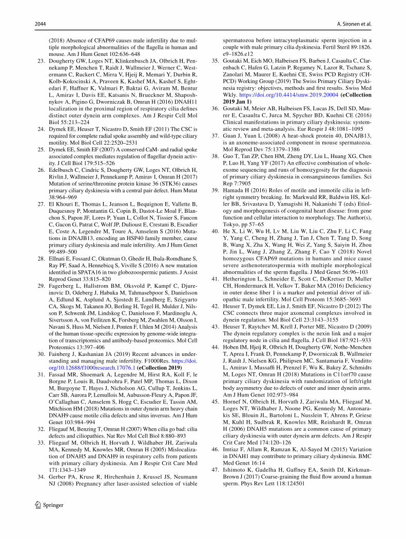
2044 A. Sironen et al.
1 3
(2018) Absence of CFAP69 causes male infertility due to mul-tiple morphological abnormalities of the flagella in human and mouse. Am J Hum Genet 102:636–648
23. Dougherty GW, Loges NT, Klinkenbusch JA, Olbrich H, Pen-nekamp P, Menchen T, Raidt J, Wallmeier J, Werner C, West-ermann C, Ruckert C, Mirra V, Hjeij R, Memari Y, Durbin R, Kolb-Kokocinski A, Praveen K, Kashef MA, Kashef S, Eght-edari F, Haffner K, Valmari P, Baktai G, Aviram M, Bentur L, Amirav I, Davis EE, Katsanis N, Brueckner M, Shaposh-nykov A, Pigino G, Dworniczak B, Omran H (2016) DNAH11 localization in the proximal region of respiratory cilia defines distinct outer dynein arm complexes. Am J Respir Cell Mol Biol 55:213–224
24. Dymek EE, Heuser T, Nicastro D, Smith EF (2011) The CSC is required for complete radial spoke assembly and wild-type ciliary motility. Mol Biol Cell 22:2520–2531
25. Dymek EE, Smith EF (2007) A conserved CaM- and radial spoke associated complex mediates regulation of flagellar dynein activ-ity. J Cell Biol 179:515–526
26. Edelbusch C, Cindric S, Dougherty GW, Loges NT, Olbrich H, Rivlin J, Wallmeier J, Pennekamp P, Amirav I, Omran H (2017) Mutation of serine/threonine protein kinase 36 (STK36) causes primary ciliary dyskinesia with a central pair defect. Hum Mutat 38:964–969
27. El Khouri E, Thomas L, Jeanson L, Bequignon E, Vallette B, Duquesnoy P, Montantin G, Copin B, Dastot-Le Moal F, Blan-chon S, Papon JF, Lores P, Yuan L, Collot N, Tissier S, Faucon C, Gacon G, Patrat C, Wolf JP, Dulioust E, Crestani B, Escudier E, Coste A, Legendre M, Toure A, Amselem S (2016) Muta-tions in DNAJB13, encoding an HSP40 family member, cause primary ciliary dyskinesia and male infertility. Am J Hum Genet 99:489–500
28. ElInati E, Fossard C, Okutman O, Ghedir H, Ibala-Romdhane S, Ray PF, Saad A, Hennebicq S, Viville S (2016) A new mutation identified in SPATA16 in two globozoospermic patients. J Assist Reprod Genet 33:815–820
29. Fagerberg L, Hallstrom BM, Oksvold P, Kampf C, Djure-inovic D, Odeberg J, Habuka M, Tahmasebpoor S, Danielsson A, Edlund K, Asplund A, Sjostedt E, Lundberg E, Szigyarto CA, Skogs M, Takanen JO, Berling H, Tegel H, Mulder J, Nils-son P, Schwenk JM, Lindskog C, Danielsson F, Mardinoglu A, Sivertsson A, von Feilitzen K, Forsberg M, Zwahlen M, Olsson I, Navani S, Huss M, Nielsen J, Ponten F, Uhlen M (2014) Analysis of the human tissue-specific expression by genome-wide integra-tion of transcriptomics and antibody-based proteomics. Mol Cell Proteomics 13:397–406
30. Fainberg J, Kashanian JA (2019) Recent advances in under-standing and managing male infertility. F1000Res. https ://doi.org/10.12688 /f1000 resea rch.17076 .1 (eCollection 2019)
31. Fassad MR, Shoemark A, Legendre M, Hirst RA, Koll F, le Borgne P, Louis B, Daudvohra F, Patel MP, Thomas L, Dixon M, Burgoyne T, Hayes J, Nicholson AG, Cullup T, Jenkins L, Carr SB, Aurora P, Lemullois M, Aubusson-Fleury A, Papon JF, O’Callaghan C, Amselem S, Hogg C, Escudier E, Tassin AM, Mitchison HM (2018) Mutations in outer dynein arm heavy chain DNAH9 cause motile cilia defects and situs inversus. Am J Hum Genet 103:984–994
32. Fliegauf M, Benzing T, Omran H (2007) When cilia go bad: cilia defects and ciliopathies. Nat Rev Mol Cell Biol 8:880–893
33. Fliegauf M, Olbrich H, Horvath J, Wildhaber JH, Zariwala MA, Kennedy M, Knowles MR, Omran H (2005) Mislocaliza-tion of DNAH5 and DNAH9 in respiratory cells from patients with primary ciliary dyskinesia. Am J Respir Crit Care Med 171:1343–1349
34. Gerber PA, Kruse R, Hirchenhain J, Krussel JS, Neumann NJ (2008) Pregnancy after laser-assisted selection of viable
spermatozoa before intracytoplasmatic sperm injection in a couple with male primary cilia dyskinesia. Fertil Steril 89:1826.e9–1826.e12
35. Goutaki M, Eich MO, Halbeisen FS, Barben J, Casaulta C, Clar-enbach C, Hafen G, Latzin P, Regamey N, Lazor R, Tschanz S, Zanolari M, Maurer E, Kuehni CE, Swiss PCD Registry (CH-PCD) Working Group (2019) The Swiss Primary Ciliary Dyski-nesia registry: objectives, methods and first results. Swiss Med Wkly. https ://doi.org/10.4414/smw.2019.20004 (eCollection 2019 Jan 1)
36. Goutaki M, Meier AB, Halbeisen FS, Lucas JS, Dell SD, Mau-rer E, Casaulta C, Jurca M, Spycher BD, Kuehni CE (2016) Clinical manifestations in primary ciliary dyskinesia: system-atic review and meta-analysis. Eur Respir J 48:1081–1095
37. Guan J, Yuan L (2008) A heat-shock protein 40, DNAJB13, is an axoneme-associated component in mouse spermatozoa. Mol Reprod Dev 75:1379–1386
38. Guo T, Tan ZP, Chen HM, Zheng DY, Liu L, Huang XG, Chen P, Luo H, Yang YF (2017) An effective combination of whole-exome sequencing and runs of homozygosity for the diagnosis of primary ciliary dyskinesia in consanguineous families. Sci Rep 7:7905
39. Hamada H (2016) Roles of motile and immotile cilia in left-right symmetry breaking. In: Markwald RR, Baldwin HS, Kel-ler BB, Srivastava D, Yamagishi H, Nakanishi T (eds) Etiol-ogy and morphogenesis of congenital heart disease: from gene function and cellular interaction to morphology. The Author(s), Tokyo, pp 57–65
40. He X, Li W, Wu H, Lv M, Liu W, Liu C, Zhu F, Li C, Fang Y, Yang C, Cheng H, Zhang J, Tan J, Chen T, Tang D, Song B, Wang X, Zha X, Wang H, Wei Z, Yang S, Saiyin H, Zhou P, Jin L, Wang J, Zhang Z, Zhang F, Cao Y (2018) Novel homozygous CFAP69 mutations in humans and mice cause severe asthenoteratospermia with multiple morphological abnormalities of the sperm flagella. J Med Genet 56:96–103
41. Hetherington L, Schneider E, Scott C, DeKretser D, Muller CH, Hondermarck H, Velkov T, Baker MA (2016) Deficiency in outer dense fiber 1 is a marker and potential driver of idi-opathic male infertility. Mol Cell Proteom 15:3685–3693
42. Heuser T, Dymek EE, Lin J, Smith EF, Nicastro D (2012) The CSC connects three major axonemal complexes involved in dynein regulation. Mol Biol Cell 23:3143–3155
43. Heuser T, Raytchev M, Krell J, Porter ME, Nicastro D (2009) The dynein regulatory complex is the nexin link and a major regulatory node in cilia and flagella. J Cell Biol 187:921–933
44. Hoben IM, Hjeij R, Olbrich H, Dougherty GW, Nothe-Menchen T, Aprea I, Frank D, Pennekamp P, Dworniczak B, Wallmeier J, Raidt J, Nielsen KG, Philipsen MC, Santamaria F, Venditto L, Amirav I, Mussaffi H, Prenzel F, Wu K, Bakey Z, Schmidts M, Loges NT, Omran H (2018) Mutations in C11orf70 cause primary ciliary dyskinesia with randomization of left/right body asymmetry due to defects of outer and inner dynein arms. Am J Hum Genet 102:973–984
45. Hornef N, Olbrich H, Horvath J, Zariwala MA, Fliegauf M, Loges NT, Wildhaber J, Noone PG, Kennedy M, Antonara-kis SE, Blouin JL, Bartoloni L, Nusslein T, Ahrens P, Griese M, Kuhl H, Sudbrak R, Knowles MR, Reinhardt R, Omran H (2006) DNAH5 mutations are a common cause of primary ciliary dyskinesia with outer dynein arm defects. Am J Respir Crit Care Med 174:120–126
46. Imtiaz F, Allam R, Ramzan K, Al-Sayed M (2015) Variation in DNAH1 may contribute to primary ciliary dyskinesia. BMC Med Genet 16:14
47. Ishimoto K, Gadelha H, Gaffney EA, Smith DJ, Kirkman-Brown J (2017) Coarse-graining the fluid flow around a human sperm. Phys Rev Lett 118:124501
2045Sperm defects in primary ciliary dyskinesia and related causes of male infertility
1 3
48. Jeanson L, Copin B, Papon JF, Dastot-Le Moal F, Duquesnoy P, Montantin G, Cadranel J, Corvol H, Coste A, Desir J, Souayah A, Kott E, Collot N, Tissier S, Louis B, Tamalet A, de Blic J, Clement A, Escudier E, Amselem S, Legendre M (2015) RSPH3 mutations cause primary ciliary dyskinesia with central-complex defects and a near absence of radial spokes. Am J Hum Genet 97:153–162
49. Jeanson L, Thomas L, Copin B, Coste A, Sermet-Gaudelus I, Dastot-Le Moal F, Duquesnoy P, Montantin G, Collot N, Tissier S, Papon JF, Clement A, Louis B, Escudier E, Amselem S, Leg-endre M (2016) Mutations in GAS8, a gene encoding a nexin-dynein regulatory complex subunit, cause primary ciliary dys-kinesia with axonemal disorganization. Hum Mutat 37:776–785
50. Kawasaki A, Okamoto H, Wada A, Ainoya Y, Kita N, Maeyama T, Edamoto N, Nishiyama H, Tsukamoto S, Joraku A, Waku N, Yoshikawa H (2015) A case of primary ciliary dyskinesia treated with ICSI using testicular spermatozoa: case report and a review of the literature. Reprod Med Biol 14:195–200
51. Kherraf ZE, Amiri-Yekta A, Dacheux D, Karaouzene T, Cout-ton C, Christou-Kent M, Martinez G, Landrein N, Le Tanno P, Fourati Ben Mustapha S, Halouani L, Marrakchi O, Makni M, Latrous H, Kharouf M, Pernet-Gallay K, Gourabi H, Robinson DR, Crouzy S, Blum M, Thierry-Mieg N, Toure A, Zouari R, Arnoult C, Bonhivers M, Ray PF (2018) A homozygous ancestral SVA-insertion-mediated deletion in WDR66 induces multiple morphological abnormalities of the sperm flagellum and male infertility. Am J Hum Genet 103:400–412
52. King SM (2016) Axonemal dynein arms. Cold Spring Harb Per-spect Biol. https ://doi.org/10.1101/cshpe rspec t.a0281 00
53. King SM (2013) A solid-state control system for dynein-based ciliary/flagellar motility. J Cell Biol 201:173–175
54. Kott E, Duquesnoy P, Copin B, Legendre M, Dastot-Le Moal F, Montantin G, Jeanson L, Tamalet A, Papon JF, Siffroi JP, Rives N, Mitchell V, de Blic J, Coste A, Clement A, Escalier D, Toure A, Escudier E, Amselem S (2012) Loss-of-function mutations in LRRC6, a gene essential for proper axonemal assembly of inner and outer dynein arms, cause primary ciliary dyskinesia. Am J Hum Genet 91:958–964
55. Kuehni CE, Lucas JS (2017) Diagnosis of primary ciliary dys-kinesia: summary of the ERS Task Force report. Breathe (Sheff) 13:166–178
56. Laiho A, Kotaja N, Gyenesei A, Sironen A (2013) Transcriptome profiling of the murine testis during the first wave of spermato-genesis. PLoS One 8:e61558
57. Lehti MS, Henriksson H, Rummukainen P, Wang F, Uusitalo-Kylmala L, Kiviranta R, Heino TJ, Kotaja N, Sironen A (2018) Cilia-related protein SPEF2 regulates osteoblast differentiation. Sci Rep 8:859
58. Lehti MS, Sironen A (2017) Formation and function of sperm tail structures in association with sperm motility defects. Biol Reprod 97:522–536
59. Li L, Sha YW, Xu X, Mei LB, Qiu PP, Ji ZY, Lin SB, Su ZY, Wang C, Yin C, Li P (2018) DNAH6 is a novel candidate gene associated with sperm head anomaly. Andrologia 50:e12953
60. Li W, He X, Yang S, Liu C, Wu H, Liu W, Lv M, Tang D, Tan J, Tang S, Chen Y, Wang J, Zhang Z, Wang H, Jin L, Zhang F, Cao Y (2019) Biallelic mutations of CFAP251 cause sperm flagellar defects and human male infertility. J Hum Genet 64:49–54
61. Li W, He X, Yang S, Liu C, Wu H, Liu W, Lv M, Tang D, Tan J, Tang S, Chen Y, Wang J, Zhang Z, Wang H, Jin L, Zhang F, Cao Y (2018) Biallelic mutations of CFAP251 cause sperm flagellar defects and human male infertility. J Hum Genet 64:49
62. Li W, Liu G (2014) DNAJB13, a type II HSP40 family member, localizes to the spermatids and spermatozoa during mouse sper-matogenesis. BMC Dev Biol 14:38
63. Li Y, Sha Y, Wang X, Ding L, Liu W, Ji Z, Mei L, Huang X, Lin S, Kong S, Lu J, Qin W, Zhang X, Zhuang J, Tang Y, Lu Z (2019) DNAH2 is a novel candidate gene associated with multi-ple morphological abnormalities of the sperm flagella (MMAF). Clin Genet 95:590–600
64. Liu C, Lv M, He X, Zhu Y, Amiri-Yekta A, Li W, Wu H, Kherraf ZE, Liu W, Zhang J, Tan Q, Tang S, Zhu YJ, Zhong Y, Li C, Tian S, Zhang Z, Jin L, Ray P, Zhang F, Cao Y (2019) Homozygous mutations in SPEF2 induce multiple morphologi-cal abnormalities of the sperm flagella and male infertility. J Med Genet. https ://doi.org/10.1136/jmedg enet-2019-10601 1
65. Liu L, Luo H (2018) Whole-exome sequencing identified a novel compound heterozygous mutation of LRRC6 in a Chinese pri-mary ciliary dyskinesia patient. Biomed Res Int 2018:1854269
66. Liu W, He X, Yang S, Zouari R, Wang J, Wu H, Kherraf ZE, Liu C, Coutton C, Zhao R, Tang D, Tang S, Lv M, Fang Y, Li W, Li H, Zhao J, Wang X, Zhao S, Zhang J, Arnoult C, Jin L, Zhang Z, Ray PF, Cao Y, Zhang F (2019) Bi-allelic mutations in TTC21A induce asthenoteratospermia in humans and mice. Am J Hum Genet 104:738–748
67. Liu W, Sha Y, Li Y, Mei L, Lin S, Huang X, Lu J, Ding L, Kong S, Lu Z (2019) Loss-of-function mutations in SPEF2 cause multiple morphological abnormalities of the sperm flagella (MMAF). J Med Genet 56(10):678–684
68. Loges NT, Antony D, Maver A, Deardorff MA, Gulec EY, Gezdi-rici A, Nothe-Menchen T, Hoben IM, Jelten L, Frank D, Werner C, Tebbe J, Wu K, Goldmuntz E, Cuturilo G, Krock B, Ritter A, Hjeij R, Bakey Z, Pennekamp P, Dworniczak B, Brunner H, Peterlin B, Tanidir C, Olbrich H, Omran H, Schmidts M (2018) Recessive DNAH9 loss-of-function mutations cause laterality defects and subtle respiratory ciliary-beating defects. Am J Hum Genet 103:995–1008
69. Loges NT, Olbrich H, Fenske L, Mussaffi H, Horvath J, Fliegauf M, Kuhl H, Baktai G, Peterffy E, Chodhari R, Chung EM, Rut-man A, O’Callaghan C, Blau H, Tiszlavicz L, Voelkel K, Witt M, Zietkiewicz E, Neesen J, Reinhardt R, Mitchison HM, Omran H (2008) DNAI2 mutations cause primary ciliary dyskinesia with defects in the outer dynein arm. Am J Hum Genet 83:547–558
70. Lores P, Coutton C, El Khouri E, Stouvenel L, Givelet M, Thomas L, Rode B, Schmitt A, Louis B, Sakheli Z, Chaudhry M, Fernandez-Gonzales A, Mitsialis A, Dacheux D, Wolf JP, Papon JF, Gacon G, Escudier E, Arnoult C, Bonhivers M, Savinov SN, Amselem S, Ray PF, Dulioust E, Toure A (2018) Homozygous missense mutation L673P in adenylate kinase 7 (AK7) leads to primary male infertility and multiple morphological anomalies of the flagella but not to primary ciliary dyskinesia. Hum Mol Genet 27:1196–1211
71. Lucas JS, Adam EC, Goggin PM, Jackson CL, Powles-Glover N, Patel SH, Humphreys J, Fray MD, Falconnet E, Blouin JL, Cheeseman MT, Bartoloni L, Norris DP, Lackie PM (2012) Static respiratory cilia associated with mutations in Dnahc11/DNAH11: a mouse model of PCD. Hum Mutat 33:495–503
72. Lucas JS, Barbato A, Collins SA, Goutaki M, Behan L, Caudri D, Dell S, Eber E, Escudier E, Hirst RA, Hogg C, Jorissen M, Latzin P, Legendre M, Leigh MW, Midulla F, Nielsen KG, Omran H, Papon JF, Pohunek P, Redfern B, Rigau D, Rindlisbacher B, San-tamaria F, Shoemark A, Snijders D, Tonia T, Titieni A, Walker WT, Werner C, Bush A, Kuehni CE (2017) European Respira-tory Society guidelines for the diagnosis of primary ciliary dys-kinesia. Eur Respir J. https ://doi.org/10.1183/13993 003.01090 -2016 (print 2017 Jan)
73. Lucas JS, Burgess A, Mitchison HM, Moya E, Williamson M, Hogg C (2014) Diagnosis and management of primary ciliary dyskinesia. Arch Dis Child 99:850–856
74. Maiti AK, Mattei MG, Jorissen M, Volz A, Zeigler A, Bou-vagnet P (2000) Identification, tissue specific expression, and
2046 A. Sironen et al.
1 3
chromosomal localisation of several human dynein heavy chain genes. Eur J Hum Genet 8:923–932
75. Martinez G, Kherraf ZE, Zouari R, Fourati Ben Mustapha S, Saut A, Pernet-Gallay K, Bertrand A, Bidart M, Hograindleur JP, Amiri-Yekta A, Kharouf M, Karaouzene T, Thierry-Mieg N, Dacheux-Deschamps D, Satre V, Bonhivers M, Toure A, Arnoult C, Ray PF, Coutton C (2018) Whole-exome sequencing identifies mutations in FSIP2 as a recurrent cause of multiple morphological abnormalities of the sperm flagella. Hum Reprod 33:1973–1984
76. Mata M, Lluch-Estelles J, Armengot M, Sarrion I, Carda C, Cor-tijo J (2012) New adenylate kinase 7 (AK7) mutation in primary ciliary dyskinesia. Am J Rhinol Allergy 26:260–264
77. Mazor M, Alkrinawi S, Chalifa-Caspi V, Manor E, Sheffield VC, Aviram M, Parvari R (2011) Primary ciliary dyskinesia caused by homozygous mutation in DNAL1, encoding dynein light chain 1. Am J Hum Genet 88:599–607
78. Merveille AC, Davis EE, Becker-Heck A, Legendre M, Amirav I, Bataille G, Belmont J, Beydon N, Billen F, Clement A, Clercx C, Coste A, Crosbie R, de Blic J, Deleuze S, Duquesnoy P, Escalier D, Escudier E, Fliegauf M, Horvath J, Hill K, Jorissen M, Just J, Kispert A, Lathrop M, Loges NT, Marthin JK, Momozawa Y, Montantin G, Nielsen KG, Olbrich H, Papon JF, Rayet I, Roger G, Schmidts M, Tenreiro H, Towbin JA, Zelenika D, Zentgraf H, Georges M, Lequarre AS, Katsanis N, Omran H, Amselem S (2011) CCDC39 is required for assembly of inner dynein arms and the dynein regulatory complex and for normal ciliary motil-ity in humans and dogs. Nat Genet 43:72–78
79. Miki K, Willis WD, Brown PR, Goulding EH, Fulcher KD, Eddy EM (2002) Targeted disruption of the Akap4 gene causes defects in sperm flagellum and motility. Dev Biol 248:331–342
80. Milara J, Armengot M, Mata M, Morcillo EJ, Cortijo J (2010) Role of adenylate kinase type 7 expression on cilia motility: pos-sible link in primary ciliary dyskinesia. Am J Rhinol Allergy 24:181–185
81. Milisav I, Jones MH, Affara NA (1996) Characterization of a novel human dynein-related gene that is specifically expressed in testis. Mamm Genome 7:667–672
82. Miranda-Vizuete A, Tsang K, Yu Y, Jimenez A, Pelto-Huikko M, Flickinger CJ, Sutovsky P, Oko R (2003) Cloning and devel-opmental analysis of murid spermatid-specific thioredoxin-2 (SPTRX-2), a novel sperm fibrous sheath protein and autoanti-gen. J Biol Chem 278:44874–44885
83. Mitchison HM, Schmidts M, Loges NT, Freshour J, Dritsoula A, Hirst RA, O’Callaghan C, Blau H, Al Dabbagh M, Olbrich H, Beales PL, Yagi T, Mussaffi H, Chung EM, Omran H, Mitchell DR (2012) Mutations in axonemal dynein assembly factor DNAAF3 cause primary ciliary dyskinesia. Nat Genet 44(381–9):S1–S2
84. Mitchison HM, Valente EM (2017) Motile and non-motile cilia in human pathology: from function to phenotypes. J Pathol 241:294–309
85. Moore DJ, Onoufriadis A, Shoemark A, Simpson MA, zur Lage PI, de Castro SC, Bartoloni L, Gallone G, Petridi S, Woollard WJ, Antony D, Schmidts M, Didonna T, Makrythanasis P, Bevillard J, Mongan NP, Djakow J, Pals G, Lucas JS, Marthin JK, Nielsen KG, Santoni F, Guipponi M, Hogg C, Antonarakis SE, Emes RD, Chung EM, Greene ND, Blouin JL, Jarman AP, Mitchison HM (2013) Mutations in ZMYND10, a gene essential for proper axonemal assembly of inner and outer dynein arms in humans and flies, cause primary ciliary dyskinesia. Am J Hum Genet 93:346–356
86. Munro NC, Currie DC, Lindsay KS, Ryder TA, Rutman A, Dewar A, Greenstone MA, Hendry WF, Cole PJ (1994) Fertility
in men with primary ciliary dyskinesia presenting with respira-tory infection. Thorax 49:684–687
87. Neesen J, Kirschner R, Ochs M, Schmiedl A, Habermann B, Mueller C, Holstein AF, Nuesslein T, Adham I, Engel W (2001) Disruption of an inner arm dynein heavy chain gene results in asthenozoospermia and reduced ciliary beat frequency. Hum Mol Genet 10:1117–1128
88. Neesse A, Gangeswaran R, Luettges J, Feakins R, Weeks ME, Lemoine NR, Crnogorac-Jurcevic T (2007) Sperm-associated antigen 1 is expressed early in pancreatic tumorigenesis and pro-motes motility of cancer cells. Oncogene 26:1533–1545
89. Nunez-Olle M, Jung C, Terre B, Balsiger NA, Plata C, Roset R, Pardo-Pastor C, Garrido M, Rojas S, Alameda F, Lloreta J, Martin-Caballero J, Flores JM, Stracker TH, Valverde MA, Munoz FJ, Gil-Gomez G (2017) Constitutive Cyclin O deficiency results in penetrant hydrocephalus, impaired growth and infertil-ity. Oncotarget 8:99261–99273
90. Oda T, Yanagisawa H, Kamiya R, Kikkawa M (2014) A molec-ular ruler determines the repeat length in eukaryotic cilia and flagella. Science 346:857–860
91. Olbrich H, Schmidts M, Werner C, Onoufriadis A, Loges NT, Raidt J, Banki NF, Shoemark A, Burgoyne T, Al Turki S, Hurles ME, UK10 K Consortium, Kohler G, Schroeder J, Nurnberg G, Nurnberg P, Chung EM, Reinhardt R, Marthin JK, Nielsen KG, Mitchison HM, Omran H (2012) Recessive HYDIN mutations cause primary ciliary dyskinesia without randomization of left-right body asymmetry. Am J Hum Genet 91:672–684
92. Olcese C, Patel MP, Shoemark A, Kiviluoto S, Legendre M, Williams HJ, Vaughan CK, Hayward J, Goldenberg A, Emes RD, Munye MM, Dyer L, Cahill T, Bevillard J, Gehrig C, Guip-poni M, Chantot S, Duquesnoy P, Thomas L, Jeanson L, Copin B, Tamalet A, Thauvin-Robinet C, Papon JF, Garin A, Pin I, Vera G, Aurora P, Fassad MR, Jenkins L, Boustred C, Cullup T, Dixon M, Onoufriadis A, Bush A, Chung EM, Antonarakis SE, Loebinger MR, Wilson R, Armengot M, Escudier E, Hogg C, UK10 K Rare Group, Amselem S, Sun Z, Bartoloni L, Blouin JL, Mitchison HM (2017) X-linked primary ciliary dyskinesia due to mutations in the cytoplasmic axonemal dynein assembly factor PIH1D3. Nat Commun 8:14279
93. Omran H, Kobayashi D, Olbrich H, Tsukahara T, Loges NT, Hagiwara H, Zhang Q, Leblond G, O’Toole E, Hara C, Mizuno H, Kawano H, Fliegauf M, Yagi T, Koshida S, Miyawaki A, Zentgraf H, Seithe H, Reinhardt R, Watanabe Y, Kamiya R, Mitchell DR, Takeda H (2008) Ktu/PF13 is required for cyto-plasmic pre-assembly of axonemal dyneins. Nature 456:611–616
94. Onoufriadis A, Paff T, Antony D, Shoemark A, Micha D, Kuyt B, Schmidts M, Petridi S, Dankert-Roelse JE, Haarman EG, Daniels JM, Emes RD, Wilson R, Hogg C, Scambler PJ, Chung EM, UK10 K, Pals G, Mitchison HM (2013) Splice-site mutations in the axonemal outer dynein arm docking complex gene CCDC114 cause primary ciliary dyskinesia. Am J Hum Genet 92:88–98
95. Onoufriadis A, Shoemark A, Schmidts M, Patel M, Jimenez G, Liu H, Thomas B, Dixon M, Hirst RA, Rutman A, Burgoyne T, Williams C, Scully J, Bolard F, Lafitte JJ, Beales PL, Hogg C, Yang P, Chung EM, Emes RD, O’Callaghan C, UK10K, Bouvag-net P, Mitchison HM (2014) Targeted NGS gene panel identifies mutations in RSPH1 causing primary ciliary dyskinesia and a common mechanism for ciliary central pair agenesis due to radial spoke defects. Hum Mol Genet 23:3362–3374
96. Osinka A, Poprzeczko M, Zielinska MM, Fabczak H, Joachim-iak E, Wloga D (2019) Ciliary proteins: filling the gaps. Recent advances in deciphering the protein composition of motile ciliary complexes. Cells. https ://doi.org/10.3390/cells 80707 30
97. Ozkavukcu S, Celik-Ozenci C, Konuk E, Atabekoglu C (2018) Live birth after laser assisted viability assessment (LAVA) to
2047Sperm defects in primary ciliary dyskinesia and related causes of male infertility
1 3
detect pentoxifylline resistant ejaculated immotile spermato-zoa during ICSI in a couple with male Kartagener’s syndrome. Reprod Biol Endocrinol 16:10
98. Paff T, Loges NT, Aprea I, Wu K, Bakey Z, Haarman EG, Dan-iels JMA, Sistermans EA, Bogunovic N, Dougherty GW, Hoben IM, Grosse-Onnebrink J, Matter A, Olbrich H, Werner C, Pals G, Schmidts M, Omran H, Micha D (2017) Mutations in PIH1D3 cause X-linked primary ciliary dyskinesia with outer and inner dynein arm defects. Am J Hum Genet 100:160–168
99. Pereira R, Oliveira J, Ferraz L, Barros A, Santos R, Sousa M (2015) Mutation analysis in patients with total sperm immotil-ity. J Assist Reprod Genet 32:893–902
100. Pereira R, Oliveira ME, Santos R, Oliveira E, Barbosa T, San-tos T, Goncalves P, Ferraz L, Pinto S, Barros A, Oliveira J, Sousa M (2019) Characterization of CCDC103 expression profiles: further insights in primary ciliary dyskinesia and in human reproduction. J Assist Reprod Genet 36:1683–1700
101. Raidt J, Werner C, Menchen T, Dougherty GW, Olbrich H, Loges NT, Schmitz R, Pennekamp P, Omran H (2015) Ciliary function and motor protein composition of human fallopian tubes. Hum Reprod 30:2871–2880
102. Rawe VY, Galaverna GD, Acosta AA, Olmedo SB, Chemes HE (2001) Incidence of tail structure distortions associated with dysplasia of the fibrous sheath in human spermatozoa. Hum Reprod 16:879–886
103. Rawe VY, Terada Y, Nakamura S, Chillik CF, Olmedo SB, Chemes HE (2002) A pathology of the sperm centriole respon-sible for defective sperm aster formation, syngamy and cleav-age. Hum Reprod 17:2344–2349
104. Roberts AJ, Kon T, Knight PJ, Sutoh K, Burgess SA (2013) Functions and mechanics of dynein motor proteins. Nat Rev Mol Cell Biol 14:713–726
105. Robson EA, Chetcuti P, Hirst RA, Mitchison H, Moya E, Peckham D, Robinson PJ, Rutman A, O’Callaghan C (2017) Update on primary ciliary dyskinesia. Paediatr Child Health 27(7):337–342
106. Rubbo B, Lucas JS (2017) Clinical care for primary ciliary dyskinesia: current challenges and future directions. Eur Respir Rev. https ://doi.org/10.1183/16000 617.0023-2017 (print 2017 Sep 30)
107. Rubbo B, Shoemark A, Jackson CL, Hirst R, Thompson J, Hayes J, Frost E, Copeland F, Hogg C, O’Callaghan C, Read-ing I, Lucas JS (2019) Accuracy of high-speed video analysis to diagnose primary ciliary dyskinesia. Chest 155:1008–1017
108. Sadek CM, Damdimopoulos AE, Pelto-Huikko M, Gustafsson JA, Spyrou G, Miranda-Vizuete A (2001) Sptrx-2, a fusion pro-tein composed of one thioredoxin and three tandemly repeated NDP-kinase domains is expressed in human testis germ cells. Genes Cells 6:1077–1090
109. Schwabe GC, Hoffmann K, Loges NT, Birker D, Rossier C, de Santi MM, Olbrich H, Fliegauf M, Failly M, Liebers U, Collura M, Gaedicke G, Mundlos S, Wahn U, Blouin JL, Niggemann B, Omran H, Antonarakis SE, Bartoloni L (2008) Primary ciliary dyskinesia associated with normal axoneme ultrastructure is caused by DNAH11 mutations. Hum Mutat 29:289–298
110. Sha YW, Wang X, Su ZY, Mei LB, Ji ZY, Bao H, Li P (2018) Patients with multiple morphological abnormalities of the sperm flagella harbouring CFAP44 or CFAP43 mutations have a good pregnancy outcome following intracytoplasmic sperm injection. Andrologia 51:e13151
111. Sha YW, Xu X, Mei LB, Li P, Su ZY, He XQ, Li L (2017) A homozygous CEP135 mutation is associated with multiple morphological abnormalities of the sperm flagella (MMAF). Gene 633:48–53
112. Shapiro AJ, Davis SD, Polineni D, Manion M, Rosenfeld M, Dell SD, Chilvers MA, Ferkol TW, Zariwala MA, Sagel SD,
Josephson M, Morgan L, Yilmaz O, Olivier KN, Milla C, Pitt-man JE, Daniels MLA, Jones MH, Janahi IA, Ware SM, Daniel SJ, Cooper ML, Nogee LM, Anton B, Eastvold T, Ehrne L, Guadagno E, Knowles MR, Leigh MW, Lavergne V, Ameri-can Thoracic Society Assembly on Pediatrics (2018) Diagnosis of primary ciliary dyskinesia. An Official American Thoracic Society Clinical Practice Guideline. Am J Respir Crit Care Med 197:e24–e39
113. Shapiro AJ, Leigh MW (2017) Value of transmission electron microscopy for primary ciliary dyskinesia diagnosis in the era of molecular medicine: genetic defects with normal and non-diagnostic ciliary ultrastructure. Ultrastruct Pathol 41:373–385
114. Shen Y, Zhang F, Li F, Jiang X, Yang Y, Li X, Li W, Wang X, Cheng J, Liu M, Zhang X, Yuan G, Pei X, Cai K, Hu F, Sun J, Yan L, Tang L, Jiang C, Tu W, Xu J, Wu H, Kong W, Li S, Wang K, Sheng K, Zhao X, Yue H, Yang X, Xu W (2019) Loss-of-function mutations in QRICH2 cause male infertility with multiple morphological abnormalities of the sperm flagella. Nat Commun 10:433
115. Shinohara K, Chen D, Nishida T, Misaki K, Yonemura S, Ham-ada H (2015) Absence of radial spokes in mouse node cilia is required for rotational movement but confers ultrastructural instability as a trade-off. Dev Cell 35:236–246
116. Shoemark A (2017) Applications of emerging transmission electron microscopy technology in PCD research and diagnosis. Ultrastruct Pathol 41:408–414
117. Shoemark A, Burgoyne T, Kwan R, Dixon M, Patel MP, Rogers AV, Onoufriadis A, Scully J, Daudvohra F, Cullup T, Loebinger MR, Wilson R, Chung EMK, Bush A, Mitchison HM, Hogg C (2018) Primary ciliary dyskinesia with normal ultrastructure: three-dimensional tomography detects absence of DNAH11. Eur Respir J. https ://doi.org/10.1183/13993 003.01809 -2017 (print 2018 Feb)
118. Shoemark A, Dixon M, Corrin B, Dewar A (2012) Twenty-year review of quantitative transmission electron microscopy for the diagnosis of primary ciliary dyskinesia. J Clin Pathol 65:267–271
119. Shoemark A, Frost E, Dixon M, Ollosson S, Kilpin K, Patel M, Scully J, Rogers AV, Mitchison HM, Bush A, Hogg C (2017) Accuracy of immunofluorescence in the diagnosis of primary ciliary dyskinesia. Am J Respir Crit Care Med 196:94–101
120. Sironen A, Hansen J, Thomsen B, Andersson M, Vilkki J, Top-pari J, Kotaja N (2010) Expression of SPEF2 during mouse sper-matogenesis and identification of IFT20 as an interacting protein. Biol Reprod 82:580–590
121. Sironen A, Kotaja N, Mulhern H, Wyatt TA, Sisson JH, Pavlik JA, Miiluniemi M, Fleming MD, Lee L (2011) Loss of SPEF2 function in mice results in spermatogenesis defects and primary ciliary dyskinesia. Biol Reprod 85:690–701
122. Sironen A, Thomsen B, Andersson M, Ahola V, Vilkki J (2006) An intronic insertion in KPL2 results in aberrant splicing and causes the immotile short-tail sperm defect in the pig. Proc Natl Acad Sci USA 103:5006–5011
123. Smith DJ, Gaffney EA, Blake JR (2008) Modelling mucociliary clearance. Respir Physiol Neurobiol 163:178–188
124. Tang S, Wang X, Li W, Yang X, Li Z, Liu W, Li C, Zhu Z, Wang L, Wang J, Zhang L, Sun X, Zhi E, Wang H, Li H, Jin L, Luo Y, Wang J, Yang S, Zhang F (2017) Biallelic mutations in CFAP43 and CFAP44 cause male infertility with multiple mor-phological abnormalities of the sperm flagella. Am J Hum Genet 100:854–864
125. Tarkar A, Loges NT, Slagle CE, Francis R, Dougherty GW, Tamayo JV, Shook B, Cantino M, Schwartz D, Jahnke C, Olbrich H, Werner C, Raidt J, Pennekamp P, Abouhamed M, Hjeij R, Kohler G, Griese M, Li Y, Lemke K, Klena N, Liu X, Gabriel G, Tobita K, Jaspers M, Morgan LC, Shapiro AJ, Letteboer SJ,
2048 A. Sironen et al.
1 3
Mans DA, Carson JL, Leigh MW, Wolf WE, Chen S, Lucas JS, Onoufriadis A, Plagnol V, Schmidts M, Boldt K, UK10K, Roep-man R, Zariwala MA, Lo CW, Mitchison HM, Knowles MR, Burdine RD, Loturco JJ, Omran H (2013) DYX1C1 is required for axonemal dynein assembly and ciliary motility. Nat Genet 45:995–1003
126. Ta-Shma A, Hjeij R, Perles Z, Dougherty GW, Abu Zahira I, Letteboer SJF, Antony D, Darwish A, Mans DA, Spittler S, Edel-busch C, Cindric S, Nothe-Menchen T, Olbrich H, Stuhlmann F, Aprea I, Pennekamp P, Loges NT, Breuer O, Shaag A, Rein AJJT, Gulec EY, Gezdirici A, Abitbul R, Elias N, Amirav I, Schmidts M, Roepman R, Elpeleg O, Omran H (2018) Homozy-gous loss-of-function mutations in MNS1 cause laterality defects and likely male infertility. PLoS Genet 14:e1007602
127. Terre B, Lewis M, Gil-Gomez G, Han Z, Lu H, Aguilera M, Prats N, Roy S, Zhao H, Stracker TH (2019) Defects in effer-ent duct multiciliogenesis underlie male infertility in GEMC1-, MCIDAS- or CCNO-deficient mice. Development. https ://doi.org/10.1242/dev.16262 8
128. Urbanska P, Song K, Joachimiak E, Krzemien-Ojak L, Koprowski P, Hennessey T, Jerka-Dziadosz M, Fabczak H, Gaertig J, Nica-stro D, Wloga D (2015) The CSC proteins FAP61 and FAP251 build the basal substructures of radial spoke 3 in cilia. Mol Biol Cell 26:1463–1475
129. Vanaken GJ, Bassinet L, Boon M, Mani R, Honore I, Papon JF, Cuppens H, Jaspers M, Lorent N, Coste A, Escudier E, Amselem S, Maitre B, Legendre M, Christin-Maitre S (2017) Infertility in an adult cohort with primary ciliary dyskinesia: phenotype-gene association. Eur Respir J. https ://doi.org/10.1183/13993 003.00314 -2017 (print 2017 Nov)
130. Vanaken GJ, Bassinet L, Boon M, Mani R, Honore I, Papon JF, Cuppens H, Jaspers M, Lorent N, Coste A, Escudier E, Amselem S, Maitre B, Legendre M, Christin-Maitre S (2017) Infertility in an adult cohort with primary ciliary dyskinesia: phenotype-gene association. Eur Respir J. https ://doi.org/10.1183/13993 003.00314 -2017 (print 2017 Nov)
131. Van’s Gravesande KS, Omran H (2005) Primary ciliary dyski-nesia: clinical presentation, diagnosis and genetics. Ann Med 37:439–449
132. Wallmeier J, Al-Mutairi DA, Chen CT, Loges NT, Pennekamp P, Menchen T, Ma L, Shamseldin HE, Olbrich H, Dougherty GW, Werner C, Alsabah BH, Kohler G, Jaspers M, Boon M, Griese M, Schmitt-Grohe S, Zimmermann T, Koerner-Rettberg C, Horak E, Kintner C, Alkuraya FS, Omran H (2014) Mutations in CCNO result in congenital mucociliary clearance disorder with reduced generation of multiple motile cilia. Nat Genet 46:646–651
133. Wallmeier J, Shiratori H, Dougherty GW, Edelbusch C, Hjeij R, Loges NT, Menchen T, Olbrich H, Pennekamp P, Raidt J, Werner C, Minegishi K, Shinohara K, Asai Y, Takaoka K, Lee C, Griese M, Memari Y, Durbin R, Kolb-Kokocinski A, Sauer S, Wallingford JB, Hamada H, Omran H (2016) TTC25 deficiency results in defects of the outer dynein arm docking machinery and primary ciliary dyskinesia with left-right body asymmetry randomization. Am J Hum Genet 99:460–469
134. Wambergue C, Zouari R, Fourati Ben Mustapha S, Martinez G, Devillard F, Hennebicq S, Satre V, Brouillet S, Halouani L, Mar-rakchi O, Makni M, Latrous H, Kharouf M, Amblard F, Arnoult C, Ray PF, Coutton C (2016) Patients with multiple morphologi-cal abnormalities of the sperm flagella due to DNAH1 mutations have a good prognosis following intracytoplasmic sperm injec-tion. Hum Reprod 31:1164–1172
135. Wang X, Jin H, Han F, Cui Y, Chen J, Yang C, Zhu P, Wang W, Jiao G, Wang W, Hao C, Gao Z (2017) Homozygous DNAH1 frameshift mutation causes multiple morphological anomalies of the sperm flagella in Chinese. Clin Genet 91:313–321
136. Wheway G, Mitchison HM (2019) Opportunities and chal-lenges for molecular understanding of ciliopathies—the 100,000 Genomes Project. Front Genet 10:127
137. Whitfield M, Thomas L, Bequignon E, Schmitt A, Stouvenel L, Montantin G, Tissier S, Duquesnoy P, Copin B, Chantot S, Dastot F, Faucon C, Barbotin AL, Loyens A, Siffroi JP, Papon JF, Escudier E, Amselem S, Mitchell V, Toure A, Legendre M (2019) Mutations in DNAH17, encoding a sperm-specific axone-mal outer dynein arm heavy chain, cause isolated male infertility due to asthenozoospermia. Am J Hum Genet 105(1):198–212
138. Wirschell M, Olbrich H, Werner C, Tritschler D, Bower R, Sale WS, Loges NT, Pennekamp P, Lindberg S, Stenram U, Carlen B, Horak E, Kohler G, Nurnberg P, Nurnberg G, Porter ME, Omran H (2013) The nexin-dynein regulatory complex subunit DRC1 is essential for motile cilia function in algae and humans. Nat Genet 45:262–268
139. Yang K, Meinhardt A, Zhang B, Grzmil P, Adham IM, Hoyer-Fender S (2012) The small heat shock protein ODF1/HSPB10 is essential for tight linkage of sperm head to tail and male fertility in mice. Mol Cell Biol 32:216–225
140. Yang L, Banerjee S, Cao J, Bai X, Peng Z, Chen H, Huang H, Han P, Feng S, Yi N, Song X, Wu J (2018) Compound heterozy-gous variants in the coiled-coil domain containing 40 gene in a chinese family with primary ciliary dyskinesia cause extreme phenotypic diversity in cilia ultrastructure. Front Genet 9:23
141. Yildirim G, Ficicioglu C, Akcin O, Attar R, Tecellioglu N, Yen-cilek F (2009) Can pentoxifylline improve the sperm motion and ICSI success in the primary ciliary dyskinesia? Arch Gynecol Obstet 279:213–215
142. Yuan S, Liu Y, Peng H, Tang C, Hennig GW, Wang Z, Wang L, Yu T, Klukovich R, Zhang Y, Zheng H, Xu C, Wu J, Hess RA, Yan W (2019) Motile cilia of the male reproductive system require miR-34/miR-449 for development and function to gener-ate luminal turbulence. Proc Natl Acad Sci USA 116:3584–3593
143. Zabeo D, Heumann JM, Schwartz CL, Suzuki-Shinjo A, Morgan G, Widlund PO, Hoog JL (2018) A lumenal interrupted helix in human sperm tail microtubules. Sci Rep 8:2727
144. Zariwala MA, Leigh MW, Ceppa F, Kennedy MP, Noone PG, Carson JL, Hazucha MJ, Lori A, Horvath J, Olbrich H, Loges NT, Bridoux AM, Pennarun G, Duriez B, Escudier E, Mitchison HM, Chodhari R, Chung EM, Morgan LC, de Iongh RU, Rut-land J, Pradal U, Omran H, Amselem S, Knowles MR (2006) Mutations of DNAI1 in primary ciliary dyskinesia: evidence of founder effect in a common mutation. Am J Respir Crit Care Med 174:858–866
145. Zheng Y, Zhang J, Wang L, Zhou Z, Xu M, Li J, Sha JH (2006) Cloning and characterization of a novel sperm tail protein, NYD-SP28. Int J Mol Med 18:1119–1125
146. Zhou J, Yang F, Leu NA, Wang PJ (2012) MNS1 is essential for spermiogenesis and motile ciliary functions in mice. PLoS Genet 8:e1002516
147. Zuccarello D, Ferlin A, Cazzadore C, Pepe A, Garolla A, Moretti A, Cordeschi G, Francavilla S, Foresta C (2008) Mutations in dynein genes in patients affected by isolated non-syndromic asthenozoospermia. Hum Reprod 23:1957–1962
148. Zur Lage P, Newton FG, Jarman AP (2019) Survey of the cili-ary motility machinery of drosophila sperm and ciliated mecha-nosensory neurons reveals unexpected cell-type specific varia-tions: a model for motile ciliopathies. Front Genet 10:24
Publisher’s Note Springer Nature remains neutral with regard to jurisdictional claims in published maps and institutional affiliations.


![Cyclin-dependent kinases and rare developmental disorders...- Missense Loss of function (haploinsufficiency) [23] CCNO Atypical; unknown Primary ciliary dyskinesia 29 (615872) Autosomal](https://static.fdocuments.net/doc/165x107/60cdaf34eef1464ff4534f9c/cyclin-dependent-kinases-and-rare-developmental-disorders-missense-loss-of.jpg)