Specific and Nonhepatotoxic Degradation of Nuclear ...
12
Research Articles / http://www.sciencemag.org/content/early/recent / 20 February 2014 / Page 1 / 10.1126/science.1243462 Hepatitis B virus (HBV) infection remains a major public health threat with more than 350 million humans chronically infected worldwide at risk of developing end-stage liver disease and hepatocellular carcinoma. Each year, more than 600,000 humans die from consequences of chronic HBV infection. A prophylactic vaccine has been available for hepatitis B for almost thirty years, but the overall number of chronic infections re- mains high. HBV is a small, enveloped DNA virus replicating via an RNA in- termediate. The encapsidated viral genome consists of a 3.2 kb partially double-stranded relaxed circular DNA (rcDNA) molecule. The virus has optimized its life-cycle for long-term persistence in the liver (1). Upon translocation to the nucleus, the rcDNA genome is converted into a co- valently closed circular DNA (cccDNA), which serves as the template for viral transcription and secures HBV persistence. Nucleos(t)ide analogs are efficient antivirals but only control and do not cure HBV infection owing to the persistence of HBV cccDNA. There- fore, long-term treatment is required, which is expensive and may lead to concomitant resistance (2). Interferon (IFN)-α is licensed for hepatitis B ther- apy and treatment with this cytokine can result in virus clearance in a pro- portion of patients; however, its effica- cy is limited and high doses are not tolerated (3). Thus, efficient and non- toxic elimination of cccDNA in hepato- cytes is a major goal of HBV research. Using animal models, it has been shown that HBV replication, and in particular the cccDNA content of the liver, can be affected by noncytopathic mechanisms involving cytokines such as interferons and tumor necrosis factor (TNF), which influence RNA and cap- sid stability (4–7). Here, we describe an antiviral mechanism that interferes with cccDNA stability and is distinct from influences of antiviral cytokines on cccDNA activity (8). High-Dose IFN-α Leads to cccDNA Degradation in HBV-Infected Hepa- tocytes IFN-α is known to exert transcrip- tional, post-transcriptional and epige- netic antiviral effects on HBV (8–12). To study the effect of IFN-α on HBV cccDNA, we used HBV-infected, diffe- rentiated HepaRG (dHepaRG) cells and primary human hepatocytes (PHH). These are human cell types susceptible to HBV infection (13, 14) and respon- sive to IFN-α treatment in vitro (fig. S1A). IFN-α treatment did not lead to detectable hepatotoxicity, even at very high doses (fig. S1B). Treating dHe- paRG cells with 500 or 1000 IU/ml IFN-α controlled HBV-DNA synthesis as efficiently as 0.5 μM (5-fold EC50) of the nucleoside analog lamivudine (LAM). IFN-α, however, unlike LAM also significantly reduced expression of HBV-RNA and hepatitis B sur- face (HBsAg) and e (HBeAg) antigens (Fig. 1A and fig. S1C). In patients, interruption of LAM treatment results in a rebound of HBV replication (2). Using IFN-α, we observed only a partial or no re- bound in HBV-infected dHepaRG cells after treatment cessation (Fig. 1A). Because dHepaRG don’t allow virus spread, reduction of HBeAg and lacking rebound indicated an effect of IFN-α on the established HBV cccDNA transcription template besides the known antiviral effects on viral replication (14). By cccDNA-specific qPCR, we determined an 80% reduction of cccDNA after 10 days of treatment (Fig. 1B). Reduc- tion of cccDNA was confirmed by Southern blot analysis (fig. S1D) and was dose dependent (fig. S1E). cccDNA reduction could be induced at any time point (Fig. 1C) and persisted over time (Fig. 1, A and C). The Specific and Nonhepatotoxic Degradation of Nuclear Hepatitis B Virus cccDNA Julie Lucifora, 1,2 * Yuchen Xia, 1 * Florian Reisinger, 1 Ke Zhang, 1 Daniela Stadler, 1 Xiaoming Cheng, 1 Martin F. Sprinzl, 1,3 Herwig Koppensteiner, 1 Zuzanna Makowska, 4 Tassilo Volz, 5 Caroline Remouchamps, 6 Wen-Min Chou, 1 Wolfgang E. Thasler, 7 Norbert Hüser, 8 David Durantel, 9 T. Jake Liang, 10 Carsten Münk, 11 Markus H. Heim, 4 Jeffrey L. Browning, 12 Emmanuel Dejardin, 6 Maura Dandri, 2,5 Michael Schindler, 1 Mathias Heikenwalder, 1 †‡ Ulrike Protzer 1,2 †‡ 1 Institute of Virology, Technische Universität München–Helmholtz Zentrum München, 81675 Munich, Germany. 2 German Center for Infection Research (DZIF), Munich and Hamburg sites, Germany. 3 1st Medical Department, University Hospital Mainz, 55131 Mainz, Germany. 4 Department of Biomedicine, University Hospital Basel, 4031 Basel, Switzerland. 5 Department of Internal Medicine, University Medical Center Hamburg-Eppendorf, 20246 Hamburg, Germany. 6 GIGA-Research Laboratory of Molecular Immunology and Signal Transduction, University of Liège, 4000 Liège, Belgium. 7 Department of General, Visceral, Transplantation, Vascular and Thoracic Surgery, Grosshadern Hospital, Ludwig Maximilians University, 81377 Munich, Germany. 8 Department of Surgery, University Hospital Rechts der Isar, Technische Universität München, 85748 Munich, Germany. 9 INSERM U1052, CNRS UMR 5286, Cancer Research Center of Lyon, University of Lyon, LabEx DEVweCAN, 69007 Lyon, France. 10 Liver Diseases Branch, National Institute of Diabetes and Digestive and Kidney Diseases, Bethesda, MD 20892, USA. 11 Clinic for Gastroenterology, Hepatology and Infectiology, Medical Faculty, Heinrich-Heine University, 40225 Düsseldorf, Germany. 12 Department of Immunobiology, Biogen Idec, Cambridge, MA 02142, USA. *These authors contributed equally to this work. †Corresponding author. E-mail: [email protected] (U.P.); [email protected] (M.H.) ‡These authors contributed equally to this work. Current antivirals can control but not eliminate hepatitis-B-virus (HBV), because HBV establishes a stable nuclear cccDNA. Interferon-α treatment can clear HBV but is limited by systemic side effects. Here we describe how interferon-α can induce specific degradation of the nuclear viral DNA without hepatotoxicity and propose lymphotoxin-β-receptor activation as a therapeutic alternative. Interferon-α and lymphotoxin-β-receptor activation up-regulated APOBEC3A and 3B cytidine- deaminases, respectively, in HBV-infected cells, primary hepatocytes and human liver-needle biopsies. HBV-core protein mediated the interaction with nuclear cccDNA resulting in cytidine-deamination, apurinic/apyrimidinic site formation and finally cccDNA degradation that prevented HBV-reactivation. Genomic DNA was not affected. Thus, inducing nuclear deaminases - e.g., by lymphotoxin-β-receptor activation - allows development of new therapeutics that combined with existing antivirals may cure hepatitis B. on February 21, 2014 www.sciencemag.org Downloaded from on February 21, 2014 www.sciencemag.org Downloaded from on February 21, 2014 www.sciencemag.org Downloaded from on February 21, 2014 www.sciencemag.org Downloaded from on February 21, 2014 www.sciencemag.org Downloaded from on February 21, 2014 www.sciencemag.org Downloaded from on February 21, 2014 www.sciencemag.org Downloaded from on February 21, 2014 www.sciencemag.org Downloaded from on February 21, 2014 www.sciencemag.org Downloaded from on February 21, 2014 www.sciencemag.org Downloaded from on February 21, 2014 www.sciencemag.org Downloaded from on February 21, 2014 www.sciencemag.org Downloaded from
Transcript of Specific and Nonhepatotoxic Degradation of Nuclear ...

Research Articles
/ http://www.sciencemag.org/content/early/recent / 20 February 2014 / Page 1 / 10.1126/science.1243462
Hepatitis B virus (HBV) infection remains a major public health threat
with more than 350 million humans chronically infected worldwide at
risk of developing end-stage liver disease and hepatocellular carcinoma.
Each year, more than 600,000 humans die from consequences of chronic
HBV infection. A prophylactic vaccine has been available for hepatitis B
for almost thirty years, but the overall number of chronic infections re-
mains high.
HBV is a small, enveloped DNA virus replicating via an RNA in-
termediate. The encapsidated viral genome consists of a 3.2 kb partially
double-stranded relaxed circular DNA (rcDNA) molecule. The virus has
optimized its life-cycle for long-term persistence in the liver (1). Upon
translocation to the nucleus, the rcDNA genome is converted into a co-
valently closed circular DNA (cccDNA), which serves as the template
for viral transcription and secures HBV
persistence. Nucleos(t)ide analogs are
efficient antivirals but only control and
do not cure HBV infection owing to the
persistence of HBV cccDNA. There-
fore, long-term treatment is required,
which is expensive and may lead to
concomitant resistance (2). Interferon
(IFN)-α is licensed for hepatitis B ther-
apy and treatment with this cytokine
can result in virus clearance in a pro-
portion of patients; however, its effica-
cy is limited and high doses are not
tolerated (3). Thus, efficient and non-
toxic elimination of cccDNA in hepato-
cytes is a major goal of HBV research.
Using animal models, it has been
shown that HBV replication, and in
particular the cccDNA content of the
liver, can be affected by noncytopathic
mechanisms involving cytokines such
as interferons and tumor necrosis factor
(TNF), which influence RNA and cap-
sid stability (4–7). Here, we describe an
antiviral mechanism that interferes with
cccDNA stability and is distinct from
influences of antiviral cytokines on
cccDNA activity (8).
High-Dose IFN-α Leads to cccDNA
Degradation in HBV-Infected Hepa-
tocytes
IFN-α is known to exert transcrip-
tional, post-transcriptional and epige-
netic antiviral effects on HBV (8–12).
To study the effect of IFN-α on HBV
cccDNA, we used HBV-infected, diffe-
rentiated HepaRG (dHepaRG) cells and
primary human hepatocytes (PHH).
These are human cell types susceptible
to HBV infection (13, 14) and respon-
sive to IFN-α treatment in vitro (fig.
S1A). IFN-α treatment did not lead to
detectable hepatotoxicity, even at very
high doses (fig. S1B). Treating dHe-
paRG cells with 500 or 1000 IU/ml
IFN-α controlled HBV-DNA synthesis
as efficiently as 0.5 μM (5-fold EC50)
of the nucleoside analog lamivudine
(LAM). IFN-α, however, unlike LAM
also significantly reduced expression of HBV-RNA and hepatitis B sur-
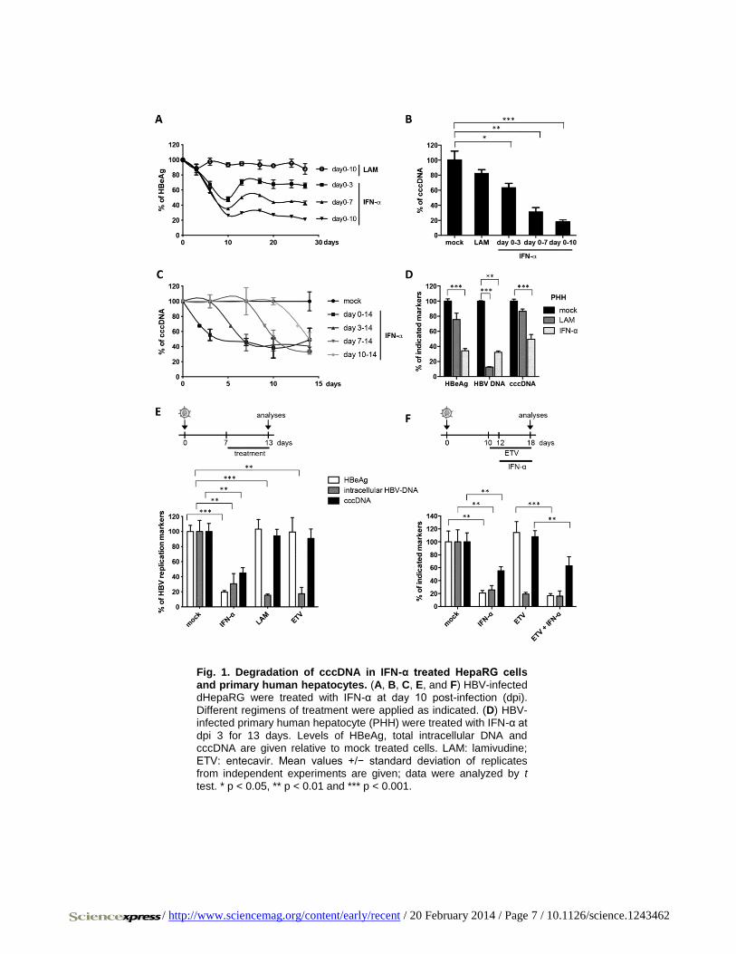
face (HBsAg) and e (HBeAg) antigens (Fig. 1A and fig. S1C).
In patients, interruption of LAM treatment results in a rebound of
HBV replication (2). Using IFN-α, we observed only a partial or no re-
bound in HBV-infected dHepaRG cells after treatment cessation (Fig.
1A). Because dHepaRG don’t allow virus spread, reduction of HBeAg
and lacking rebound indicated an effect of IFN-α on the established
HBV cccDNA transcription template besides the known antiviral effects
on viral replication (14). By cccDNA-specific qPCR, we determined an
80% reduction of cccDNA after 10 days of treatment (Fig. 1B). Reduc-
tion of cccDNA was confirmed by Southern blot analysis (fig. S1D) and
was dose dependent (fig. S1E). cccDNA reduction could be induced at
any time point (Fig. 1C) and persisted over time (Fig. 1, A and C). The
Specific and Nonhepatotoxic Degradation of Nuclear Hepatitis B Virus cccDNA
Julie Lucifora,1,2* Yuchen Xia,
1* Florian Reisinger,1 Ke Zhang,1 Daniela
Stadler,1 Xiaoming Cheng,1 Martin F. Sprinzl,1,3
Herwig Koppensteiner,1
Zuzanna Makowska,4 Tassilo Volz,
5 Caroline Remouchamps,
6 Wen-Min
Chou,1 Wolfgang E. Thasler,7 Norbert Hüser,8 David Durantel,9 T. Jake
Liang,10
Carsten Münk,11
Markus H. Heim,4 Jeffrey L. Browning,
12
Emmanuel Dejardin,6 Maura Dandri,2,5
Michael Schindler,1 Mathias
Heikenwalder,1†‡ Ulrike Protzer1,2†‡
1Institute of Virology, Technische Universität München–Helmholtz Zentrum München, 81675 Munich,
Germany. 2German Center for Infection Research (DZIF), Munich and Hamburg sites, Germany.
31st
Medical Department, University Hospital Mainz, 55131 Mainz, Germany. 4Department of Biomedicine,
University Hospital Basel, 4031 Basel, Switzerland. 5Department of Internal Medicine, University Medical
Center Hamburg-Eppendorf, 20246 Hamburg, Germany. 6GIGA-Research Laboratory of Molecular
Immunology and Signal Transduction, University of Liège, 4000 Liège, Belgium. 7Department of General,
Visceral, Transplantation, Vascular and Thoracic Surgery, Grosshadern Hospital, Ludwig Maximilians
University, 81377 Munich, Germany. 8Department of Surgery, University Hospital Rechts der Isar,
Technische Universität München, 85748 Munich, Germany. 9INSERM U1052, CNRS UMR 5286, Cancer
Research Center of Lyon, University of Lyon, LabEx DEVweCAN, 69007 Lyon, France. 10
Liver Diseases
Branch, National Institute of Diabetes and Digestive and Kidney Diseases, Bethesda, MD 20892, USA. 11
Clinic for Gastroenterology, Hepatology and Infectiology, Medical Faculty, Heinrich-Heine University,
40225 Düsseldorf, Germany. 12
Department of Immunobiology, Biogen Idec, Cambridge, MA 02142, USA.
*These authors contributed equally to this work.
†Corresponding author. E-mail: [email protected] (U.P.); [email protected] (M.H.)
‡These authors contributed equally to this work.
Current antivirals can control but not eliminate hepatitis-B-virus (HBV), because HBV establishes a stable nuclear cccDNA. Interferon-α treatment can clear HBV but is limited by systemic side effects. Here we describe how interferon-α can induce specific degradation of the nuclear viral DNA without hepatotoxicity and propose lymphotoxin-β-receptor activation as a therapeutic alternative. Interferon-α and lymphotoxin-β-receptor activation up-regulated APOBEC3A and 3B cytidine-deaminases, respectively, in HBV-infected cells, primary hepatocytes and human liver-needle biopsies. HBV-core protein mediated the interaction with nuclear cccDNA resulting in cytidine-deamination, apurinic/apyrimidinic site formation and finally cccDNA degradation that prevented HBV-reactivation. Genomic DNA was not affected. Thus, inducing nuclear deaminases - e.g., by lymphotoxin-β-receptor activation - allows development of new therapeutics that combined with existing antivirals may cure hepatitis B.
on
Feb
ruar
y 21
, 201
4w
ww
.sci
ence
mag
.org
Dow
nloa
ded
from
o
n F
ebru
ary
21, 2
014
ww
w.s
cien
cem
ag.o
rgD
ownl
oade
d fr
om
on
Feb
ruar
y 21
, 201
4w
ww
.sci
ence
mag
.org
Dow
nloa
ded
from
o
n F
ebru
ary
21, 2
014
ww
w.s
cien
cem
ag.o
rgD
ownl
oade
d fr
om
on
Feb
ruar
y 21
, 201
4w
ww
.sci
ence
mag
.org
Dow
nloa
ded
from
o
n F
ebru
ary
21, 2
014
ww
w.s
cien
cem
ag.o
rgD
ownl
oade
d fr
om
on
Feb
ruar
y 21
, 201
4w
ww
.sci
ence
mag
.org
Dow
nloa
ded
from
o
n F
ebru
ary
21, 2
014
ww
w.s
cien
cem
ag.o
rgD
ownl
oade
d fr
om
on
Feb
ruar
y 21
, 201
4w
ww
.sci
ence
mag
.org
Dow
nloa
ded
from
o
n F
ebru
ary
21, 2
014
ww
w.s
cien
cem
ag.o
rgD
ownl
oade
d fr
om
on
Feb
ruar
y 21
, 201
4w
ww
.sci
ence
mag
.org
Dow
nloa
ded
from
o
n F
ebru
ary
21, 2
014
ww
w.s
cien
cem
ag.o
rgD
ownl
oade
d fr
om

/ http://www.sciencemag.org/content/early/recent / 20 February 2014 / Page 2 / 10.1126/science.1243462
effect was corroborated in HBV-infected primary human hepatocytes
(PHH) (Fig. 1D). In contrast to IFN-α, LAM and even more potent nuc-
leoside analog entecavir (ETV) at very high doses (0.5 μM, 1000-fold
IC-50) only inhibited reverse transcription and thus HBV replication, but
not viral persistence (Fig. 1E). Pretreatment with ETV did not enhance
the effect of IFN-α (Fig. 1F) indicating that IFN-α induces the decay of
established HBV cccDNA. Since the doses of IFN-α used to achieve this
effect were high, we screened for other cytokines showing similar anti-
viral effects at moderate doses.
LTβR Activation Controls HBV and Leads to cccDNA Degradation
in HBV-Infected Cells
IFN-γ and TNF-α are known to control HBV in a noncytopathic fashion
(4, 7), but cannot be used as therapeutics because they cause severe side
effects. We tested the effect of lymphotoxin (LT) β receptor (LTβR)
activation as an alternative therapeutic option. TNF superfamily mem-
bers LTα, LTβ and CD258 are the physiological ligands for LTβR and
activate several inflammatory, anti-inflammatory, pro- and anti-survival
pathways (15). Like hepatocytes (16), dHepaRG (14) and HepG2-H1.3
cells permit HBV replication (17) and express the LTβR (fig. S2, A and
B). To activate LTβR, we used a super-agonistic tetravalent bispecific
antibody (BS1) and a bivalent anti-LTβR monoclonal antibody (CBE11)
(18, 19). As expected, LTβR agonists activated canonical (20) and non-
canonical nuclear factor kappa-light-chain-enhancer of activated B cells
(NF-κB) pathways to trigger p100 cleavage (fig. S2C), RelA phosphory-
lation (fig. S2D), nuclear RelB and RelA translocation (fig. S2, E and F),
and up-regulation of known target genes (fig. S2G) without causing any
detectable hepatocytotoxicity (fig. S2H).
To test the effect of LTβR-activation on HBV infection, dHepaRG
cells were treated with BS1 for 12 days starting 24 hours prior to HBV
infection. LTβR-activation decreased levels of all HBV-markers, includ-
ing cccDNA by approximately 90% without toxicity (Fig. 2A). The anti-
viral effect was highly potent with an EC5O of approximately 0.01
μg/mL (fig. S3A). Inhibition of apoptosis did not alter antiviral activity
(fig. S4). Neither IFN-β nor classic IFN-stimulated genes were up-
regulated upon BS1-treatment (fig. S2G) and antiviral activity was inde-
pendent of IFN-induction (fig. S5).
In vivo, activation of the murine LTβR by systemic application of an
agonistic antibody (ACH6) induced RelA and RelB nuclear translocation
in hepatocytes of HBV-transgenic mice (fig. S6A), reduced HBV vire-
mia (fig. S6B), HBV RNA (fig. S6C) and HBV core (HBc) protein ex-
pression in the liver (fig. S6, D and E). Neither signs of hepatocyte
apoptosis (fig. S6F) nor elevation of aminotransferases (ALT) (fig. S6G,
right panel) were observed indicating good in vivo tolerability of LTβR-
activation. Since HBV-transgenic mice do not establish HBV cccDNA,
this indicated additional antiviral effects of LTβR-activation on HBV
RNA transcription or stability. Accordingly, discontinuation of LTβR-
activation induced an immediate, strong rebound of HBV replication
(fig. S6G).
To investigate whether LTβR-activation would affect established
HBV cccDNA in the context of a persistent infection and prevent HBV
reactivation, dHepaRG cells were treated with LTβR agonists BS1 or
CBE11 when a stable, nuclear cccDNA pool had established. All HBV
markers, including HBV cccDNA, were reduced upon LTβR-activation
in HBV-infected dHepaRG cells (Fig. 2, B and C, and fig. S3) as well as
in stably transfected HepG2H1.3 cells containing high levels of cccDNA
(Fig. 2C). In HBV-infected primary human hepatocytes (PHH), LTβR
agonisation reduced HBV cccDNA, HBeAg secretion and even more
pronounced HBV-DNA replication (Fig. 2D). cccDNA degradation was
more effective (up to 95%) when treatment was prolonged (fig. S3, C
and D). Treatment interruption for 10 days was almost as efficient as
continuous treatment (fig. S3C) indicating that LTβR agonists induce a
persistent antiviral effect. In contrast to LAM treatment, no rebound of
HBV-replication was observed when BS1 treatment stopped (Fig. 2E).
Hence, LTβR activation not only suppressed HBV replication but also
caused nuclear cccDNA degradation, needed to achieve virus elimina-
tion.
LTβR Activation and IFN-α Treatment Induce Deamination and
Apurinic/Apyrimidinic (AP) Site Formation in cccDNA
To investigate if cccDNA degradation upon LTβR-activation or
IFN-α treatment was a result of DNA damage, we examined cccDNA
deamination by differential DNA denaturation PCR (3D-PCR) (21). Low
denaturing temperatures were sufficient for cccDNA amplification from
HBV-infected dHepaRG cells and for PHH treated with IFN-α or BS1,
compared with untreated, LAM- or ETV-treated cells (Fig. 3A and fig.
S7, C and D). Using a cocktail of recombinant proteins containing all
enzymes necessary for DNA repair (preCR mix), we could reverse the
denaturation of cccDNA (Fig. 3A, lower panels). The fact, that denatura-
tion temperatures of mock, LAM and ETV treated cells also shifted,
indicated that this modification of HBV cccDNA existed even without
exposure to exogenous drugs. Deamination of cccDNA (Fig. 3A, right
panel) and a drop in cccDNA levels after treatment with CBE11 (table
S1) was confirmed in vivo in human liver chimeric uPA-SCID mice
infected with HBV. Sequencing analyses showed G/A transitions oc-
curred under treatment (Fig. 3B and fig. S7, A and B) indicating deami-
nation of cytidines to uridine in the HBV cccDNA minus strand. At
lower denaturation temperatures G/A transitions became more obvious
(Fig. 3C and fig. S7A). These data showed that both LTβR-activation
and IFN-α treatment led to cccDNA deamination in vitro and in vivo,
and help to explain the G/A hypermutation observed in patient samples
(21).
Importantly, neither deamination nor mutations of genomic DNA
were observed by 3D-PCR (fig. S8A) or by deep sequencing of selected
housekeeping or IFN- and LTβR-target genes (fig. S8B). This indicated
that DNA modifications were specifically targeted to viral cccDNA.
After cytidine deamination, DNA-glycosylases recognize the dam-
aged DNA and cleave N-glycosidic bonds to release the base and create
an accessible AP site that can then be cleaved by endonucleases (22).
These AP sites can either be repaired, can lead to mutations upon DNA
replication or can induce DNA degradation (23). We quantified AP sites
created by LTβR-activation or IFN-α treatment. However, no increase of
AP sites in total DNA extracts from dHepaRG cells or PHH treated with
IFN-α or LTβR-agonists (fig. S8C) was found, reassuring that our treat-
ments did not lead to detectable damage in genomic DNA. Because AP
sites in the small (3.2 kb) cccDNA are very likely to be missed by this
analysis, we digested total DNA extracts with an AP-endonuclease
(APE1) and then amplified cccDNA by qPCR. APE digestion further
decreased cccDNA extracted from dHepaRG cells and PHH treated with
IFN-α or LTβR-agonists but not with LAM (Fig. 3D). Taken together,
our data indicate that both, LTβR-activation or IFN-α treatment induced
deamination and AP-site formation in HBV cccDNA leading to its de-
gradation, but did not affect genomic DNA.
LTβR Activation and IFN-α Treatment Up-Regulate Expression of
Nuclear APOBEC3 Deaminases
IFN-α is known to induce several cytidine deaminases (23, 24). We
performed genome-wide expression profiling of HBV-infected dHe-
paRG cells after LTβR-activation (fig. S9A) and classified regulated
genes according to their activity and properties (fig. S9B). Hereby,
APOBEC3B (A3B) was identified to be the most up-regulated gene with
nucleic acid binding properties (fig. S9C).
Analysis of all APOBEC3 family members showed that LTβR-
activation leads to strong up-regulation of A3B and to minor extent A3G
in HBV-infected dHepaRG and PHH, and after systemic application in
human liver chimeric uPA-SCID mice (fig. S10A). A3B expression was

/ http://www.sciencemag.org/content/early/recent / 20 February 2014 / Page 3 / 10.1126/science.1243462
induces by LTβR-activation in a dose-dependent manner and expression
levels steadily increased during continuous treatment (fig. S11) correlat-
ing with a concomitant increase in treatment efficacy over time (fig.
S3C). Treatment of PHH isolated from different donors with LTβR-
agonist BS1 resulted in cccDNA degradation at different levels (Fig. 3E
and fig. S10B), which could neither be explained by the level of A3B
upregulation (Fig. 3E) nor by detection of a previously described (25)
genomic deletion of the A3B allele, which seems to correlate with HBV
persistence in infected patients (fig. S10, B and C).
In contrast to LTβR-activation, IFN-α treatment induced mainly
A3A, but also A3F and A3G expression in HBV-infected dHepaRG cells
and PHH (fig. S12A), and A3D expression in isolated PHH. By systemic
IFN treatment of chimpanzees (26), A3A was strongly upregulated in
liver needle biopsies (fig. S12B). Activation of A3A, A3F and A3G after
IFN-α treatment was dose- and time-dependent, and decreased after an
initial peak despite continuous treatment indicating that cells become
refractory to IFN-α (fig. S13). In patients treated with subcutaneous
pegylated IFN-α, needle biopsies obtained at different time points con-
firmed a rapid, strong upregulation of A3A and to a lower extend of
A3G in the liver peaking at 16 hours post treatment (fig. S12C). Expres-
sion levels declined after this time point and remained low until day 6
post treatment confirming a fast but only transient induction of A3A by
IFN-α treatment. Interestingly, the level of A3B or A3A induction in
BS-1 and IFN-α treated PHH, respectively, did not directly correlate
with the level of cccDNA degradation (Fig. 3E). The fact that IFN-α
only induces a transient A3A induction and cells rapidly become refrac-
tory to IFN-α may account for the limited effect of IFN-α treatment in
HBV-infected patients (3).
APOBEC3A or APOBEC3B Activity Is Essential to Induce cccDNA
Degradation
Among the APOBEC3 family members up-regulated in our experi-
ments, only A3A and A3B located to the nucleus (fig. S14) where they
can gain access to cccDNA. To verify that they are indeed responsible
for the induction of cccDNA degradation, we overexpressed the HIV-Vif
protein (known to promote the degradation of all APOBEC3 proteins
except A3B (27, 28)) in dHepaRG cells in a tetracycline-regulated fa-
shion. Expression of HIV-Vif reduced A3A, A3F and A3G expression
(fig. S15A), reverted IFN-α-induced cccDNA deamination and pre-
vented cccDNA degradation induced by IFN-α treatment (Fig. 4A).
However, expression of HIV-Vif did not alter A3B levels (fig. S15B)
and had no impact on cccDNA degradation by LTβR-activation (fig.
S15C). To specifically address the role of A3A or A3B in cccDNA de-
gradation we further knocked down A3A and A3B in dHepaRG cells
under IFN-α or LTβR-agonist treatment, respectively, and observed
reduced cccDNA deamination (Fig. 4, B and C, left panels). A3A as well
as A3B knock-down completely reverted cccDNA degradation, but
could not rescue the additional effect of IFN-α or LTβR-activation on
HBV replication (Fig. 4, B and C, right panels).
To confirm the impact of A3A and A3B on cccDNA deamination,
we overexpressed A3A and A3B, respectively, in HBV-replicating
HepG2-H1.3 (Fig. 4, D and E). Cytidine-deamination of nuclear
cccDNA by A3A and A3B is in accordance with other studies showing
that both localize to the nucleus (29) and may be involved in the elimina-
tion of foreign DNA (23).
APOBEC3A Interacts with the HBV Core Protein and Binds to
cccDNA
APOBECs have evolved to restrict retroviral replication (30) as well
as DNA transfer into cells. They are able to clear foreign nuclear DNA
(23, 31), but it remains unclear how HBV cccDNA DNA was recognized
and whether it was specifically targeted in our experiments. To assess
specificity, we generated cell lines replicating a mammalian replicon
plasmid pEpi containing a linear HBV 1.3-fold overlength sequence.
From the linear HBV-genome, HBV replication was initiated and in
addition to the pEpi-H1.3 replicon HBV cccDNA was established in the
nucleus. Treatment with either IFN-α or LTβR-agonist BS1 inhibited
HBV replication and resulted in deamination and degradation of HBV
cccDNA, but not of the HBV-sequence containing replicon (fig. S16).
This indicated that deamination and subsequent degradation induced by
both treatments is HBV cccDNA specific.
HBV core protein associates with A3G (32) and HBV cccDNA (33)
and thus was a candidate to mediate the targeting of A3 deaminases to
HBV cccDNA. Confocal microscopy indicated a co-localization of A3A
and A3B with HBV core in different cell lines and PHH (Fig. 5 and fig.
S17). Chromatin immunoprecipitation (ChIP) experiments using stably
(fig. S18A) or transiently transfected HepG2H1.3 cells or HBV-infected
and IFN-α treated dHepaRG cells, showed that HBV core protein and
A3A both bind to the cccDNA minichromosome (Fig. 6A). Supporting
the possibility that a guardian protein prevents A3A direct binding to
DNA (34), we could not detect A3A binding to genomic DNA (fig.
S18B) even in the presence of HBV core, which has been reported to
also bind to cellular DNA (35).
HBV core protein co-immunoprecipitated A3A in HepG2H1.3 cells
and transfected HuH7 cells indicating physical interaction with A3A
(fig. S19). Direct interaction of HBV core expressed after HBV infection
and A3A induced by IFN-α was confirmed by proximity ligation assay
(PLA) (Fig. 6B and fig. S20) and fluorescence resonance energy transfer
(FRET) analysis (Fig. 6C). By deletion analysis, we determined that the
central region of HBc (aa 77 to 149) is involved in the interaction with
A3A (Fig. 6C and fig. S21).
These data suggest that A3A is targeted to cccDNA by interaction
with HBV core. No such targeting to genomic DNA has been described
so far. Since APOBEC3 deaminases are thought to act on single stranded
DNA (36), one possibility is that A3A and A3B act on cccDNA when it
is transiently rendered single-stranded by RNA polymerase II before
transcription initiation.
We suggest, therefore, the following mechanism of APOBEC-
dependent degradation of HBV cccDNA (Fig. 6D). High dose IFN-α
treatment or LTβR-activation up-regulate the expression of A3A and
A3B, respectively, which subsequently co-localize or directly interact
with HBV core in infected hepatocytes, translocate to the nucleus, where
they are brought into close contact with cccDNA by HBV core. Now,
APOBECs can deaminate cccDNA that is transiently rendered single-
stranded during transcription. Uracils in HBV cccDNA are recognized
and excised by cellular DNA glycosylases leading to formation of AP
sites, which are then recognized by cellular AP endonculeases (23) lead-
ing to cccDNA digestion. Why cccDNA is degraded instead of being
repaired by the cellular DNA repair machinery remains elusive so far.
Using a mixture of various enzymes, we were able to repair deaminated
cccDNA in tubo (Fig. 3A) suggesting induction of an additional factor
promoting DNA degradation or an impaired function of the repair ma-
chinery rather than a lack of recognition by the repair machinery. Thus,
we can only speculate that either the number of AP sites introduced after
treatment is too high and exceeds the capacity of the cellular repair ma-
chinery or that IFN-α treatment or LTβR-activation or even HBV itself
(37) modulate the repair machinery. This may shift the equilibrium from
cccDNA repair (38) to degradation.
Ideally, a cure for HBV infection needs to eliminate cccDNA. There-
fore, cytokines or cytokine-receptor agonists that can trigger HBV
cccDNA deamination and its degradation are interesting antiviral candi-
dates. Antivirals that induce A3A/B activity should be combined with
nucleos(t)ide analogs to avoid the replenishment of nuclear cccDNA
after degradation. LTβR-agonists were active at low doses and we did
not observe any toxicity in vitro or in vivo nor did we detect any modifi-
cation of genomic DNA. Constitutive overexpression of LTα/β for more

/ http://www.sciencemag.org/content/early/recent / 20 February 2014 / Page 4 / 10.1126/science.1243462
than one year has been associated with inflammatory liver disease and
hepatocellular carcinoma (16). As antivirals, however, LTβR-agonists
would only be used for a limited period of time minimizing the risk of
side effects. Moreover, LTβR-activation was already explored as a can-
cer treatment (18).
A recent study has shown a significantly higher frequency of an A3B
deletion allele in persistent HBV carriers and hepatocellular carcinoma
patients compared with healthy controls (25). This finding was further
supported by the moderate deamination of cccDNA even in absence of
treatment, and by the observation that knockdown of A3B in the absence
of any treatment increased cccDNA levels. Although deregulated ex-
pression of A3A and A3B has been shown to correlate with genomic
DNA mutations (39, 40), we did not detect any alterations of genomic
DNA using analyses of AP sites, 3D-PCR analysis and deep sequencing
of a set of human genes.
Our data indicate that cccDNA degradation is possible and can be
induced without side-effects on the infected host cell. An important task
will be testing of combinations of nucleos(t)ide analogs with novel anti-
viral strategies (e.g., LTβR agonists or adoptive T-cell therapy (41)) to
activate A3A or A3B to cure hepatitis B.
References and Notes
1. U. Protzer, M. K. Maini, P. A. Knolle, Living in the liver: hepatic infections.
Nat. Rev. Immunol. 12, 201–213 (2012). doi:10.1038/nri3169 Medline
2. F. Zoulim, Hepatitis B virus resistance to antiviral drugs: where are we going?
Liver Int. 31, (Suppl 1), 111–116 (2011). doi:10.1111/j.1478-
3231.2010.02399.x Medline
3. K. Wursthorn, M. Lutgehetmann, M. Dandri, T. Volz, P. Buggisch, B. Zollner,
T. Longerich, P. Schirmacher, F. Metzler, M. Zankel, C. Fischer, G. Currie,
C. Brosgart, J. Petersen, Peginterferon alpha-2b plus adefovir induce strong
cccDNA decline and HBsAg reduction in patients with chronic hepatitis B.
Hepatology 44, 675–684 (2006). doi:10.1002/hep.21282 Medline
4. L. G. Guidotti, K. Ando, M. V. Hobbs, T. Ishikawa, L. Runkel, R. D.
Schreiber, F. V. Chisari, Cytotoxic T lymphocytes inhibit hepatitis B virus
gene expression by a noncytolytic mechanism in transgenic mice. Proc. Natl.
Acad. Sci. U.S.A. 91, 3764–3768 (1994). doi:10.1073/pnas.91.9.3764 Medline
5. L. G. Guidotti, R. Rochford, J. Chung, M. Shapiro, R. Purcell, F. V. Chisari,
Viral clearance without destruction of infected cells during acute HBV
infection. Science 284, 825–829 (1999). doi:10.1126/science.284.5415.825
Medline
6. S. F. Wieland, H. C. Spangenberg, R. Thimme, R. H. Purcell, F. V. Chisari,
Expansion and contraction of the hepatitis B virus transcriptional template in
infected chimpanzees. Proc. Natl. Acad. Sci. U.S.A. 101, 2129–2134 (2004).
doi:10.1073/pnas.0308478100 Medline
7. H. McClary, R. Koch, F. V. Chisari, L. G. Guidotti, Relative sensitivity of
hepatitis B virus and other hepatotropic viruses to the antiviral effects of
cytokines. J. Virol. 74, 2255–2264 (2000). doi:10.1128/JVI.74.5.2255-
2264.2000 Medline
8. L. Belloni, L. Allweiss, F. Guerrieri, N. Pediconi, T. Volz, T. Pollicino, J.
Petersen, G. Raimondo, M. Dandri, M. Levrero, IFN-α inhibits HBV
transcription and replication in cell culture and in humanized mice by
targeting the epigenetic regulation of the nuclear cccDNA minichromosome.
J. Clin. Invest. 122, 529–537 (2012). doi:10.1172/JCI58847 Medline
9. A. Rang, S. Günther, H. Will, Effect of interferon alpha on hepatitis B virus
replication and gene expression in transiently transfected human hepatoma
cells. J. Hepatol. 31, 791–799 (1999). doi:10.1016/S0168-8278(99)80279-7
Medline
10. V. Pasquetto, S. F. Wieland, S. L. Uprichard, M. Tripodi, F. V. Chisari,
Cytokine-sensitive replication of hepatitis B virus in immortalized mouse
hepatocyte cultures. J. Virol. 76, 5646–5653 (2002).
doi:10.1128/JVI.76.11.5646-5653.2002 Medline
11. S. F. Wieland, L. G. Guidotti, F. V. Chisari, Intrahepatic induction of
alpha/beta interferon eliminates viral RNA-containing capsids in hepatitis B
virus transgenic mice. J. Virol. 74, 4165–4173 (2000).
doi:10.1128/JVI.74.9.4165-4173.2000 Medline
12. S. L. Uprichard, S. F. Wieland, A. Althage, F. V. Chisari, Transcriptional and
posttranscriptional control of hepatitis B virus gene expression. Proc. Natl.
Acad. Sci. U.S.A. 100, 1310–1315 (2003). doi:10.1073/pnas.252773599
Medline
13. P. Gripon, C. Diot, N. Thézé, I. Fourel, O. Loreal, C. Brechot, C. Guguen-
Guillouzo, Hepatitis B virus infection of adult human hepatocytes cultured in
the presence of dimethyl sulfoxide. J. Virol. 62, 4136–4143 (1988). Medline
14. P. Gripon, S. Rumin, S. Urban, J. Le Seyec, D. Glaise, I. Cannie, C.
Guyomard, J. Lucas, C. Trepo, C. Guguen-Guillouzo, Infection of a human
hepatoma cell line by hepatitis B virus. Proc. Natl. Acad. Sci. U.S.A. 99,
15655–15660 (2002). doi:10.1073/pnas.232137699 Medline
15. M. J. Wolf, G. M. Seleznik, N. Zeller, M. Heikenwalder, The unexpected role
of lymphotoxin beta receptor signaling in carcinogenesis: from lymphoid
tissue formation to liver and prostate cancer development. Oncogene 29,
5006–5018 (2010). doi:10.1038/onc.2010.260 Medline
16. J. Haybaeck, N. Zeller, M. J. Wolf, A. Weber, U. Wagner, M. O. Kurrer, J.
Bremer, G. Iezzi, R. Graf, P. A. Clavien, R. Thimme, H. Blum, S. A.
Nedospasov, K. Zatloukal, M. Ramzan, S. Ciesek, T. Pietschmann, P. N.
Marche, M. Karin, M. Kopf, J. L. Browning, A. Aguzzi, M. Heikenwalder, A
lymphotoxin-driven pathway to hepatocellular carcinoma. Cancer Cell 16,
295–308 (2009). doi:10.1016/j.ccr.2009.08.021 Medline
17. S. Jost, P. Turelli, B. Mangeat, U. Protzer, D. Trono, Induction of antiviral
cytidine deaminases does not explain the inhibition of hepatitis B virus
replication by interferons. J. Virol. 81, 10588–10596 (2007).
doi:10.1128/JVI.02489-06 Medline
18. M. Lukashev, D. LePage, C. Wilson, V. Bailly, E. Garber, A. Lukashin, A.
Ngam-ek, W. Zeng, N. Allaire, S. Perrin, X. Xu, K. Szeliga, K. Wortham, R.
Kelly, C. Bottiglio, J. Ding, L. Griffith, G. Heaney, E. Silverio, W. Yang, M.
Jarpe, S. Fawell, M. Reff, A. Carmillo, K. Miatkowski, J. Amatucci, T.
Crowell, H. Prentice, W. Meier, S. M. Violette, F. Mackay, D. Yang, R.
Hoffman, J. L. Browning, Targeting the lymphotoxin-beta receptor with
agonist antibodies as a potential cancer therapy. Cancer Res. 66, 9617–9624
(2006). doi:10.1158/0008-5472.CAN-06-0217 Medline
19. X. Hu, M. A. Zimmerman, K. Bardhan, D. Yang, J. L. Waller, G. B. Liles, J.
R. Lee, R. Pollock, D. Lev, C. F. Ware, E. Garber, V. Bailly, J. L. Browning,
K. Liu, Lymphotoxin β receptor mediates caspase-dependent tumor cell
apoptosis in vitro and tumor suppression in vivo despite induction of NF-κB
activation. Carcinogenesis 34, 1105–1114 (2013). doi:10.1093/carcin/bgt014
Medline
20. E. Dejardin, N. M. Droin, M. Delhase, E. Haas, Y. Cao, C. Makris, Z. W. Li,
M. Karin, C. F. Ware, D. R. Green, The lymphotoxin-beta receptor induces
different patterns of gene expression via two NF-kappaB pathways. Immunity
17, 525–535 (2002). doi:10.1016/S1074-7613(02)00423-5 Medline
21. R. Suspène, D. Guétard, M. Henry, P. Sommer, S. Wain-Hobson, J. P.
Vartanian, Extensive editing of both hepatitis B virus DNA strands by
APOBEC3 cytidine deaminases in vitro and in vivo. Proc. Natl. Acad. Sci.
U.S.A. 102, 8321–8326 (2005). doi:10.1073/pnas.0408223102 Medline
22. J. I. Friedman, J. T. Stivers, Detection of damaged DNA bases by DNA
glycosylase enzymes. Biochemistry 49, 4957–4967 (2010).
doi:10.1021/bi100593a Medline
23. M. D. Stenglein, M. B. Burns, M. Li, J. Lengyel, R. S. Harris, APOBEC3
proteins mediate the clearance of foreign DNA from human cells. Nat. Struct.
Mol. Biol. 17, 222–229 (2010). doi:10.1038/nsmb.1744 Medline
24. M. Bonvin, F. Achermann, I. Greeve, D. Stroka, A. Keogh, D. Inderbitzin, D.
Candinas, P. Sommer, S. Wain-Hobson, J. P. Vartanian, J. Greeve, Interferon-
inducible expression of APOBEC3 editing enzymes in human hepatocytes
and inhibition of hepatitis B virus replication. Hepatology 43, 1364–1374
(2006). doi:10.1002/hep.21187 Medline
25. T. Zhang, J. Cai, J. Chang, D. Yu, C. Wu, T. Yan, K. Zhai, X. Bi, H. Zhao, J.
Xu, W. Tan, C. Qu, D. Lin, Evidence of associations of APOBEC3B gene
deletion with susceptibility to persistent HBV infection and hepatocellular
carcinoma. Hum. Mol. Genet. 22, 1262–1269 (2013).
doi:10.1093/hmg/dds513 Medline
26. Y. Huang, J. J. Feld, R. K. Sapp, S. Nanda, J. H. Lin, L. M. Blatt, M. W.
Fried, K. Murthy, T. J. Liang, Defective hepatic response to interferon and
activation of suppressor of cytokine signaling 3 in chronic hepatitis C.
Gastroenterology 132, 733–744 (2007). doi:10.1053/j.gastro.2006.11.045
Medline
27. B. P. Doehle, A. Schäfer, B. R. Cullen, Human APOBEC3B is a potent
inhibitor of HIV-1 infectivity and is resistant to HIV-1 Vif. Virology 339,
281–288 (2005). doi:10.1016/j.virol.2005.06.005 Medline

/ http://www.sciencemag.org/content/early/recent / 20 February 2014 / Page 5 / 10.1126/science.1243462
28. G. Berger, J. Turpin, S. Cordeil, K. Tartour, X. N. Nguyen, R. Mahieux, A.
Cimarelli, Functional analysis of the relationship between Vpx and the
restriction factor SAMHD1. J. Biol. Chem. 287, 41210–41217 (2012).
doi:10.1074/jbc.M112.403816 Medline
29. H. Muckenfuss, M. Hamdorf, U. Held, M. Perkovic, J. Löwer, K. Cichutek, E.
Flory, G. G. Schumann, C. Münk, APOBEC3 proteins inhibit human LINE-1
retrotransposition. J. Biol. Chem. 281, 22161–22172 (2006).
doi:10.1074/jbc.M601716200 Medline
30. C. Münk, A. Willemsen, I. G. Bravo, An ancient history of gene duplications,
fusions and losses in the evolution of APOBEC3 mutators in mammals. BMC
Evol. Biol. 12, 71 (2012). doi:10.1186/1471-2148-12-71 Medline
31. M. A. Carpenter, M. Li, A. Rathore, L. Lackey, E. K. Law, A. M. Land, B.
Leonard, S. M. Shandilya, M. F. Bohn, C. A. Schiffer, W. L. Brown, R. S.
Harris, Methylcytosine and normal cytosine deamination by the foreign DNA
restriction enzyme APOBEC3A. J. Biol. Chem. 287, 34801–34808 (2012).
doi:10.1074/jbc.M112.385161 Medline
32. P. Turelli, B. Mangeat, S. Jost, S. Vianin, D. Trono, Inhibition of hepatitis B
virus replication by APOBEC3G. Science 303, 1829 (2004).
doi:10.1126/science.1092066 Medline
33. C. T. Bock, S. Schwinn, S. Locarnini, J. Fyfe, M. P. Manns, C. Trautwein, H.
Zentgraf, Structural organization of the hepatitis B virus minichromosome. J.
Mol. Biol. 307, 183–196 (2001). doi:10.1006/jmbi.2000.4481 Medline
34. M. M. Aynaud, R. Suspène, P. O. Vidalain, B. Mussil, D. Guétard, F. Tangy,
S. Wain-Hobson, J. P. Vartanian, Human Tribbles 3 protects nuclear DNA
from cytidine deamination by APOBEC3A. J. Biol. Chem. 287, 39182–39192
(2012). doi:10.1074/jbc.M112.372722 Medline
35. Y. Guo, W. Kang, X. Lei, Y. Li, A. Xiang, Y. Liu, J. Zhao, J. Zhang, Z. Yan,
Hepatitis B viral core protein disrupts human host gene expression by binding
to promoter regions. BMC Genomics 13, 563 (2012). doi:10.1186/1471-2164-
13-563 Medline
36. H. C. Smith, R. P. Bennett, A. Kizilyer, W. M. McDougall, K. M. Prohaska,
Functions and regulation of the APOBEC family of proteins. Semin. Cell Dev.
Biol. 23, 258–268 (2012). doi:10.1016/j.semcdb.2011.10.004 Medline
37. T. H. Lee, S. J. Elledge, J. S. Butel, Hepatitis B virus X protein interacts with
a probable cellular DNA repair protein. J. Virol. 69, 1107–1114 (1995).
Medline
38. K. Kitamura, Z. Wang, S. Chowdhury, M. Simadu, M. Koura, M. Muramatsu,
Uracil DNA glycosylase counteracts APOBEC3G-induced hypermutation of
hepatitis B viral genomes: excision repair of covalently closed circular DNA.
PLoS Pathog. 9, e1003361 (2013). doi:10.1371/journal.ppat.1003361 Medline
39. S. Landry, I. Narvaiza, D. C. Linfesty, M. D. Weitzman, APOBEC3A can
activate the DNA damage response and cause cell-cycle arrest. EMBO Rep.
12, 444–450 (2011). doi:10.1038/embor.2011.46 Medline
40. M. B. Burns, L. Lackey, M. A. Carpenter, A. Rathore, A. M. Land, B.
Leonard, E. W. Refsland, D. Kotandeniya, N. Tretyakova, J. B. Nikas, D.
Yee, N. A. Temiz, D. E. Donohue, R. M. McDougle, W. L. Brown, E. K.
Law, R. S. Harris, APOBEC3B is an enzymatic source of mutation in breast
cancer. Nature 494, 366–370 (2013). doi:10.1038/nature11881 Medline
41. K. Krebs, N. Böttinger, L. R. Huang, M. Chmielewski, S. Arzberger, G.
Gasteiger, C. Jäger, E. Schmitt, F. Bohne, M. Aichler, W. Uckert, H. Abken,
M. Heikenwalder, P. Knolle, U. Protzer, T cells expressing a chimeric antigen
receptor that binds hepatitis B virus envelope proteins control virus replication
in mice. Gastroenterology 145, 456–465 (2013).
doi:10.1053/j.gastro.2013.04.047 Medline
42. J. Lucifora, S. Arzberger, D. Durantel, L. Belloni, M. Strubin, M. Levrero, F.
Zoulim, O. Hantz, U. Protzer, Hepatitis B virus X protein is essential to
initiate and maintain virus replication after infection. J. Hepatol. 55, 996–
1003 (2011). doi:10.1016/j.jhep.2011.02.015 Medline
43. H. Schulze-Bergkamen, A. Untergasser, A. Dax, H. Vogel, P. Büchler, E.
Klar, T. Lehnert, H. Friess, M. W. Büchler, M. Kirschfink, W. Stremmel, P.
H. Krammer, M. Müller, U. Protzer, Primary human hepatocytes—a valuable
tool for investigation of apoptosis and hepatitis B virus infection. J. Hepatol.
38, 736–744 (2003). doi:10.1016/S0168-8278(03)00120-X Medline
44. S. M. Lee, C. Schelcher, M. Demmel, M. Hauner, W. E. Thasler, Isolation of
human hepatocytes by a two-step collagenase perfusion procedure. J. Vis.
Exp. (79): (2013). Medline
45. W. E. Thasler, T. S. Weiss, K. Schillhorn, P. T. Stoll, B. Irrgang, K. W. Jauch,
Charitable State-Controlled Foundation Human Tissue and Cell Research:
Ethic and Legal Aspects in the Supply of Surgically Removed Human Tissue
For Research in the Academic and Commercial Sector in Germany. Cell
Tissue Bank. 4, 49–56 (2003). doi:10.1023/A:1026392429112 Medline
46. O. Hantz, R. Parent, D. Durantel, P. Gripon, C. Guguen-Guillouzo, F. Zoulim,
Persistence of the hepatitis B virus covalently closed circular DNA in
HepaRG human hepatocyte-like cells. J. Gen. Virol. 90, 127–135 (2009).
doi:10.1099/vir.0.004861-0 Medline
47. M. Quasdorff, M. Hösel, M. Odenthal, U. Zedler, F. Bohne, P. Gripon, H. P.
Dienes, U. Drebber, D. Stippel, T. Goeser, U. Protzer, A concerted action of
HNF4alpha and HNF1alpha links hepatitis B virus replication to hepatocyte
differentiation. Cell. Microbiol. 10, 1478–1490 (2008). doi:10.1111/j.1462-
5822.2008.01141.x Medline
48. U. Protzer, S. Seyfried, M. Quasdorff, G. Sass, M. Svorcova, D. Webb, F.
Bohne, M. Hösel, P. Schirmacher, G. Tiegs, Antiviral activity and
hepatoprotection by heme oxygenase-1 in hepatitis B virus infection.
Gastroenterology 133, 1156–1165 (2007). doi:10.1053/j.gastro.2007.07.021
Medline
49. A. Untergasser, U. Zedler, A. Langenkamp, M. Hösel, M. Quasdorff, K.
Esser, H. P. Dienes, B. Tappertzhofen, W. Kolanus, U. Protzer, Dendritic
cells take up viral antigens but do not support the early steps of hepatitis B
virus infection. Hepatology 43, 539–547 (2006). doi:10.1002/hep.21048
Medline
50. J. Summers, P. M. Smith, A. L. Horwich, Hepadnavirus envelope proteins
regulate covalently closed circular DNA amplification. J. Virol. 64, 2819–
2824 (1990). Medline
51. W. Gao, J. Hu, Formation of hepatitis B virus covalently closed circular
DNA: removal of genome-linked protein. J. Virol. 81, 6164–6174 (2007).
doi:10.1128/JVI.02721-06 Medline
52. G. K. Smyth, Linear models and empirical bayes methods for assessing
differential expression in microarray experiments. Stat. Appl. Genet. Mol.
Biol. 3, Article3 (2004).
53. C. Banning, J. Votteler, D. Hoffmann, H. Koppensteiner, M. Warmer, R.
Reimer, F. Kirchhoff, U. Schubert, J. Hauber, M. Schindler, A flow
cytometry-based FRET assay to identify and analyse protein-protein
interactions in living cells. PLoS ONE 5, e9344 (2010).
doi:10.1371/journal.pone.0009344 Medline
54. K. Arnold, L. Bordoli, J. Kopp, T. Schwede, The SWISS-MODEL
workspace: a web-based environment for protein structure homology
modelling. Bioinformatics 22, 195–201 (2006).
doi:10.1093/bioinformatics/bti770 Medline
55. S. A. Wynne, R. A. Crowther, A. G. Leslie, The crystal structure of the human
hepatitis B virus capsid. Mol. Cell 3, 771–780 (1999). doi:10.1016/S1097-
2765(01)80009-5 Medline
56. I. J. Byeon et al., NMR structure of human restriction factor APOBEC3A
reveals substrate binding and enzyme specificity. Nat. Commun. 4, 1890
(2013).
57. A. W. Ghoorah, M. D. Devignes, M. Smaïl-Tabbone, D. W. Ritchie, Protein
docking using case-based reasoning. Proteins 81, 2150–2158 (2013).
doi:10.1002/prot.24433 Medline
58. R. A. Sayle, E. J. Milner-White, RASMOL: biomolecular graphics for all.
Trends Biochem. Sci. 20, 374–376 (1995). doi:10.1016/S0968-
0004(00)89080-5 Medline
59. L. G. Guidotti, B. Matzke, H. Schaller, F. V. Chisari, High-level hepatitis B
virus replication in transgenic mice. J. Virol. 69, 6158–6169 (1995). Medline
60. M. Dandri, M. R. Burda, E. Török, J. M. Pollok, A. Iwanska, G. Sommer, X.
Rogiers, C. E. Rogler, S. Gupta, H. Will, H. Greten, J. Petersen, Repopulation
of mouse liver with human hepatocytes and in vivo infection with hepatitis B
virus. Hepatology 33, 981–988 (2001). doi:10.1053/jhep.2001.23314 Medline
61. M. Lütgehetmann, T. Bornscheuer, T. Volz, L. Allweiss, J. H. Bockmann, J.
M. Pollok, A. W. Lohse, J. Petersen, M. Dandri, Hepatitis B virus limits
response of human hepatocytes to interferon-α in chimeric mice.
Gastroenterology 140, 2074–2083, e1–e2 (2011).
doi:10.1053/j.gastro.2011.02.057 Medline
62. M. Sarasin-Filipowicz, E. J. Oakeley, F. H. Duong, V. Christen, L.
Terracciano, W. Filipowicz, M. H. Heim, Interferon signaling and treatment
outcome in chronic hepatitis C. Proc. Natl. Acad. Sci. U.S.A. 105, 7034–7039
(2008). doi:10.1073/pnas.0707882105 Medline
Acknowledgments: We would like to thank Romina Bester, Theresa Asen,
Kerstin Ackermann, Kathrin Kappes, Martin Feuerherd, Robert Baier, Ruth
Hillermann, Ute Finkel, Aikatherini Krikoni and Fang Zhang for their
http://www.ncbi.nlm.nih.gov/entrez/query.fcgi?cmd=Retrieve&db=PubMed&list_uids=7815490&dopt=Abstract
http://www.ncbi.nlm.nih.gov/entrez/query.fcgi?cmd=Retrieve&db=PubMed&list_uids=7815490&dopt=Abstract
http://www.ncbi.nlm.nih.gov/entrez/query.fcgi?cmd=Retrieve&db=PubMed&list_uids=2335817&dopt=Abstract

/ http://www.sciencemag.org/content/early/recent / 20 February 2014 / Page 6 / 10.1126/science.1243462
technical support, Prof. Luigi Terracciano for analysis of acute hepatitis
patients, Prof. Frank Chisari for providing HBV transgenic mice (HBV
1.3.32), Prof. Thorsten Buch and Olivia Prazeres da Costa for help with array
analysis and data discussions, Lena Allweiss and Anne Groth for help by
generating and treating humanized uPA/SCID mice, as well as Siemens
Healthcare Diagnostics for providing reagents. The study was supported by
grants from FCC (Fédération belge Contre le Cancer) to ED, an ERC Starting
grant (LiverCancerMechanism) to MH, the German Research Foundation
(SFB 841 to MD, SFB TR 36 to MH, SFB TR 22), the Peter-Hans
Hofschneider foundation and the Helmholtz Alliances HAIT (to UP) and
PCCC (to MH). We acknowledge the support of the nonprofit foundation
HTCR, which holds human tissue on trust, making it broadly available for
research on an ethical and legal basis. Patent application EP12006XXX filed
at the European patent office: ‘Lymphotoxin signaling activation and its
downstream mediators eliminate HBV ccc DNA’. Microarray data have been
submitted to the GEO database (http://www.ncbi.nlm.nih.gov/geo/) and have
the accession number GSE46667. Human liver-chimeric UPA/SCID mice
were handled in accordance with protocols approved by the Ethical
Committee of the city and state of Hamburg (permission number G12/015).
Experiments with HBV-transgenic mice were performed in accordance to the
German legislation governing animal studies and the Principles of Laboratory
Animal Care guidelines, NIH (55.1-1-54-2531.3-27-08). The study protocol
for the experiment with Chimpanzee was approved at the Southwest
Foundation for Biomedical Research, San Antonio, TX (IACUC 869 PT,
approved in 2004).
Supplementary Materials
www.sciencemag.org/cgi/content/full/science.1243462/DC1
Materials and Methods
Figs. S1 to S21
Table S1
References (42–62)
18 July 2013; accepted 5 February 2014
Published online 20 February 2014
10.1126/science.1243462
/ http://www.sciencemag.org/content/early/recent / 20 February 2014 / Page 7 / 10.1126/science.1243462
Fig. 1. Degradation of cccDNA in IFN-α treated HepaRG cells
and primary human hepatocytes. (A, B, C, E, and F) HBV-infected dHepaRG were treated with IFN-α at day 10 post-infection (dpi).
Different regimens of treatment were applied as indicated. (D) HBV-infected primary human hepatocyte (PHH) were treated with IFN-α at
dpi 3 for 13 days. Levels of HBeAg, total intracellular DNA and cccDNA are given relative to mock treated cells. LAM: lamivudine;
ETV: entecavir. Mean values +/− standard deviation of replicates from independent experiments are given; data were analyzed by t
test. * p < 0.05, ** p < 0.01 and *** p < 0.001.

/ http://www.sciencemag.org/content/early/recent / 20 February 2014 / Page 8 / 10.1126/science.1243462
Fig. 2. LTβR-activation inhibits HBV infection and leads to cccDNA degradation in HepaRG
cells and PHH. (A and B) HBV-infected dHepaRG were treated with BS1, CBE11, hu-IgG control or lamivudine (LAM). (A) Treatment started 24h before infection for 12 days or (B) at 18 dpi for 10
days. Levels of the indicated HBV markers as well as cell viability are given relative to untreated controls (mock). (C) cccDNA levels were analyzed after 14 days of BS1 treatment by Southern
blot in HBV-infected dHepaRG and HBV-replicating HepG2H1.3 cells. Supercoiled cccDNA bands were identified by their expected size and linearization upon EcoRI digestion (3,2 kb). (D) PHH
were infected with HBV and treated with BS1 at 7 dpi for 10 days. Levels of the indicated HBV markers were compared to untreated PHH of the same donor (donor 3) (mock). (E) HBV-infected
dHepaRG were treated with BS1, hu-IgG control or LAM. Intracellular HBV-DNA was analyzed 8 and 14 days after treatment cessation. Mean values +/− standard deviation of replicates from
independent experiments are given; data were analyzed by t test. * p < 0.05, *** p < 0.001.

/ http://www.sciencemag.org/content/early/recent / 20 February 2014 / Page 9 / 10.1126/science.1243462
Fig. 3. Deamination and AP-site formation in cccDNA upon IFN-α treatment and LTβR-activation. (A) dHepaRG (left) and PHH (middle panel) were infected with HBV and treated with IFN-α, BS1 or LAM. Human chimeric uPA/SCID mice were treated with
CBE11 or hu-IgG control (right panel). 3D-PCR analyses were performed on cccDNA left either untreated (upper panels) or treated with a PreCR mix (lower panels). (B and C) 3D-PCR products from HBV-infected dHepaRG cells treated as indicated (IFN-α, BS1
or mock) were cloned and sequenced and mutations were analyzed. (D) Total DNA extracts from HBV-infected cells treated as indicated were digested with APE1, and cccDNA content was compared to mock-treated cells. In (B), (C), and (D), mean values +/−
standard deviation of biological triplicates from two independent experiments are given; data were analyzed by t test. * p < 0.05, ** p < 0.01. (E) PHH were infected with HBV and treated with BS1 or IFN-α at 7 dpi for 10 days. Levels of the indicated cccDNA as well
as A3A and A3B mRNA expression were compared to untreated PHH (mock) of the same donor.

/ http://www.sciencemag.org/content/early/recent / 20 February 2014 / Page 10 / 10.1126/science.1243462
Fig. 4. Analysis of cccDNA deamination and degradation. (A to C) cccDNA denaturation was
analyzed by 3D-PCR (left panels); levels of HBeAg, total intracellular DNA and cccDNA are given relative to mock treated cells (right panels). (A) dHepaRG-tA-Vif cells treated with IFN-α for 10
days with and without doxycycline (dox)-induced HIV-Vif expression. HBV-infected dHepaRG cells treated with (B) IFN-α or (C) BS1 transfected with siRNA against A3A or A3B, respectively,
or sequence nonspecific siRNA (sicontrol). Mean values +/− standard deviation of independent replicates and experiments are given; data were analyzed by t test. * p < 0.05, ** p < 0.01 and ***
p < 0.001. (D) cccDNA denaturation analysis by 3D-PCR in HepG2-H1.3 cells overexpressing A3A or (E) A3B from lentiviral vector plasmid pLenti6.3 or pTR600, respectively, for 5 days.

/ http://www.sciencemag.org/content/early/recent / 20 February 2014 / Page 11 / 10.1126/science.1243462
Fig. 5. Co-localization of A3A and A3B with HBV core
protein (HBc). (A) HuH7 cells were co-transfected with an HBV1.1-fold genome and A3A-Flag or A3B-Flag expressing
plasmids and stained using DAPI, anti-HBc and anti-FLAG antibodies. (B) HBV-infected dHepaRG and PHH were
treated with IFN-α at day 7 post infection for 3 days. A3A and HBc were analyzed by immunofluorescence staining. Right
panels indicate z stacks taken at the dotted lines.

/ http://www.sciencemag.org/content/early/recent / 20 February 2014 / Page 12 / 10.1126/science.1243462
Fig. 6. Interaction of A3A, HBV core protein (HBc) and cccDNA. (A) Chromatin
immunoprecipitation (ChIP) was performed using lysates of HepG2H1.3 cells transfected with A3A-expressing plasmid, or HBV-infected dHepaRG cells treated with IFN-α for 3 days. IPs using
antibodies against histone H3, A3A, HBc and control rabbit IgG (RIgG) were analyzed by qPCR for cccDNA. (B) Interaction between HBc and A3A was assessed by proximity ligation assay (PLA) in
HBV-infected, IFN-α treated dHepaRG. PLA-spots were quantified in single cells by software-based spot-counting. Data were analyzed by one-way ANOVA. ** p < 0.01 and *** p < 0.001. (C) Serial HBV
core-deletion mutants (left panel) were fused to CFP and interaction with A3A-YFP was assessed by FACS-FRET in HuH7.5 hepatoma cells (right panel). Cells cotransfected with CFP and YFP served as
controls to exclude false positive FRET and subtract background signals. A CFP-YFP fusion construct was used as positive control. Mean values ± standard deviation of FRET-positive cells from 3-4
independent experiments are given. Black boxes indicate shared regions of HBc mutants giving a FRET signal. (D) Model of cccDNA degradation induced by IFN-α treatment or LTβR-activation.