Speciality and Variability of Embalming Methods Seen in ...1993... · Speciality and Variability of...
15
Speciality and Variability of Embalming Methods Seen in Human Mummies from Qurna, Egypt IWATARO MORIMOTO St. Marianna University School of Medicine Based on observations of human mummies from Tombs No . 317 and 178 at Qurna, Egypt, two important items of the mummification process were studied. First, the removal of the brain through the nostrils was characteristic of the ancient Egyptians. This was found in 55 out of 60 mummies (91. 7 %) from Tomb No . 317 , but in 5 out of 19 mummies (26 . 3%) from Tomb No. 178. The low incidence in the latter tomb could be due to the moderated style of embalming adopted to avoid heavy expense. The hooked rod used in extracting the brain remained in and around the median plane in 28 (46 . 7%) of the 60 mummies , but was shifted to the left side in 22 (36 . 7%) . The brain was rarely extracted through an opening for ventilation made on the left side of the skull or in the roof of the right orbit , or through the foramen magnum. In this pro- cess, the embalmers would feel no reluctance about standing on one particular side of the corpse. Second, the male external genital organs were specially treated . The organs were partially or totally cut off in 5 (50.0%), and deformed in 1 (10 . 0%), out of 10 mummies from Tomb No. 317, while they were cut off in 1 (25.0%), and deformed in 3 (75 . 0%) , out of 4 mummies from Tomb No . 178. No prepuce was found on those penises of adult mummies which were left intact, suggesting that the ancient Egyp- tians practiced circumcision. In one mummy, the removed genitals had been replaced with an artificial phallus made of gilded resin . In another child mummy, the penis was serpentine in shape, a symbol of new life and resurrection . The special treatment of the external genital organs in males may indicate that the Egyptians sought eternity on the basis of beliefs associated with the mythical tales oflsis and Osiris . The various em- balming techniques presented here probably originated in the special crafts for this task for which the embalmers are emplo yed under the direction of hereditary master practitioners . Key words: mummification process, brain, external genital organs , lsis and Osiris, Egypt. 1. INTRODUCTION As shown by Herodotus in the fifth century B.C. (Godley 1981: 371 ), in ancient Egypt of the 18th to 21st Dynasty, when the mummification process was in full flower, there were professionals whose whole business was embalming human corpses and who had special crafts for this task . Since their treatment of the body at workshops in the necropolices was not always of the same quality and their methods of embalming varied from era to era, it is understandable that experts frequently disagree about details of the mummification pro- cess. Even the order of operations is not fully agreed, but Leca (1980 : 162-163) has reconstructed a dozen of steps in the routine procedure, starting with removal of the brain N ilo-Ethiopion Studies 1: 27-41 (1993) Copyright © 1993 . Japan Association for Nilo-Ethiopian Studies 27
Transcript of Speciality and Variability of Embalming Methods Seen in ...1993... · Speciality and Variability of...

Speciality and Variability of Embalming Methods Seen in Human Mummies from Qurna, Egypt
IWATARO MORIMOTO
St. Marianna University School of Medicine
Based on observations of human mummies from Tombs No. 317 and 178 at Qurna, Egypt, two important items of the mummification process were studied. First, the removal of the brain through the nostrils was characteristic of the ancient Egyptians. This was found in 55 out of 60 mummies (91. 7 %) from Tomb No. 317 , but in 5 out of 19 mummies (26 .3%) from Tomb No. 178. The low incidence in the latter tomb could be due to the moderated style of embalming adopted to avoid heavy expense. The hooked rod used in extracting the brain remained in and around the median plane in 28 (46 .7%) of the 60 mummies , but was shifted to the left side in 22 (36 .7%) . The brain was rarely extracted through an opening for ventilation made on the left side of the skull or in the roof of the right orbit , or through the foramen magnum. In this process, the embalmers would feel no reluctance about standing on one particular side of the corpse. Second , the male external genital organs were specially treated . The organs were partially or totally cut off in 5 (50.0%), and deformed in 1 (10 .0%), out of 10 mummies from Tomb No. 317, while they were cut off in 1 (25.0%), and deformed in 3 (75 .0%) , out of 4 mummies from Tomb No . 178. No prepuce was found on those penises of adult mummies which were left intact, suggesting that the ancient Egyptians practiced circumcision . In one mummy, the removed genitals had been replaced with an artificial phallus made of gilded resin . In another child mummy, the penis was serpentine in shape, a symbol of new life and resurrection . The special treatment of the external genital organs in males may indicate that the Egyptians sought eternity on the basis of beliefs associated with the mythical tales oflsis and Osiris . The various embalming techniques presented here probably originated in the special crafts for this task for which the embalmers are employed under the direction of hereditary master practitioners.
Key words: mummification process, brain, external genital organs , lsis and Osiris, Egypt.
1. INTRODUCTION
As shown by Herodotus in the fifth century B .C . (Godley 1981: 371 ), in ancient Egypt of the 18th to 21st Dynasty, when the mummification process was in full flower, there were professionals whose whole business was embalming human corpses and who had special crafts for this task . Since their treatment of the body at workshops in the necropolices was not always of the same quality and their methods of embalming varied from era to era, it is understandable that experts frequently disagree about details of the mummification process . Even the order of operations is not fully agreed, but Leca (1980 : 162-163) has reconstructed a dozen of steps in the routine procedure, starting with removal of the brain
N ilo-Ethiopion Studies 1: 27-41 (1993) Copyright © 1993 . Japan Association for Nilo-Ethiopian Studies
27

28 Nilo-Ethiopian Studies
and ending with the wrapping of the body, and including special treatment of the external genital organs.
The present paper concerns with two important steps in the mummification process, the removal of the brain and the special treatment of the external genital organs in males, with some remarks upon the speciality and variability of the embalming methods adopted by the ancient Egyptians.
2. MATERIALS AND METHODS
Previous papers, Morimoto (1985 : 1-11; 1989: 169-187) and Morimoto et al. (1986: 1-11; 1988: 1-16), contained scattered data on both the removal of the brain and the special treatment of the external genital organs. Here , these data are brought together, and some errors in the previous articles are rectified.
The present study on the removal of the brain employed the heads or skulls of 60 human mummies (19 males, 32 females and 9 children) from Tomb No. 317 of the so-called "Tombs of the Nobles" at Qurna, West Bank ofThebes (old name ofLuxor), Egypt, which came to light during the 1983-84, 1984-85 and 1985-86 seasons of fieldwork by a team from Waseda University, Japan . In addition, the heads or skulls of 19 mummies (10 males, 8 females and 1 child) from Tomb No. 178 at the same Qurna, which were discovered by Dr. Mahmud Abd al Raziq, were made available by the kind permission of the Egyptian Antiquities Organization. For the special treatment of the external genital organs, the organs of mummies of 7 males and 3 children from Tomb No. 314 and of 4 males from Tomb No. 178 were examined . It seems that tomb robbers had carried the mummies into these two tombs from other graves in search of art treasures with gold and jewels. In these circumstances, it might be safely assumed that the mummies examined dated widely from the 18th Dynasty of the New Kingdom to the Ptolemaic stage of the Greco-Roman period, i.e. , ea. 1550-30 B.C. Although there was no clear chronological differences between the mummies from Tombs No. 314 and No . 178, the location of the tombs from which the mummies had been stolen was considered to be so restricted that the mummies examined could be full of local col or.
Since dissection or unwrapping of the mummies was strictly forbidden by the Egyptian Government, the examination was performed only by naked eye and X-ray within the limits. However, all the mummies examined had been so broken up by tomb robbers that , fortunately, not only their surfaces, but also their internal structures could easily be observed without dissection or unwrapping.
3. OBSERVATIONS
3 .1. Removal of the brain It is well known that ancient Egyptian embalmers usually extracted the brain through the
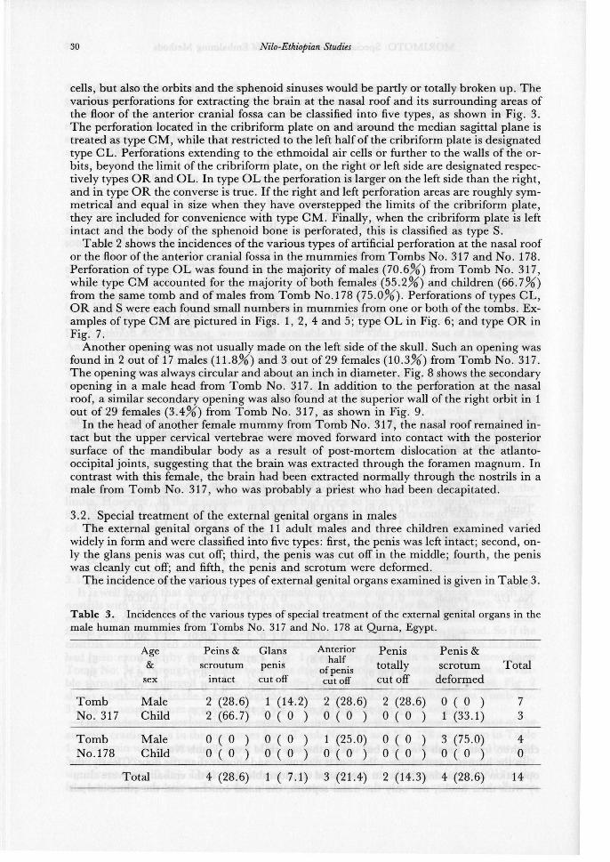
nostrils with the aid of a long, hooked rod such as that illustrated by Bucaille (1990 : 5). The roof of the nasal cavities chiefly formed by the cribriform plate of the ethmoid bone, which forms part of the floor of the anterior cranial fossa, was thus perforated by this rod . So if the nostrils were enlarged and the cribriform plate was perforated, it can be said that the brain had been extracted by the embalmers. Fig. 1 shows the nostrils in a male mummy from Tomb No . 317, through which the brain was drained . Perforations of the nasal roof are visible through the enlarged nostrils, which differ from each other in shape and size. Fig . 2 shows a perforation in the median sagittal plane of the nasal roof in a child from Tomb No. 317. The nasal septum remains intact but is slightly shifted to right side .
The incidences of perforations for extracting the brain at the nasal roof or the floor of the anterior cranial fossa in the mummies from Tomb No. 317 and No. 178 are shown in Table 1. The brain was extracted in 55 out of 60 mummies (91. 7 %) from Tomb No. 317, but only 5 out of 19 (26 .3%) from Tomb No . 178, the difference in the incidences being statistically significant (:x:2=30.26). There was, however, no significant sex difference (:x: 2=0.001) in the frequency of perforation . The table also indicates that the brain was extracted in all nine

MORIMOTO: Speciality and Variability of Embalming Methods 29
Table 1. lncidences of artificial perforation for extracting the brain at the nasal roof of the floor of the anterior cranial fossa in human mummies from Tombs No. 317 and No. 178 at Qurna, Egypt .
Age & sex N Perforated (%)
Tomb No. 317 Male 19 17 ( 89.5± 7.03) Female 32 29 ( 90.6± 5.16) Child 9 9 (100.0± 0 )
Total 60 55 ( 91.7± 3. 72)
Tomb No. 178 Male 10 4 ( 40.0± 15.49) Female 8 1 ( 12.5±11.69) Child 0 ( 0 ± 0 )
Total 19 5 ( 26.3±10.10)
Total Male 29 21 ( 72.4± 8.30) Female 40 30 ( 75.0± 6.85) Child 10 9 ( 90.0± 9.49)
Total 79 60 ( 75.9± 4.81)
Diff. between tombs: x2=30 .26 > x2.95 (1)=3 .84 Diff. between sexes: x2=0.001 < x2.95 (1)=3.84
Table 2. lncidences of the various types of artificial perforation for extracting the brain at the nasal roof or the floor of the anterior cranial fossa in human mummies from Tombs No. 317 and No . 178 at Qurna, Egypt.
Age& Type of artificial perforation Total
sex OL (%) CM(%) CL(%) OR(%) s (%)
Tomb Male 12 (70 .6) 2 (11.8) ( 5.9) 2 (11.8) 0 ( 0 ) 17 No. 317 Female 9 (31.0) 16 (55 .2) 1 ( 3 .4) 2 ( 6.9) 1 ( 3.4) 29
Child 1 (11.1) 6 (66 . 7) 2 (22 .2) 0 ( 0 ) 0 ( 0 ) 9
Total 22 (40 .0) 24 (43.6) 4 ( 7 .3) 4 ( 7 .3) 1 ( 1.8) 55
Tomb Male 0 ( 0 3 (75.0) 0 ( 0 1 (25 .0) 0 ( 0 ) 4 No . 178 Female 0 ( 0 0 ( 0 ) 0 ( 0 0 ( 0 ) 1 (100 .0) 1
Child 0 ( 0 0 ( 0 ) 0 ( 0 0 ( 0 ) 0 ( 0 ) 0
Total 0 ( 0 3 (60.0) 0 ( 0 1 (20.0) 1 ( 20 .0) 5
Total Male 12 (57 .1) 5 (23 .8) 1 ( 4.8) 3 (14.3) 0 ( 0 ) 21 Female 9 (30.0) 16 (53.3) 1 ( 3.3) 2 ( 6. 7) 2 ( 6.7) 30 Child 1 (11.1) 6 (66.7) 2 (22 .2) 0 ( 0 ) 0 ( 0 ) 9
Total 22 (36 . 7) 27 (45.0) 4 ( 6. 7) 5 ( 8. 3) 2 (3.3) 60
children from Tomb No . 317 but not in the one child from Tomb No. 178 . In the human nasal cavities , the roof is narrower and shorter than the floor. Clearly, the
operation of extracting the brain demanded considerable skill . If the embalmers were clumsy with their hooks, not only the nasal septum, the nasal conchae and the ethmoidal air
30 Nilo-Ethiopian Studies
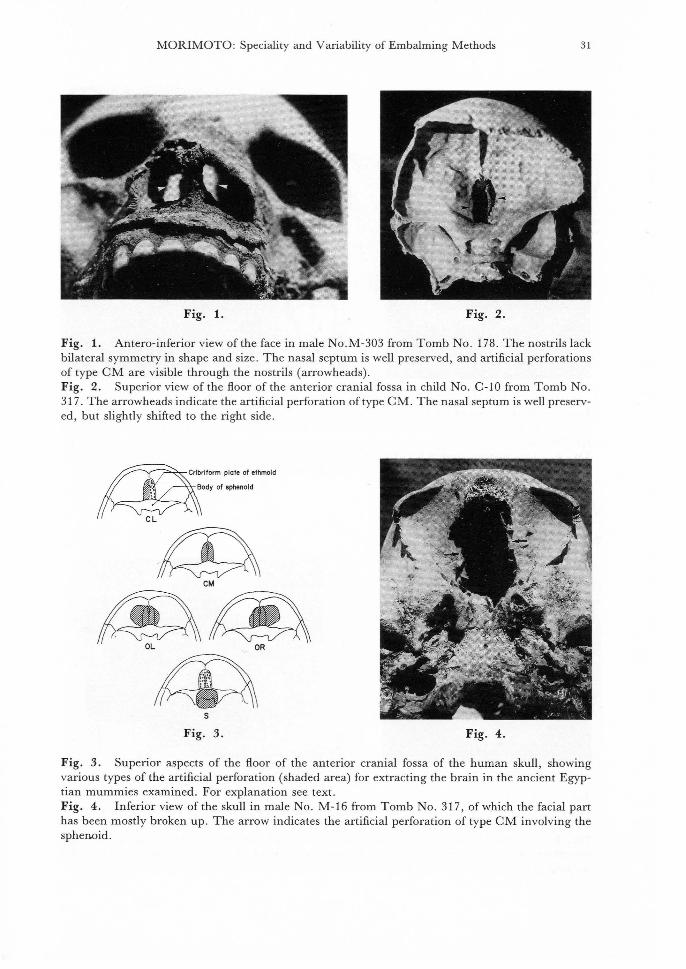
cells, but also the orbits and the sphenoid sinuses would be partly or totally broken up. The various perforations for extracting the brain at the nasal roof and its surrounding areas of the floor of the anterior cranial fossa can be classified into five types, as shown in Fig. 3. The perforation located in the cribriform plate on and around the median sagittal plane is treated as type CM, while that restricted to the left half of the cribriform plate is designated type CL. Perforations extending to the ethmoidal air cells or further to the walls of the orbits, beyond the limit of the cribriform plate, on the right or left side are designated respectively types OR and OL. In type OL the perforation is larger on the left side than the right, and in type OR the converse is true. If the right and left perforation areas are roughly symmetrical and equal in size when they have overstepped the limits of the cribriform plate, they are included for convenience with type CM. Finally, when the cribriform plate is left intact and the body of the sphenoid bone is perforated, this is classified as type S.
Table 2 shows the incidences of the various types of artificial perforation at the nasal roof or the floor of the anterior cranial fossa in the mummies from Tombs No. 317 and No. 178. Perforation of type OL was found in the majority of males (70.6%) from Tomb No. 317, while type CM accounted for the majority of both females (55.2%) and children (66.7%) from the same tomb and of males from Tomb No.178 (75.0%). Perforations of types CL, OR and S were each found small numbers in mummies from one or both of the tombs. Examples of type CM are pictured in Figs. 1, 2, 4 and 5; type OL in Fig. 6; and type OR in Fig. 7.
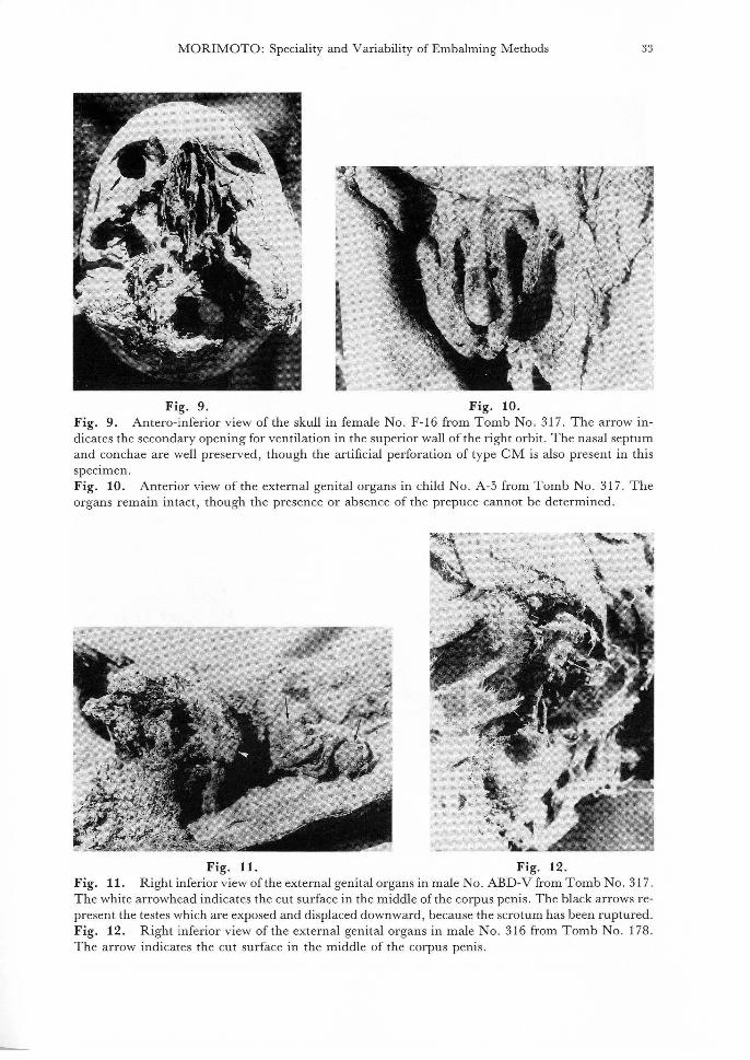
Another opening was not usually made on the left side of the skull. Such an opening was found in 2 out of 17 males (11.8%) and 3 out of29 females (10.3%) from Tomb No. 317. The opening was always circular and about an inch in diameter. Fig. 8 shows the secondary opening in a male head from Tomb No. 317. In addition to the perforation at the nasal roof, a similar secondary opening was also found at the superior wall of the right orbit in 1 out of 29 females (3.4%) from Tomb No. 317, as shown in Fig. 9.
In the head of another female mummy from Tomb No. 317, the nasal roof remained intact but the upper cervical vertebrae were moved forward into contact with the posterior surface of the mandibular body as a result of post-mortem dislocation at the atlantooccipital joints, suggesting that the brain was extracted through the foramen magnum. In contrast with this female, the brain had been extracted normally through the nostrils in a male from Tomb No. 317, who was probably a priest who had been decapitated.
3. 2. Special treatment of the external genital organs in males The external genital organs of the 11 adult males and three children examined varied
widely in form and were classified into five types: first, the penis was left intact; second, only the glans penis was cut off; third, the penis was cut off in the middle; fourth, the penis was cleanly cut off; and fifth, the penis and scrotum were deformed.
The incidence of the various types of external genital organs examined is given in Table 3.
Table 3. lncidences of the various types of special treatment of the external genital organs in the male human mummies from Tombs No. 317 and No. 178 at Qurna, Egypt.
Tomb No. 317
Tomb No.178
Age &
sex
Male Child
Male Child
Total
Peins & Glans scroutum penis
intact cut off
2 (28.6) 2 (66. 7)
0 ( 0 0 ( 0
1 (14 .2) 0 ( 0 )
0 ( 0 ) 0 ( 0 )
Anterior half
of penis cutoff
2 (28.6) 0 ( 0 )
1 (25.0) 0 ( 0 )
Penis totally cut off
2 (28.6) 0 ( 0 )
0 ( 0 0 ( 0
Penis & scrotum
deformed
0 ( 0 ) 1 (33.1)
3 (75 .0) 0 ( 0 )
4 (28.6) ( 7.1) 3 (21.4) 2 (14.3) 4 (28.6)
Total
7 3
4 0
14
MORIMOTO: Speciality and Variability of Embalming Methods 31
Fig. 1. Fig. 2.
Fig. 1. Antero-inferior view of the face in male No .M-303 from Tomb No. 178. The nostrils lack bilateral symmetry in shape and size. The nasal septum is well preserved, and artificial perforations of type CM are visible through the nostrils (arrowheads) . Fig. 2. Superior view of the floor of the anterior cranial fossa in child No. C-10 from Tomb No. 317 . The arrowheads indicate the artificial perforation of type CM. The nasal septum is well preserved, but slightly shifted to the right side .
rn:i:riform plate of ethmoid
~ody of sphenold
CL
CM
s
Fig. 3. Fig. 4.
Fig. 3. Superior aspects of the floor of the anterior cranial fossa of the human skull , showing various types of the artificial perforation (shaded area) for extracting the brain in the ancient Egyptian mummies examined. For explanation see text. Fig. 4. Inferior view of the skull in male No. M-16 from Tomb No. 317, of which the facial part has been mostly broken up. The arrow indicates the artificial perforation of type CM involving the sphenoid.

32 Nilo-Ethiopian Studies
Fig. 5. Fig. 6.
Fig. 5. Inferior view ofthe skull in female No. F-9 from Tomb No. 317, ofwhich the facial part has been mostly broken up. The arrows indicate the artificial perforation of type CM involving the sphenoid bone . Fig. 6. Left anterior view of the skull in female No . Head-B from Tomb No. 317. The arrow indicates the artificial perforation of type OL in both medial and superior walls of the left orbit .
Fig. 7. Fig. 8.
Fig. 7. Inferior view of the skull in female No. F-26 from Tomb No. 317, of which the facial part has been mostly broken up. The arrows indicates the artificial perforation of type OR extending into both medial and superior walls of the orbits on both sides. The nasal septum remains intact. Fig. 8. Lateral view of the head in male No. A-30 from Tomb No. 317. The arrows indicate the secondary opening for ventilation on the left side of the head in the posterior part of the frontal squama just above the left temporal line.
MORIMOTO : Speciality and Variability of Embalming Methods 33
Fig. 9. Fig. 10. Fig. 9. Antero-inferior view of the skull in female No. F-16 from Tomb No. 317. The arrow indicates the secondary opening for ventilation in the superior wall of the right orbit. The nasal septum and conchae are well preserved, though the artificial perforation of type CM is also present in this speCimen. Fig. 10. Anterior view of the external genital organs in child No. A-5 from Tomb No. 317 . The organs remain intact , though the presence or absence of the prepuce cannot be determined .
Fig. 11. Fig. 12. Fig. 11. Right inferior view of the external genital organs in male No. ABD-V from Tomb No. 317. The white arrowhead indicates the cut surface in the middle of the corpus penis . The black arrows represent the testes which are exposed and displaced downward, because the scrotum has been ruptured. Fig. 12. Right inferior view of the external genital organs in male No. 316 from Tomb No. 178. The arrow indicates the cut surface in the middle of the corpus penis.

34 Nilo-Ethiopian Studies
Fig. 13. Fig. 14.
Fig. 13. Antero-inferior view of the pelvis and perineum in male No. 8 from Tomb No . 317. The external genital organs were cleanly removed.
Fig. 14. Right lateral view of a model penis and scrotum made of gilded resin, as seen in male No. 17 from Tomb No. 317, of which the true external genital organs has been cleanly removed.
Fig. 15. Fig. 16.
Fig. 15. Right lateral view of the external genital organs in the supine child No. A-7 from Tomb No. 317. The elongated penis, which is flexed near the glans penis and serpentine in shape, is laid on the flattened scrotum.
Fig. 16. Antero-inferior view of the external genital organs in male No. 321 from Tomb No . 178. The elongated penis is supported by the deformed scrotum.
MORIMOTO: Speciality and Variability of Embalming Methods 35
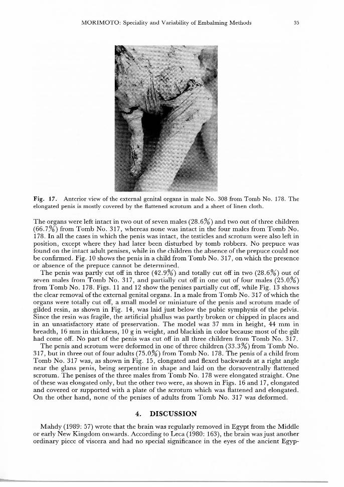
Fig. 17. Anterior view of the external genital organs in male No . 308 from Tomb No. 178. The elongated penis is mostly covered by the flattened scrotum and a sheet of linen cloth .
The organs were left intact in two out of seven males (28 . 6%) and two out of three children (66 .7%) from Tomb No. 317, whereas none was intact in the four males from Tomb No . 178 . In all the cases in which the penis was intact, the testicles and scrotum were also left in position, except where they had later been disturbed by tomb robbers. No prepuce was found on the intact adult penises, while in the children the absence of the prepuce could not be confirmed. Fig. 10 shows the penis in a child from Tomb No . 317, on which the presence or absence of the prepuce cannot be determined.
The penis was partly cut off in three (42.9%) and totally cut off in two (28.6%) out of seven males from Tomb No. 317, and partially cut off in one out of four males (25 .0%) from Tomb No. 178. Figs . 11 and 12 show the penises partially cut off, while Fig. 13 shows the clear removal of the external genital organs. In a male from Tomb No. 317 of which the organs were totally cut off, a small model or miniature of the penis and scrotum made of gilded resin, as shown in Fig. 14, was laid just below the pubic symphysis of the pelvis. Since the resin was fragile, the artificial phallus was partly broken or chipped in places and in an unsatisfactory state of preservation. The model was 37 mm in height, 44 mm in breadth, 16 mm in thickness, 10 gin weight, and blackish in color because most of the gilt had come off. No part of the penis was cut off in all three children from Tomb No. 317 .
The penis and scrotum were deformed in one of three children (33.3%) from Tomb No. 317, but in three out offour adults (75.0%) from Tomb No . 178. The penis of a child from Tomb No . 317 was, as shown in Fig. 15, elongated and flexed backwards at a right angle near the glans penis , being serpentine in shape and laid on the dorsoventrally flattened scrotum. The penises of the three males from Tomb No. 178 were elongated straight . One of these was elongated only, but the other two were, as shown in Figs. 16 and 17, elongated and covered or supported with a plate of the scrotum which was flattened and elongated . On the other hand, none of the penises of adults from Tomb No. 317 was deformed .
4. DISCUSSION
Mahdy (1989: 57) wrote that the brain was regularly removed in Egypt from the Middle or early New Kingdom onwards . According to Leca (1980: 163), the brain was just another ordinary piece of viscera and had no special significance in the eyes of the ancient Egyp-

36 Nilo-Ethiopian Studies
tians. Peck (1980: 21) reported that the brain was apparently not preserved, and that no mention was found of any container used in the mummification process or any burial material associated with the brain.
Other ethnic groups also removed the brain by different methods: the Canary Islanders by unknown methods; the Scythians of Siberia by trephination (Ascenzi et al, 1980: 229-231 ); and the Australians (Pretty & Calder 1980: 202) and Peruvians (VreelandJr. & Cockburn 1980: 139) through the foramen magnum. In no known case was the brain extracted through the nostrils among non-Egyptian populations. Thus the removal of the brain through the nostrils was a method of embalming peculiar to the ancient Egyptians.
This method of extracting the brain was always accompanied by an artificial perforation in the roof of the nasal cavities. The incidences of the perforations were, as shown in Table 1, respectively 91.7% in the mummies from the Tomb No. 317 and 26.3% in those from the Tomb No. 178, the difference in the incidences being statistically significant. These two tombs, however, belonged to the same "Tombs of the Nobles" at Qurna and were within a short walk of each other. Since almost all the mummies examined were probably brought into the two tombs from other graves by tomb robbers, it must be reiterated that the chronological difference between the mummies from tombs No. 314 and No. 178 could not be exactly determined. It appears, however, that all the mummies examined came within the range from the New Kingdom to the Ptolemaic period. The difference in the incidences of removal of the brain between the two tombs might be thus only a chronological variation due to the long course of time. According to Herodotus (Godley 1981: 371 ), three categories of mummification existed. The most elaborate process involved the removal of the brain, but the second method did not. These traditional methods of embalming had, according to Cottrell (1960: 223-225), been used for thousands of years. It might be assumed therefore, that the mummies from Tomb No . 178, in which the incidence of the perforation at the nasal roof was low, were of those who often selected a moderate style of embalming to avoid heavy expense.
Leca (1980: 162) explained that there were roughly three eras of mummification in Egypt: the first lasted up to the 18th Dynasty, when the process was still in the experimental stage; the second up to the 21st Dynasty, when it was in full flower; and the third when, with the Persian invasion, it went into decline. Spencer (1982: 117) maintained that significant advances in mummification were made in the 18th Dynasty. The removal of the brain was, according to him, first practiced during this period. Based on the difference in the incidences of perforations for extracting the brain between the mummies from Tombs No. 317 and No. 178, it might be said that the mummies from the former tomb, showing a high incidence, were earlier than those from the latter, which showed a low incidence. In this connection, Leca (1980: 164) believed that not every corpus had its brain removed, for the process was a costly one, but that it became more common as the centuries passed, as Nicolaeff demonstrated in his study of 492 skulls from Saqqara that less than 5 percent of the 4th Dynasty cases had had the brains removed, compared to 50 percent during the Greek period.
Leca (1980: 163-164) wrote that the embalmer inserted the needle through the left nostril. The present observations confirm the main use of the left nostril. As shown in Fig. 1 , the nostrils were asymmetrical in shape and size. Usually the left nostril was broader and more enlarged than the right. Such a collateral asymmetry of the nostrils can be also seen in an Egyptian mummy exhibited at the Wellcome Museum of the History of Medicine in the Science Museum, London. The pressure of the bandages applied to the face ·seemed to have little influence on the form of the nostrils, because none of the nostrils was collapsed and only the left nostril was usually broadened and enlarged . The nasal septum, if it remained, was generally shifted to the right in the nasal cavities, as shown in Fig. 2, probably due to the repeated pressure of the hooked rod used. And the nasal conchae on the left side were sometimes broken up, whereas those on the right were preserved. It should be recalled here that the perforation of type CL was rarely found in the mummies examined, though there was no case in which only the right half of the cribriform plate was perforated. It is notable that in the rare cases in which an opening for ventilating the brain was made, this was
MORIMOTO : Speciality and Variability of Embalming Methods 37
found only on the left side of the skull, and an incision for evisceration was also made only on the left side of the anterior abdominal wall.
The operation for extracting the brain through the nostrils was very difficult and demanded considerable skill. If the hooked rod failed to follow the correct course, a large hole could be made at the floor of the anterior cranial fossa , and the ethmoidal air cells, the orbital walls and the sphenoidal body could be extensively destroyed . In the mummies from Tombs No . 317 and No. 178, as shown in Table 2, the artificial perforation of type CM was the most frequent. Together the perforations of types CM and CL accounted for 31 out of 60 cases (51. 7 %) , a majority of cases. This indicates that the operation was not always done accurately . Even when the artificial perforation involved the sphenoid bone, the hook moved in and around the median sagittal plane, as shown in Fig. 4.
Leca (1980: 163-164) mentioned that usually the ethmoid bone was perforated on the right side, from which one can deduce that the embalmer placed himself on the left of the body and, if he was right-handed , as is likely, inserted the needle through the left nostril; and as he scraped , the instrument would have had a natural tendency to veer to the right. In the present study, as shown in Table 2, the perforation of type OL was the second frequent and occupied 22 of60 specimens (36. 7%) . There was no perforation of type CR, and type OR was found only in 5 out of 60 cases (8 .3%). The difference in the incidences between the perforations of types OL and OR seems to suggest that the instrument would have had a tendency to veer to the left side rather than the right. This is supported by the findings that the nasal septum was often well preserved in the mummy heads , as shown Figs. 2, 7 and 9; and that in several cases of type CL, only the left half of the cribriform plate was broken while the nasal septum remained intact.
The fact that perforations of types CM and OL accounted for the great majority of specimens indicates that the hooked rod inserted into the left nostrils was almost parallel to the nasal septum or, occasionally, inclined to the left side rather than the right. In this situation, it would be natural for the right-handed embalmer to have placed himself on the right side of the body. In a wall painting of the tomb ofSennediem at Deir el-Medina in the 19th Dynasty, the divine embalmer Anubis was , as shown by Adams (1984: 28), David (1978: 91), Glubok & Tamarin (1978: 51), Hobson (1990: 119) and Mahdy (1989: 108), standing on the left side of the mummy. Doubt remains, however , about whether the embalmer used to work on the left side of the corpse . For instance, pictures of the burial chamber illustrated in The Ancient Egyptian Book of the Dead (Faulkner, 1985: 146-148) represented Anubis standing either on the left side of the mummy of Nakht or on the right side of the mummy of Muthetepti. Budge (1972: 214), Ceram (1958 : 114-115), Spencer (1982 : 132-134), Ions (1982 : 83) , Grilletto (1988: 182) and Mahdy (1989: 56) placed Anubis apparently on the right side of the embalmed mummy in their books. It is, therefore, impossible to suppose that the Egyptian embalmers felt reluctant to stand on one particular side of the body.
Besides the perforation in the nasal roof, another opening was cut through the left lateral wall of the skull. The opening was located on and around the left temporal line from the pterion to the posterior part of the frontal bone just above the line. This opening, in association with the perforation in the nasal roof, was considered to be used for draining off the brain, according to Leca (1980 : 164), perhaps by using water. He wrote that such a second opening was found in a skull dating from the Greek epoch .
Leca (1980 : 164) said that another little-used method consisted in taking out an eye and breaking through the upper wall of the orbit , which again resulted in a larger opening for extracting the brain. One female mummy showed both perforation in the nasal roof and opening at the right orbital wall (Fig. 9), with the left eyeball remaining in situ . In this case, the opening at the right orbital wall played the same role as the opening on the left side of the skull.
The head of a female mummy from the Tomb No. 317 showed no artificial perforation in the nasal roof. However, the upper cervical vertebrae were displaced forward to expose the foramen magnum, and the brain tissue would have flowed out of this foramen without any other treatment. This specimen is reminiscent of the mummy of Ahmose I, in which, according to Adams (1984: 35), Bucaille (1990: 5), Pace (1974: 101) and Harris & Wente
38 Nilo-Ethiopian Studies
(1980 : 168), the brain was extracted through the foramen magnum in association with surgical removal of the atlas or first cervical vertebra. The removal of the brain not through the nostrils but at the base of the skull was, according to Mahdy ( 1989: 86), a method occasionally employed by embalmers during the Old Kingdom. Again, if the embalmers intentionally decapitated the corpse, this might be regarded as an atypical method of extracting the brain. In a male mummy, probably of a decapitated priest, from Tomb No . 317, however, the brain was extracted in the normal way through the nostrils.
There were a variety of special treatments of the external genital organs in the ancient Egyptian mummies examined. Leca (1980: 169) stated that in the mummification process the external male genital organs were nearly always left in position. In the present study, however, the o_rgans were intact in only 2 out of 11 adults (18.2%) and in two out of three children (66. 7%).
Although Kamal (1967: 104-106) mentioned that circumcised mummies were few, the present study shows that the prepuce was cleanly removed from all penises that were left intact. This indicates that the habit of circumcision was deeply rooted among the Egyptians of ancient times. Circumcision was, according to Kamal, practiced between six and twelve years of age, but not in all males at any time. Harris & Weeks (1973: 126) wrote that Ahmose I was not circumcised, unlike most Egyptian males, because this pharaoh was in ill health, perhaps even a hemophiliac or "bleeder." Herodotus (Godley 1981: 319) described that the Egyptians and those who had learned it from them were the only people who practiced circumcision. Rowling (1967: 536-537) said that circumcision was practiced almost universally in Egypt, and that the custom probably dated from Predynastic times and finally seemed to disappear in Coptic times . He also maintained that circumcision was almost certainly performed just before puberty. Bunson (1991: 53) wrote that male circumcision, which was practiced as part of traditional methods of hygiene, was not performed at birth but during adolescence. The famous relief in the tomb of Ankh-ma Hor at Saqqara, seen in the figure given by Kamal (1967: 467), and dating from the 6th Dynasty, showed the operation being performed. Chabas (1861 : 307) introduced another ancient Egyptian relief where circumcision was also practiced (cited by Bryk 1934). The results of the present study, in which the absence of the prepuce was ascertained in all the adults, might suggest that circumcision was usually performed during adolescence.
The results in Table 3 show that the penises were partially or totally cut off in 6 out of 11 adults (54.5%), whereas none of them was removed in three children. The studies of the royal mummies by Smith (1912) were, according to Sandison & Wells (1967 : 515) , frustrated by the fact that tomb robbers sometimes broke off the phallus, as in the cases of Ramesses II and Seti II ; by the apparent removal of the pudenda during embalming, in the cases ofThutmose Ill and the unknown person C and E; by pressure of the pudendum into a leaf-like structure against the perineum, as in Thutmose I and II; and the probable removal of scrotum and subsequent damage to the penis, in the case of Merenptah . Bucaille (1990 : 10) wrote that the external genital organs ofRamesses II were removed completely, while only a segment of the penis was missing in the case of Merenptah. Leca (1980 : 169), however, explained that the external genital organs in Seti I and Ramesses II were cleanly removed with the aid of a sharp instrument. Smith's view of the removal of the organs in Ramesses II differs from that of Leca in whether the organs had been accidentally removed by tomb robbers or intentionally cut off by the embalmers. In this regard, Bard et al. ( 1985: 7 4) offered no explanation for the fact that their radiological and xeroradiographic investigations of the mummy of Ramesses II showed an absence of images corresponding to the external genital organs .
In a male from Tomb No. 317 whose external genital organs were clearly removed, a model or miniature of the same organs was applied to the pelvis . Adams (1984: 15) illustrated a penis modelled in cloth from Meydum which dates back to the Old Kingdom. Since David (1978: 98, 115) also reported that an artificial phallus was supplied for a Manchester mummy, it is clear that a false phallus was sometimes applied to a mummy with the organs off.
Leca (1980 : 169) gave no hint of the meaning of the ritual amputation of the external
MORIMOTO: Speciality and Variability of Embalming Methods 39
genital organs in the mummification process. Regarding the wide prevalence of taking special care over the genital organs, however, Egyptian mythology offers the following suggestion. In the Egyptian myth, according to Ions (1982: 57), Isis sought her dead husband and brother, Osiris, and went to Byblos in Phoenicia. On her return to Egypt after gaining the body of Osiris, Isis hid in the Delta marshes, in order to conceal from her brother Set both the recovery of Osiris's body and fact that she was expecting a child. Set, however, discovered the coffer, and tore the body of his dead brother Osiris into fourteen parts and scattered them throughout the kingdom. Isis patiently began another search for her husband's body and, finding the parts one by one, preserved them carefully. At the place where each was found she held a funeral and set up a stela, hoping that Set would believe that the parts had really been buried in separate places. She found them all except the phallus, which Set had cast into the River Nile, where it had been eaten by the Nile crab, which for this reason was accursed. But Isis modelled another and reconstituted her husband's body. She thus performed the rites of embalmment for the first time, and thereby restored Osiris to eternal life, for it was always considered in Egypt that eternal life for the soul depended on the preservation intact of the physical body. Based an belief in the mythical tales of Isis and Osiris, therefore, the ancient Egyptians who sought eternity may have come to treat the external genital organs with special care and attention in the mummification process. The fish which swallowed the phallus ofOsiris when Set hacked his brother into pieces was, according to Budge (1972: 98), a species of mormoyrus and worshiped at Oxyrhynchus (Behnesa). Leca (1980: 169) described that the penis and testicles were cleanly removed, then separately wrapped and preserved inside a statuette ofOsiris in gilded wood, which was kept with the body inside the tomb. Recently Bunson (1991 : 97) has suggested that during the Ramessid Dynasties the external genital organs were surgically removed and placed in a special casket in the shape of the god Osiris, and that this was performed, perhaps, in commemoration of the god's loss of his own genitals or as a mystical ceremony.
The present observations, on the other hand, showed that the penises were elongated straight in three adults from Tomb No. 178, as shown in Figs. 16 and 17. In two of them, the elongated penises were covered or supported with the flattened scrotum. The penis of a child from Tomb No. 317 was, as shown in Fig. 15, elongated, flexed and serpentine. Snakes were, according to Budge (1972: 96), regarded as friends and protectors by the ancient Egyptians . The snake Sa-ta, mentioned in the "Book of the Dead," was a symbol of new life and resurrection.
Leca (1980: 149) mentioned that in Egypt about five hundred million bodies were mummified up to the Roman period, more than in any other civilization in antiquity. It is easy to understand that the mummification methods and techniques varied in different localities. The materials used in the present study consisted of mummies from two of the "Tombs of the Nobles" at Qurna, which dated from the New Kingdom to the Ptolemaic period . The mummification process, which was characterized by both the removal of the brain through the nostrils and the special treatment of the male genitals , employed a variety of techniques, as mentioned above . Some of these largely answered their purpose, but some of them ended in failure . Leca (1980: 156) wrote that each embalmer was limited by contract to a certain geographical area, and that the embalming profession was hereditary. The embalmers had a special craft of a guild-like organization for this task through which workers were employed under the direction of the master practioners or chief embalmers , so that their work would not always be of the same quality, and the embalming techniques they used might vary greatly from the originals . It would seem that, as the years went by, the necropolices on the West Bank of Thebes prospered or declined according to prevailing conditions.
40 Nilo-Ethiopian Studies
ACKNOWLEDGEMENTS
The author wishes to express his gratitude to the Egyptian Antiquities Organization, Cairo, for courtesy and permission to use materials in the present work. He is also much indebted to Professor Kiyohiko Sakurai and his team members of Waseda University, Tokyo, for their active interest, valuable advice and constant encouragement.
REFERENCES
Adams, B. 1984 Egyptian Mummies . Aylesbury: Shire Publications.
Ascenzi , A ., A . Cockburn & E. Kleiss 1980 Miscellaneous mummies. In Cockburn & Cockburn (eds.) Mummies, Disease and Ancient
Cultures. London: Cambridge Univ. Press, pp.224-238 . Bard, M., C . Faun~ & Massare
1985 Etude radiologique . La momie de Ramses !I. Contribution scientifique a l 'Egyptologie . Paris: Museum d'Histoire Naturelle, Musee de l'Homme, pp. 68-81.
Bryk, F. 1934 Circumcision in Man and Woman: Its History, Psychology and Ethnology . Berger (tr.) New York:
American Ethnological Press. Bucaille, M .
1990 Mummies of the Pharaohs: Modern Medical Investigations. Pannell and the author (tr.), New York: St. Martin's Press .
Budge, E. A. W. 1972 From Fetish to God in Ancient Egypt . New York: Benjamin Blom.
Bunson, M . 1991 The Encyclopendia of Ancient Egypt . New York & London : Facts On File .
Cerum, C . W. 1958 The March of Archaeology. New York: Alfred A. Knopf.
Chabas , F . D. 1861 La circoncision chez les Egyptiens. Revue Archeologique. N. S., 11, 3: 298-300.
Cottrell, L. 1960 Life under the Phalaohs. New York: Holt, Rinehart and Winston.
David, R. (ed.) 1978 Mysteries of the Mummies: The Story of the Manchester University Investigation. London: Cassell .
Faulkner, R . 0. (tr.) 1985 The Ancient Egyptian Book of the Dead. London: British Museum Publications.
Glubok, S. & A. Tamarin 1978 The Mummy of Ramose: The life and Death of an Ancient Egyptian Nobleman . New York: Harper
&Row. Godley, A. D.
1981 Herodotus I, Books I-ll, Loeb Classical Library No . l17. London: Harvard Univ. Press & William Heinemann .
Grilletto, R. 1988 Mummification and embalming. In Roveri (ed.) Egyptian Civilization, Religious Beliefs .
Turin: lnstituto Bancario San Paulo di Torino. Harris, J. E. & K . R . Weeks
1973 X-raying the Pharaohs . New York: Charles Scribner's Sons. Harris, J. E. & E. F. Wente (eds.)
1980 An X-ray Atlas of the Royal Mummies. Chicago : Univ. Chicago Press . Hobson, C .
1990 Exploring the World of the Pharaohs: A Complete Guide to Ancient Egypt. London: Thames and Hudson.
Ions, V . 1982 Egyptian Mythology. Middlesex: Newnes .
Kamal, H . 1967 Dictionary of Pharaohnic Medicine. Cairo: National Pub . House .
Leca, A.-P. 1980 The Cult of the Immortal: Mummies and the Ancient Egyptian Way of Death. Asmal (tr.) London:
MORIMOTO: Speciality and Variability of Embalming Methods 41
Souvenir Press. Mahdy, C. E .
1989 Mummies, Myth and Magic in Ancient Egypt. London: Thames and Hadson. Morimoto, I.
1985 The human mummies from the 1983 excavations at Qurna, Egypt. Studies in Egyptian Culture, 2:1-11.
Morimoto, I. 1989 External genital organs in male mummies from Qurna, Egypt.]. Anthrop. Soc. Nippon.,
97 :169-187. Morimoto, I., Y. Naito, K. Hirata & T . Wakebe
1986 Ancient human mummies from Qurna, Egypt, 1. Method of extracting brain. Studies in Egyptian Culture, 4: 1-11.
Morimoto, I., Y. Naito, K. Hirata & T . Wakebe 1988 Ancient human mummies from Qurna, Egypt, 2. Head and Neck, Studies in Egyptian
Culture, 7:1-16. Pace, M. M .
1974 Wrapped for Eternity: The Story of the Egyptian Mummy. London: Lutterworth. Peck, W . H .
1980 Mummies of ancient Egypt, In Cockburn & Cockburn (eds.) Mummies, Disease and Ancient Cultures. London: Cambridge Univ. Press, pp.11-28.
Pretty, G. L. & A. Calder 1980 Mummification in Australia and Melanesia. In Cockburn & Cockburn (eds .) Mummies,
Disease and Ancient Cultures. London: Cambridge Univ. Press, pp .194-210. Row ling, J. T.
1967 Urology in Egypt. In Broth well & Sandison ( eds.) Disease in Antiquity . Springfield: Charles C. Thomas, pp.532-537.
Sandison, A. T. & C. Wells 1967 Diseases of the reproductive system. In Brothwell & Sandison (eds.) Diseases in Antiquity.
Springfield: Charles C Thomas, pp .498-520. Smith, E.
1912 The Royal Mummies. Cairo: Musee du Cairo Spencer, A. J.
1982 Death in Ancient Egypt . Middlesex: Penguin Books. Vreeland Jr., J. M. & A. Cockburn
1980 Mummies of Peru. In Cockburn & Cockburn (eds.) Mummies, Disease and Ancient Cultures. London: Cambridge Univ . Press, pp.135-174.
IWATORO MORIMOTO: St. Marianna University School of Medicine, 2-16-1. Sugao Miyamae, Kawasaki Tokyo 216 Japan