Spatial Coding: Receptive Fields and Tactile...
42
Spatial Coding: Receptive Fields and Tactile Discrimination
Transcript of Spatial Coding: Receptive Fields and Tactile...

Spatial Coding:Receptive Fields and Tactile Discrimination

Where is it?
What is it?
How strong is it ?
What must sensory systems encode?

Different sensory modalities are ‘represented’ in different lobes..
When the brain wants to keep certain types of information distinct, one major strategy is to put that information in different places.
This strategy is also evident within topographic sensory modalities, i.e. modalities in which a sensory surface is mapped to a sheet of neural tissue like the cortex, preserving near-neighbor relationships.

Right Occipital LobeMedial Surface
Calcarine Sulcus
Left Visual Field
Fixation Point
Superior
Inferior
Anterior
Inferior visual field
Superior visual field
Retinotopic Map of V1

Tonotopic Maps

Somatic
Sensory
Motor
Somatotopic Maps
Wilder Penfield

Dermatome: cutaneous area supplied by a single spinal nerve root
BCP for review of segmental organization

CN V
C2
T1L1 S1 S3
S4-5

Herpes Zoster (Shingles)Reactivated chicken pox virusfrom within cells of nerve root
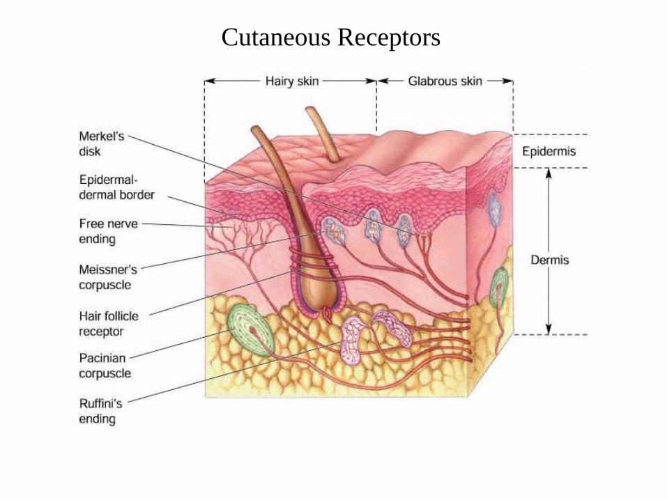
Cutaneous Receptors

Sir Henry Head (1861-1940)

“Protopathic System”“Epicritic System”

Large Diameter Afferents (Aa or I, Ab or II)
All are mechanoreceptors
Encapsulated Receptors
Small Diameter Afferents (Ad or III, C or IV)
Free nerve endings
Mechanoreceptors (crude or non-discriminative touch)
Nociceptors (damaging mechanical, thermal, chemical)
Chemoreceptors („spicy‟)
Thermoreceptors (warm, cold)
Discriminative touch
Joint position sense (proprioception)
Serve different submodalities

100 m/sec = 225 miles/hour
„Fine‟ touch „Crude‟ touchPosition sense
Encapsulated receptors Free nerve endings
Differentially sensitive to pressure and local anesthetics
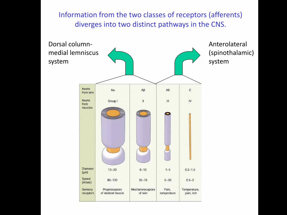
Dorsal column-medial lemniscus system
Anterolateral (spinothalamic) system
Information from the two classes of receptors (afferents) diverges into two distinct pathways in the CNS.

Dorsal root ganglion
Dorsal columns To brain
THALAMUS
(Discriminative Touch, Position Sense)
Dorsal Root Fiber
CORTEX
MEDULLA
SPINAL CORD
Dorsal Columns
VPL
DORSAL COLUMN
MEDIAL LEMNISCUS SYSTEM
SI
MIDBRAIN
PONS
Dorsal Column Nuclei
MedialLemniscus
I (A-alpha)
II (A-beta)
Dorsal root ganglion
Synapse
DecussationAnterolateral system
To brain
Dorsal Root Fiber
CORTEX
THALAMUS
SPINAL CORD
(Pain, Temperature, Low-acuity or "Crude" Touch)
ANTEROLATERAL SYSTEM
Intralaminar Nuc.
Posterior Grp.
SI, SII & Others
VPL
BRAIN STEMReticular
Formation
AnterolateralColumn
III (A-delta)
IV (C)
Be able to sketch these pathways
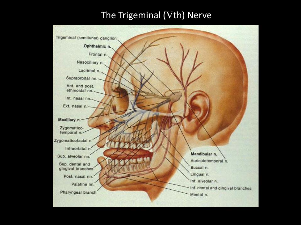
The Trigeminal (Vth) Nerve

What is the basis for this?
Two-Point Discrimination Thresholds (or Minimum Separable)
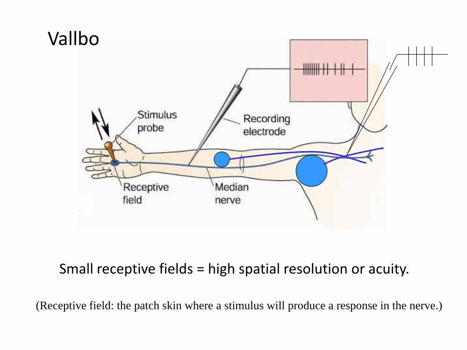
Small receptive fields = high spatial resolution or acuity.
Vallbo
(Receptive field: the patch skin where a stimulus will produce a response in the nerve.)

Skin resolution
Palm Finger Finger tip
0
0.3
0.6
0
40
80
120
Spatial re
solu
tion
Innerv
ation d
ensity
units/s
q. m
m.
1/m
m
Meissner’s corpuscles
Two-point discrimination
High receptive field density = high spatial resolution or acuity.

Differential Cortical Magnification of the Receptor Surface
S1
Regions of high spatial acuity have:
Small receptive fields
High receptive field density
Large representations in sensory cortex

Critical separation of activity in cortex

Cutaneous receptive fieldsA
C D
S
T
I
M
Receptive fieldB
ResponseSkin

Feed-Forward Inhibition
Feedback Inhibition
Lateral Inhibition
Recurrent collateral

Excitatory
synapseInhibitory synapse
STIM
peak
peak
trough
11
22
33
4
5
4
5
First order neurons Second order neurons
Lateral Inhibition
A similar diagram using feed-forward inhibition is in BCP
Lateral inhibition increases spatial contrast in the neural activity pattern

Which receptors mediate the two-point discrimination thresholds?
(neurospeak)

Physiological
Class
Adaptation Receptive Field Probable Morphology
PC (RA 1 ) Fast Large Pacinian corpuscle
RA (RA II) Fast Small Meissner’s corpuscle
SA I Slow Small Merkel’s disc
SA II Slow Large Ruffini’s ending
t
Raised dots
Move pattern
Record from all cells simultaneouslyA
Snapshot of spike activity at one instantReceptive fields
Stored spike activity
t
Record from one cell, assume it is identical to the othersB
Receptive field

Receptive field
Spike trains on
sequential sweeps
Step this way after each sweep
„Reconstruct‟ pattern for
a continuous sheet of
cells with identical
receptive fields
Merkel‟s receptors
Meissner‟s corpuscles
Pacinian corpuscles
Patch of skin with
a sheet of receptors
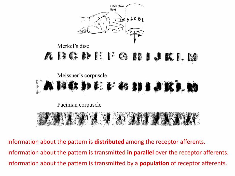
Merkel‟s disc
Meissner‟s corpuscle
Pacinian corpuscle
Information about the pattern is distributed among the receptor afferents.
Information about the pattern is transmitted in parallel over the receptor afferents.
Information about the pattern is transmitted by a population of receptor afferents.

Mueller’s “Law of Specific Nerve Energies”
“Labeled lines”
“Grandmother cells”
“Feature detectors”
Etc.
• Each type of sensory nerve ending, however stimulated (electrically, mechanically, etc.), gives rise to its own specific sensation
• Each type of sensation depends not upon any special character of the different nerves but upon the part of the brain in which their fibers terminate.
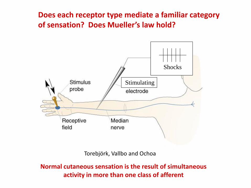
Does each receptor type mediate a familiar category of sensation? Does Mueller’s law hold?
Torebjörk, Vallbo and Ochoa
Stimulating
Shocks
Normal cutaneous sensation is the result of simultaneous activity in more than one class of afferent
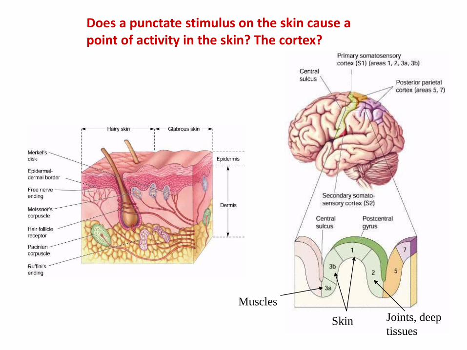
Does a punctate stimulus on the skin cause a point of activity in the skin? The cortex?
Muscles
Joints, deep
tissuesSkin

3a 3b 1 2
S1
Hand area
Vibrating probe
Muscle receptors
Cutaneous
receptors
Deep tissue
receptors
Information from a point on the skin is distributed to a zone or zones of neural circuitry in the cortex.
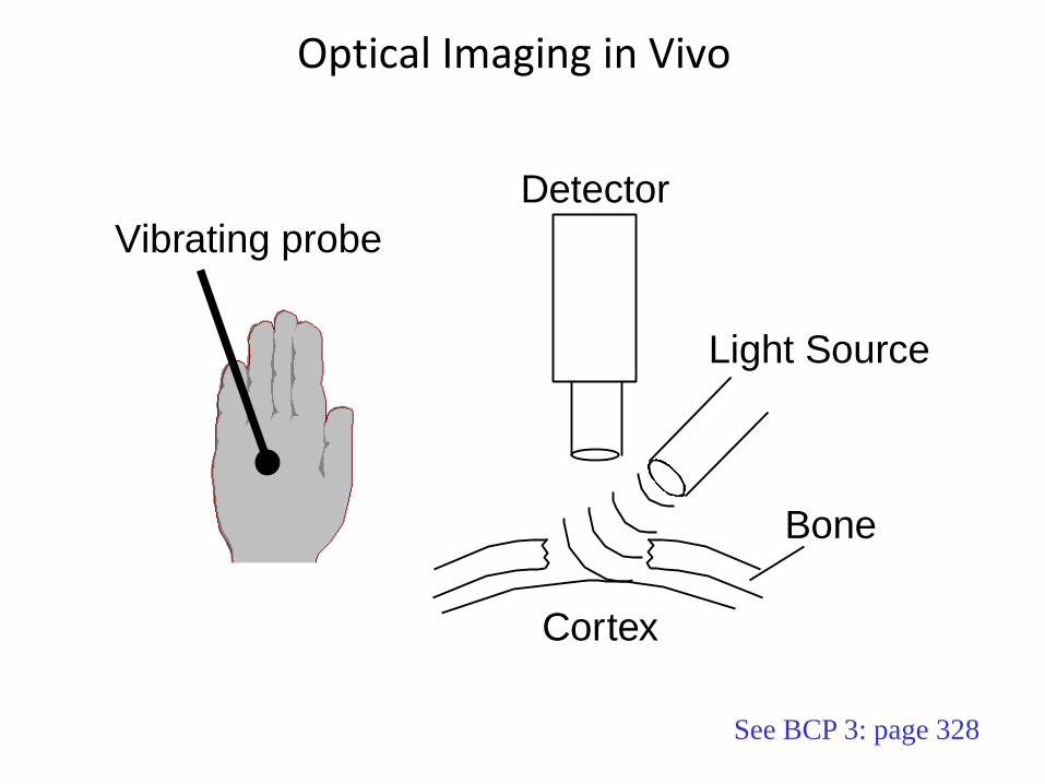
Cortex
Light Source
Detector
Bone
Vibrating probe
Optical Imaging in Vivo
See BCP 3: page 328

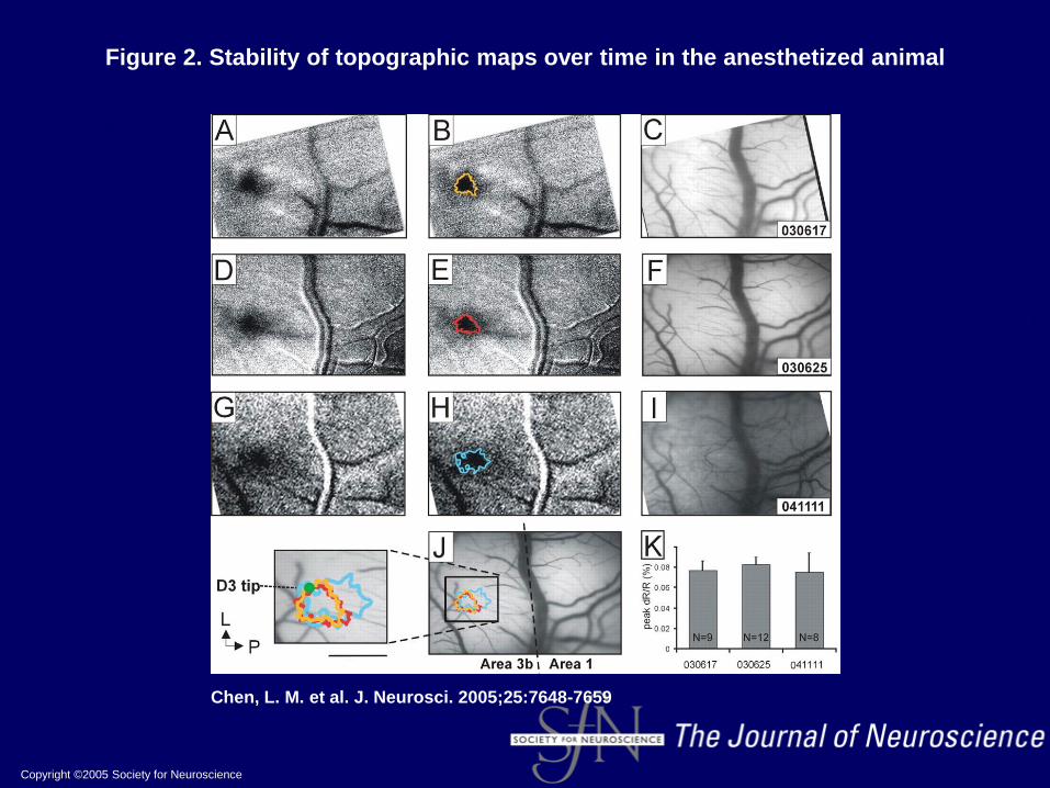
Copyright ©2005 Society for Neuroscience
Chen, L. M. et al. J. Neurosci. 2005;25:7648-7659
Figure 2. Stability of topographic maps over time in the anesthetized animal

Are neural representations in S1 fixed?

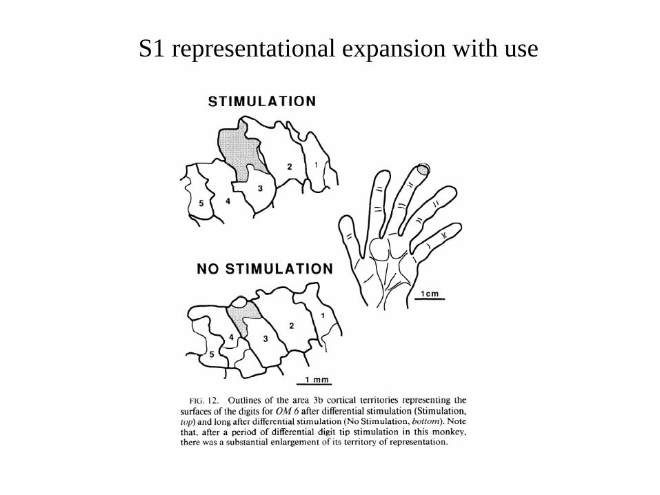
S1 representational expansion with use

Some Major Principles of Somatic-Sensory Spatial Coding
Spatial acuity varies inversely with receptive-field size
Lateral inhibition in the CNS enhances spatial contrast
Information from a point on the skin is “dissected” by differentclasses of receptors and transmitted in parallel channels that influence a region or regions of cortex
Spatial acuity varies directly with receptive-field (receptor) density
Spatial acuity varies directly with the cortical magnification