
Sources, Extraction and Biomedical Properties of ...
23
foods Review Sources, Extraction and Biomedical Properties of Polysaccharides Samee Ullah 1,2 , Anees Ahmed Khalil 2 , Faryal Shaukat 1 and Yuanda Song 1, * 1 Colin Ratledge Center for Microbial Lipids, Center for Functional Foods and Health, School of Agriculture Engineering and Food Science, Shandong University of Technology, Zibo 255049, China 2 University Institute of Diet and Nutritional Sciences, Faculty of Allied Health Sciences, The University of Lahore, Lahore 54000, Pakistan * Correspondence: [email protected]; Tel.: +86-139-0617-4047 Received: 16 July 2019; Accepted: 28 July 2019; Published: 1 August 2019 Abstract: In the recent era, bioactive compounds from plants have received great attention because of their vital health-related activities, such as antimicrobial activity, antioxidant activity, anticoagulant activity, anti-diabetic activity, UV protection, antiviral activity, hypoglycemia, etc. Previous studies have already shown that polysaccharides found in plants are not likely to be toxic. Based on these inspirational comments, most research focused on the isolation, identification, and bioactivities of polysaccharides. A large number of biologically active polysaccharides have been isolated with varying structural and biological activities. In this review, a comprehensive summary is provided of the recent developments in the physical and chemical properties as well as biological activities of polysaccharides from a number of important natural sources, such as wheat bran, orange peel, barely, fungi, algae, lichen, etc. This review also focused on biomedical applications of polysaccharides. The contents presented in this review will be useful as a reference for future research as well as for the extraction and application of these bioactive polysaccharides as a therapeutic agent. Keywords: bioactive polysaccharides; extraction; biomedical applications 1. Introduction Polysaccharides are considered as vital bio-macromolecules for all living organisms, which are structurally comprised of homo or hetero monosaccharides and uronic acids connected with glycosidic linkages [1–3]. They are predominantly found in various parts of plants, animals, fungi, bacteria, and seaweed, and play a critical role in numerous physiological functions of life [4]. Polysaccharides along with lipids, proteins, and polynucleotides are classified as the most pivotal four macromolecules in life sciences. Bioactive polysaccharides are known as polysaccharides produced from living organisms, and/or are functionalized from sugar-based materials possessing biological effects on organisms. Moreover, during the last decades, bioactive polysaccharides have been investigated as therapeutic agents against many chronic diseases due to their biodegradability, non-toxic nature, and biocompatibility [5]. Studies have shown that polysaccharides possess a wide range of pharmacological perspectives such as antioxidative, antitumor, antimicrobial, anti-obesity, hypolipidemic, antidiabetic, and hepato-protective properties [6–8]. They have been investigated extensively for the development of novel products in the field of cosmetics, food, medicine, petrochemicals, and paper [3,9,10]. Particularly, in the medicinal industry, polysaccharides are mostly used as pharmaceuticals and medical biomaterials (hypoglycemic, anti-osteoarthritic, and anticancer products) to curtail the effect of respective metabolic syndromes [9,11]. The potentiality of bioactive polysaccharides is strongly influenced depending upon their configuration and chemical structure. Nevertheless, the macromolecular configurations of plant Foods 2019, 8, 304; doi:10.3390/foods8080304 www.mdpi.com/journal/foods
Transcript of Sources, Extraction and Biomedical Properties of ...

foods
Review
Sources, Extraction and Biomedical Propertiesof Polysaccharides
Samee Ullah 1,2 , Anees Ahmed Khalil 2, Faryal Shaukat 1 and Yuanda Song 1,*1 Colin Ratledge Center for Microbial Lipids, Center for Functional Foods and Health, School of Agriculture
Engineering and Food Science, Shandong University of Technology, Zibo 255049, China2 University Institute of Diet and Nutritional Sciences, Faculty of Allied Health Sciences,
The University of Lahore, Lahore 54000, Pakistan* Correspondence: [email protected]; Tel.: +86-139-0617-4047
Received: 16 July 2019; Accepted: 28 July 2019; Published: 1 August 2019�����������������
Abstract: In the recent era, bioactive compounds from plants have received great attention because oftheir vital health-related activities, such as antimicrobial activity, antioxidant activity, anticoagulantactivity, anti-diabetic activity, UV protection, antiviral activity, hypoglycemia, etc. Previous studieshave already shown that polysaccharides found in plants are not likely to be toxic. Based on theseinspirational comments, most research focused on the isolation, identification, and bioactivities ofpolysaccharides. A large number of biologically active polysaccharides have been isolated withvarying structural and biological activities. In this review, a comprehensive summary is provided ofthe recent developments in the physical and chemical properties as well as biological activities ofpolysaccharides from a number of important natural sources, such as wheat bran, orange peel, barely,fungi, algae, lichen, etc. This review also focused on biomedical applications of polysaccharides.The contents presented in this review will be useful as a reference for future research as well as forthe extraction and application of these bioactive polysaccharides as a therapeutic agent.
Keywords: bioactive polysaccharides; extraction; biomedical applications
1. Introduction
Polysaccharides are considered as vital bio-macromolecules for all living organisms, whichare structurally comprised of homo or hetero monosaccharides and uronic acids connected withglycosidic linkages [1–3]. They are predominantly found in various parts of plants, animals, fungi,bacteria, and seaweed, and play a critical role in numerous physiological functions of life [4].Polysaccharides along with lipids, proteins, and polynucleotides are classified as the most pivotal fourmacromolecules in life sciences. Bioactive polysaccharides are known as polysaccharides producedfrom living organisms, and/or are functionalized from sugar-based materials possessing biologicaleffects on organisms. Moreover, during the last decades, bioactive polysaccharides have beeninvestigated as therapeutic agents against many chronic diseases due to their biodegradability,non-toxic nature, and biocompatibility [5]. Studies have shown that polysaccharides possess a widerange of pharmacological perspectives such as antioxidative, antitumor, antimicrobial, anti-obesity,hypolipidemic, antidiabetic, and hepato-protective properties [6–8]. They have been investigatedextensively for the development of novel products in the field of cosmetics, food, medicine,petrochemicals, and paper [3,9,10]. Particularly, in the medicinal industry, polysaccharides are mostlyused as pharmaceuticals and medical biomaterials (hypoglycemic, anti-osteoarthritic, and anticancerproducts) to curtail the effect of respective metabolic syndromes [9,11].
The potentiality of bioactive polysaccharides is strongly influenced depending upon theirconfiguration and chemical structure. Nevertheless, the macromolecular configurations of plant
Foods 2019, 8, 304; doi:10.3390/foods8080304 www.mdpi.com/journal/foods
Foods 2019, 8, 304 2 of 23
cellular polysaccharides, particularly hetero polysaccharides (hemicelluloses), are very complex owingto the occurrence of various monosaccharides acting as isobaric stereoisomers [12,13]. Additionally,polysaccharides present in plants, microorganisms (bacteria, fungi, and yeasts), animals, and algaeare chemically and/or physically bound with various other biomolecules like lignin, proteins,lipids, polynucleotides, and a few minerals [14]. Hence, to understand the importance of bioactivepolysaccharides in the domain of life sciences resulted in the multi-disciplinary collaborationof scientists from the fields of microbiology, phytology, glycol-biology, nutrition, food sciences,and glycol-medicine [5]. Polysaccharides both in the simple and complex glycol-conjugated form arerenowned for various bioactive functions, for instance, antivirus, antioxidant, antimicrobial, anticancer,antidiabetic, reno-protective, immunomodulatory, and anti-stress perspectives.
2. Sources of Bioactive Polysaccharides
Bioactive polysaccharides are categorized broadly depending upon their sources, structure,applications, solubility, and chemical composition. On the basis of chemical composition, they arecharacterized as homopolysaccharides (homoglycans) and heteropolysaccharides (heteroglycans).Homoglycans comprise of a single type of monosaccharide, such as glycogen and cellulose whichconsists of glucose molecules, whereas heteroglycans are made up of a different type of monosaccharides,for example, heparin and chondroitin sulfate (CS) [15].
According to the glycosides linked on to the glycan, polysaccharides can also be classified asproteoglycans and glycoproteins, glycolipids, and glycoconjugates [16,17]. According to groupingbased on origins, bioactive polysaccharides are usually classified as those derived from animals(chondroitin sulfate, heparin, and hyaluronan), plants (pectin, inulin, Ginseng polysaccharides, xylans,and arabinans), bacteria (exopolysaccharides, capsular polysaccharides, and peptidoglycans), lichen,fungi, and algae. In the section below, bioactive properties associated with polysaccharides arereviewed depending upon their natural sources in order to understand their functionality.
2.1. Bioactive Polysaccharides in Dietary Fibers
The FAO (Food and Agriculture Organization) defined dietary fibers as a variety of indigestibleplant polysaccharides including pectins, cellulose, gums, hemicelluloses, oligosaccharides, and variouslignified compounds. Dietary fibers are the type of polysaccharides that are biologically active in theirinnate form and/or after enzymatic and chemical treatments. For instance, celluloses and hemicellulosesdirectly stimulate the bowel movement, whereas inulin is fermented by microflora with the intentto prevent various gastrointestinal disorders [18]. Dietary fibers play a vital role in prevention fromvarious diseases especially hyperlipidemia, cardiovascular complications, and obesity. Gums, inulin,and pectin are capable of slowing down the passage of food in the digestive tract, reducing serumcholesterol concentrations, hindering the absorption of sugar from food to bloodstream and, therefore,avoiding sudden hyperglycemic conditions post food ingestion.
Among dietary fibers, lignin, cellulose, and hemicellulose are classified as insoluble fibers whichhelp in the stimulation of the bowel movement, speeds up the removal of waste via the digestivetract, and helps in prevention of hemorrhoids, constipation, and diverticulosis [19,20]. Results ofvarious epidemiological investigations and clinical trials have suggested that consumption of moderateor higher levels of dietary fiber efficiently reduces the risk of numerous metabolic syndromes likediabetes [21], strokes [22], hypertension [23,24], hyperlipidemia/hypercholesterolemia, [25,26], obesity,and cancer [27]. Usually, ingestion of dietary fiber has health modulating benefits, like increased stoolbulk, reduced intestinal transit time, a decline in levels of total cholesterol (TC) and postprandialblood glucose, and/or insulin contents [27–29]. Keeping in view the significance of dietary fibers withspecial reference to human health and their associated mechanism of actions, these are consideredas an abundant source of bioactive polysaccharides. Table 1 summarizes the types, names, sources,composition, and physiological effects of dietary fiber polysaccharides from various studies and trials.

Foods 2019, 8, 304 3 of 23
2.2. Bioactive Polysaccharides in Herbs
Herbs have attained a significant position in traditional medicines (Indian Ayurveda, ancientChinese medicine, phytomedicine in western countries, and Japanese Kamp medicine) of numerouscountries owing to their associated curing benefits against numerous diseases. Outcomes ofrecent pharmacological studies have illuminated that the chief component of herbal medicinesusually comprises of tannins, polysaccharides, high molar mass proteins, and low molar massconstituents like terpenoids (quassinoids, sesquiterpenes, and Rabdosia diterpenes), saponins, alkaloids(protoberberine alkaloid, phenanthridine, etc.), and flavonoids (Scutellaria flavones) [30]. Amongthe above-mentioned compounds in herbal medicines, polysaccharides are believed to be mainbioactive compounds responsible for various pharmacological potentials like antitumor, antioxidant,hepato-protective, antiviral, radio-protective, immuno-stimulatory, and anti-fatigue activities [31–35].Innate polysaccharides present in numerous herbs are known for stimulation of the humanimmune system, inhibition of viral replication, scavenging free radicals, and inhibition of lipidperoxidation [33,36,37]. Recent advancements regarding the application of polysaccharides present inherbal medicines for prevention and cure of diseases are documented in Table 1.
Table 1. Biological activities of bioactive polysaccharides.
Polysaccharides Biological Activities Mechanism of Action References
β-glucan Anti-obesity activity• Reduced energy intake• Increase in fullness and satiety• Decreased hunger and increased satiety and fullness.
[38]
Pectin, AlgalFucoidan
Anti-microbialactivity
• Inhibitory effects on the entry of enveloped virusesincluding herpes and HIV into cells
• Inhibit the formation of syncytium formation[39,40]
Fucoidan,PolygonummultiflorumThunbPolysaccharides
Anti-oxidant activity
• Inhibited ERK and p38-MAPK signaling pathwaysscavenging free radicals (e.g., superoxide anion radical,hydroxyl radical, and hydroxyl peroxide),
• Prevents lipid oxidation and protein glycation• Inhibits the formation of ROS and RNS
[40,41]
Red algae sulfatedpolysaccharides,Carrageenan,Fucoidan,Chondroitin sulfate
Anti-inflammatoryactivity
• Lowered the expression of inducible nitric oxidesynthase (iNOS)
• Inhibited the expressions of TNF-a, interleukin-1b (IL-1b)and interferon-c (IFN-c)
• Repressed pro-inflammatory cytokines• Suppresses the activity of COX-2
[40,42,43]
β-glucan Anti-diabetic activity• Suppressed the formation of AGE• Reduced postprandial glucose and insulin responses• Increases the level of antioxidant enzymes in the body
[38]
Konjac glucomannan Hypo-cholesterolemicactivity
• Reduced the plasma cholesterol• Significantly lowered Serum total, HDL-C, and LDL-C• Reduces the concentration of serum MDA
[44]
Pectin, Ginsengpolysaccharides,Heparan sulfate
Anti-tumour activity
• Constrains the production of prostaglandin E2• Prevents from the oxidation of DNA• Stimulates macrophages to produce helper types 1 and 2
(Th1 and Th2) cytokines• Promotes the formation of ternary complexes• Displaces growth factors
[39,45,46]
Ginsengpolysaccharides
Immune modulatoryactivity
• Downregulates the secretion of inflammation relatedmediator nitric oxide (NO) and cytokines (TNF-a, IL-6,and IL-1b)
• Reduces the activation of neutrophils
[45]
β-glucan, Pectin,Gums, Konjacglucomannan
Gastro-protectiveactivity
• Supplement increased faecal bulk• Alter postprandial lipid and lipoprotein composition
[19,44]
Acanthopanaxpolysaccharides
Neuro-protectiveactivity
• Increase SOD and GSH-Px activities and IL-10 levels• Reduces the levels of MDA, IL-1, and TNF-α• Prevents the formation of inflammatory cytokines
[47]

Foods 2019, 8, 304 4 of 23
2.3. Bioactive Polysaccharides in Algae and Lichens
Polysaccharides present in lichens and algae have been of great interest to the food scientistsowing to their associated exceptional physical properties (stabilizing, gelling, and thickening capability)and biological potentials (immunomodulating, antiviral, anticoagulant, antitumor, antioxidative,anti-inflammatory, and anti-thrombotic activity) [48,49]. Basically, sulfated polysaccharides are themain group of the most attractive and interesting constituents in marine algae i.e., laminarans andfucoidans in the Phaeophyceae (brown algae), carrageenans in the Rhodophyceae (red algae), andulvans in the Chlorophyceae (green algae) [40]. One of the most vital properties linked with sulfatedpolysaccharides is their anticoagulant characteristics. For instance, carrageenan (sulfated galactans)isolated from red algae and sulfated fucoidans extracted from brown algae are known to possessexcellent anti-coagulant properties. Sulfated polysaccharides identified in algae have been stated toown equal or even stronger activities than those linked with heparin [32]. Various other biologicalproperties of sulfated polysaccharides present in algae have been intensively investigated.
Experimentally, fucoidan has been reported to possess maximum antioxidative propertiesfollowed by alginate and laminaran, therefore protecting human health from the damage of ROS(reactive oxygen species). Likewise, the anticancer and anti-tumor characteristics of algae-basedsulfated polysaccharides are found to be due to their antioxidative and free radical scavengingproperties [50]. Moreover, inhibition of HIV and herpes viruses in cells owing to anti-viral potential ofsulfated rhamnogalactans, carrageenans, and fucoidans have also been scientifically proven [40].Immuno-modulating characteristics associated with algae-based sulfated polysaccharides weredemonstrated by increasing the secreting and phagocytic activities of macrophages [51].
Polysaccharides derived from lichens (composite organism formed due to a symbiotic partnershipamong alga and fungus) are principally linear or rarely substituted α-glucans and/or β-glucans.This type of glucans isolated from lichens is reported to have a wide range of bioactive functionssuch as immunomodulatory perspectives, anti-tumor potential, and anti-viral characteristics [49,52,53].The immuno-modulating activity of polysaccharides (β-glucans) derived from lichenin and lichensare linked to stimulation of a variety of immune responses like production of NO (nitric oxide) andROS (reactive oxygen species) and release of cytokines and arachidonic acid metabolites [51]. Lichenin,also known as lichenan, consists of a galactoglucomannan (water-soluble hemicellulose) structure thatis responsible for anti-thrombotic and anti-coagulant properties [54]. Bioactive properties related tosulfated polysaccharides from lichens and algae are highly dependent on their structural properties,for instance, sulfate concentration and distribution of sulfate groups on the main chain, stereochemistry,and molar masses. Hence, there is a need for the modification of innate sulfate polysaccharides toattain biologically active polysaccharides having desired molecular size and functionality for bioactivefurther application [40,55]. An overview of some recent advancements in this context are depicted inTable 2.
2.4. Bioactive Polysaccharides in Fungi
A wide range of bioactive saccharides is present in fungi ranging from monosaccharides topolysaccharides. They can be summarized as the intracellular or extracellular polysaccharidesdepending upon their presence in the fungi [56]. The bioactive polysaccharides derived frombasidiomycetes are reported to have antitumor and anticancer activities and can prevent breastcancer in post-menopausal women. The compound which can inhibit tumors was first isolated fromthe basidiomycete Boletus edulis in 1957. In traditional medicine, several basidiomycete mushrooms areused as an immunosuppressant to treat cancer and even for treating AIDS [57]. Polysaccharopeptide(PSP) isolated from Ganoderma lucidum induces the inhibition of oncogenes and kill the tumor cellsdirectly. Similarly, the bioactive polysaccharides schizophyllan, lentinan, and polysaccharide-K isolatedfrom Grifola frondosa and Trametes versicolor, respectively, improve the immune function of the hostand showed a synergistic effect with chemotherapy [58]. Just like mushrooms, another source ofbioactive polysaccharides is Cordyceps militaris, and they possess a wide range of structurally different

Foods 2019, 8, 304 5 of 23
bioactive polysaccharides. Water-soluble polysaccharides from C. militaris contain a (1→ 4) galactoselinkage and (1→ 3,6)-mannose linkage and branching usually arising from (1→ 4)-linked glucose.These polysaccharides were reported for their numerous biological activities including anti-tumor,immunomodulatory, antioxidant, and anti-inflammatory activities [56,59].
2.5. Bioactive Polysaccharides from Bacteria
Bacteria are a rich source of biologically active polysaccharides. On the base of structural properties,bacterial polysaccharides can be defined into five major classes: Exopolysaccharides (EPSs), capsularpolysaccharides (CPSs), lipopolysaccharides (LPSs), peptidoglycans, and teichoic acids [60].
A lot of research has been done on hyperbranched bacterial polysaccharides (HBPSs). For example,highly branched dextran with a backbone of 75% (α-(1→6)-D-Glcp) with 19% of α-(1→3) and a fewα-(1→2) branching [61], was separated from Leuconostoc citreum B-2 and studied extensively, and showeda water-holding capacity of up to 450% and an 80% water solubility index, consequently havingpotential applications in cosmetic, food, and pharmaceutical industries [62,63]. From an industrialpoint of view, EPSs producing Gram-negative prokaryotes (cyanobacteria) are important because oftheir ability to produce novel molecules. These EPSs usually linger as a sheath/capsule with the cellsand when they are liberated from the cells they are termed as released polysaccharides. Some bacteriafrom the genus cyanobacterium, namely Anabaena, Aphanocapsa, Cyanothece, Phormidium, Synechocystis,and Nostoc are useful to produce sulfated EPSs, which also contain uronic acids [64]. Upon fermentationin the laboratory, polysaccharide yield from 0.5–4 g/L can be obtained from three known genera ofmarine bacteria, namely Alteromonas sp., Pseudoalteromonas sp. and Vibrio sp. [65]. ExopolysaccharideHE 800 is secreted by Vibrio diabolicus and it contains an equal amount of hexosamine and glucuronicacid, which can be depolymerized, N-deacetylated, and after chemical sulfurylation produce newderivatives of HE800. These derivatives are referred to as DRS HE800 and are structurally very closeto heparan-sulfate [66]. Moreover, acidic and highly branched heteropolysaccharide (EPS GY785) isproduced by the bacterium Alteromonas infernus. Non-saccharide units of EPS GY785 are composed ofuronic acid and sugar (glucose and galactose) [67].
EPSs derived in the laboratory showed widespread biological activities like being an antioxidant,having cholesterol-lowering and inflammation-regulating properties, as well as anti-tumor, anti-coagulant, and antivirus activities [62,68]. Moreover, bacterial polysaccharides are used in ProteinGlycan Coupling Technology (PGCT) to produce glycoconjugate vaccines against Gram-positive andGram-negative bacteria [69].
2.6. Bioactive Polysaccharides in Wood
Bioactive polysaccharides present in wood primarily consists of celluloses and some primarygroups of hemicelluloses (glucans, arabinans, xylans, glucomannans, and galactans) [70,71]. Pectinsand galacto-glucomannans derived from wood are stated to have radical scavenging properties andimmune-stimulating activities [72,73]. Xylo-oligosaccharides (xylans) derived from dietary fibers,hard and/or softwood have been reported to be used efficiently as prebiotics by nutraceutical andmedicinal industries [74]. Similarly, pentosane polysulfate—a xylan derivative being extractedfrom beech-wood—is also known as a biomedical agent for the treatment of interstitial cystitis,a bladder pain syndrome [75]. While most polysaccharides do not reveal biological propertiesunless they are subjected to some modification; derivatives of cellulose-like hydroxypropyl-cellulose(HPC), hydroxypropyl-methylcellulose (HPMC), hydroxyethyl-cellulose (HEC), and methyl-cellulosehave evidently proven their functional roles in numerous fields (medicinal, cosmetics, food,and pharmaceutical) [76].
2.7. Bioactive Polysaccharides from other Sources
Sulfated glycosaminoglycans (GAGs) like keratin sulfate (KS), heparan sulfate (HS), dermatansulfate (DS), and chondroitin sulfate (CS) belongs to a class known as animal-derived bioactive

Foods 2019, 8, 304 6 of 23
polysaccharides. Heparan sulfate comprises of repeated units of N-acetylated and sulfated disaccharides(glucosamine and glucuronic/iduronic acid) units [77–79]. Evidently, heparin is known to be themost effectual clinical anti-coagulant used in medical sciences. The anti-coagulation potential ofheparin is mainly due to its particular structural arrangement, in which specifically the bindingsite of anti-thrombin III (ATIII, a non-vitamin K-dependent protease) is of central importance in theprevention of fibrin clot formation which is generated owing to the enzymatic activity of thrombin [80].Better knowledge regarding the interlinkage among structural properties and activity of heparincreates an opportunity for the development of new drugs having more specificity and improvedanti-coagulating properties. Furthermore, this structural-activity connection is also helpful in exploringvarious biomedical applications like anti-viral, anti-inflammatory, anti-cancer, and wound healingproperties [78,79,81,82]. DS and CS chains comprise of sulfated N-acetylgalactosamine and iduronicacid (in case of DS)/glucuronic acid (in case of CS) disaccharide repeating units [83,84]. Both of these arefunctionally characterized as bioactive polysaccharides owing to their presence as a vital molecule in theextracellular matrix of the brain, which helps in regulation of cell migration, adhesion, and proliferationwhile healing of wounds, as well as proper signaling of growth factor in the skeleton [85–88].
On the other hand, HA (hyaluronic acid) primarily isolated form animal tissues compriseof the linear structure of non-sulfated glycosaminoglycan molecules with repeated units ofβ-(1-3)-N-acetyl-d-glucosamine, and β-(1-4)-d-glucuronic acid. It is also a vital constituent of the brain’sextracellular matrix, which promotes mediation of cellular signaling, helps in healing of wounds, andmorphogenesis. Owing to these properties, HA and their associated derivatives are predominantlybeing applied by the medical industry in eye surgery, viscosupplementation, and as a biomedical toolfor drug delivery [89–91]. Chitin is known to be the most abundant polysaccharide present in theexoskeleton of shrimps and crabs, the cell walls of yeast and fungi, and the cuticles of insects. It isby far the second most copious polymer after cellulose and comprises of a random distribution ofβ-(1-4)-linked N-acetyl-glucosamine and N-glucosamine [92,93]. Industrially, chitosan is characterizedas a vital derivative of chitin and is practically obtained due to partial deacetylation of chitin eitherby the action of enzyme chitin deacetylase or under alkaline conditions [94]. Bioactive properties(anti-cancer, anti-tumor, anti-bacterial, anti-inflammatory, etc.) associated with oligomers of chitosanand chitin, yielded owing to partial acidic hydrolysis, have earlier been documented [95].
3. Extraction and Quantification of Bioactive Polysaccharides
In general, bioactive potentials of naturally occurring polysaccharides are greatly dependentupon their molar mass and level and distribution of groups/side chains on the backbone. Therefore,isolation of polysaccharides from complex cellular plant matrices while keeping their bioactivityintact is of great significance. During the last decade, various innovative green extraction techniques(microwave-assisted, ultra-sonication, supercritical fluid extraction, and hot water extraction) are inpractice for isolation of bioactive polysaccharides. These techniques have acquired great attentionof scientists and researchers mainly due to their increased extraction rates, cost-effective nature,enviro-friendly characteristics, and structure preservative potentials [73,96,97].
Numerous scientific investigations have been implemented for the extraction and isolation ofbioactive polysaccharides. The type of method adopted defines the physicochemical propertiesand antioxidant potential of isolated polysaccharides. For this purpose, a group of scientistscompared the structural and antioxidant properties of bioactive polysaccharides extracted fromBarrenwort (Epimedium acuminatum) through various extraction techniques (microwave-assisted,enzymatic, hot water extraction, and ultrasound-assisted extraction). They were of the view thatbioactive polysaccharides isolated from hot water extraction had the highest antioxidant propertiesas compared to those extracted from other techniques, while their physicochemical properties werethe same [98]. The hot water extraction method used in combination with various latest techniques(enzymatic pre-treatment, microwave, and ultrasound-assisted) are useful in increasing the yield andextraction productivity of polysaccharides. Likewise, enzymatic pre-treatment of raw material prior to
Foods 2019, 8, 304 7 of 23
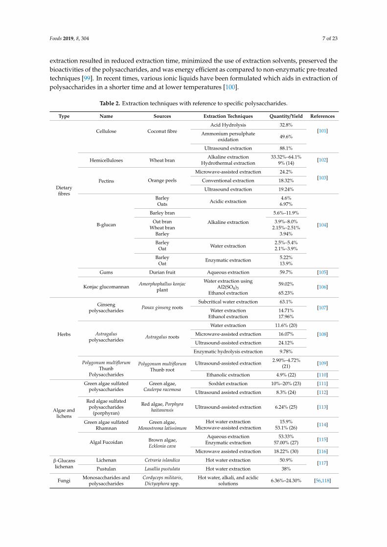
extraction resulted in reduced extraction time, minimized the use of extraction solvents, preserved thebioactivities of the polysaccharides, and was energy efficient as compared to non-enzymatic pre-treatedtechniques [99]. In recent times, various ionic liquids have been formulated which aids in extraction ofpolysaccharides in a shorter time and at lower temperatures [100].
Table 2. Extraction techniques with reference to specific polysaccharides.
Type Name Sources Extraction Techniques Quantity/Yield References
Dietaryfibres
Cellulose Coconut fibreAcid Hydrolysis 32.8%
[101]Ammonium persulphateoxidation 49.6%
Ultrasound extraction 88.1%
Hemicelluloses Wheat branAlkaline extraction 33.32%–64.1% [102]
Hydrothermal extraction 9% (14)
Pectins Orange peels
Microwave-assisted extraction 24.2%[103]Conventional extraction 18.32%
Ultrasound extraction 19.24%
B-glucan
BarleyAcidic extraction
4.6%
[104]
Oats 6.97%
Barley bran
Alkaline extraction
5.6%–11.9%
Oat bran 3.9%–8.0%Wheat bran 2.15%–2.51%
Barley 3.94%
BarleyWater extraction
2.5%–5.4%Oat 2.1%–3.9%
Barley Enzymatic extraction 5.22%Oat 13.9%
Gums Durian fruit Aqueous extraction 59.7% [105]
Konjac glucomannan Amorphophallus konjacplant
Water extraction usingAl2(SO4)3
59.02% [106]Ethanol extraction 65.23%
Herbs
Ginsengpolysaccharides
Panax ginseng rootsSubcritical water extraction 63.1%
[107]Water extraction 14.71%Ethanol extraction 17.96%
Astragaluspolysaccharides Astragalus roots
Water extraction 11.6% (20)
[108]Microwave-assisted extraction 16.07%
Ultrasound-assisted extraction 24.12%
Enzymatic hydrolysis extraction 9.78%
Polygonum multiflorumThunb
Polysaccharides
Polygonum multiflorumThunb root
Ultrasound-assisted extraction 2.90%–4.72%(21) [109]
Ethanolic extraction 4.9% (22) [110]
Algae andlichens
Green algae sulfatedpolysaccharides
Green algae,Caulerpa racemosa
Soxhlet extraction 10%–20% (23) [111]
Ultrasound assisted extraction 8.3% (24) [112]
Red algae sulfatedpolysaccharides
(porphyran)
Red algae, Porphyrahaitanensis Ultrasound-assisted extraction 6.24% (25) [113]
Green algae sulfatedRhamnan
Green algae,Monostroma latissimum
Hot water extraction 15.9% [114]Microwave-assisted extraction 53.1% (26)
Algal Fucoidan Brown algae,Ecklonia cava
Aqueous extraction 53.33% [115]Enzymatic extraction 57.00% (27)
Microwave assisted extraction 18.22% (30) [116]
β-Glucanslichenan
Lichenan Cetraria islandica Hot water extraction 50.9% [117]Pustulan Lasallia pustulata Hot water extraction 38%
Fungi Monosaccharides andpolysaccharides
Cordyceps militaris,Dictyophora spp.
Hot water, alkali, and acidicsolutions 6.36%–24.30% [56,118]

Foods 2019, 8, 304 8 of 23
In addition, this purification of polysaccharides from the crude extract is really of great importanceas the linkage among structure and safety of products formed for food, pharmaceutical, and biomedicalapplication depends on this. Purification could be achieved by using various techniques (gel filtration,ion exchange and affinity chromatography, ethanolic precipitation, and fractional precipitation),individually or in combination [32].
4. Biomedical Applications
Polysaccharides and their derived compounds are medicinally more preferred as comparedto synthetic polymers owing to their biodegradability, non-toxic nature, biocompatibility, and lowprocessing expenses. Mentioned benefits related to polysaccharides isolated from natural sourcesmake them a valuable ingredient in the fields of pharmaceuticals, nutraceuticals, food, and cosmeticindustries. At the present time, polysaccharides are been used in healthcare and disease control, whilevarious novel areas have also been discovered like in cancer diagnosis, inhibition, and treatment;in drug delivery; in anti-bacterial and anti-viral perspectives; and in tissue engineering [92,119].Therefore, this segment highlights the use of bioactive polysaccharides against various metabolicsyndromes and in the above-mentioned novel areas.
4.1. Anti-Microbial and Antiviral
Various clinical investigations have authenticated that oral administration of pectin to infants andchildren significantly reduced diarrhea and other intestinal infections. This may be because of thedecreased concentration of pathogenic bacteria like Citrobacter, Salmonella, Enterobacter, Shigella, Proteus,and Klebsiella [120]. A linear relationship has been documented among the concentration of probioticsand intestinal health [28].
The bioactive potential of fucoidans—a sulphated polysaccharide derived from marine brownseaweeds—have demonstrated noteworthy anti-viral potential against the cytomegalovirus, HIV, andHSV (herpes simplex virus) [121]. Additionally, few other seaweed-extracted polysaccharides likesulphated rhamnogalactans, carrageenans, and fucoidans have shown an inhibitory effect on viruses(HSV and HIV). Fucoidan comprises of a large quantity of L-fucose and sulphate groups along withfractions of galaturonic acid, xylose, mannose, and galactose. Undaria pinnatifida (marine brown alga)contains fucoidans and have been used in bone health supplementation mainly due to stimulationof osteoblastic cell differentiation. This sulphated polysaccharide has also been known to possesspreventing action on UV-B-induced matrix metalloproteinase-1 (MMP-1) expression by inhibiting theERK (extra-cellular signal regulated kinases) pathways. Therefore, it could be utilized as a functionalingredient in dermal ointments to prevent from skin photo-aging [40].
Some of the other fractions of algae have properties of virucidal and enzyme inhibitory activityinhibiting the formation of the syncytium. Besides, the sulfate group present is necessary for theanti-HIV activity and potency increases with the degree of sulfation.
4.2. Anti-Tumor/Cancer
Numerous scientists have explored dietary fibers as possessing potent anti-cancer properties.Amongst all, pectin has been investigated to reduce cancerous cell migration and tumor growth ina rat model that were administrated with modified citrus pectin [122]. This may be due to bindingof pectin to galectin-3, which results in inhibitory action on some of its functional activities [123].Anti-tumor mode of actions associated with dietary pectin are related to their immune-potentiation,probiotic properties, tumor growth inhibition, anti-mutagenic potential, and regulatory action oftransformation-related oncogenes [124,125]. Anti-tumor mechanisms associated with pectin could bedue to cellular immunological potential [126].
According to a study, ginseng polysaccharides were found to have a stimulating effect on DCs(dendritic cells) causing an elevated formation of IFN-g (interferon-g) [127]. It has also been documentedthat acidic ginseng polysaccharides (GPs) enhanced the production of cytotoxic cells against tumors

Foods 2019, 8, 304 9 of 23
and promoted macrophages for the production of Th1 and Th2 (helper type 1 and 2) cytokines [128,129].Depending upon disease environment or timing of treatments, ginseng polysaccharides extracted fromPanax ginseng demonstrated immuno-modulating perspectives mainly in an immunosuppressing orimmuno-stimulating manner [130]. Acidic GP also revealed modulating action on the concentrationsof antioxidative enzymes like GPx (glutathione peroxidase) and SOD (superoxide dismutase) probablydue to induction of regulating cytokines [131,132]. Likewise, Lemmon et al. [132] found that theimmuno-stimulating potential of acidic GPs isolated from American ginseng (Panax quinquefolius) wasactually mediated by polysaccharides having molecular weight more than 100 kDa [34].
Furthermore, scientists have proven the fact that heparin administration may also have a beneficialimpact on cancer and inflammation. Anti-cancerous, anti-inflammatory, and anti-tumor propertiesassociated with heparin and its low molecular weight species are owing to the pathological functions ofheparan sulfate (HS) chains of proteoglycan structure (HSPGs). Outcomes of an investigation validatedthat heparin transfers GRs (growth factors) stored by HS chains of HSPGs in the ECM (extracellularmatrix) and on cell surfaces. Full-size heparin has potent pro-angiogenic properties as it increases theproduction of ternary complexes of heparin bound FGF2 and VEGF with GF receptors [45].
4.3. Anti-Obesity and Hypocholesterolemia
Numerous trials have shown a direct relationship between consumption of dietary fiber, richdiet/dietary fiber supplementation, and weight loss [133–137]. According to a meta-analysis comprisingof 22 clinical trials, it was documented that a 12 g increase in the content of daily fiber intake resultedin a 10% decrease in energy intake along with a 1900 g decline in body weight [138]. More precisely,the administration of glucomannan (1.24 g/day) along with energy-restricted diet for five consecutiveweeks caused a significant decrease in body weight as compared to the placebo group [139].
In a clinical trial on healthy volunteers, a drink containing oat β-glucan (10.5 g/400 g and 2.5 and5 g/300 g) enhanced fullness sensation as compared to fiber-free drink [140,141]. Likewise, in healthyadolescents subjected to biscuits enriched with barley β-glucan (5.2%) helped in suppressing appetiteratings as compared to control biscuits [142]. Similarly, administration of bread formulated by barleyβ-glucan (3%) to volunteers resulted in decrease of hunger and increased satiety and fullness. This alsoresulted in a noteworthy decrease in energy intake at successive lunches [143]. On the other hand, abar prepared from barley β-glucan (1.2 g) subjected to healthy volunteers did not change scores forenergy intake and appetite scores as compared to control bars [144]. Effects of β-glucan on satietydepends upon the concentration, molecular weight (31–3100 kDa), solubility, and food carrying it [38].
Furthermore, a group of scientists investigated the hypocholesterolemic perspectives of adietary supplement comprising equal content of konjac glucomannan (KGM) and chitosan [145].The concentration of serum total cholesterol and low-density lipoprotein cholesterol (LDL-c)significantly reduced at the end of the trial (28th day). Fecal excretion of bile acids and neutralsterol were observed more at the commencement of the study as compared to the initiation of the study.Similarly, Chen et al. [146] investigated the impact of KGM supplementation (3.6 g/day) on levels ofglucose and lipid biomarkers in hypercholesterolemic type-2 diabetic patients. Twenty-two diabeticpatients having increased serum cholesterol content were selected for this study. As compared to theplacebo group, KGM supplemented group showed decreased levels of LDL-c (20.7%), fasting glucose(23.2%), serum cholesterol (11.1%), and Apo-B (12.9%). Fecal bile acid and neutral sterol content wereelevated significantly by 75.4% and 18.0%, respectively. Results of all the mentioned trials revealedthat KGM supplementation could assist in the treatment of hypercholesterolemic diabetic patients [44].
4.4. Anti-Diabetic
Scientific evidences have shown that β-glucan can contribute to control glycemic responses.Numerous factors are found to affect such interactions like the nature of the food, concentration, andmolecular weight of β-glucan. Among all these, the dose of β-glucan is considered to be the mostimportant factor in regulating the impact of fiber on glycemic responses. As compared to other fibers,

Foods 2019, 8, 304 10 of 23
a small dose of β-glucan is sufficient to reduce the insulin and postprandial glucose responses in type2 diabetic [147,148], healthy [149,150], and hyperlipidemic subjects [151]. Studies have revealed thatconsumption of breakfasts comprising of 4, 6, and 8.6 g of β-glucan momentously reduced the meanconcentration of serum insulin and glucose as compared to control non-insulin-dependent diabeticmellitus subjects [147]. The content of exogenous glucose was noticed as 18% less in a polenta mealcontaining oat β-glucan (5 g) as compared to a control polenta meal without oat β-glucan-subjectedindividuals [152]. Likewise, consumption of a meal consisting of 13C-labelled glucose and β-glucan(8.9 g), for a period of three days, reduced (21%) the levels of exogenous 13C-glucose in plasma ascompared to control meal having no β-glucan [38,153].
4.5. Gastro-Protective
An experimental trial conducted by means of two diverse types of resistant starches (one ahigh amylose granular resistant corn starch and the other was high amylose non-granular, dispersed,and retrograded resistant corn starch) to evaluate the influence on blood lipid concentration, fecalSCFA and bulking, and glycemic indexes. This study also comprised of supplements containing lowfiber control and high fiber control. Outcomes of this trial revealed that high fiber control (wheat bran)and both resistance starches subjected groups showed an elevation in the fecal bulk as compared to thelow fiber control group. Likewise, the average ratio of fecal SCFAs and butyrate had progressive effectson colon health. Xanthan gum may also be used in milk as a prebiotic for lactic acid bacteria. Similartrials regarding prebiotics have demonstrated protective implications on the sustainability of culturesunder the presence of bile salts and refrigeration and low pH conditions. According to a study, guargum has the capability to change lipoprotein and postprandial lipid compositions. Supplementation ofguar gum has an influence on lipoprotein composition, lipemia, and postprandial glycaemia [19].
Chen et al. [154] explored the effect of KGM supplementation on the gastrointestinal responsein volunteer subjects. They were of the view that KGM supplementation significantly elevated thedry and wet stool weight and defecation frequency to 21.7%, 30.2%, and 27.0%, correspondingly.The improved dry fecal mass may be due to the existence of plant soluble materials. Nevertheless,the bacterial biomass of total bacteria, bifido-bacteria, and lactobacilli increased in fecal mass in KGMsupplemented groups. Furthermore, reduction in fecal pH and elevation in fecal short chain fatty acids(SCFAs) resulted in increased colonic fermentation owing to KGM supplementation [44].
4.6. Immune Modulatory
Ginseng polysaccharides (GPs) have not only been known to possess immune-stimulatingperspectives but also are found to suppress the proinflammatory responses. According to a recentstudy, novel neutral polysaccharide (PPQN) derived from an American ginseng root was documentedto have a suppressing effect against inflammation. This activity was reported due to the inhibitoryeffect of isolated polysaccharide on inflammatory-related mediators such as cytokines (IL-1b, IL-6,TNF-a) and NO (nitric oxide) in comparison with LPS (lipopolysaccharide) treatment. Owing tothis mode of action, novel neutral polysaccharide isolated from an American ginseng root could beused in modulating numerous inflammatory-related health implications (tumor, cancer, etc.) [155].Similarly, another study reported the inhibitory influence of ginseng polysaccharides on immunologicalresponses noticed in collagen-induced arthritic subject [156]. P. quinquefolius (American ginseng) isextensively used for the preparation of numerous herbal products. Extracts of P. quinquefolius werefound to suppress the immune-inflammatory response, reduced the activity of neutrophils, inducedthe formation of cytokines in the spleen, and elevated the production of splenic-B lymphocytes andbone marrow [157–160].
Platycodon grandifloras is an herbaceous plant which is used as folk medicines since ancient timesto curb various diseases like asthma, bronchitis, and pulmonary tuberculosis. Proximate compositionof P. grandifloras reveals that it is a rich source of carbohydrates (90%), protein (2.4%), ash (1.5%),and fat (0.1%). Polysaccharides extracted from roots of P. grandifloras have been reported to possess

Foods 2019, 8, 304 11 of 23
antidiabetic, hypolipidemic and hypocholesterolemic properties [161]. Furthermore, the inulin-typepolysaccharides isolated from P. grandifloras (PGs) roots validated the immune-modulating impact onmacrophages and B-cells, but had no effect on T-cells [162].
4.7. Anti-Inflammatory
Astragalus polysaccharides (APS) are known to possess anti-inflammatory effects on cytokines ofCD4+ Th (T-helper) cells. In in-vitro antidiabetic models, an Astragalus polysaccharide has potentiatedthe lowering effect on the expression of T-helper 1 (Th1) and regulated the imbalance of Th1 and Th2. APShas reported to significantly enhance the gene expression of peroxisome-proliferator-activated receptorgamma (PPAR-γ) in a concentration-time dependent manner [163] and stimulated superoxide dismutase(SOD) anti-oxidative mechanism in type-1 diabetes mellitus (DM) models [164,165]. Moreover, APSreduced the expression of iNOS (inducible nitric oxide synthase) [122]. These inflammatory markers(NO, PPAR-γ, SOD, and iNOS) amongst diverse roles also perform numerous functions in regulatingand stimulating inflammatory response [42].
Water-soluble sulfated polysaccharides (WSSPs) isolated from marine algae are also classified asanti-inflammatory compounds. On the other hand, very few pieces of evidence are present regardinganti-inflammatory perspectives of seaweed based sulfated polysaccharides. In vitro and in vivostudies have revealed that Gracilaria verrucose- and Porphyra yezoensis-derived sulfated polysaccharidesstimulated the respiratory burst and phagocytosis in experimented mouse macrophages [40]. Orallyadministrated chondroitin sulfate (CS) isolated from cartilage of Skate (Raja kenojei) affected arthriticconditions in a dose-dependent manner in chondroitin sulfate-treated groups. Pre- and post-treatedgroups that were subjected to CS (1000 mg kg−1) revealed momentously decreased clinical scores ascompared to vehicle treated groups. CS administration decreased the infiltration of inflammatory cellsand prohibited from paw and knee joint destruction. Moreover, the results of RT-PCR showed thatCS ingestion significantly repressed the expression of IL-1b (interleukin-1b), IFN-c (interferon-c), andTNF-α as compared to vehicle administrated group. The CS-treated group reduced the formationof rheumatoid arthritis responses (IgG and IgM) in collagen-induced arthritic mice (CIA) model.Outcomes of this study authenticate the shielding potential of chondroitin sulfate in CIA mice mainlydue to the inhibitory effect of pro-inflammatory cytokines formation [43].
4.8. Neuro-Protective
Acanthopanax senticosus derived polysaccharides comprised of uronic acid (22.5%), proteins (18.7%),and carbohydrates (58.3%). It could be established that Acanthopanax-based polysaccharides may notonly help in improving symptoms regarding nervous defects but also reduced the infarct volumeand water content of the brain in rats having cerebral ischemia–reperfusion injury. Additionally,polysaccharides isolated from A. senticosus elevated SOD, IL-10, and GSH-Px concentration and reducesthe levels of TNF-α, IL-1, and MDA in brains tissues of experimented rats. Conclusively, bioactivepolysaccharides extracted from A. senticosus protected brain damage due to antioxidative potential andinhibitory action on stimulation of inflammatory cytokines [47].
4.9. Anti-Oxidant
Bioactive acidic polysaccharides extracted from Polygonum multiflorum showed significantantioxidative properties (hydroxyl peroxide, superoxide anion radical, and hydroxyl radical),protein glycation and lipid oxidation. In addition to this, the intraperitoneal (i.p.) administrationof P. multiflorium-based polysaccharides may increase the serum concentration of antioxidativecharacteristics in cyclophosphamide-induced anemic mice. Results of this study validate the useof P. multiflorium as a novel antioxidant tool to prevent oxidation [41]. Sulfated polysaccharidesnot only act as dietary fiber but also act as a natural antioxidant agent. They are responsible forthe antioxidant properties possessed by marine algae. Various studies have recognized the useof numerous classes of SPs (alginic acid, Fucoidan, and laminaran) as potent antioxidative agents.

Foods 2019, 8, 304 12 of 23
Antioxidative potential of SPs has classified by multiple in-vitro methods such as DPPH, FRAP, NO,ABTS radical scavenging, superoxide radical scavenging assay, and the hydroxyl radical scavengingassay. Additionally, Xue et al. [166] stated that many marine-based sulfated polysaccharides haveshown antioxidant potential in organic solvents and a phosphatidylcholine-liposomal suspension [40].
4.10. Tissue Engineering
Application of bioactive polysaccharides and their derivatives in the field of tissue engineering(cell differentiation, cell adhesion, cell remodeling, cell proliferation, and cell responsive degradation)has opened new horizons in medical research, and therefore impelled the researchers to regenerate newtissues and define the structure of cellular growth [92]. Various bioactive polysaccharides includingstarch, chitosan, chondroitin sulfate, alginate, cellulose, chitin, hyaluronic acid, and their derivatives arebeing used as biomaterials in applications for tissue engineering [167]. Application of these bioactivepolysaccharides as scaffolds in tissue engineering are required to accomplish some requirements suchas non-toxicity, biodegradability having controlled the rate of degradation, biocompatibility, structuralintegrity, and suitable porosity [92].
Chitosan and chitin have all the required potential to act as scaffolds for tissue engineeringmainly due to their mechanical strength, degradability, and immunogenicity. Hence, for tissueengineering they are being developed as 3D-hydrogels, free standing films, porous sponges, andfibrous scaffolds, inside which for in-vitro/in-vivo cultures the most suitable cell types are needed [168].Designing of 3D-chitin/chitosan-based hydrogels and sponge scaffolds, and 2D-scaffolds for thepurpose of cartilage and tendon regenerations, for encapsulation of stem cells ensuring their therapeuticapplication, and for utilizing these as a tool for regenerative medicine have been reported in numerousresearches [169,170]. Furthermore, for bone regeneration purpose, the tissue engineering industryhas formulated combinations of chitosan and hydroxyapatite and grafted chitosan and carbonnanotubes [171]. Along with this, numerous other bioactive polysaccharides like cellulose, hyaluronicacid, and starch have also been studied in detail to validate their use as a biomaterial for skin, bone,and cartilage tissue engineering [95].
4.11. Wound Healing and Wound Dressing
Numerous bioactive polysaccharides (alginate, chitin, hyaluronan, chitosan, and cellulose) areused for the preparation of wound healing materials owing to their intrinsic bio-compatible, less toxic,and pharmaceutical activities [172,173]. For instance, hyaluronan is a vital extracellular componentpossessing distinctive viscoelastic, hygroscopic, and rheological characteristics are well known for itstissue repairing properties owing to their physicochemical potentials and specific interaction withcells and extracellular matrices. It is documented earlier that hyaluronan has a multidimensionalrole regarding the repairing process of cell or wound healing specifically inflammation, granulation,formation of tissues, re-epithelialization, and remodeling. Various hyaluronan-derived products likeesterified, cross-linked, or chemically modified products are medicinally used for wound healing andtissue repairing purposes [174]. While designing bioactive material for tissue engineering their woundhealing properties is of great interest.
Naturally available wound dressing films either prepared by encapsulation or simply dispersionof the sodium alginate matrix in essential oils from cinnamon, lemon, tea, lemongrass, lavender,elicriso italic, peppermint, chamomile blue, and eucalyptus have demonstrated exceptional anti-fungaland anti-microbial activities, and therefore their application in disposable dressings for woundscould also be found [175]. Development of wound dressings obtained from cross-linkage betweenchitosan/silk fibroin blending membranes and di-aldehyde alginate have found to enhance cellularproliferative properties, suggesting their applications as wound healing agents [176]. Preparation offreestanding sodium alginate films or Ca2þ cross-linked alginate beads was achieved by mixing aqueousdispersions of PVPI (povidone iodine) and Na-Alg. These films/beads showed anti-fungal/anti-bacterialproperties along with control release of povidone iodine into wounds as these products came into

Foods 2019, 8, 304 13 of 23
direct contact with the moist environment [175]. These applications validate the use of these productstherapeutically in wound dressings. Some innovative wound dressings were prepared for externaltreatment of wounds by in situ injection of nanocomposite hydrogels that actually comprised ofoxidized alginate, curcumin, and N, O-carboxymethyl chitosan. Results of various in vitro, in vivo,and histological investigations have proven the use of nanocurcumin, N, O-carboxymethyl chitosan,and oxidized alginate-based hydrogels as novel tools in wound dressings for their application as woundrepairing agents. Furthermore, gamma radiations were successfully employed for the synthesis of silvernanoparticles comprising of alginate and polyvinyl pyrrolidone (PvP)-based hydrogels. These productshave scientifically shown their capability regarding the prevention of fluid accumulations in exudatewounds [177]. The amalgamation of nano-silver particles provides a promising anti-microbial propertyand hence made these PvP-alginate hydrogels most appropriate for wound healing and dressing. Otherthan alginate and their associated derivatives, numerous other naturally occurring polysaccharideslike hyaluronic acid, cellulose, chitosan, and chitin have been investigated by researchers to assesstheir wound healing applications [178].
4.12. Drug Delivery and Controlled Release
Application of bioactive polysaccharides as a novel agent in drug delivery and controlledrelease has also been studied by scientists owing to their least toxicity, minimum immunogenicity,and biocompatibility. Various naturally occurring polysaccharide-based drug delivery systems are inpractice due to their targeted delivery/controlled release, shielding effect against premature degradationof drugs, improvement of intracellular transportation, enhancement of bioavailability of drugs, as wellas delivery of small interfering RNA, antigens, and genes [179]. Delivery systems mentioned hereusually possess covalent/ionic cross-linkages, poly-electrolyte complexes, conjugates of polysaccharidesand drugs, and self-assembly [179]. Release of 3-D cross-linked drugs could be triggered by varyingredox potential, pH, light, ions, temperature, and application of magnetic and/or electric fields [180].Mainly the three most abundantly used polysaccharides i.e., alginate, chitin, cellulose, and chitosan areoverviewed in detail as under in this portion.
Pharmaceutic application of cellulose and their associated derivatives could be classified either aspharmaceutical excipients for protecting purposes or as bioactive molecules themselves. Applicationof bioactive polysaccharides as pharmaceutical excipients in orally administrated drug deliverysystems have been explored to enhance the solubility and bioavailability of drugs, to increase thefinal product (drug) stability, and to attain release profile from final formulations [181]. These days,microcrystalline cellulose, rice, and corn starches have been broadly engaged in formulations of capsulediluents, tablet dis-integrants, and glidants. Various cellulose derivatives like HPMC (hydroxypropylmethyl cellulose), MC (methyl cellulose), HPC (hydroxypropyl cellulose), and HEC (hydroxyethylcellulose) possessing better physiochemical properties as compared to cellulose are evidently beingused in pharmaceutical industries [182]. For instance, HPMC phthalate has significant pH dependingsolubility, specifically, stability under acidic conditions of the stomach while soluble in mild acidicto slight alkaline solutions and, hence, are being applied for controlled release of intestinal targeteddrugs. In recent times, nanocellulose-based drug delivery systems comprising of CNCs (cellulosenanocrystals), NFC (nanofibrillated cellulose), and BC (bacterial cellulose) have been investigatedcomprehensively [183]. For example, the binding and release of the hydrochloride salt of doxorubicinand tetracycline have been explored extensively due to ionic cross-linked systems, in which sulfategroups on cellulose nanocrystals possessing negative charge are reversibly cross-linked ionicallyto counterpart positively charged drugs. Likewise, nanofibrillated cellulose-based films have alsobeen investigated for entrapment of drugs and are being used in pharmaceutical industries for theproduction of long-lasting drug release systems [184].
Reconnoitering the application of chitin/chitosan as bio-molecular delivery vectors have impelledthe scientists for the development of therapeutic drug delivery systems like siRNA (small interferingRNA) carriers, antigens, and genes [185]. In vivo, therapeutic application of chitosan-based siRNA

Foods 2019, 8, 304 14 of 23
carries has shown great potential as a tool for gene expression associated diseases. Inhibitory influenceon human colorectal cancer gene expression due to the application of chitosan-siRNA nanoparticleshave been studied in an earlier study [186]. It was noticed that chitosan-siRNA nanoparticles developedby ionic gelation with Na-tri-polyphosphate demonstrated a more targeted dene inhibiting impactowing to increased binding and loading effectiveness. Long-lasting delivery of encapsulated antigensor intra-dermal vaccines administrated through chitosan microneedles transdermal delivery systemsare documented to deliver more sustainable immune stimulation [187]. Though, the sensitivity ofpH could also affect the stability issues of the drug delivery systems [179]. Various other bioactivepolysaccharides like chondroitin, pectin, xanthan gum, dextran, chitin, gellan gum, chitosan, anddextran are also being used for controlled drug delivery [1,181].
5. Conclusions
Bioactive polysaccharides have acquired significant attention from scientists as functionalbiomolecules for the development of innovative and value-added products in the fields of pharmaceutics,food, cosmetics, and the biomedical industry. Their therapeutic application is mainly due to theirbio-degradable, non-toxic, and bio-compatible nature. Extraction and isolation of naturally occurringbioactive polysaccharides possessing high purity with maximum extraction yield, meanwhile keepingin view that the native structure remains intact, are of great future concern and remains a field forfurther exploration. Momentous results to authenticate the use of these polysaccharides as a novel toolin the pharmaceutical and medicinal industry will require a multidimensional approach from scientistsof various fields like healthcare, food science, organic chemistry, material science and engineering,as well as plant biology.
Author Contributions: S.U. and A.A.K. drafted this manuscript; Y.S. edited and reviewed the whole manuscriptand provided suggestions to main authors with critical input and corrections; F.S. assisted in locating andinterpreting the literature sources whenever or/and wherever was necessary; and all authors read and approvedthe final manuscript.
Funding: This work was supported by the National Natural Science Foundation of China (Grant Nos. 31670064and 31271812), and TaiShan Industrial Experts Program.
Conflicts of Interest: Authors declare no conflict of interest.
References
1. Zhang, Y.; Wang, F. Carbohydrate drugs: Current status and development prospect. Drug Discov. Ther. 2015,9, 79–87. [CrossRef] [PubMed]
2. Li, P.; Wang, F. Polysaccharides: Candidates of promising vaccine adjuvants. Drug Discov. Ther. 2015, 9,88–93. [CrossRef] [PubMed]
3. Do Amaral, A.E.; Petkowicz, C.L.O.; Mercê, A.L.R.; Iacomini, M.; Martinez, G.R.; Rocha, M.E.M.;Cadena, S.M.S.C.; Noleto, G.R. Leishmanicidal activity of polysaccharides and their oxovanadium (iv/v)complexes. Eur. J. Med. Chem. 2015, 90, 732–741. [CrossRef] [PubMed]
4. Zong, A.; Cao, H.; Wang, F. Anticancer polysaccharides from natural resources: A review of recent research.Carbohydr. Polym. 2012, 90, 1395–1410. [CrossRef] [PubMed]
5. Colegate, S.M.; Molyneux, R.J. Bioactive Natural Products: Detection, Isolation, and Structural Determination;CRC Press: Boca Raton, FL, USA, 2007.
6. Zhang, C.; Gao, Z.; Hu, C.; Zhang, J.; Sun, X.; Rong, C.; Jia, L. Antioxidant, antibacterial and anti-agingactivities of intracellular zinc polysaccharides from grifola frondosa sh-05. Int. J. Biol. Macromol. 2017, 95,778–787. [CrossRef] [PubMed]
7. Sinha, V.; Kumria, R. Polysaccharides in colon-specific drug delivery. Int. J. Pharm. 2001, 224, 19–38.[CrossRef]
8. Dong, B.; Hadinoto, K. Direct comparison between millifluidic and bulk-mixing platform in the synthesis ofamorphous drug-polysaccharide nanoparticle complex. Int. J. Pharm. 2017, 523, 42–51. [CrossRef]

Foods 2019, 8, 304 15 of 23
9. Jung, B.; Shim, M.-K.; Park, M.-J.; Jang, E.H.; Yoon, H.Y.; Kim, K.; Kim, J.-H. Hydrophobically modifiedpolysaccharide-based on polysialic acid nanoparticles as carriers for anticancer drugs. Int. J. Pharm. 2017,520, 111–118. [CrossRef]
10. Nuti, E.; Santamaria, S.; Casalini, F.; Yamamoto, K.; Marinelli, L.; La Pietra, V.; Novellino, E.; Orlandini, E.;Nencetti, S.; Marini, A.M. Arylsulfonamide inhibitors of aggrecanases as potential therapeutic agents forosteoarthritis: Synthesis and biological evaluation. Eur. J. Med. Chem. 2013, 62, 379–394. [CrossRef]
11. Chen, Q.; Mei, X.; Han, G.; Ling, P.; Guo, B.; Guo, Y.; Shao, H.; Wang, G.; Cui, Z.; Bai, Y. Xanthan gum protectsrabbit articular chondrocytes against sodium nitroprusside-induced apoptosis in vitro. Carbohydr. Polym.2015, 131, 363–369. [CrossRef]
12. An, H.J.; Lebrilla, C.B. Structure elucidation of native n-and o-linked glycans by tandem mass spectrometry(tutorial). Mass Spectrom. Rev. 2011, 30, 560–578. [CrossRef]
13. MaKi-Arvela, P.I.; Salmi, T.; Holmbom, B.; Willfor, S.; Murzin, D.Y. Synthesis of sugars by hydrolysis ofhemicelluloses-a review. Chem. Rev. 2011, 111, 5638–5666. [CrossRef] [PubMed]
14. Yang, L.; Zhang, L.-M. Chemical structural and chain conformational characterization of some bioactivepolysaccharides isolated from natural sources. Carbohydr. Polym. 2009, 76, 349–361. [CrossRef]
15. Xiao, Z.; Tappen, B.R.; Ly, M.; Zhao, W.; Canova, L.P.; Guan, H.; Linhardt, R.J. Heparin mapping usingheparin lyases and the generation of a novel low molecular weight heparin. J. Med. Chem. 2010, 54, 603–610.[CrossRef]
16. Gatti, G.; Casu, B.; Hamer, G.; Perlin, A. Studies on the conformation of heparin by 1h and 13c nmrspectroscopy. Macromolecules 1979, 12, 1001–1007. [CrossRef]
17. Varki, A.; Cummings, R.; Esko, J.; Stanley, P.; Hart, G.; Aebi, M.; Darvill, A.; Kinoshita, T.; Packer, N.;Prestegard, J. Oligosaccharides and Polysaccharides—Essentials of Glycobiology, 3rd ed.; Cold Spring HarborLaboratory Press: Cold Spring Harbor, NY, USA, 2017.
18. Pool-Zobel, B.L. Inulin-type fructans and reduction in colon cancer risk: Review of experimental and humandata. Br. J. Nutr. 2005, 93, S73–S90. [CrossRef]
19. Chawla, R.; Patil, G. Soluble dietary fiber. Compr. Rev. Food Sci. Food Saf. 2010, 9, 178–196. [CrossRef]20. Tungland, B.; Meyer, D. Nondigestible oligo-and polysaccharides (dietary fiber): Their physiology and role
in human health and food. Compr. Rev. Food Sci. Food Saf. 2002, 1, 90–109. [CrossRef]21. Weng, L.-C.; Lee, N.-J.; Yeh, W.-T.; Ho, L.-T.; Pan, W.-H. Lower intake of magnesium and dietary fiber
increases the incidence of type 2 diabetes in taiwanese. J. Formos. Med. Assoc. 2012, 111, 651–659. [CrossRef]22. Casiglia, E.; Tikhonoff, V.; Caffi, S.; Boschetti, G.; Grasselli, C.; Saugo, M.; Giordano, N.; Rapisarda, V.;
Spinella, P.; Palatini, P. High dietary fiber intake prevents stroke at a population level. Clin. Nutr. 2013, 32,811–818. [CrossRef]
23. Viuda-Martos, M.; López-Marcos, M.; Fernández-López, J.; Sendra, E.; López-Vargas, J.; Pérez-Álvarez, J.Role of fiber in cardiovascular diseases: A review. Food Sci. Food Saf. 2010, 9, 240–258. [CrossRef]
24. Whelton, S.P.; Hyre, A.D.; Pedersen, B.; Yi, Y.; Whelton, P.K.; He, J. Effect of dietary fiber intake on bloodpressure: A meta-analysis of randomized, controlled clinical trials. LWW 2005, 23, 475–481. [CrossRef]
25. Chau, C.-F.; Huang, Y.-L.; Lin, C.-Y. Investigation of the cholesterol-lowering action of insoluble fibre derivedfrom the peel of citrus sinensis l. Cv. Liucheng. Food Chem. 2004, 87, 361–366. [CrossRef]
26. Kendall, C.W.; Esfahani, A.; Jenkins, D.J. The link between dietary fibre and human health. Food Hydrocoll.2010, 24, 42–48. [CrossRef]
27. Lunn, J.; Buttriss, J. Carbohydrates and dietary fibre. Nutr. Bull. 2007, 32, 21–64. [CrossRef]28. Anderson, J.W.; Baird, P.; Davis, R.H.; Ferreri, S.; Knudtson, M.; Koraym, A.; Waters, V.; Williams, C.L. Health
benefits of dietary fiber. Nutr. Rev. 2009, 67, 188–205. [CrossRef]29. Brown, L.; Rosner, B.; Willett, W.W.; Sacks, F.M. Cholesterol-lowering effects of dietary fiber: A meta-analysis.
Am. J. Clin. Nutr. 1999, 69, 30–42. [CrossRef]30. Tang, W.; Hemm, I.; Bertram, B. Recent development of antitumor agents from chinese herbal medicines.
Part ii. High molecular compounds. Planta Med. 2003, 69, 193–201. [CrossRef]31. Harlev, E.; Nevo, E.; Lansky, E.P.; Ofir, R.; Bishayee, A. Anticancer potential of aloes: Antioxidant,
antiproliferative, and immunostimulatory attributes. Planta Med. 2012, 78, 843–852. [CrossRef]32. Thakur, M.; Weng, A.; Fuchs, H.; Sharma, V.; Bhargava, C.S.; Chauhan, N.S.; Dixit, V.K.; Bhargava, S.
Rasayana properties of ayurvedic herbs: Are polysaccharides a major contributor. Carbohydr. Polym. 2012,87, 3–15. [CrossRef]

Foods 2019, 8, 304 16 of 23
33. Tian, L.; Zhao, Y.; Guo, C.; Yang, X. A comparative study on the antioxidant activities of an acidicpolysaccharide and various solvent extracts derived from herbal houttuynia cordata. Carbohydr. Polym. 2011,83, 537–544. [CrossRef]
34. Jin, M.; Huang, Q.; Zhao, K.; Shang, P. Biological activities and potential health benefit effects ofpolysaccharides isolated from lycium barbarum l. Int. J. Biol. Macromol. 2013, 54, 16–23. [CrossRef]
35. Li, T.; Peng, T. Traditional chinese herbal medicine as a source of molecules with antiviral activity. Antivir. Res.2013, 97, 1–9. [CrossRef]
36. Harhaji, T.L.M.; Mijatovic, S.A.; Maksimovic-Ivanic, D.D.; Stojanovic, I.D.; Momcilovic, M.B.; Tufegdžic, S.J.;Maksimovic, V.M.; Marjanovi, Ž.S.; Stošic-Grujicic, S.D. Anticancer properties of ganoderma lucidummethanol extracts in vitro and in vivo. Nutr. Cancer 2009, 61, 696–707. [CrossRef]
37. Ke, M.; Zhang, X.-J.; Han, Z.-H.; Yu, H.-Y.; Lin, Y.; Zhang, W.-G.; Sun, F.-H.; Wang, T.-J. Extraction, purificationof lycium barbarum polysaccharides and bioactivity of purified fraction. Carbohydr. Polym. 2011, 86, 136–141.[CrossRef]
38. El Khoury, D.; Cuda, C.; Luhovyy, B.; Anderson, G. Beta glucan: Health benefits in obesity and metabolicsyndrome. J. Nutr. Metab. 2011, 2012. [CrossRef]
39. Zhang, W.; Xu, P.; Zhang, H. Pectin in cancer therapy: A review. Trends Food Sci. Technol. 2015, 44, 258–271.[CrossRef]
40. Wijesekara, I.; Pangestuti, R.; Kim, S.-K. Biological activities and potential health benefits of sulfatedpolysaccharides derived from marine algae. Carbohydr. Polym. 2011, 84, 14–21. [CrossRef]
41. Zhu, W.; Xue, X.; Zhang, Z. Structural, physicochemical, antioxidant and antitumor property of an acidicpolysaccharide from polygonum multiflorum. Int. J. Biol. Macromol. 2017, 96, 494–500. [CrossRef]
42. Agyemang, K.; Han, L.; Liu, E.; Zhang, Y.; Wang, T.; Gao, X. Recent advances in astragalus membranaceusanti-diabetic research: Pharmacological effects of its phytochemical constituents. Evid.-Based Complement.Altern. Med. 2013, 2013. [CrossRef]
43. Volpi, N. Anti-inflammatory activity of chondroitin sulphate: New functions from an old naturalmacromolecule. Inflammopharmacology 2011, 19, 299–306. [CrossRef] [PubMed]
44. Behera, S.S.; Ray, R.C. Konjac glucomannan, a promising polysaccharide of amorphophallus konjac k. Kochin health care. Int. J. Biol. Macromol. 2016, 92, 942–956. [CrossRef] [PubMed]
45. Loh, S.H.; Park, J.-Y.; Cho, E.H.; Nah, S.-Y.; Kang, Y.-S. Animal lectins: Potential receptors for ginsengpolysaccharides. J. Ginseng Res. 2017, 41, 1–9. [CrossRef] [PubMed]
46. Casu, B.; Naggi, A.; Torri, G. Heparin-derived heparan sulfate mimics to modulate heparan sulfate-proteininteraction in inflammation and cancer. Matrix Biol. 2010, 29, 442–452. [CrossRef] [PubMed]
47. Xie, Y.; Zhang, B.; Zhang, Y. Protective effects of acanthopanax polysaccharides on cerebral ischemia–reperfusion injury and its mechanisms. Int. J. Biol. Macromol. 2015, 72, 946–950. [CrossRef] [PubMed]
48. Kim, S.-K.; Li, Y.-X. Medicinal benefits of sulfated polysaccharides from sea vegetables. Adv. Food Nutr. Res.2011, 64, 391–402. [PubMed]
49. Olafsdottir, E.S.; Ingólfsdottir, K. Polysaccharides from lichens: Structural characteristics and biologicalactivity. Planta Medica 2001, 67, 199–208. [CrossRef] [PubMed]
50. Chattopadhyay, N.; Ghosh, T.; Sinha, S.; Chattopadhyay, K.; Karmakar, P.; Ray, B. Polysaccharides fromturbinaria conoides: Structural features and antioxidant capacity. Food Chem. 2010, 118, 823–829. [CrossRef]
51. Schepetkin, I.A.; Quinn, M.T. Botanical polysaccharides: Macrophage immunomodulation and therapeuticpotential. Int. Immunopharmacol. 2006, 6, 317–333. [CrossRef] [PubMed]
52. Omarsdottir, S.; Freysdottir, J.; Olafsdottir, E.S. Immunomodulating polysaccharides from the lichen thamnoliavermicularis var. Subuliformis. Phytomedicine 2007, 14, 179–184. [CrossRef]
53. Zambare, V.P.; Christopher, L.P. Biopharmaceutical potential of lichens. Pharm. Biol. 2012, 50, 778–798.[CrossRef] [PubMed]
54. Martinichen-Herrero, J.; Carbonero, E.; Sassaki, G.; Gorin, P.; Iacomini, M. Anticoagulant and antithromboticactivities of a chemically sulfated galactoglucomannan obtained from the lichen cladoniaibitipocae. Int. J.Biol. Macromol. 2005, 35, 97–102. [CrossRef] [PubMed]
55. Ngo, D.-H.; Kim, S.-K. Sulfated polysaccharides as bioactive agents from marine algae. Int. J. Biol. Macromol.2013, 62, 70–75. [CrossRef] [PubMed]

Foods 2019, 8, 304 17 of 23
56. Zhang, J.; Wen, C.; Duan, Y.; Zhang, H.; Ma, H. Advance in cordyceps militaris (linn) link polysaccharides:Isolation, structure, and bioactivities: A review. Int. J. Biol. Macromol. 2019, 132, 906–914. [CrossRef][PubMed]
57. Meng, X.; Liang, H.; Luo, L. Antitumor polysaccharides from mushrooms: A review on the structuralcharacteristics, antitumor mechanisms and immunomodulating activities. Carbohydr. Res. 2016, 424, 30–41.[CrossRef] [PubMed]
58. Yu, Y.; Shen, M.; Song, Q.; Xie, J. Biological activities and pharmaceutical applications of polysaccharide fromnatural resources: A review. Carbohydr. Polym. 2018, 183, 91–101. [CrossRef] [PubMed]
59. Negre-Salvayre, A.; Coatrieux, C.; Ingueneau, C.; Salvayre, R. Advanced lipid peroxidation end products inoxidative damage to proteins. Potential role in diseases and therapeutic prospects for the inhibitors. Br. J.Pharmacol. 2008, 153, 6–20. [CrossRef] [PubMed]
60. Kamerling, J.P.; Gerwig, G.J. Strategies for the structural analysis of carbohydrates. Compr. Glycosci. 2007,1–68. [CrossRef]
61. Feng, F.; Zhou, Q.; Yang, Y.; Zhao, F.; Du, R.; Han, Y.; Xiao, H.; Zhou, Z. Characterization of highly brancheddextran produced by leuconostoc citreum b-2 from pineapple fermented product. Int. J. Biol. Macromol. 2018,113, 45–50. [CrossRef]
62. Chen, L.; Ge, M.-D.; Zhu, Y.-J.; Song, Y.; Cheung, P.C.K.; Zhang, B.-B.; Liu, L.-M. Structure, bioactivity andapplications of natural hyperbranched polysaccharides. Carbohydr. Polym. 2019, 223, 115076. [CrossRef]
63. Cerning, J. Exocellular polysaccharides produced by lactic acid bacteria. FEMS Microbiol. Rev. 1990, 7,113–130. [CrossRef] [PubMed]
64. Pereira, S.; Micheletti, E.; Zille, A.; Santos, A.; Moradas-Ferreira, P.; Tamagnini, P.; De Philippis, R. Usingextracellular polymeric substances (eps)-producing cyanobacteria for the bioremediation of heavy metals:Do cations compete for the eps functional groups and also accumulate inside the cell? Microbiology 2011, 157,451–458. [CrossRef] [PubMed]
65. Guezennec, J. Deep-sea hydrothermal vents: A new source of innovative bacterial exopolysaccharides ofbiotechnological interest? J. Ind. Microbiol. Biotechnol. 2002, 29, 204–208. [CrossRef] [PubMed]
66. Senni, K.; Pereira, J.; Gueniche, F.; Delbarre-Ladrat, C.; Sinquin, C.; Ratiskol, J.; Godeau, G.; Fischer, A.-M.;Helley, D.; Colliec-Jouault, S. Marine polysaccharides: A source of bioactive molecules for cell therapy andtissue engineering. Mar. Drugs 2011, 9, 1664–1681. [CrossRef] [PubMed]
67. Roger, O.; Kervarec, N.; Ratiskol, J.; Colliec-Jouault, S.; Chevolot, L. Structural studies of the mainexopolysaccharide produced by the deep-sea bacterium alteromonas infernus. Carbohydr. Res. 2004, 339,2371–2380. [CrossRef] [PubMed]
68. Zhou, Y.; Cui, Y.; Qu, X. Exopolysaccharides of lactic acid bacteria: Structure, bioactivity and associations:A review. Carbohydr. Polym. 2019, 207, 317–332. [CrossRef] [PubMed]
69. Kay, E.; Cuccui, J.; Wren, B.W. Recent advances in the production of recombinant glycoconjugate vaccines.NPJ Vaccines 2019, 4, 16. [CrossRef]
70. Gatenholm, P.; Tenkanen, M. Industrially Isolated Hemicellulose; ACS Symposium Series: Washington, DC,USA, 2004; pp. 1–2.
71. Barsett, H.; Ebringerová, A.; Harding, S.; Heinze, T.; Hromádková, Z.; Muzzarelli, C.; Muzzraelli, R.;Paulsen, B.; Elseoud, O. Polysaccharides I: Structure, Characterisation and Use; Springer Science & BusinessMedia: Berlin, Germany, 2005; Volume 186.
72. Ebringerová, A.; Hromádková, Z.; Hríbalová, V.; Xu, C.; Holmbom, B.; Sundberg, A.; Willför, S. Norwayspruce galactoglucomannans exhibiting immunomodulating and radical-scavenging activities. Int. J. Biol.Macromol. 2008, 42, 1–5. [CrossRef]
73. Le Normand, M.; Mélida, H.; Holmbom, B.; Michaelsen, T.E.; Inngjerdingen, M.; Bulone, V.; Paulsen, B.S.;Ek, M. Hot-water extracts from the inner bark of norway spruce with immunomodulating activities.Carbohydr. Polym. 2014, 101, 699–704. [CrossRef]
74. Aachary, A.A.; Prapulla, S.G. Xylooligosaccharides (xos) as an emerging prebiotic: Microbial synthesis,utilization, structural characterization, bioactive properties, and applications. Food Sci. Food Saf. 2011, 10,2–16. [CrossRef]
75. Van Ophoven, A.; Vonde, K.; Koch, W.; Auerbach, G.; Maag, K.P. Efficacy of pentosan polysulfate for thetreatment of interstitial cystitis/bladder pain syndrome: Results of a systematic review of randomizedcontrolled trials. Curr. Med. Res. Opin. 2019, 1–9. [CrossRef] [PubMed]
Foods 2019, 8, 304 18 of 23
76. Li, J.; Mei, X. Applications of Cellulose and Cellulose Derivatives in Immediate Release Solid Dosage; ACS Publications:Washington, DC, USA, 2006.
77. Chappell, E.P.; Liu, J. Use of biosynthetic enzymes in heparin and heparan sulfate synthesis. Bioorg. Med.Chem. 2013, 21, 4786–4792. [CrossRef] [PubMed]
78. Linhardt, R.J. 2003 Claude S. Hudson award address in carbohydrate chemistry. Heparin: Structure andactivity. J. Med. Chem. 2003, 46, 2551–2564. [CrossRef] [PubMed]
79. Sakiyama-Elbert, S.E. Incorporation of heparin into biomaterials. Acta Biomater. 2014, 10, 1581–1587.[CrossRef] [PubMed]
80. Schedin-Weiss, S.; Richard, B.; Hjelm, R.; Olson, S.T. Antiangiogenic forms of antithrombin specifically bindto the anticoagulant heparin sequence. Biochemistry 2008, 47, 13610–13619. [CrossRef] [PubMed]
81. Rajangam, K.; Behanna, H.A.; Hui, M.J.; Han, X.; Hulvat, J.F.; Lomasney, J.W.; Stupp, S.I. Heparin bindingnanostructures to promote growth of blood vessels. Nano Lett. 2006, 6, 2086–2090. [CrossRef] [PubMed]
82. Zhang, F.; Walcott, B.; Zhou, D.; Gustchina, A.; Lasanajak, Y.; Smith, D.F.; Ferreira, R.S.; Correia, M.T.S.;Paiva, P.M.; Bovin, N.V. Structural studies of the interaction of crataeva tapia bark protein with heparin andother glycosaminoglycans. Biochemistry 2013, 52, 2148–2156. [CrossRef] [PubMed]
83. Silbert, J.E.; Sugumaran, G. Biosynthesis of chondroitin/dermatan sulfate. IUBMB Life 2002, 54, 177–186.[CrossRef]
84. Takegawa, Y.; Araki, K.; Fujitani, N.; Furukawa, J.-I.; Sugiyama, H.; Sakai, H.; Shinohara, Y. Simultaneousanalysis of heparan sulfate, chondroitin/dermatan sulfates, and hyaluronan disaccharides by glycoblotting-assisted sample preparation followed by single-step zwitter-ionic-hydrophilic interaction chromatography.Anayticall Chem. 2011, 83, 9443–9449. [CrossRef]
85. Sugahara, K.; Mikami, T.; Uyama, T.; Mizuguchi, S.; Nomura, K.; Kitagawa, H. Recent advances in thestructural biology of chondroitin sulfate and dermatan sulfate. Curr. Opin. Struct. Biol. 2003, 13, 612–620.[CrossRef]
86. Kwok, J.; Warren, P.; Fawcett, J. Chondroitin sulfate: A key molecule in the brain matrix. Int. J. Biochem.Cell Biol. 2012, 44, 582–586. [CrossRef]
87. Zou, X.; Foong, W.; Cao, T.; Bay, B.; Ouyang, H.; Yip, G. Chondroitin sulfate in palatal wound healing.J. Dent. Res. 2004, 83, 880–885. [CrossRef]
88. Alliston, T. Chondroitin sulfate and growth factor signaling in the skeleton: Possible links to mps vi. J. Pediatr.Rehabil. Med. 2010, 3, 129–138.
89. Balazs, E.A. Therapeutic use of hyaluronan. Struct. Chem. 2009, 20, 341–349. [CrossRef]90. Gaffney, J.; Matou-Nasri, S.; Grau-Olivares, M.; Slevin, M. Therapeutic applications of hyaluronan.
Mol. BioSyst. 2010, 6, 437–443. [CrossRef]91. Prestwich, G.D. Hyaluronic acid-based clinical biomaterials derived for cell and molecule delivery in
regenerative medicine. J. Control. Release 2011, 155, 193–199. [CrossRef]92. Khan, F.; Ahmad, S.R. Polysaccharides and their derivatives for versatile tissue engineering application.
Macromol. Biosci. 2013, 13, 395–421. [CrossRef]93. Logesh, A.; Thillaimaharani, K.; Sharmila, K.; Kalaiselvam, M.; Raffi, S. Production of chitosan from
endolichenic fungi isolated from mangrove environment and its antagonistic activity. Asian Pac. J.Trop. Biomed. 2012, 2, 140–143. [CrossRef]
94. Khor, E.; Lim, L.Y. Implantable applications of chitin and chitosan. Biomaterials 2003, 24, 2339–2349. [CrossRef]95. Rinaudo, M. Main properties and current applications of some polysaccharides as biomaterials. Polym. Int.
2008, 57, 397–430. [CrossRef]96. Chao, Z.; Ri-Fu, Y.; Tai-Qiu, Q. Ultrasound-enhanced subcritical water extraction of polysaccharides from
lycium barbarum l. Sep. Purif. Technol. 2013, 120, 141–147. [CrossRef]97. Song, T.; Pranovich, A.; Holmbom, B. Separation of polymeric galactoglucomannans from hot-water extract
of spruce wood. Bioresour. Technol. 2013, 130, 198–203. [CrossRef]98. Cheng, H.; Feng, S.; Jia, X.; Li, Q.; Zhou, Y.; Ding, C. Structural characterization and antioxidant activities of
polysaccharides extracted from epimedium acuminatum. Carbohydr. Polym. 2013, 92, 63–68. [CrossRef]99. Chen, S.; Chen, H.; Tian, J.; Wang, J.; Wang, Y.; Xing, L. Enzymolysis-ultrasonic assisted extraction, chemical
characteristics and bioactivities of polysaccharides from corn silk. Carbohydr. Polym. 2014, 101, 332–341.[CrossRef]

Foods 2019, 8, 304 19 of 23
100. Abe, M.; Fukaya, Y.; Ohno, H. Extraction of polysaccharides from bran with phosphonate or phosphinate-derived ionic liquids under short mixing time and low temperature. Green Chem. 2010, 12, 1274–1280.[CrossRef]
101. Do Nascimento, D.M.; Dias, A.F.; De Araújo Junior, C.P.; De Freitas Rosa, M.; Morais, J.P.S.;De Figueirêdo, M.C.B. A comprehensive approach for obtaining cellulose nanocrystal from coconut fiber.Part II: Environmental assessment of technological pathways. Ind. Crops Prod. 2016, 93, 58–65. [CrossRef]
102. Matavire, T.O. Extraction and Modification of Hemicellulose from Wheat Bran to Produce EntrapmentMaterials for the Controlled Release of Chemicals and Bioactive Substances. Master’s Thesis, StellenboschUniversity, Stellenbosch, South Africa, March 2018.
103. Boukroufa, M.; Boutekedjiret, C.; Petigny, L.; Rakotomanomana, N.; Chemat, F. Bio-refinery of orange peelswaste: A new concept based on integrated green and solvent free extraction processes using ultrasound andmicrowave techniques to obtain essential oil, polyphenols and pectin. Ultrason. Sonochem. 2015, 24, 72–79.[CrossRef]
104. Maheshwari, G.; Sowrirajan, S.; Joseph, B. Extraction and isolation of β-glucan from grain sources—A review.J. Food Sci. 2017, 82, 1535–1545. [CrossRef]
105. Amid, B.T.; Mirhosseini, H. Optimisation of aqueous extraction of gum from durian (durio zibethinus) seed:A potential, low cost source of hydrocolloid. Food Chem. 2012, 132, 1258–1268. [CrossRef]
106. Yanuriati, A.; Marseno, D.W.; Harmayani, E. Characteristics of glucomannan isolated from fresh tuber ofporang (amorphophallus muelleri blume). Carbohydr. Polym. 2017, 156, 56–63. [CrossRef]
107. Zhang, Y.; Zhang, Y.; Taha, A.A.; Ying, Y.; Li, X.; Chen, X.; Ma, C. Subcritical water extraction of bioactivecomponents from ginseng roots (panax ginseng ca mey). Ind. Crops Prod. 2018, 117, 118–127. [CrossRef]
108. Guo, Z.; Lou, Y.; Kong, M.; Luo, Q.; Liu, Z.; Wu, J. A systematic review of phytochemistry, pharmacologyand pharmacokinetics on astragali radix: Implications for astragali radix as a personalized medicine. Int. J.Mol. Sci. 2019, 20, 1463. [CrossRef]
109. Zhu, W.; Xue, X.; Zhang, Z. Ultrasonic-assisted extraction, structure and antitumor activity of polysaccharidefrom polygonum multiflorum. Int. J. Biol. Macromol. 2016, 91, 132–142. [CrossRef]
110. Lv, L.; Cheng, Y.; Zheng, T.; Li, X.; Zhai, R. Purification, antioxidant activity and antiglycation ofpolysaccharides from polygonum multiflorum thunb. Carbohydr. Polym. 2014, 99, 765–773. [CrossRef]
111. Alves, A.; Caridade, S.G.; Mano, J.F.; Sousa, R.A.; Reis, R.L. Extraction and physico-chemical characterizationof a versatile biodegradable polysaccharide obtained from green algae. Carbohydr. Res. 2010, 345, 2194–2200.[CrossRef]
112. Xu, S.-Y.; Huang, X.; Cheong, K.-L. Recent advances in marine algae polysaccharides: Isolation, structure,and activities. Mar. Drugs 2017, 15, 388. [CrossRef]
113. Yu, X.; Zhou, C.; Yang, H.; Huang, X.; Ma, H.; Qin, X.; Hu, J. Effect of ultrasonic treatment on the degradationand inhibition cancer cell lines of polysaccharides from porphyra yezoensis. Carbohydr. Polym. 2015, 117,650–656. [CrossRef]
114. Tsubaki, S.; Oono, K.; Hiraoka, M.; Onda, A.; Mitani, T. Microwave-assisted hydrothermal extraction ofsulfated polysaccharides from ulva spp. and monostroma latissimum. Food Chem. 2016, 210, 311–316.[CrossRef]
115. Lee, W.-W.; Ahn, G.; Wijesinghe, W.; Yang, X.; Ko, C.-I.; Kang, M.-C.; Lee, B.-J.; Jeon, Y.-J. Enzyme-assistedextraction of ecklonia cava fermented with lactobacillus brevis and isolation of an anti-inflammatorypolysaccharide. Algae 2011, 26, 343–350. [CrossRef]
116. Rodriguez-Jasso, R.M.; Mussatto, S.I.; Pastrana, L.; Aguilar, C.N.; Teixeira, J.A. Microwave-assisted extractionof sulfated polysaccharides (fucoidan) from brown seaweed. Carbohydr. Polym. 2011, 86, 1137–1144.[CrossRef]
117. Murray, P.G.; Grassick, A.; Laffey, C.D.; Cuffe, M.M.; Higgins, T.; Savage, A.V.; Planas, A.; Tuohy, M.G.Isolation and characterization of a thermostable endo-β-glucanase active on 1, 3-1, 4-β-d-glucans from theaerobic fungus talaromyces emersonii cbs 814.70. Enzym. Microb. Technol. 2001, 29, 90–98. [CrossRef]
118. Nie, S.; Cui, S.W.; Xie, M.; Phillips, A.O.; Phillips, G.O. Bioactive polysaccharides from cordyceps sinensis:Isolation, structure features and bioactivities. Bioact. Carbohydr. Diet. Fibre 2013, 1, 38–52. [CrossRef]
119. Klein, S. Polysaccharides in Oral Drug Delivery: Recent Applications and Future Perspectives; ACS SymposiumSeries: Washington, DC, USA, 2009; pp. 13–30.

Foods 2019, 8, 304 20 of 23
120. Olano-Martin, E.; Gibson, G.R.; Rastall, R. Comparison of the in vitro bifidogenic properties of pectins andpectic-oligosaccharides. J. Appl. Microbiol. 2002, 93, 505–511. [CrossRef]
121. Witvrouw, M.; De Clercq, E. Sulfated polysaccharides extracted from sea algae as potential antiviral drugs.Gen. Pharmacol. Vasc. Syst. 1997, 29, 497–511. [CrossRef]
122. Nangia-Makker, P.; Hogan, V.; Honjo, Y.; Baccarini, S.; Tait, L.; Bresalier, R.; Raz, A. Inhibition of humancancer cell growth and metastasis in nude mice by oral intake of modified citrus pectin. J.Natl. Cancer Inst.2002, 94, 1854–1862. [CrossRef]
123. Lattimer, J.M.; Haub, M.D. Effects of dietary fiber and its components on metabolic health. Nutrients 2010, 2,1266–1289. [CrossRef]
124. Georgiev, Y.; Ognyanov, M.; Yanakieva, I.; Kussovski, V.; Kratchanova, M. Isolation, characterization andmodification of citrus pectins. J. BioSci. Biotechnol. 2012, 1, 223–233.
125. Cheng, H.; Li, S.; Fan, Y.; Gao, X.; Hao, M.; Wang, J.; Zhang, X.; Tai, G.; Zhou, Y. Comparative studies of theantiproliferative effects of ginseng polysaccharides on ht-29 human colon cancer cells. Med. Oncol. 2011, 28,175–181. [CrossRef]
126. Jeon, C.; Kang, S.; Park, S.; Lim, K.; Hwang, K.W.; Min, H. T cell stimulatory effects of korean red ginsengthrough modulation of myeloid-derived suppressor cells. J. Ginseng Res. 2011, 35, 462. [CrossRef]
127. Kim, M.-H.; Byon, Y.-Y.; Ko, E.-J.; Song, J.-Y.; Yun, Y.-S.; Shin, T.; Joo, H.-G. Immunomodulatory activity ofginsan, a polysaccharide of panax ginseng, on dendritic cells. Korean J. Physiol. Pharmacol. 2009, 13, 169–173.[CrossRef]
128. Kim, K.-H.; Lee, Y.-S.; Jung, I.-S.; Park, S.-Y.; Chung, H.-Y.; Lee, I.-R.; Yun, Y.-S. Acidic polysaccharide frompanax ginseng, ginsan, induces th1 cell and macrophage cytokines and generates lak cells in synergy withril-2. Planta Medica 1998, 64, 110–115. [CrossRef]
129. Lee, Y.; Chung, I.; Lee, I.; Kim, K.; Hong, W.; Yun, Y. Activation of multiple effector pathways of immunesystem by the antineoplastic immunostimulator acidic polysaccharide ginsan isolated from panax ginseng.Anticancer Res. 1997, 17, 323–331.
130. Yoo, D.-G.; Kim, M.-C.; Park, M.-K.; Park, K.-M.; Quan, F.-S.; Song, J.-M.; Wee, J.J.; Wang, B.-Z.; Cho, Y.-K.;Compans, R.W. Protective effect of ginseng polysaccharides on influenza viral infection. PLoS ONE 2012, 7,e33678. [CrossRef]
131. Sun, Y. Structure and biological activities of the polysaccharides from the leaves, roots and fruits of panaxginseng ca meyer: An overview. Carbohydr. Polym. 2011, 85, 490–499. [CrossRef]
132. Lemmon, H.R.; Sham, J.; Chau, L.A.; Madrenas, J. High molecular weight polysaccharides are keyimmunomodulators in north american ginseng extracts: Characterization of the ginseng genetic signature inprimary human immune cells. J. Ethnopharmacol. 2012, 142, 1–13. [CrossRef]
133. Rigaud, D.; Ryttig, K.; Angel, L.; Apfelbaum, M. Overweight treated with energy restriction and a dietaryfibre supplement: A 6-month randomized, double-blind, placebo-controlled trial. Int. J. Obes. 1990, 14,763–769.
134. Birketvedt, G.; Aaseth, J.; Florholmen, J.; Ryttig, K. Long-term effect of fibre supplement and reduced energyintake on body weight and blood lipids in overweight subjects. Acta Medica (Hradec Kralove) 2000, 43, 129–132.
135. Pittler, M.H.; Ernst, E. Guar gum for body weight reduction: Meta-analysis of randomized trials. Am. J. Med.2001, 110, 724–730. [CrossRef]
136. Mueller-Cunningham, W.M.; Quintana, R.; Kasim-Karakas, S.E. An ad libitum, very low-fat diet resultsin weight loss and changes in nutrient intakes in postmenopausal women. J. Am. Diet. Assoc. 2003, 103,1600–1606. [CrossRef]
137. Hays, N.P.; Starling, R.D.; Liu, X.; Sullivan, D.H.; Trappe, T.A.; Fluckey, J.D.; Evans, W.J. Effects of an adlibitum low-fat, high-carbohydrate diet on body weight, body composition, and fat distribution in older menand women: A randomized controlled trial. Arch. Int. Med. 2004, 164, 210–217. [CrossRef]
138. Howarth, N.C.; Saltzman, E.; Roberts, S.B. Dietary fiber and weight regulation. Nutr. Rev. 2001, 59, 129–139.[CrossRef]
139. Birketvedt, G.S.; Shimshi, M.; Thom, E.; Florholmen, J. Experiences with three different fi ber supplements inweight reduction. Med. Sci. Monit. 2005, 11, PI5–PI8.
140. Lyly, M.; Liukkonen, K.-H.; Salmenkallio-Marttila, M.; Karhunen, L.; Poutanen, K.; Lähteenmäki, L. Fibre inbeverages can enhance perceived satiety. Eur. J. Nutr. 2009, 48, 251–258. [CrossRef]

Foods 2019, 8, 304 21 of 23
141. Lyly, M.; Ohls, N.; Lähteenmäki, L.; Salmenkallio-Marttila, M.; Liukkonen, K.-H.; Karhunen, L.; Poutanen, K.The effect of fibre amount, energy level and viscosity of beverages containing oat fibre supplement onperceived satiety. Food Nutr. Res. 2010, 54, 2149. [CrossRef]
142. Vitaglione, P.; Lumaga, R.B.; Montagnese, C.; Messia, M.C.; Marconi, E.; Scalfi, L. Satiating effect of a barleybeta-glucan–enriched snack. J. Am. Coll. Nutr. 2010, 29, 113–121. [CrossRef]
143. Vitaglione, P.; Lumaga, R.B.; Stanzione, A.; Scalfi, L.; Fogliano, V. B-glucan-enriched bread reduces energyintake and modifies plasma ghrelin and peptide yy concentrations in the short term. Appetite 2009, 53,338–344. [CrossRef]
144. Peters, H.P.; Boers, H.M.; Haddeman, E.; Melnikov, S.M.; Qvyjt, F. No effect of added β-glucan or offructooligosaccharide on appetite or energy intake. Am. J. Clin. Nutr. 2008, 89, 58–63. [CrossRef]
145. Gallaher, D.D.; Gallaher, C.M.; Mahrt, G.J.; Carr, T.P.; Hollingshead, C.H.; Hesslink , R., Jr.; Wise, J.A glucomannan and chitosan fiber supplement decreases plasma cholesterol and increases cholesterolexcretion in overweight normocholesterolemic humans. J. Am. Col. Nutr. 2002, 21, 428–433. [CrossRef]
146. Chen, H.-L.; Sheu, W.H.-H.; Tai, T.-S.; Liaw, Y.-P.; Chen, Y.-C. Konjac supplement alleviated hypercholesterolemiaand hyperglycemia in type 2 diabetic subjects—A randomized double-blind trial. J. Am. Col. Nutr. 2003, 22,36–42. [CrossRef]
147. Tappy, L.; Gügolz, E.; Würsch, P. Effects of breakfast cereals containing various amounts of β-glucan fiberson plasma glucose and insulin responses in niddm subjects. Diabetes Care 1996, 19, 831–834. [CrossRef]
148. Tapola, N.; Karvonen, H.; Niskanen, L.; Mikola, M.; Sarkkinen, E. Glycemic responses of oat bran productsin type 2 diabetic patients. Nutr. Metab. Cardiovasc. Dis. 2005, 15, 255–261. [CrossRef]
149. Mäkeläinen, H.; Anttila, H.; Sihvonen, J.; Hietanen, R.; Tahvonen, R.; Salminen, E.; Mikola, M.;Sontag-Strohm, T. The effect of β-glucan on the glycemic and insulin index. Eur. J. Clin. Nutr. 2007,61, 779. [CrossRef]
150. Maki, K.; Galant, R.; Samuel, P.; Tesser, J.; Witchger, M.; Ribaya-Mercado, J.; Blumberg, J.; Geohas, J. Effects ofconsuming foods containing oat β-glucan on blood pressure, carbohydrate metabolism and biomarkers ofoxidative stress in men and women with elevated blood pressure. Eur. J. Clin. Nutr. 2007, 61, 786. [CrossRef]
151. Hallfrisch, J.; Scholfield, D.J.; Behall, K.M. Diets containing soluble oat extracts improve glucose and insulinresponses of moderately hypercholesterolemic men and women. Am. J. Clin. Nutr. 1995, 61, 379–384.[CrossRef]
152. Nazare, J.A.; Normand, S.; Oste Triantafyllou, A.; Brac De La Perrière, A.; Desage, M.; Laville, M. Modulationof the postprandial phase by β-glucan in overweight subjects: Effects on glucose and insulin kinetics.Mol. Nutr. Food Res. 2009, 53, 361–369. [CrossRef]
153. Battilana, P.; Ornstein, K.; Minehira, K.; Schwarz, J.; Acheson, K.; Schneiter, P.; Burri, J.; Jequier, E.; Tappy, L.Mechanisms of action of β-glucan in postprandial glucose metabolism in healthy men. Eur. J. Clin. Nutr.2001, 55, 327. [CrossRef]
154. Chen, H.-L.; Cheng, H.-C.; Liu, Y.-J.; Liu, S.-Y.; Wu, W.-T. Konjac acts as a natural laxative by increasing stoolbulk and improving colonic ecology in healthy adults. Nutrition 2006, 22, 1112–1119. [CrossRef]
155. Wang, L.; Yu, X.; Yang, X.; Li, Y.; Yao, Y.; Lui, E.M.K.; Ren, G. Structural and anti-inflammatory characterizationof a novel neutral polysaccharide from north american ginseng (panax quinquefolius). Int. J. Biol. Macromol.2015, 74, 12–17. [CrossRef]
156. Zhao, H.; Zhang, W.; Xiao, C.; Lu, C.; Xu, S.; He, X.; Li, X.; Chen, S.; Yang, D.; Chan, A. Effect of ginsengpolysaccharide on tnf-alpha and ifn-gamma produced by enteric mucosal lymphocytes in collagen inducedarthritic rats. J. Med. Plant Res. 2011, 5, 1536–1542.
157. Pillai, R.; Lacy, P. Inhibition of neutrophil respiratory burst and degranulation responses by cvt-e002, the mainactive ingredient in cold-fx. Allergy Asthma Clin. Immunol. 2011, 7, A31. [CrossRef]
158. Biondo, P.D.; Goruk, S.; Ruth, M.R.; O’connell, E.; Field, C.J. Effect of cvt-e002™(cold-fx®) versus aginsenoside extract on systemic and gut-associated immune function. Int. Immunopharmacol. 2008, 8,1134–1142. [CrossRef]
159. Wang, M.; Guilbert, L.J.; Li, J.; Wu, Y.; Pang, P.; Basu, T.K.; Shan, J.J. A proprietary extract from north americanginseng (panax quinquefolium) enhances il-2 and ifn-γ productions in murine spleen cells induced by con-a.Int. Immunopharmacol. 2004, 4, 311–315. [CrossRef]

Foods 2019, 8, 304 22 of 23
160. Wang, M.; Guilbert, L.J.; Ling, L.; Li, J.; Wu, Y.; Xu, S.; Pang, P.; Shan, J.J. Immunomodulating activity ofcvt-e002, a proprietary extract from north american ginseng (panax quinquefolium). J. Pharm. Pharmacol.2001, 53, 1515–1523. [CrossRef]
161. Kim, K.-S.; Seo, E.-K.; Lee, Y.-C.; Lee, T.-K.; Cho, Y.-W.; Ezaki, O.; Kim, C.-H. Effect of dietary platycodongrandiflorum on the improvement of insulin resistance in obese zucker rats. J. Nutr. Biochem. 2000, 11,420–424. [CrossRef]
162. Han, S.B.; Park, S.H.; Lee, K.H.; Lee, C.W.; Lee, S.H.; Kim, H.C.; Kim, Y.S.; Lee, H.S.; Kim, H.M. Polysaccharideisolated from the radix of platycodon grandiflorum selectively activates b cells and macrophages but not tcells. Int. Immunopharmacol. 2001, 1, 1969–1978. [CrossRef]
163. Li, R.; Qiu, S.; Chen, H.; Wang, L. Immunomodulatory effects of astragalus polysaccharide in diabetic mice.J. Chin. Integr. Med. 2008, 6, 166–170. [CrossRef]
164. Chen, W.; Li, Y.-M.; Yu, M.-H. Astragalus polysaccharides: An effective treatment for diabetes prevention innod mice. Exp. Clin. Endocrinol. Diabetes 2008, 116, 468–474. [CrossRef]
165. Chen, W.; Yu, M.; Li, Y. Effects of astragalus polysaccharides on ultrastructure and oxidation/apoptosisrelated cytokines’ gene expression of non-obese diabetic mice’s islets. Available online: https://europepmc.org/abstract/cba/636484 (accessed on 15 June 2019).
166. Xue, C.; Yu, G.; Hirata, T.; Terao, J.; Lin, H. Antioxidative activities of several marine polysaccharidesevaluated in a phosphatidylcholine-liposomal suspension and organic solvents. Biosci. Biotechnol. Biochem.1998, 62, 206–209. [CrossRef]
167. Oliveira, J.T.; Reis, R. Polysaccharide-based materials for cartilage tissue engineering applications. J. TissueEng. Regen. Med. 2011, 5, 421–436. [CrossRef]
168. Wan, A.C.; Tai, B.C. Chitin—a promising biomaterial for tissue engineering and stem cell technologies.Biotechnol. Adv. 2013, 31, 1776–1785. [CrossRef]
169. Croisier, F.; Jérôme, C. Chitosan-based biomaterials for tissue engineering. Eur. Polymer J. 2013, 49, 780–792.[CrossRef]
170. Lu, H.F.; Lim, S.-X.; Leong, M.F.; Narayanan, K.; Toh, R.P.; Gao, S.; Wan, A.C. Efficient neuronal differentiationand maturation of human pluripotent stem cells encapsulated in 3d microfibrous scaffolds. Biomaterials 2012,33, 9179–9187. [CrossRef]
171. Venkatesan, J.; Kim, S.-K. Chitosan composites for bone tissue engineering—An overview. Mar. Drugs 2010,8, 2252–2266. [CrossRef]
172. Barud, H.d.S.; De Araújo Júnior, A.M.; Saska, S.; Mestieri, L.B.; Campos, J.A.D.B.; De Freitas, R.M.;Ferreira, N.U.; Nascimento, A.P.; Miguel, F.G.; Vaz, M.M.d.O.L.L. Antimicrobial brazilian propolis (epp-af)containing biocellulose membranes as promising biomaterial for skin wound healing. Evid.-Based Complement.Altern. Med. 2013, 2013. [CrossRef]
173. Hrynyk, M.; Martins-Green, M.; Barron, A.E.; Neufeld, R.J. Alginate-peg sponge architecture and role in thedesign of insulin release dressings. Biomacromolecules 2012, 13, 1478–1485. [CrossRef]
174. Anilkumar, T.; Muhamed, J.; Jose, A.; Jyothi, A.; Mohanan, P.; Krishnan, L.K. Advantages of hyaluronic acidas a component of fibrin sheet for care of acute wound. Biologicals 2011, 39, 81–88. [CrossRef]
175. Liakos, I.; Rizzello, L.; Scurr, D.J.; Pompa, P.P.; Bayer, I.S.; Athanassiou, A. All-natural composite wounddressing films of essential oils encapsulated in sodium alginate with antimicrobial properties. Int. J. Pharm.2014, 463, 137–145. [CrossRef]
176. Gu, Z.; Xie, H.; Huang, C.; Li, L.; Yu, X. Preparation of chitosan/silk fibroin blending membrane fixed withalginate dialdehyde for wound dressing. Int. J. Biol. Macromol. 2013, 58, 121–126. [CrossRef]
177. Singh, R.; Singh, D. Radiation synthesis of pvp/alginate hydrogel containing nanosilver as wound dressing.J. Mate. Sci. Mater. Med. 2012, 23, 2649–2658. [CrossRef]
178. Anisha, B.; Biswas, R.; Chennazhi, K.; Jayakumar, R. Chitosan–hyaluronic acid/nano silver composite spongesfor drug resistant bacteria infected diabetic wounds. Int. J. Biol. Macromol. 2013, 62, 310–320. [CrossRef]
179. Mizrahy, S.; Peer, D. Polysaccharides as building blocks for nanotherapeutics. Chem. Soc. Rev. 2012, 41,2623–2640. [CrossRef]
180. Alvarez-Lorenzo, C.; Blanco-Fernandez, B.; Puga, A.M.; Concheiro, A. Crosslinked ionic polysaccharides forstimuli-sensitive drug delivery. Adv. Drug Deliv. Rev. 2013, 65, 1148–1171. [CrossRef]
181. Reddy, K.; Krishna Mohan, G.; Satla, S.; Gaikwad, S. Natural polysaccharides: Versatile excipients forcontrolled drug delivery systems. Asian J. Pharm. Sci. 2011, 6, 275–286.

Foods 2019, 8, 304 23 of 23
182. Jain, A.K.; Söderlind, E.; Viridén, A.; Schug, B.; Abrahamsson, B.; Knopke, C.; Tajarobi, F.; Blume, H.;Anschütz, M.; Welinder, A. The influence of hydroxypropyl methylcellulose (hpmc) molecular weight,concentration and effect of food on in vivo erosion behavior of hpmc matrix tablets. J. Control. Release 2014,187, 50–58. [CrossRef]
183. Plackett, D.V.; Letchford, K.; Jackson, J.K.; Burt, H.M. A review of nanocellulose as a novel vehicle for drugdelivery. Nord. Pulp. Pap. Res. J. 2014, 29, 105–118. [CrossRef]
184. Kolakovic, R.; Peltonen, L.; Laukkanen, A.; Hirvonen, J.; Laaksonen, T. Nanofibrillar cellulose films forcontrolled drug delivery. Eur. J. Pharm. Biopharm. 2012, 82, 308–315. [CrossRef]
185. Shelke, N.B.; James, R.; Laurencin, C.T.; Kumbar, S.G. Polysaccharide biomaterials for drug delivery andregenerative engineering. Polym. Adv. Technol. 2014, 25, 448–460. [CrossRef]
186. Ji, A.; Su, D.; Che, O.; Li, W.; Sun, L.; Zhang, Z.; Yang, B.; Xu, F. Functional gene silencing mediated bychitosan/sirna nanocomplexes. Nanotechnology 2009, 20, 405103. [CrossRef]
187. Chen, M.-C.; Huang, S.-F.; Lai, K.-Y.; Ling, M.-H. Fully embeddable chitosan microneedles as a sustainedrelease depot for intradermal vaccination. Biomaterials 2013, 34, 3077–3086. [CrossRef]
© 2019 by the authors. Licensee MDPI, Basel, Switzerland. This article is an open accessarticle distributed under the terms and conditions of the Creative Commons Attribution(CC BY) license (http://creativecommons.org/licenses/by/4.0/).


![Biomedical Information Extraction. Outline Intro to biomedical information extraction PASTA [Demetriou and Gaizauskas] Biomedical named entities Name.](https://static.fdocuments.net/doc/165x107/56649d4e5503460f94a2cf57/biomedical-information-extraction-outline-intro-to-biomedical-information.jpg)


