![Editorial - Open Access Journal · 2019-07-12 · differential diagnosis between a Charcot joint with and without osteomyelitis [14–16]. In conclusion, Charcot foot is a clinical](https://static.fdocuments.net/doc/165x107/5ee210a6ad6a402d666cb48a/editorial-open-access-journal-2019-07-12-differential-diagnosis-between-a-charcot.jpg)
Sources Charcot-Marie-Tooth (CMT) - AACPDMGait Analysis Is… • The systematicand...
9
AACPDM 2016 - BRK 21 Treatment of CMT - Pierz/Õunpuu 1 The Role of Gait Analysis in Patients with Charcot-Marie-Tooth Disease Kristan Pierz, MD and Sylvia Õunpuu, MSc Center for Motion Analysis Division of Orthopaedics Connecticut Children’s Medical Center Farmington, Connecticut, USA Overview • Utility of Gait Analysis • Ankle motion – the basics • CMT ankle – Gait characteristics (video, kinematics, kinetics) – Case studies based on CMT ankle subtypes • Discussion Sources • Literature • Our experience of examining 68 patients with CMT with comprehensive motion analysis • Current study of orthopaedic outcomes (no treatment control group) • Õunpuu et al., Gait and Posture, 2013. Charcot-Marie-Tooth (CMT) (Hereditary Sensory and Motor Neuropathy) • Most commonly inherited neurological disorder = de-myelination of large peripheral nerves • Myelin & axonal subtypes • Characterized by: • distal muscle weakness and imbalance • foot and ankle deformities • associated gait implications • impairment progression at varying rates Textbook Gait Description • Foot drop (excessive equinus) in swing • Steppage (hyper- flexion of knee and hip in swing) • Circumduction and pelvic hiking in swing (Fenton, JOPA 1984) (Morrisy, Pediatric Orthopedics) (Vinci, Archives of Physical Medicine & Rehab 2002) Textbook Clinical Description • Forefoot equinus and adductus • Hindfoot varus • Pes cavus • Toe deformities – claw toes (Guyton; Foot and Ankle 2000)
Transcript of Sources Charcot-Marie-Tooth (CMT) - AACPDMGait Analysis Is… • The systematicand...
AACPDM 2016 - BRK 21 Treatment of CMT - Pierz/Õunpuu 1
The Role of Gait Analysis in Patients with Charcot-Marie-Tooth Disease
Kristan Pierz, MD and Sylvia Õunpuu, MScCenter for Motion Analysis
Division of Orthopaedics
Connecticut Children’s Medical Center
Farmington, Connecticut, USA
Overview
• Utility of Gait Analysis
• Ankle motion – the basics
• CMT ankle
– Gait characteristics (video, kinematics, kinetics)
– Case studies based on CMT ankle subtypes
• Discussion
Sources
• Literature
• Our experience of examining 68 patients with CMT with comprehensive motion analysis
• Current study of orthopaedic outcomes (no treatment control group)
• Õunpuu et al., Gait and Posture, 2013.
Charcot-Marie-Tooth (CMT)(Hereditary Sensory and Motor Neuropathy)
• Most commonly inherited neurological disorder = de-myelination of large peripheral nerves• Myelin & axonal subtypes
• Characterized by: • distal muscle weakness and imbalance• foot and ankle deformities• associated gait implications • impairment progression at varying rates
Textbook Gait Description
• Foot drop (excessive equinus) in swing
• Steppage (hyper-flexion of knee and hip in swing)
• Circumduction and pelvic hiking in swing
(Fenton, JOPA 1984)
(Morrisy, Pediatric Orthopedics)
(Vinci, Archives of Physical Medicine & Rehab 2002)
Textbook Clinical Description• Forefoot equinus
and adductus
• Hindfoot varus
• Pes cavus
• Toe deformities
– claw toes
(Guyton; Foot and Ankle 2000)
AACPDM 2016 - BRK 21 Treatment of CMT - Pierz/Õunpuu 2
• Clinical experience:
– Persons with CMT do not all have the same clinical presentation (impairments)
– Therefore, there are a variety of gait patterns and deformity…
Background
• The optimal treatment of gait pathology requires a detailed understanding of the pathomechanics during gait
• Visual assessment is limited in providing a full understanding of movement pathology
– It is just too complicated!
Gait Analysis Is…
• The systematic and objective documentation of gait function in terms of the following:
– Joint angles (joint kinematics) in 3D
– Joint moments and powers (joint kinetics) in 3D
– Muscle activity
• Includes integration of gait analysis data with the impairments such as:
– Weakness
– Limited range of motion
– Bony deformity
Plantar-Dorsi flexion
Gait Cycle
40
-40
Dor
Pla
deg
25% 50% 75%
Plantar flexor weakness
The First Step
• Understand the gait analysis data.
– Know your angle definitions and how they correlate with the clinical exam
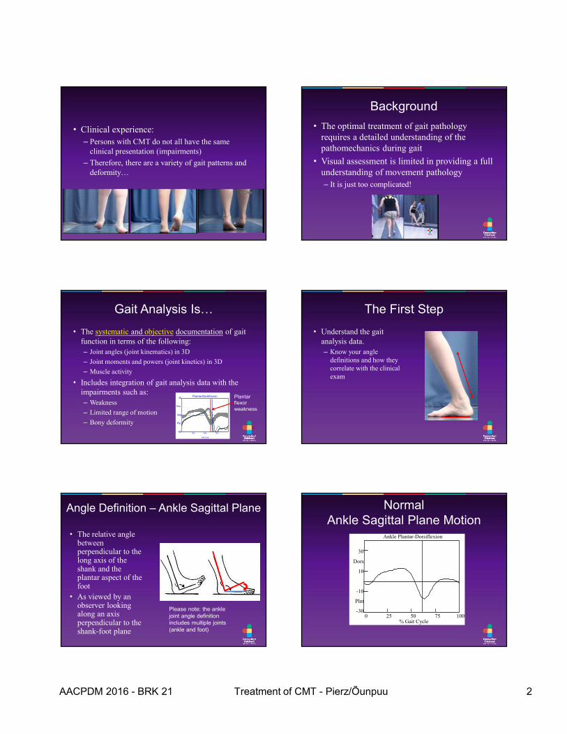
Angle Definition – Ankle Sagittal Plane
• The relative angle between perpendicular to the long axis of the shank and the plantar aspect of the foot
• As viewed by an observer looking along an axis perpendicular to the shank-foot plane
Please note: the ankle joint angle definition includes multiple joints (ankle and foot)
Normal Ankle Sagittal Plane Motion
0 25 50 75 100 % Gait Cycle
Ankle Plantar-Dorsiflexion
30
Dors
10
-10
Plnt
-30
AACPDM 2016 - BRK 21 Treatment of CMT - Pierz/Õunpuu 3
Left Right
Plantar-Dorsiflexion
Gait Cycle
40
-40
Dor
Pla
deg
25% 50% 75%
STANCE SWINGDORSIFLEXION
PLANTAR FLEXION
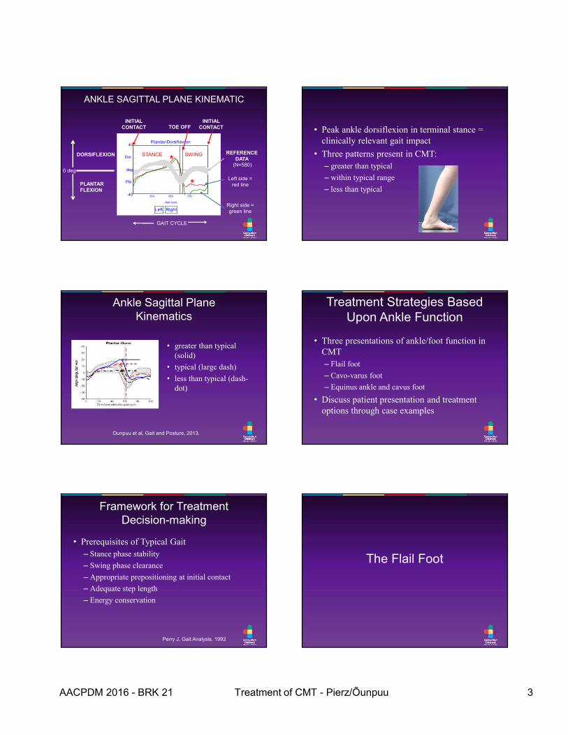
ANKLE SAGITTAL PLANE KINEMATIC
TOE OFFINITIAL
CONTACTINITIAL
CONTACT
GAIT CYCLE
Right side = green line
Left side = red line*
*REFERENCE
DATA(N=580)
0 deg
• Peak ankle dorsiflexion in terminal stance = clinically relevant gait impact
• Three patterns present in CMT:
– greater than typical
– within typical range
– less than typical
Ankle Sagittal Plane Kinematics
• greater than typical (solid)
• typical (large dash)
• less than typical (dash-dot)
Ounpuu et al, Gait and Posture, 2013.
Treatment Strategies Based Upon Ankle Function
• Three presentations of ankle/foot function in CMT
– Flail foot
– Cavo-varus foot
– Equinus ankle and cavus foot
• Discuss patient presentation and treatment options through case examples
Framework for Treatment Decision-making
• Prerequisites of Typical Gait
– Stance phase stability
– Swing phase clearance
– Appropriate prepositioning at initial contact
– Adequate step length
– Energy conservation
Perry J, Gait Analysis, 1992
The Flail Foot
AACPDM 2016 - BRK 21 Treatment of CMT - Pierz/Õunpuu 4
The Flail Foot –Compromised Prerequisites of Gait
• Stance phase stability
• Swing phase clearance
• Appropriate prepositioning at initial contact
• Adequate step lengths Treatment Goals: Address
these compromised prerequisites of gait
The Flail Foot
• Clinical Examination Findings
– Limited passive dorsiflexion range of motion
• Knee flexed (1 ± 7 degrees)
• Knee extended (8 ± 7 degrees)
– Full plantar flexion and forefoot inversion/eversion
– Strength: (median/maximum/minumum)
• Plantar Flexors (2/5/2)
• Dorsiflexors (4/5/0)
• Forefoot Invertors (5/5/0)
• Forefoot Evertors (4/5/2)
Flail Foot
• Functional outcome of ankle weakness includes instability in standing and during gait due to limited ability to bear weight over the forefoot
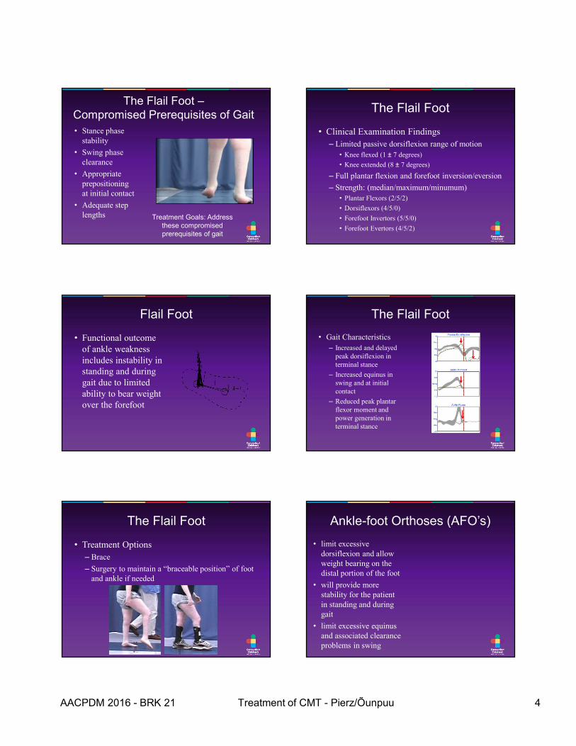
The Flail Foot
• Gait Characteristics
– Increased and delayed peak dorsiflexion in terminal stance
– Increased equinus in swing and at initial contact
– Reduced peak plantar flexor moment and power generation in terminal stance
The Flail Foot
• Treatment Options
– Brace
– Surgery to maintain a “braceable position” of foot and ankle if needed
Ankle-foot Orthoses (AFO’s)
• limit excessive dorsiflexion and allow weight bearing on the distal portion of the foot
• will provide more stability for the patient in standing and during gait
• limit excessive equinus and associated clearance problems in swing
AACPDM 2016 - BRK 21 Treatment of CMT - Pierz/Õunpuu 5
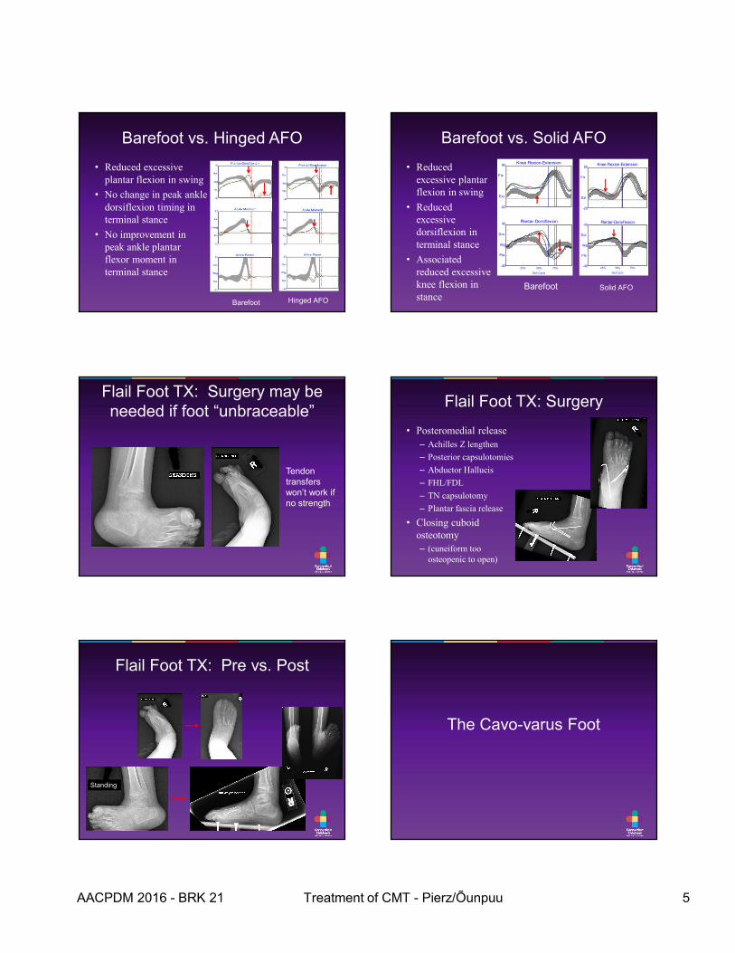
Barefoot vs. Hinged AFO
• Reduced excessive plantar flexion in swing
• No change in peak ankle dorsiflexion timing in terminal stance
• No improvement in peak ankle plantar flexor moment in terminal stance
Barefoot Hinged AFO
Barefoot vs. Solid AFO
• Reduced excessive plantar flexion in swing
• Reduced excessive dorsiflexion in terminal stance
• Associated reduced excessive knee flexion in stance
Barefoot Solid AFO
Knee Flexion-Extension80
-20
Flx
Ext
Plantar-Dorsiflexion
Gait Cycle
40
-40
Dor
Pla
deg
25% 50% 75%
Knee Flexion-Extension80
-20
Flx
Ext
Plantar-Dorsif lexion
Gait Cycle
40
-40
Dor
Pla
deg
25% 50% 75%
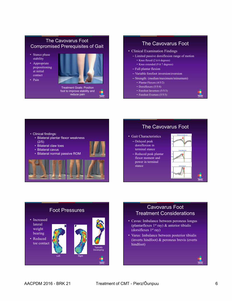
Flail Foot TX: Surgery may be needed if foot “unbraceable”
Tendon transfers won’t work if no strength
Flail Foot TX: Surgery
• Posteromedial release
– Achilles Z lengthen
– Posterior capsulotomies
– Abductor Hallucis
– FHL/FDL
– TN capsulotomy
– Plantar fascia release
• Closing cuboid osteotomy
– (cuneiform too osteopenic to open)
Flail Foot TX: Pre vs. Post
Standing
The Cavo-varus Foot
AACPDM 2016 - BRK 21 Treatment of CMT - Pierz/Õunpuu 6
The Cavovarus FootCompromised Prerequisites of Gait
• Stance phase stability
• Appropriate prepositioning at initial contact
• Pain
Treatment Goals: Position foot to improve stability and
reduce pain
The Cavovarus Foot
• Clinical Examination Findings
– Limited passive dorsiflexion range of motion
• Knee flexed (2 ± 6 degrees)
• Knee extended (9 ± 7 degrees)
– Full plantar flexion
– Variable forefoot inversion/eversion
– Strength: (median/maximum/minumum)
• Plantar Flexors (4/5/2)
• Dorsiflexors (5/5/4)
• Forefoot Invertors (5/5/3)
• Forefoot Evertors (5/5/3)
• Clinical findings:• Bilateral plantar flexor weakness
(2/5)• Bilateral claw toes• Bilateral cavus• Bilateral normal passive ROM
The Cavovarus Foot
• Gait Characteristics
– Delayed peak dorsiflexion in terminal stance
– Reduced peak plantar flexor moment and power in terminal stance
Foot Pressures
• Increased lateral weight bearing
• Reduced toe contact
Typically Developing
Left Right
Cavovarus FootTreatment Considerations
• Cavus: Imbalance between peroneus longus (plantarflexes 1st ray) & anterior tibialis (dorsiflexes 1st ray)
• Varus: Imbalance between posterior tibialis (inverts hindfoot) & peroneus brevis (everts hindfoot)
AACPDM 2016 - BRK 21 Treatment of CMT - Pierz/Õunpuu 7
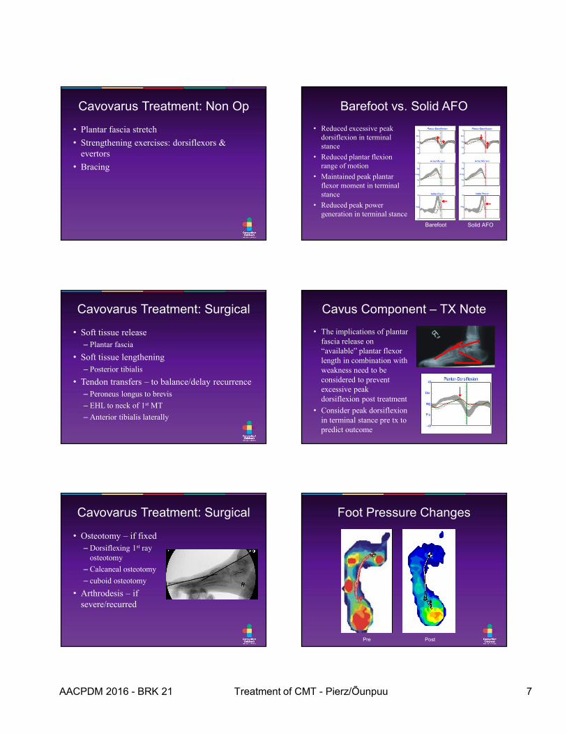
Cavovarus Treatment: Non Op
• Plantar fascia stretch
• Strengthening exercises: dorsiflexors & evertors
• Bracing
Barefoot vs. Solid AFO
• Reduced excessive peak dorsiflexion in terminal stance
• Reduced plantar flexion range of motion
• Maintained peak plantar flexor moment in terminal stance
• Reduced peak power generation in terminal stance
Barefoot Solid AFO
Cavovarus Treatment: Surgical
• Soft tissue release
– Plantar fascia
• Soft tissue lengthening
– Posterior tibialis
• Tendon transfers – to balance/delay recurrence
– Peroneus longus to brevis
– EHL to neck of 1st MT
– Anterior tibialis laterally
Cavus Component – TX Note
• The implications of plantar fascia release on “available” plantar flexor length in combination with weakness need to be considered to prevent excessive peak dorsiflexion post treatment
• Consider peak dorsiflexion in terminal stance pre tx to predict outcome
Cavovarus Treatment: Surgical
• Osteotomy – if fixed
– Dorsiflexing 1st ray osteotomy
– Calcaneal osteotomy
– cuboid osteotomy
• Arthrodesis – if severe/recurred
Foot Pressure Changes
Pre Post
AACPDM 2016 - BRK 21 Treatment of CMT - Pierz/Õunpuu 8
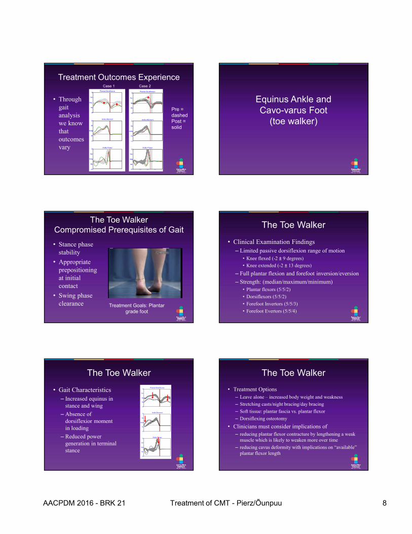
Treatment Outcomes Experience
• Through gait analysis we know that outcomes vary
Pl antar-Dorsiflexi on40
-40
Dor
Pla
Deg
Ankl e Moment2.0
-1.0
Pfl
Dfl
Nm/kg
Ankl e Power3.0
-2.0
Gen
Abs
W /kg
Plantar-Dorsif lexion40
-40
Dor
Pla
deg
Ankl e Moment2.0
-1.0
Ext
Flx
Nm/kg
Ankle Power3.0
-2.0
Gen
Abs
W/kg
25 % 50 % 75%
Case 1 Case 2
Pre = dashedPost = solid
Equinus Ankle and Cavo-varus Foot
(toe walker)
The Toe WalkerCompromised Prerequisites of Gait
• Stance phase stability
• Appropriate prepositioning at initial contact
• Swing phase clearance Treatment Goals: Plantar
grade foot
The Toe Walker
• Clinical Examination Findings
– Limited passive dorsiflexion range of motion
• Knee flexed (-2 ± 9 degrees)
• Knee extended (-2 ± 13 degrees)
– Full plantar flexion and forefoot inversion/eversion
– Strength: (median/maximum/minimum)
• Plantar flexors (5/5/2)
• Dorsiflexors (5/5/2)
• Forefoot Invertors (5/5/3)
• Forefoot Evertors (5/5/4)
The Toe Walker
• Gait Characteristics
– Increased equinus in stance and wing
– Absence of dorsiflexior moment in loading
– Reduced power generation in terminal stance
Plantar-Dorsiflexion40
-40
Dor
Pla
deg
Ankle Moment2.0
-1.0
Ext
Flx
Nm/kg
Ankle Power3.0
-2.0
Gen
Abs
W/kg
The Toe Walker
• Treatment Options
– Leave alone – increased body weight and weakness
– Stretching casts/night bracing/day bracing
– Soft tissue: plantar fascia vs. plantar flexor
– Dorsiflexing osteotomy
• Clinicians must consider implications of
– reducing plantar flexor contracture by lengthening a weak muscle which is likely to weaken more over time
– reducing cavus deformity with implications on “available” plantar flexor length
AACPDM 2016 - BRK 21 Treatment of CMT - Pierz/Õunpuu 9
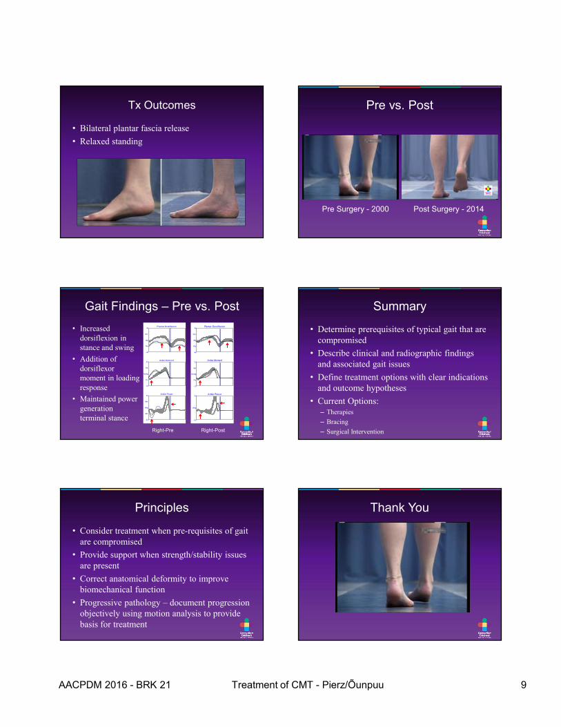
Tx Outcomes
• Bilateral plantar fascia release
• Relaxed standing
Pre vs. Post
Pre Surgery - 2000 Post Surgery - 2014
Plantar-Dorsif lexion40
-40
Dor
Pla
deg
Ankle Moment2.0
-1.0
Ext
Flx
Nm/kg
Ankle Pow er3.0
-2.0
W/kg
Gait Findings – Pre vs. Post
• Increased dorsiflexion in stance and swing
• Addition of dorsiflexor moment in loading response
• Maintained power generation terminal stance
Plantar-Dorsiflexion40
-40
Dor
Pla
deg
Ankle Moment2.0
-1.0
Ext
Flx
Nm/kg
Ankle Power3.0
-2.0
Gen
Abs
W/kg
Right-Pre Right-Post
Summary
• Determine prerequisites of typical gait that are compromised
• Describe clinical and radiographic findings and associated gait issues
• Define treatment options with clear indications and outcome hypotheses
• Current Options:– Therapies
– Bracing
– Surgical Intervention
Principles
• Consider treatment when pre-requisites of gait are compromised
• Provide support when strength/stability issues are present
• Correct anatomical deformity to improve biomechanical function
• Progressive pathology – document progression objectively using motion analysis to provide basis for treatment
Thank You