
SOP: Anion Exchange Chromatography of Green Fluorescent...
12
Montgomery County Community College Document Number: SU DP 20 340 DeKalb Pike Revision Number: 0 Blue Bell, PA Effective Date: 01APR16 Page 1 of 12 SOP: Anion Exchange Chromatography of Green Fluorescent Protein (GFP) using the AKTA Pure system Approvals Preparer: David Frank Date: 21APR16 Reviewer: Hetal Doshi Date: 21APR16 Reviewer: Dr. Maggie Bryans Date: 21APR16 1. Purpose 1.1. This procedure describes the operation of the ÄKTApure Chromatography System, controlled by Unicorn 6.3 software, for the purpose of anion exchange chromatography of samples containing green fluorescent protein (GFP). 2. Scope and Applicability 2.1. Applies to purification of GFP from diverse origins, including bacterially expressed recombinant GFP, which has been concentrated and its pH adjusted to 8.0, using a HiTrap CaptoQ HP 5 ml column installed on the GE ÄKTApure Chromatography System and controlled by Unicorn 6.3 software. 3. Summary of Method 3.1. Preparation of buffer(s) 3.2. Equilibration of system and column 3.3. Fraction collector setup 3.4. Application of sample 3.5. Washing and elution of column 3.6. Regeneration of system in preparation for subsequent run 3.7. Procedures for short or long term storage of the system 4. References 4.1. Unicorn 6.3 Users Guide (electronic) 4.2. AKTApure 25 Users Guide (electronic) 4.3. HiTrap Capto Q HP 5 information booklet 5. Definitions N/A 6. Precautions
Transcript of SOP: Anion Exchange Chromatography of Green Fluorescent...

Montgomery County Community College Document Number: SU DP 20 340 DeKalb Pike Revision Number: 0 Blue Bell, PA Effective Date: 01APR16 Page 1 of 12 SOP: Anion Exchange Chromatography of Green Fluorescent Protein (GFP) using the AKTA Pure system
Approvals
Preparer: David Frank Date: 21APR16 Reviewer: Hetal Doshi Date: 21APR16 Reviewer: Dr. Maggie Bryans Date: 21APR16 1. Purpose
1.1. This procedure describes the operation of the ÄKTApure Chromatography System, controlled by Unicorn 6.3 software, for the purpose of anion exchange chromatography of samples containing green fluorescent protein (GFP).
2. Scope and Applicability
2.1. Applies to purification of GFP from diverse origins, including bacterially expressed recombinant GFP, which has been concentrated and its pH adjusted to 8.0, using a HiTrap CaptoQ HP 5 ml column installed on the GE ÄKTApure Chromatography System and controlled by Unicorn 6.3 software.
3. Summary of Method
3.1. Preparation of buffer(s) 3.2. Equilibration of system and column 3.3. Fraction collector setup 3.4. Application of sample 3.5. Washing and elution of column 3.6. Regeneration of system in preparation for subsequent run 3.7. Procedures for short or long term storage of the system
4. References 4.1. Unicorn 6.3 Users Guide (electronic) 4.2. AKTApure 25 Users Guide (electronic) 4.3. HiTrap Capto Q HP 5 information booklet
5. Definitions
N/A
6. Precautions
Montgomery County Community College Document Number: SU DP 20 340 DeKalb Pike Revision Number: 0 Blue Bell, PA Effective Date: 01APR16 Page 2 of 12 SOP: Anion Exchange Chromatography of Green Fluorescent Protein (GFP) using the AKTA Pure system
6.1. Routine care should be exercised in handling of buffers and samples of biological materials, which may have harmful biological activity in the case of accidental ingestion, needle stick, etc.
6.2. User should read and be familiar with general good practice as outlined in the AKTApure Cue Cards located near the instrument.
6.3. Avoid damaging the threads through the use of excessive force when connecting plastic fasteners.
6.4. Care must be taken to avoid air in the fluid path, which could damage the pumps or give spurious and uninterpretable readout from the UV and/or conductivity detectors.
6.5. Gloves and protective eyewear should be worn when handling samples and reagents (buffers), however it is preferable that the user remove gloves prior to entering commands via the computer keyboard or mouse.
6.6. Buffers must be degassed and filtered prior to use with the AKTApure instrument. Samples should be centrifuged at 10000xg for 5 min before injection/introduction into the fluid path.
6.7. Equipment calibration check: The AKTApure system calibration of A280 and conductivity are automatic; baseline for measurements of A280 and conductivity are zeroed at the beginning of a chromatography run. However, calibration of the pH detector must be performed prior to use of the instrument each day, using standard calibration buffers and the automated routine in Unicorn. It is assumed that calibration is performed according to the Equipment SOP for the AKTApure 25 instrument. Further adjustment is beyond the scope of this document and should be referred to a qualified technician.
7. Responsibilities 7.1. It is the responsibility of the course instructor/lab assistant to ensure that this SOP is
performed as described and to update the procedure when necessary. 7.2. It is the responsibility of the students/technician to follow the SOP as described and to
inform the instructor about any deviations or problems that may occur while performing the procedure.
8. Equipment and Materials 8.1. AKTApure chromatography system 8.2. Additional Lab Equipment: pH meter, balance 8.3. Lab Utensils: Beakers (250, 500ml), 500 ml graduated cylinders 8.4. Reagents: Tris, hydrochloric acid, sodium chloride, filtered deionized water (MilliQ or
similar). 10 % w/v Tween 80, 20% ethanol. 8.5. Lab Supplies: Filters (0.2 m) and bottles for vacuum filtration and degassing of all
chromatography buffers. Syringe (1ml). Tubes for fraction collector.
Montgomery County Community College Document Number: SU DP 20 340 DeKalb Pike Revision Number: 0 Blue Bell, PA Effective Date: 01APR16 Page 3 of 12 SOP: Anion Exchange Chromatography of Green Fluorescent Protein (GFP) using the AKTA Pure system
9. Procedure 9.1. Sample Collection and Preparation is described elsewhere. It is assumed that the
sample is available in a suitable, compatible buffer such as 20 mM Tris-HCl, pH 8. Please refer to that document (XX.0, 13Apr15). The operator will require 0.6 ml of sample per sample injection (see below).
9.2. Reagent Preparation: Buffers should be prepared dependent on the mode of separation employed; in this instance anion exchange chromatography provides good separation of GFP from contaminating protein at pH 8.0.
9.2.1. Anion Exchange Buffer: 20 mM Tris-HCl, pH 8.0 9.2.1.1. Dissolve 2.423 gm Tris in 950 ml filtered deionized water in a one liter
beaker, with stir bar. 9.2.1.2. Titrate the pH of the Tris solution to 8.0 by addition of concentrated HCl or
1M HCl, carefully adding the appropriate acid dropwise to obtain pH 8.0. 9.2.1.3. Adjust the final volume to 1000 ml. Set aside 500 ml of this solution and
label ‘Buffer A’ along with its precise composition and date of preparation. Filter and degas the buffer by passage through a vacuum filter device attached to house vacuum, leaving the filtered solution under vacuum for 15-20 minutes.
9.2.1.4. Use the remaining 500 ml to dissolve 29.22 gm of NaCl in a 400 ml beaker. Following dissolution, filter and degas this mixture and label the bottle ‘Buffer B’, along with the actual contents (20mM Tris-HCl, pH 8.0, 1 M NaCl).
9.3. Start-up and preparation of AKTApure Instrument and computer:
Degassed buffers should be in place prior to turning on the AKTApure instrument. Equipment start-up requires turning on the instrument and, separately, the computer connected to it.
9.3.1. Place the degassed buffers A and B on top of the AKTApure instrument. 9.3.2. Locate Inlet tubing A1 and B1 (atop the instrument and resting in water or 20%
ethanol). Each has a filter unit attached, which distinguishes them from A2 and B2; those end in a male threaded fitting and will not be used for this procedure.
9.3.3. Transfer tubing Inlet A1 to the buffer A bottle. 9.3.4. Transfer tubing Inlet B1 to the buffer B bottle.

Montgomery County Community College Document Number: SU DP 20 340 DeKalb Pike Revision Number: 0 Blue Bell, PA Effective Date: 01APR16 Page 4 of 12 SOP: Anion Exchange Chromatography of Green Fluorescent Protein (GFP) using the AKTA Pure system
9.3.5. The On/Off switch for the instrument is located on the right side toward the rear of the housing. Switch to the ‘On’ position. Audible emanations from within the instrument cabinet indicate that the AKTApure system is going through its brief initialization sequence.
9.3.6. The computer On/Off switch is located on the front of the Dell desktop computer unit, near the top of the case. Press in to turn on the computer.
9.3.7. Login to the computer using credentials provided by the College. 9.3.8. Double click the Unicorn 6.3 icon on the desktop to open the software which
controls the instrument functions. Click OK in the “Log In – Unicorn” dialog box that appears.
9.3.9. Open the System Control window (under Tools menu, if not opened automatically on startup).
9.3.10. The top pane of the window will show the current state of the instrument, and the bottom pane shows the fluid path and manual controls. If the window is blank, go to the System menu and select Connect to Systems, check the box by AKTApure 25 and click OK.
9.3.11. Confirm that the correct column (HiTrap CaptoQ 5 ml) is attached to the system. If not, refer to Section 9.4 (Installing/Changing a Chromatography Column on the AKTApure Chromatography System).
9.3.12. Under the File menu, choose Open and select the method with file name “HiTrap SP 5ml Equilibration”.
9.3.13. A dialog box appears that allows the method to be run. Click Start to initiate flushing of the pumps and equilibration of the column.
9.3.14. While the equilibration method is running, prepare the fraction collector for later steps by filling the carousel with clean tubes.
9.3.15. Allow the program to run to completion, about 15 minutes.
9.4. Installing/Changing a Chromatography Column on the AKTApure Chromatography System.
It is imperative that the following operations be performed in such a way as to prevent the introduction of air bubbles into the column, which is achieved by making liquid-to-liquid (drop-to-drop) contact prior to inserting the threaded fitting into its position.
9.4.1. Have on hand a few paper lab towels and a 250 ml beaker to catch waste. 9.4.2. Remove tube connector from the UV detector inlet by unscrewing the knurled
fastener. 9.4.3. Initiate flow manually at 0.5 ml/min collecting waste in the beaker or towel. 9.4.4. Remove the plug from the column inlet and place a few drops of 20% ethanol in
the inlet, filling it to insure the absence of air.

Montgomery County Community College Document Number: SU DP 20 340 DeKalb Pike Revision Number: 0 Blue Bell, PA Effective Date: 01APR16 Page 5 of 12 SOP: Anion Exchange Chromatography of Green Fluorescent Protein (GFP) using the AKTA Pure system
9.4.5. As a droplet emerges from the inlet tubing, touch it to the liquid in the column inlet and begin to thread the fitting in, leaving slight looseness of threads so that liquid escapes around the fitting and pressure buildup in the column is prevented.
9.4.6. Remove the column bottom plug and screw the column directly into the UV detector inlet.
9.4.7. Tighten the column inlet fitting just enough to prevent leaking. 9.4.8. The column is now ready to equilibrate in buffer (step 9.3.12) prior to performing
a chromatography run.
Performing a chromatography run:
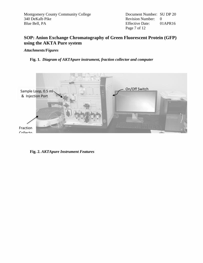
9.4.9. Place the fraction collector tube 1 near the outlet tubing from the instrument (refer to attachment Fig 1) so that it will touch the arrow on the white paddle of the fraction collector arm. Note: To rotate the carousel, reach around the left side of the collector to find a rubber roller pressing against the carousel (Fig 2). Pull the roller away from the carousel (Fig. 3); the carousel will rotate freely as long as the roller is held. When the first tube is in the correct position, release the roller.
9.4.10. Gently raise the arm and swing it into position against tube 1. 9.4.11. Place all ‘Waste’ tubing, labeled W, W1 & W2 in 1 L Ehrlenmeyer flask. 9.4.12. Place the tube labeled Outlet in a 125 ml Ehrlenmeyer flask. 9.4.13. Using a 1 ml syringe, aspirate 0.6 ml of the tPA sample into the syringe, expel
any bubbles and insert the loaded syringe into the injection port. 9.4.14. Inject the sample into the port to fill the 0.5 ml sample loop. 9.4.15. Open the Unicorn software and navigate to the System Control window. 9.4.16. Under the File menu, choose Open and select the method with file name “GFP
Capto Q 5 method”. 9.4.17. In the dialog box that opens, enter operator’s name, sample notes. 9.4.18. Click Next; note the time and and volume for the run; make sure there is excess
buffer A and B. 9.4.19. Click Next. Record the buffer composition of each buffer and the sample
identity. 9.4.20. Click Next. Enter a filename composed of the method name, date, operator or
group initials, for example HiTrapQ GFP AEX 16May15 MyGroupName. 9.4.21. Click Start.The instrument should begin to execute the method, as evidenced by a
soothing hum from the pumps and drops of liquid falling into tube 1 from the fraction collector outlet.
9.4.22. Observe that the fraction collector is receiving drops. 9.4.23. Monitor the computer screen for error messages or warnings. 9.4.24. Allow the method to run to completion, at which time the system will be re-
equilibrated and ready for subsequent runs by repeating section 9.4.

Montgomery County Community College Document Number: SU DP 20 340 DeKalb Pike Revision Number: 0 Blue Bell, PA Effective Date: 01APR16 Page 6 of 12 SOP: Anion Exchange Chromatography of Green Fluorescent Protein (GFP) using the AKTA Pure system
9.5. Equipment shut-down and short term (less than 3 days) storage 9.5.1. After completion of the final separation of the day, transfer Inlet tubing A1 and
B1 to a flask of degassed Milli-Q water (250 ml or greater). 9.5.2. In the Unicorn software, opent the System Control window. 9.5.3. Under the File menu, choose Open, then select the method ‘System Short Term
Storage’. 9.5.4. Click Start. 9.5.5. Allow the method to run to completion, as indicated by an audible tone and
onscreen window. 9.5.6. Turn off the instrument or perform the long term storage routine as needed
(section 9.6).
9.6. Equipment shut-down and long term (3 days or more) storage 9.6.1. After completion of the System Short Term Storage method, transfer Inlet tubing
A1 and B1 to a flask of degassed 20% ethanol (250 ml or greater). 9.6.2. In the Unicorn software, open the System Control window. 9.6.3. Under the File menu, choose Open, then select the method ‘System Long Term
Storage’. 9.6.4. Click Start. 9.6.5. Allow the method to run to completion, as indicated by an audible tone and
onscreen window. 9.6.6. Turn off the instrument.
9.7. Chromatogram printout 9.7.1. In the Unicorn software interface, open the Evaluation window. 9.7.2. In the Result Navigator pane, click the Results tab. 9.7.3. Locate the file of interest and double click its name to display your chromatogram
in the right pane. 9.7.4. Optional: Click the Customize button to open a dialog box that allows you to
specifiy what curves display and the scale of each axis. Recommended are the UV Chrom curve, Conductivity, and Fraction Number.
9.7.5. Click the Report button, check the Default report in the selection window and click Preview.
9.7.6. Under File, choose to Print (or Save as PDF to use a different printer).
Montgomery County Community College Document Number: SU DP 20 340 DeKalb Pike Revision Number: 0 Blue Bell, PA Effective Date: 01APR16 Page 7 of 12 SOP: Anion Exchange Chromatography of Green Fluorescent Protein (GFP) using the AKTA Pure system
Attachments/Figures
Fig. 1. Diagram of AKTApure instrument, fraction collector and computer
Fig. 2. AKTApure Instrument Features
On/Off Switch Sample Loop, 0.5 ml & Injection Port
Fraction Collecto
Montgomery County Community College Document Number: SU DP 20 340 DeKalb Pike Revision Number: 0 Blue Bell, PA Effective Date: 01APR16 Page 8 of 12 SOP: Anion Exchange Chromatography of Green Fluorescent Protein (GFP) using the AKTA Pure system
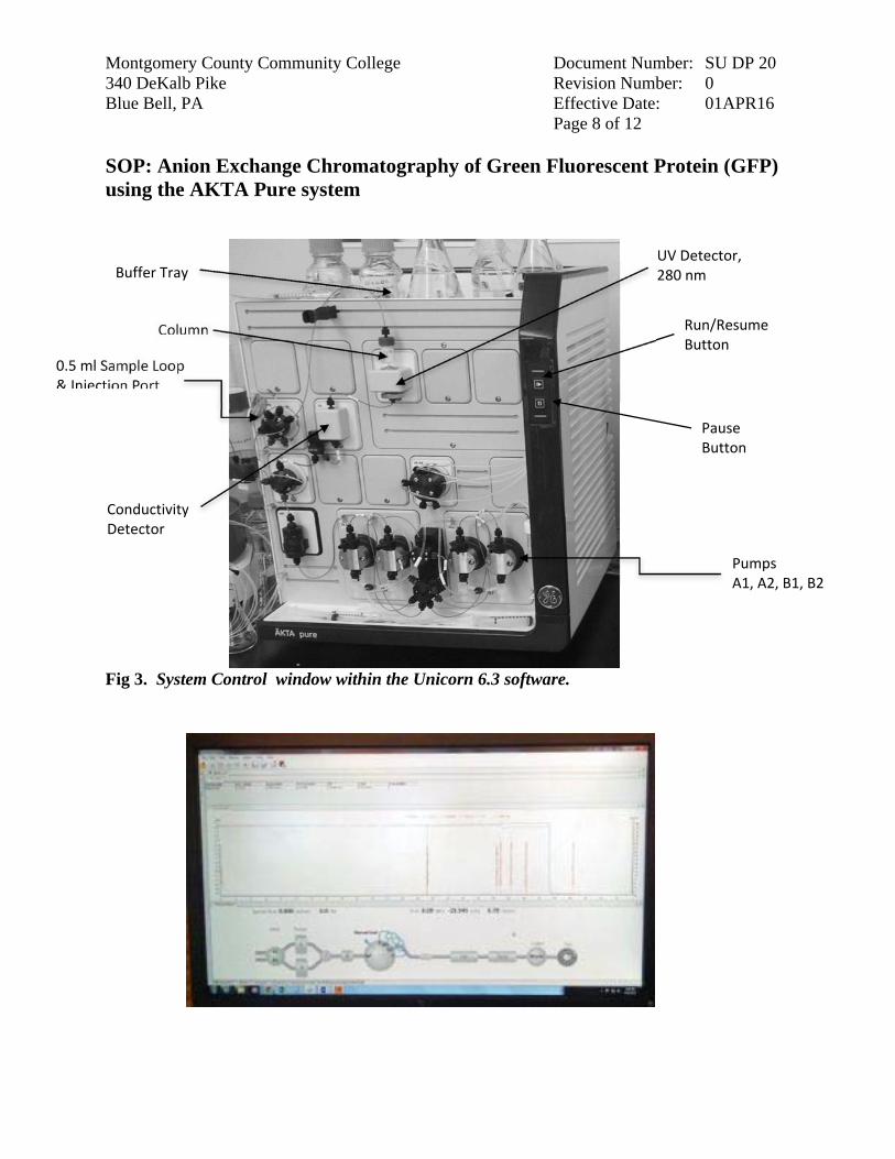
Fig 3. System Control window within the Unicorn 6.3 software.
Run/Resume Button
Pause Button
0.5 ml Sample Loop & Injection Port
Column
UV Detector, 280 nm
Conductivity Detector
Pumps A1, A2, B1, B2
Buffer Tray
Montgomery County Community College Document Number: SU DP 20 340 DeKalb Pike Revision Number: 0 Blue Bell, PA Effective Date: 01APR16 Page 9 of 12 SOP: Anion Exchange Chromatography of Green Fluorescent Protein (GFP) using the AKTA Pure system
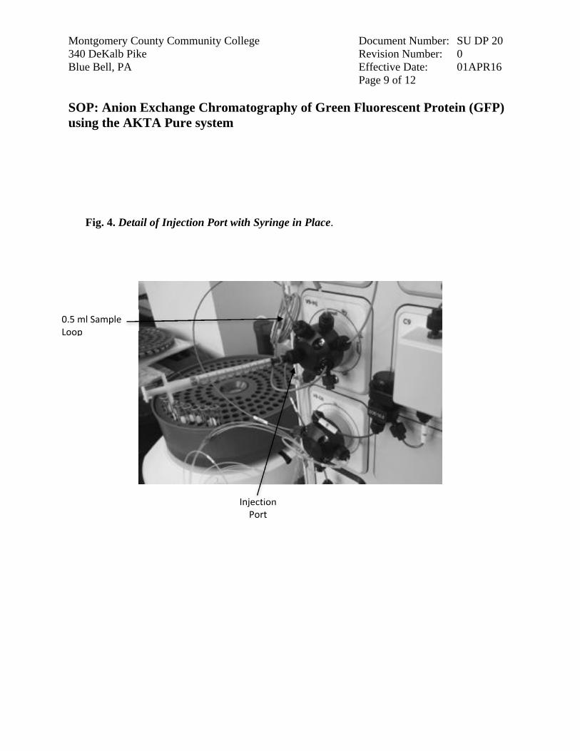
Fig. 4. Detail of Injection Port with Syringe in Place.
Injection Port
0.5 ml Sample Loop

Montgomery County Community College Document Number: SU DP 20 340 DeKalb Pike Revision Number: 0 Blue Bell, PA Effective Date: 01APR16 Page 10 of 12 SOP: Anion Exchange Chromatography of Green Fluorescent Protein (GFP) using the AKTA Pure system
Fig. 5. Fraction collector carousel rubber advancement roller/gear.
Fig. 6. Release of roller to allow free rotation of the carousel.
Roller/gear

Montgomery County Community College Document Number: SU DP 20 340 DeKalb Pike Revision Number: 0 Blue Bell, PA Effective Date: 01APR16 Page 11 of 12 SOP: Anion Exchange Chromatography of Green Fluorescent Protein (GFP) using the AKTA Pure system
Fig. 7. Location of tube #1 under the fraction collector drip outlet.
Montgomery County Community College Document Number: SU DP 20 340 DeKalb Pike Revision Number: 0 Blue Bell, PA Effective Date: 01APR16 Page 12 of 12 SOP: Anion Exchange Chromatography of Green Fluorescent Protein (GFP) using the AKTA Pure system
10. History
Revision Number
Effective Date Preparer Description of Change
0 21/04/2016 David Frank Initial release


![Dipalladium(II,II) Corners Cages from Fluorescent ...Self-Assembly and Anion sensing of Metal-organic [M6L2] Cages from Fluorescent Triphenylamine Tri-pyrazoles with Dipalladium(II,II)](https://static.fdocuments.net/doc/165x107/60f9086e83e213280d2b5e01/dipalladiumiiii-corners-cages-from-fluorescent-self-assembly-and-anion-sensing.jpg)


