Sonographic Pictorial Review of Benign Palpable · PDF fileSonographic Pictorial Review of...
10
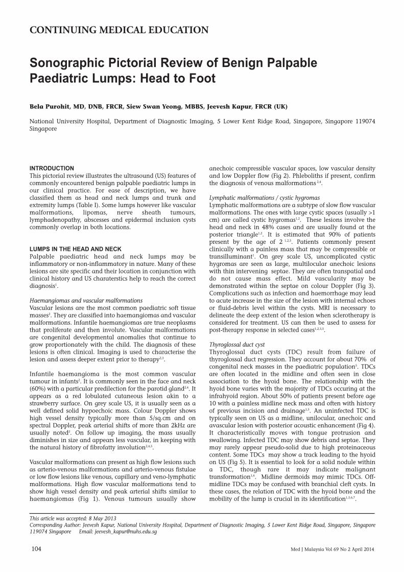
104 Med J Malaysia Vol 69 No 2 April 2014 INTRODUCTION This pictorial review illustrates the ultrasound (US) features of commonly encountered benign palpable paediatric lumps in our clinical practice. For ease of description, we have classified them as head and neck lumps and trunk and extremity lumps (Table I). Some lumps however like vascular malformations, lipomas, nerve sheath tumours, lymphadenopathy, abscesses and epidermal inclusion cysts commonly overlap in both locations. LUMPS IN THE HEAD AND NECK Palpable paediatric head and neck lumps may be inflammatory or non-inflammatory in nature. Many of these lesions are site specific and their location in conjunction with clinical history and US charaterstics help to reach the correct diagnosis 1 . Haemangiomas and vascular malformations Vascular lesions are the most common paediatric soft tissue masses 2 . They are classified into haemangiomas and vascular malformations. Infantile haemangiomas are true neoplasms that proliferate and then involute. Vascular malformations are congenital developmental anomalies that continue to grow proportionately with the child. The diagnosis of these lesions is often clinical. Imaging is used to characterise the lesion and assess deeper extent prior to therapy 2,3 . Infantile haemangioma is the most common vascular tumour in infants 2 . It is commonly seen in the face and neck (60%) with a particular predilection for the parotid gland 2,4 . It appears as a red lobulated cutaneous lesion akin to a strawberry surface. On grey scale US, it is usually seen as a well defined solid hypoechoic mass. Colour Doppler shows high vessel density typically more than 5/sq.cm and on spectral Doppler, peak arterial shifts of more than 2kHz are usually noted 2 . On follow up imaging, the mass usually diminishes in size and appears less vascular, in keeping with the natural history of fibrofatty involution 2,4,5 . Vascular malformations can present as high flow lesions such as arterio-venous malformations and arterio-venous fistulae or low flow lesions like venous, capillary and veno-lymphatic malformations. High flow vascular malformations tend to show high vessel density and peak arterial shifts similar to haemangiomas (Fig 1). Venous tumours usually show anechoic compressible vascular spaces, low vascular density and low Doppler flow (Fig 2). Phleboliths if present, confirm the diagnosis of venous malformations 2,4 . Lymphatic malformations / cystic hygromas Lymphatic malformations are a subtype of slow flow vascular malformations. The ones with large cystic spaces (usually >1 cm) are called cystic hygromas 1,2 . These lesions involve the head and neck in 48% cases and are usually found at the posterior triangle 1,2 . It is estimated that 90% of patients present by the age of 2 1,2,5 . Patients commonly present clinically with a painless mass that may be compressible or transilluminant 1 . On grey scale US, uncomplicated cystic hygromas are seen as large, multilocular anechoic lesions with thin intervening septae. They are often transpatial and do not cause mass effect. Mild vascularity may be demonstrated within the septae on colour Doppler (Fig 3). Complications such as infection and haemorrhage may lead to acute increase in the size of the lesion with internal echoes or fluid-debris level within the cysts. MRI is necessary to delineate the deep extent of the lesion when sclerotherapy is considered for treatment. US can then be used to assess for post-therapy response in selected cases 1,2,3,5 . Thyroglossal duct cyst Thyroglossal duct cysts (TDC) result from failure of thyroglossal duct regression. They account for about 70% of congenital neck masses in the paediatric population 5 . TDCs are often located in the midline and often seen in close association to the hyoid bone. The relationship with the hyoid bone varies with the majority of TDCs occuring at the infrahyoid region. About 50% of patients present before age 10 with a painless midline neck mass and often with history of previous incision and drainage 1,5 . An uninfected TDC is typically seen on US as a midline, unilocular, anechoic and avascular lesion with posterior acoustic enhancement (Fig 4). It characteristically moves with tongue protrusion and swallowing. Infected TDC may show debris and septae. They may rarely appear pseudo-solid due to high proteinaceous content. Some TDCs may show a track leading to the hyoid on US (Fig 5). It is essential to look for a solid nodule within a TDC, though rare it may indicate malignant transformation 1,6 . Midline dermoids may mimic TDCs. Off- midline TDCs may be confused with branchial cleft cysts. In these cases, the relation of TDC with the hyoid bone and the mobility of the lump is crucial in its identification 1,5,6,7 . Sonographic Pictorial Review of Benign Palpable Paediatric Lumps: Head to Foot Bela Purohit, MD, DNB, FRCR, Siew Swan Yeong, MBBS, Jeevesh Kapur, FRCR (UK) National University Hospital, Department of Diagnostic Imaging, 5 Lower Kent Ridge Road, Singapore, Singapore 119074 Singapore CONTINUING MEDICAL EDUCATION This article was accepted: 8 May 2013 Corresponding Author: Jeevesh Kapur, National University Hospital, Department of Diagnostic Imaging, 5 Lower Kent Ridge Road, Singapore, Singapore 119074 Singapore Email: [email protected]
Transcript of Sonographic Pictorial Review of Benign Palpable · PDF fileSonographic Pictorial Review of...
104 Med J Malaysia Vol 69 No 2 April 2014
INTRODUCTIONThis pictorial review illustrates the ultrasound (US) features ofcommonly encountered benign palpable paediatric lumps inour clinical practice. For ease of description, we haveclassified them as head and neck lumps and trunk andextremity lumps (Table I). Some lumps however like vascularmalformations, lipomas, nerve sheath tumours,lymphadenopathy, abscesses and epidermal inclusion cystscommonly overlap in both locations.
LUMPS IN THE HEAD AND NECKPalpable paediatric head and neck lumps may beinflammatory or non-inflammatory in nature. Many of theselesions are site specific and their location in conjunction withclinical history and US charaterstics help to reach the correctdiagnosis1.
Haemangiomas and vascular malformationsVascular lesions are the most common paediatric soft tissuemasses2. They are classified into haemangiomas and vascularmalformations. Infantile haemangiomas are true neoplasmsthat proliferate and then involute. Vascular malformationsare congenital developmental anomalies that continue togrow proportionately with the child. The diagnosis of theselesions is often clinical. Imaging is used to characterise thelesion and assess deeper extent prior to therapy2,3.
Infantile haemangioma is the most common vasculartumour in infants2. It is commonly seen in the face and neck(60%) with a particular predilection for the parotid gland2,4. Itappears as a red lobulated cutaneous lesion akin to astrawberry surface. On grey scale US, it is usually seen as awell defined solid hypoechoic mass. Colour Doppler showshigh vessel density typically more than 5/sq.cm and onspectral Doppler, peak arterial shifts of more than 2kHz areusually noted2. On follow up imaging, the mass usuallydiminishes in size and appears less vascular, in keeping withthe natural history of fibrofatty involution2,4,5.
Vascular malformations can present as high flow lesions suchas arterio-venous malformations and arterio-venous fistulaeor low flow lesions like venous, capillary and veno-lymphaticmalformations. High flow vascular malformations tend toshow high vessel density and peak arterial shifts similar tohaemangiomas (Fig 1). Venous tumours usually show
anechoic compressible vascular spaces, low vascular densityand low Doppler flow (Fig 2). Phleboliths if present, confirmthe diagnosis of venous malformations 2,4.
Lymphatic malformations / cystic hygromasLymphatic malformations are a subtype of slow flow vascularmalformations. The ones with large cystic spaces (usually >1cm) are called cystic hygromas1,2. These lesions involve thehead and neck in 48% cases and are usually found at theposterior triangle1,2. It is estimated that 90% of patientspresent by the age of 2 1,2,5. Patients commonly presentclinically with a painless mass that may be compressible ortransilluminant1. On grey scale US, uncomplicated cystichygromas are seen as large, multilocular anechoic lesionswith thin intervening septae. They are often transpatial anddo not cause mass effect. Mild vascularity may bedemonstrated within the septae on colour Doppler (Fig 3).Complications such as infection and haemorrhage may leadto acute increase in the size of the lesion with internal echoesor fluid-debris level within the cysts. MRI is necessary todelineate the deep extent of the lesion when sclerotherapy isconsidered for treatment. US can then be used to assess forpost-therapy response in selected cases1,2,3,5.
Thyroglossal duct cyst Thyroglossal duct cysts (TDC) result from failure ofthyroglossal duct regression. They account for about 70% ofcongenital neck masses in the paediatric population5. TDCsare often located in the midline and often seen in closeassociation to the hyoid bone. The relationship with thehyoid bone varies with the majority of TDCs occuring at theinfrahyoid region. About 50% of patients present before age10 with a painless midline neck mass and often with historyof previous incision and drainage1,5. An uninfected TDC istypically seen on US as a midline, unilocular, anechoic andavascular lesion with posterior acoustic enhancement (Fig 4).It characteristically moves with tongue protrusion andswallowing. Infected TDC may show debris and septae. Theymay rarely appear pseudo-solid due to high proteinaceouscontent. Some TDCs may show a track leading to the hyoidon US (Fig 5). It is essential to look for a solid nodule withina TDC, though rare it may indicate malignanttransformation1,6. Midline dermoids may mimic TDCs. Off-midline TDCs may be confused with branchial cleft cysts. Inthese cases, the relation of TDC with the hyoid bone and themobility of the lump is crucial in its identification1,5,6,7.
Sonographic Pictorial Review of Benign PalpablePaediatric Lumps: Head to Foot
Bela Purohit, MD, DNB, FRCR, Siew Swan Yeong, MBBS, Jeevesh Kapur, FRCR (UK)
National University Hospital, Department of Diagnostic Imaging, 5 Lower Kent Ridge Road, Singapore, Singapore 119074Singapore
CONTINUING MEDICAL EDUCATION
This article was accepted: 8 May 2013Corresponding Author: Jeevesh Kapur, National University Hospital, Department of Diagnostic Imaging, 5 Lower Kent Ridge Road, Singapore, Singapore119074 Singapore Email: [email protected]
Sonographic Pictorial Review of Benign Palpable Paediatric Lumps: Head to Foot
Med J Malaysia Vol 69 No 2 April 2014 105
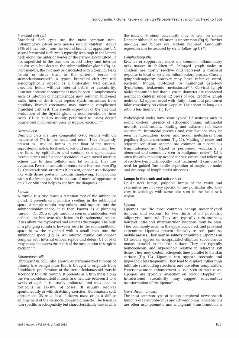
Branchial cleft cystBranchial cleft cysts are the most common non-inflammatory lateral neck masses seen in children5. About95% of these arise from the second branchial apparatus 1. Asecond branchial cleft cyst is typically seen high in the lateralneck along the anterior border of the sternocleidomastoid. Itlies superficial to the common carotid artery and internaljugular vein but deep to the submandibular gland (Fig 6).Occasionally, the cyst may be associated with a tonsillar fossafistula or sinus tract to the anterior border ofsternocleidomastoid1,5. A typical branchial cleft cyst willsonographically appear as a unilocular, well defined,anechoic lesion without internal debris or vascularity.Posterior acoustic enhancement may be seen. Complicationssuch as infection or haemorrhage may result in thickenedwalls, internal debris and septae. Cystic metastases frompapillary thyroid carcinoma may mimic a complicatedbranchial cleft cyst. Fine needle aspiration of the cyst andevaluation of the thyroid gland is recommended in thesecases. CT or MRI is usually performed to assess deeperpharyngeal involvement prior to surgical excision1,5,6.
Dermoid cystDermoid cysts are rare congenital cystic lesions with anincidence of 7% in the head and neck1. They frequentlypresent as midline lumps in the floor of the mouth,suprasternal notch, forehead, orbits and nasal cavities. Theyare lined by epithelium and contain skin appendages.Dermoid cysts on US appear pseudosolid with mixed internalechoes due to their cellular and fat content. They areavascular. Posterior acoustic enhancement is uncommon (Fig7). Osseous-dental structures if present, appear as echogenicfoci with dense posterior acoustic shadowing. Fat globuleswithin the lesion give rise to the ‘sac of marbles’ appearanceon CT or MRI that helps to confirm the diagnosis 1,5,6.
RanulaA ranula is a true mucous retention cyst of the sublingualgland. It presents as a painless swelling in the sublingualspace. A simple ranula may enlarge and rupture into thesubmandibular space, it is then known as a plungingranula1. On US, a simple ranula is seen as a unilocular, welldefined, anechoic avascular lesion in the submental region.It lies above the mylohyoid and elevates the tongue. The bulkof a plunging ranula is however seen in the submandibularspace below the mylohoid with a small beak into thesublingual space (Fig 8). An infected ranula can appearcomplex with internal echoes, septae and debris. CT or MRImay be used to assess the depth of the ranula prior to surgicalexcision 1,6,8.
Fibromatosis colliFibromastosis colli, also known as sternomastoid tumour ofinfancy is a benign mass that is thought to originate fromfibroblastic proliferation of the sternocleidomastoid musclesecondary to birth trauma. It presents as a firm mass alongthe sternocleidomastoid muscle in a neonate between 2 to 4weeks of age2. It is usually unilateral and may lead totorticollis in 14-30% of cases2. It usually resolvesspontaneously or with stretching exercises. Fibromatosis colliappears on US as a focal fusiform mass or as a diffuseenlargement of the sternocleidomastoid muscle. The lesion isnon-specific in echogenicity but characteristically moves with
the muscle. Minimal vascularity may be seen on colourDoppler although calcification is uncommon (Fig 9). Furtherimaging and biopsy are seldom required. Graduallyregression can be assessed by serial follow up US 2,9.
LymphadenopathyReactive or suppurative nodes are common inflammatoryneck masses in children 10,11. Enlarged lymph nodes inchildren are mostly reactive and represent a transientresponse to local or systemic inflammatory process. Chroniclymphadenopathy however may have infective (viral,bacterial, fungal, protozoal) or malignant aetiology(lymphoma, leukaemia, metastases)10,12. Cervical lymphnodes measuring less than 1 cm in diameter are considerednormal in children under 12 years of age. Reactive lymphnodes on US appear ovoid with fatty hilum and prominenthilar vascularity on colour Doppler. Their short to long axisratio is less than 0.5 (Fig 10) 10,11.
Pathological nodes have some typical US features such asround contour, absence of echogenic hilum, intranodalnecrosis, calcifications, matting and adjacent soft tissueoedema10,11. Intranodal necrosis and calcifications may beseen in tuberculous nodes and nodal metastases frompapillary thyroid carcinoma (Fig 11). Matting of nodes andadjacent soft tissue oedema are common in tuberculouslymphadenopathy. Mixed or peripheral vascularity isabnormal and commonly seen in malignant nodes10. US isoften the only modality needed for assessment and follow upof reactive lymphadenopathy post treatment. It can also beused for guided fine needle aspiration of suspicious nodesand drainage of lymph nodal abscesses.
Lumps in the trunk and extremities:Unlike neck lumps, palpable lumps of the trunk andextermities are not very specific to any particular site. Theyvary in aetiology with some also seen in the head neckregion.
LipomaLipomas are the most common benign mesenchymaltumours and account for two thirds of all paediatricadipocytic tumours2. They are typically subcutaneous,however intra-and intermuscular locations may be seen.They commonly occur in the upper back, neck and proximalextremities. Lipomas present clinically as soft, painless,mobile masses. They may be solitary or multiple. Lipomas onUS usually appear as encapsulated elliptical subcutaneousmasses parallel to the skin surface. They are typicallyhomogeneous and hyperechoic relative to adjacent softtissue. They may contain echogenic lines parallel to the skinsurface (Fig 12). Lipomas can appear isoechoic andhypoechoic less frequently. They tend to displace rather thaninfiltrate surrounding structures and are often compressible.Posterior acoustic enhancement is not seen in most cases.Lipomas are typically avascular on colour Doppler13,14,15.Intralesional vascularity may suggest sarcomatoustransformation of the lipoma15.
Nerve sheath tumoursThe most common type of benign peripheral nerve sheathtumours are neurofibromas and schwannomas. These lesionsare often asymptomatic and malignant transformation is
Continuing Medical Education
106 Med J Malaysia Vol 69 No 2 April 2014
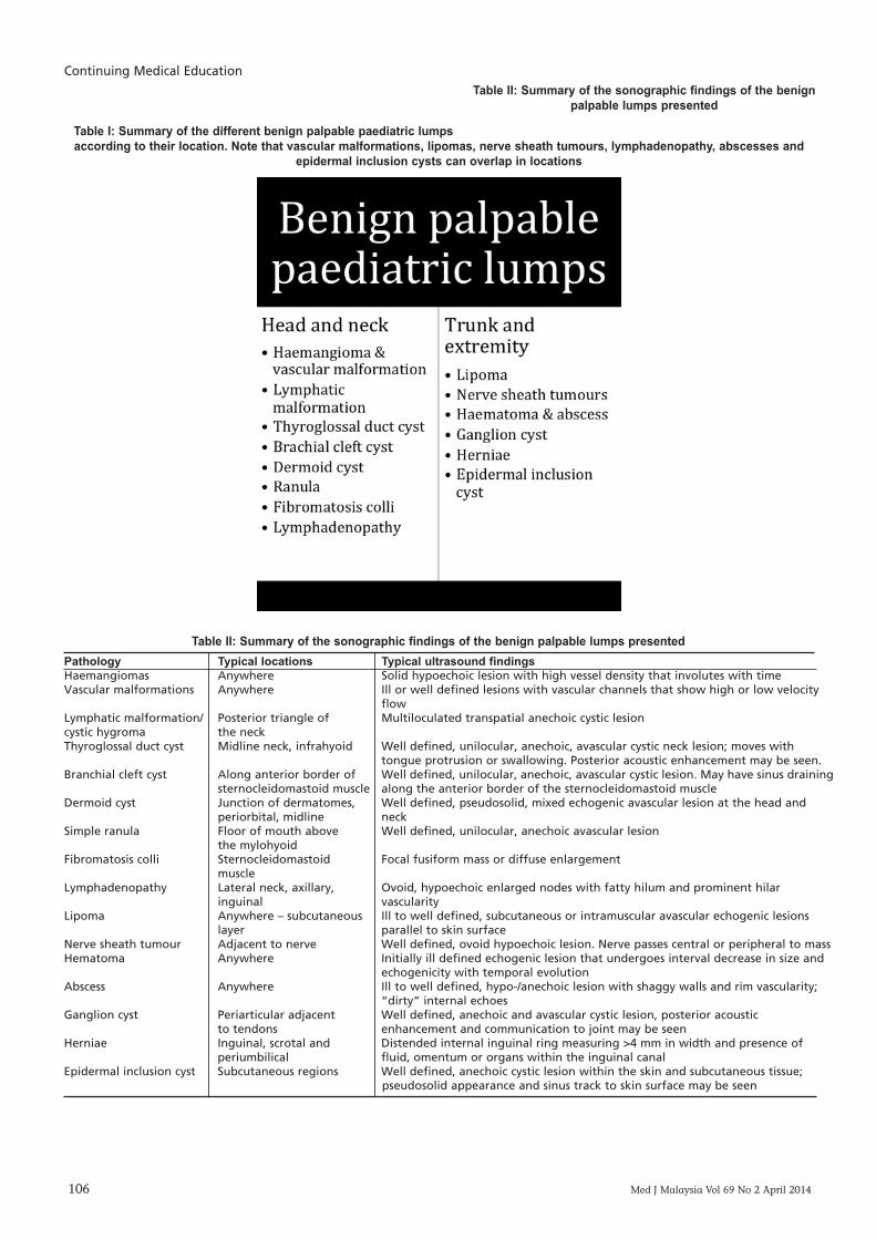
Table I: Summary of the different benign palpable paediatric lumpsaccording to their location. Note that vascular malformations, lipomas, nerve sheath tumours, lymphadenopathy, abscesses and
epidermal inclusion cysts can overlap in locations
Table II: Summary of the sonographic findings of the benignpalpable lumps presented
Table II: Summary of the sonographic findings of the benign palpable lumps presentedPathology Typical locations Typical ultrasound findingsHaemangiomas Anywhere Solid hypoechoic lesion with high vessel density that involutes with timeVascular malformations Anywhere Ill or well defined lesions with vascular channels that show high or low velocity
flowLymphatic malformation/ Posterior triangle of Multiloculated transpatial anechoic cystic lesioncystic hygroma the neckThyroglossal duct cyst Midline neck, infrahyoid Well defined, unilocular, anechoic, avascular cystic neck lesion; moves with
tongue protrusion or swallowing. Posterior acoustic enhancement may be seen.Branchial cleft cyst Along anterior border of Well defined, unilocular, anechoic, avascular cystic lesion. May have sinus draining
sternocleidomastoid muscle along the anterior border of the sternocleidomastoid muscleDermoid cyst Junction of dermatomes, Well defined, pseudosolid, mixed echogenic avascular lesion at the head and
periorbital, midline neckSimple ranula Floor of mouth above Well defined, unilocular, anechoic avascular lesion
the mylohyoidFibromatosis colli Sternocleidomastoid Focal fusiform mass or diffuse enlargement
muscleLymphadenopathy Lateral neck, axillary, Ovoid, hypoechoic enlarged nodes with fatty hilum and prominent hilar
inguinal vascularityLipoma Anywhere – subcutaneous Ill to well defined, subcutaneous or intramuscular avascular echogenic lesions
layer parallel to skin surfaceNerve sheath tumour Adjacent to nerve Well defined, ovoid hypoechoic lesion. Nerve passes central or peripheral to massHematoma Anywhere Initially ill defined echogenic lesion that undergoes interval decrease in size and
echogenicity with temporal evolutionAbscess Anywhere Ill to well defined, hypo-/anechoic lesion with shaggy walls and rim vascularity;
“dirty” internal echoesGanglion cyst Periarticular adjacent Well defined, anechoic and avascular cystic lesion, posterior acoustic
to tendons enhancement and communication to joint may be seenHerniae Inguinal, scrotal and Distended internal inguinal ring measuring >4 mm in width and presence of
periumbilical fluid, omentum or organs within the inguinal canalEpidermal inclusion cyst Subcutaneous regions Well defined, anechoic cystic lesion within the skin and subcutaneous tissue;
pseudosolid appearance and sinus track to skin surface may be seen
Sonographic Pictorial Review of Benign Palpable Paediatric Lumps: Head to Foot
Med J Malaysia Vol 69 No 2 April 2014 107
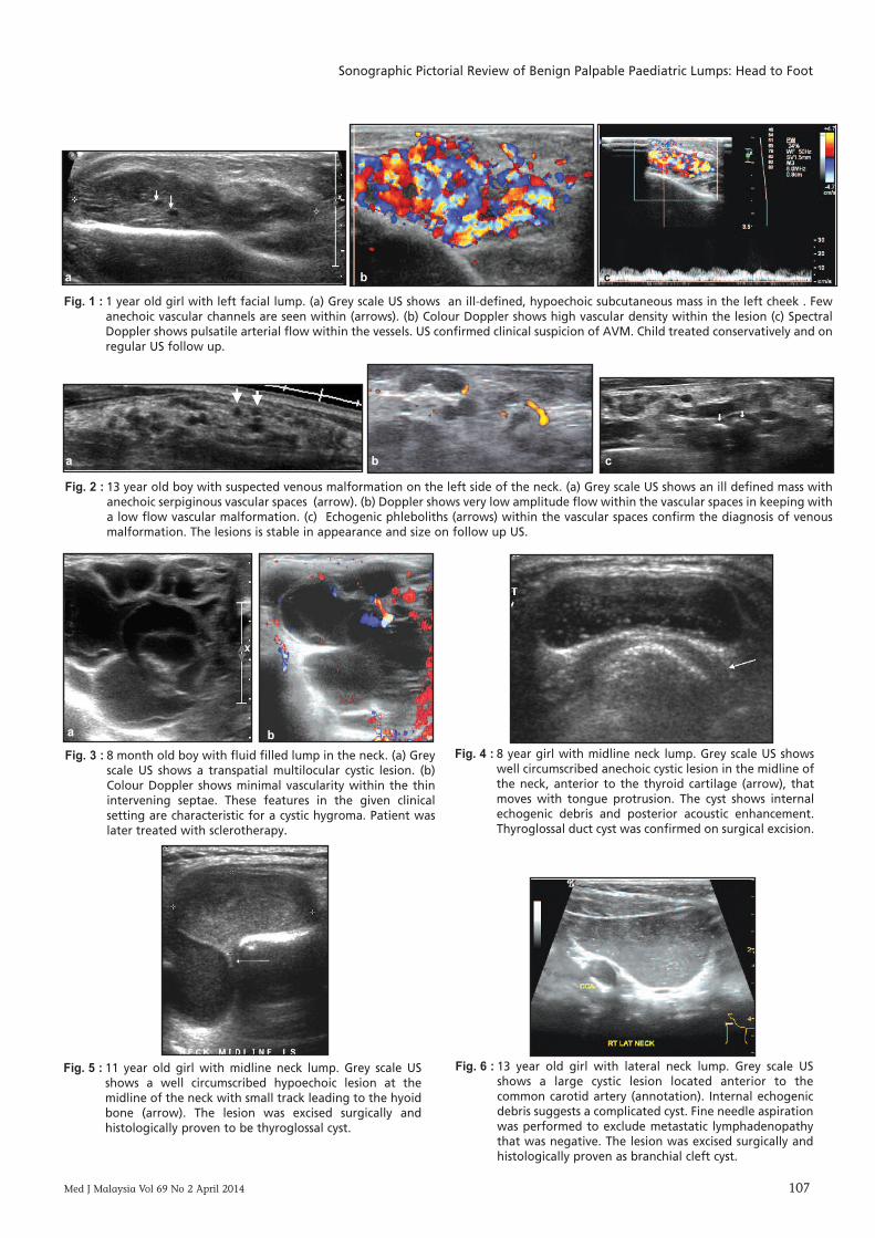
Fig. 1 : 1 year old girl with left facial lump. (a) Grey scale US shows an ill-defined, hypoechoic subcutaneous mass in the left cheek . Fewanechoic vascular channels are seen within (arrows). (b) Colour Doppler shows high vascular density within the lesion (c) SpectralDoppler shows pulsatile arterial flow within the vessels. US confirmed clinical suspicion of AVM. Child treated conservatively and onregular US follow up.
Fig. 2 : 13 year old boy with suspected venous malformation on the left side of the neck. (a) Grey scale US shows an ill defined mass withanechoic serpiginous vascular spaces (arrow). (b) Doppler shows very low amplitude flow within the vascular spaces in keeping witha low flow vascular malformation. (c) Echogenic phleboliths (arrows) within the vascular spaces confirm the diagnosis of venousmalformation. The lesions is stable in appearance and size on follow up US.
Fig. 3 : 8 month old boy with fluid filled lump in the neck. (a) Greyscale US shows a transpatial multilocular cystic lesion. (b)Colour Doppler shows minimal vascularity within the thinintervening septae. These features in the given clinicalsetting are characteristic for a cystic hygroma. Patient waslater treated with sclerotherapy.
Fig. 4 : 8 year girl with midline neck lump. Grey scale US showswell circumscribed anechoic cystic lesion in the midline ofthe neck, anterior to the thyroid cartilage (arrow), thatmoves with tongue protrusion. The cyst shows internalechogenic debris and posterior acoustic enhancement.Thyroglossal duct cyst was confirmed on surgical excision.
Fig. 5 : 11 year old girl with midline neck lump. Grey scale USshows a well circumscribed hypoechoic lesion at themidline of the neck with small track leading to the hyoidbone (arrow). The lesion was excised surgically andhistologically proven to be thyroglossal cyst.
Fig. 6 : 13 year old girl with lateral neck lump. Grey scale USshows a large cystic lesion located anterior to thecommon carotid artery (annotation). Internal echogenicdebris suggests a complicated cyst. Fine needle aspirationwas performed to exclude metastatic lymphadenopathythat was negative. The lesion was excised surgically andhistologically proven as branchial cleft cyst.
a b
a b
a b
c
c
Continuing Medical Education
108 Med J Malaysia Vol 69 No 2 April 2014
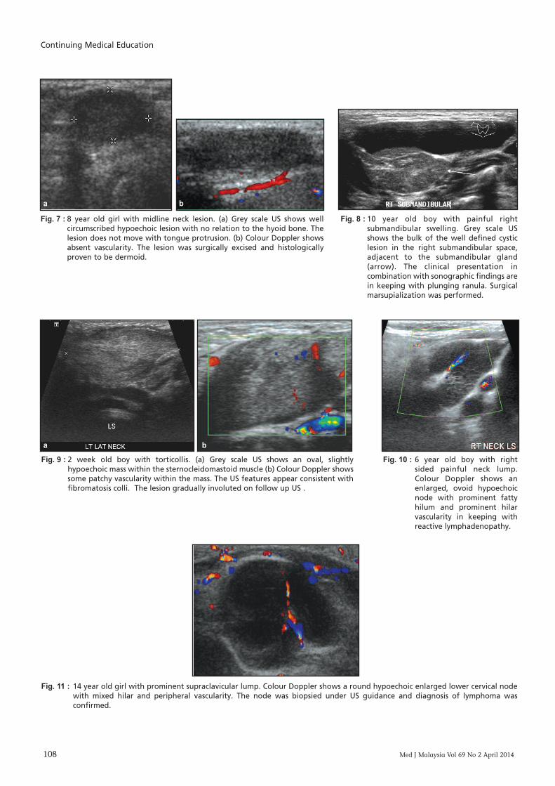
Fig. 7 : 8 year old girl with midline neck lesion. (a) Grey scale US shows wellcircumscribed hypoechoic lesion with no relation to the hyoid bone. Thelesion does not move with tongue protrusion. (b) Colour Doppler showsabsent vascularity. The lesion was surgically excised and histologicallyproven to be dermoid.
Fig. 8 : 10 year old boy with painful rightsubmandibular swelling. Grey scale USshows the bulk of the well defined cysticlesion in the right submandibular space,adjacent to the submandibular gland(arrow). The clinical presentation incombination with sonographic findings arein keeping with plunging ranula. Surgicalmarsupialization was performed.
Fig. 9 : 2 week old boy with torticollis. (a) Grey scale US shows an oval, slightlyhypoechoic mass within the sternocleidomastoid muscle (b) Colour Doppler showssome patchy vascularity within the mass. The US features appear consistent withfibromatosis colli. The lesion gradually involuted on follow up US .
Fig. 10 : 6 year old boy with rightsided painful neck lump.Colour Doppler shows anenlarged, ovoid hypoechoicnode with prominent fattyhilum and prominent hilarvascularity in keeping withreactive lymphadenopathy.
Fig. 11 : 14 year old girl with prominent supraclavicular lump. Colour Doppler shows a round hypoechoic enlarged lower cervical nodewith mixed hilar and peripheral vascularity. The node was biopsied under US guidance and diagnosis of lymphoma wasconfirmed.
a b
a b
Sonographic Pictorial Review of Benign Palpable Paediatric Lumps: Head to Foot
Med J Malaysia Vol 69 No 2 April 2014 109
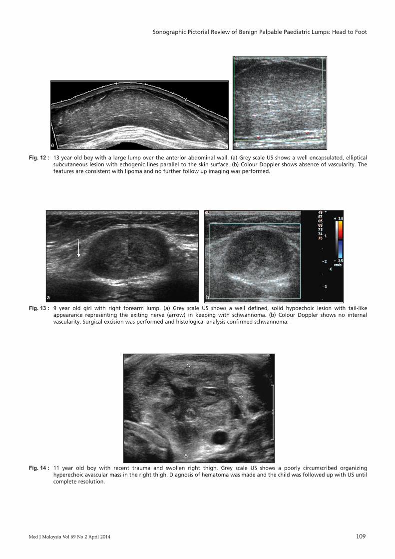
Fig. 12 : 13 year old boy with a large lump over the anterior abdominal wall. (a) Grey scale US shows a well encapsulated, ellipticalsubcutaneous lesion with echogenic lines parallel to the skin surface. (b) Colour Doppler shows absence of vascularity. Thefeatures are consistent with lipoma and no further follow up imaging was performed.
Fig. 13 : 9 year old girl with right forearm lump. (a) Grey scale US shows a well defined, solid hypoechoic lesion with tail-likeappearance representing the exiting nerve (arrow) in keeping with schwannoma. (b) Colour Doppler shows no internalvascularity. Surgical excision was performed and histological analysis confirmed schwannoma.
Fig. 14 : 11 year old boy with recent trauma and swollen right thigh. Grey scale US shows a poorly circumscribed organizinghyperechoic avascular mass in the right thigh. Diagnosis of hematoma was made and the child was followed up with US untilcomplete resolution.
a b
a b
Continuing Medical Education
110 Med J Malaysia Vol 69 No 2 April 2014
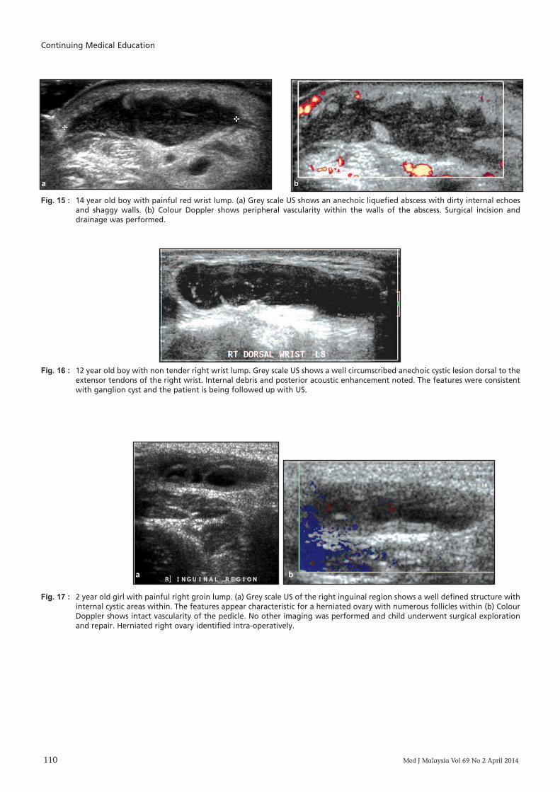
Fig. 15 : 14 year old boy with painful red wrist lump. (a) Grey scale US shows an anechoic liquefied abscess with dirty internal echoesand shaggy walls. (b) Colour Doppler shows peripheral vascularity within the walls of the abscess. Surgical incision anddrainage was performed.
Fig. 16 : 12 year old boy with non tender right wrist lump. Grey scale US shows a well circumscribed anechoic cystic lesion dorsal to theextensor tendons of the right wrist. Internal debris and posterior acoustic enhancement noted. The features were consistentwith ganglion cyst and the patient is being followed up with US.
Fig. 17 : 2 year old girl with painful right groin lump. (a) Grey scale US of the right inguinal region shows a well defined structure withinternal cystic areas within. The features appear characteristic for a herniated ovary with numerous follicles within (b) ColourDoppler shows intact vascularity of the pedicle. No other imaging was performed and child underwent surgical explorationand repair. Herniated right ovary identified intra-operatively.
a b
a b
Sonographic Pictorial Review of Benign Palpable Paediatric Lumps: Head to Foot
Med J Malaysia Vol 69 No 2 April 2014 111
rare16. US is poor at distinguishing between neurofibromasand schwannomas. Both commonly appear as well defined,ovoid or fusiform, homogeneous and hypoechoic masses (Fig13). Posterior acoustic enhancement and ‘target’ appearancei.e. hyperechoic centre with hypoechoic periphery may beoccasionally visualized. The nerve is seen to pass through thecentre of the mass in neurofibroma whereas the nerve isrelated to the periphery of the mass in a schwannoma.Schwannomas may appear cystic and more vascular thanneurofibromas 15,16,17,18.
Haematoma and AbscessHaematomas and abscesses are common in the extremities.Clinical history is crucial to distinguish between the two asthey have similar sonographic features. The sonographicappearance of haematoma changes with time. Acutehaematoma is usually ill defined and hyperechoic whilstchronic haematoma reduces in echogenicity and size (Fig 14).Vascularity is often absent in a haematoma. An infectedhaematoma can progress onto an abscess.
An abscess on US usually appears as an ill to well definedhypo or anechoic lesion with shaggy walls. It may show‘dirty’ internal echoes, fluid-fluid levels, septae and rim
vascularity on colour Doppler (Fig 15). An abscess isfrequently associated with adjacent oedematoussubcutaneous tissue and enlarged reactive regional nodes. USmay be used for guided aspiration and follow up tillresolution 18,19.
Ganglion cystAbout 50-70% of soft tissue masses in the wrist region areganglion cysts 18. They are less common at the shoulder,elbow, hip, knee and ankle. A wrist ganglion on US typicallyappears as a well defined, anechoic and avascular lesion,lying dorsal to the extensor tendons (Fig 16). Posterioracoustic enhancement is common. Some ganglions may becompressible. Lobulations, septations and internal echoesmay be seen. An anechoic duct leading to the joint may bevisualized. An infected ganglion can mimic a solid mass andappear heterogeneous with internal echoes 18,20.
HerniaeCongenital indirect inguinal herniae are the most commonexternal herniae in children. These usually contain omentalfat, bowel loops and peritoneal fluid 21. Unusual sac contentsmay include the vermiform appendix, part of the urinarybladder and ovaries and/or fallopian tubes in female infants.
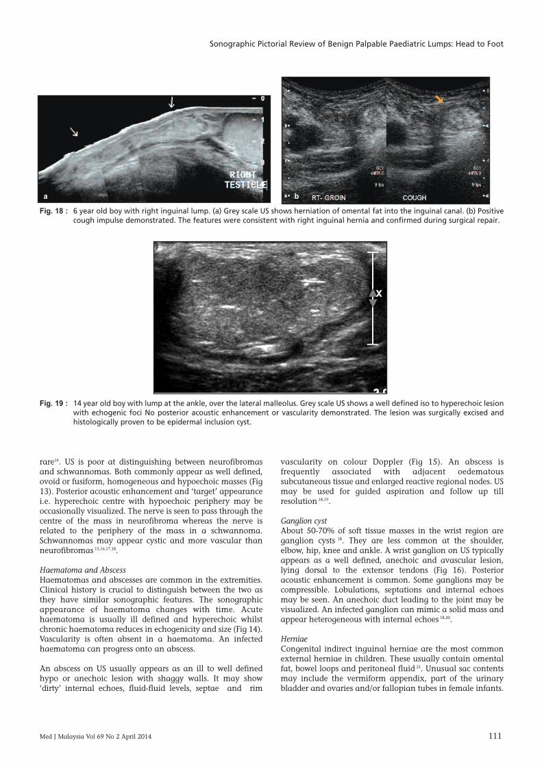
Fig. 18 : 6 year old boy with right inguinal lump. (a) Grey scale US shows herniation of omental fat into the inguinal canal. (b) Positivecough impulse demonstrated. The features were consistent with right inguinal hernia and confirmed during surgical repair.
Fig. 19 : 14 year old boy with lump at the ankle, over the lateral malleolus. Grey scale US shows a well defined iso to hyperechoic lesionwith echogenic foci No posterior acoustic enhancement or vascularity demonstrated. The lesion was surgically excised andhistologically proven to be epidermal inclusion cyst.
a b
Continuing Medical Education
112 Med J Malaysia Vol 69 No 2 April 2014
A herniated ovary if detected on US must be differentiatedfrom a round ligament cyst. Torsion of the ovarian vascularpedicle must be ruled out in all cases of herniated ovary (Fig17) 22. US criteria that have been decribed for paediatricinguinal herniae include internal inguinal ring greater than4 mm in diameter and presence of fluid or organs in theinguinal canal at rest or during straining 23. Herniatedomental fat appears hyperechoic whilst bowel loops willdemonstrate peristalsis (Fig 18). Bowel viability must beassessed in all cases by evaluating for peristalsis, reducibilityand mucosal vascularity. Valsalva manoeuvre andexamination in the standing position helps to assess slidingmotion and reducibility of herniae in older cooperativechildren 24.
Epidermal inclusion cystEpidermal inclusion cyst results from the implantation ofepidermal components into the dermis. The aetiology may becongenital (secondary to misplacement of the remnantectodermal tissues during embryogenesis) traumatic or post-surgical 25. They usually occur in the back, neck, scalp andextremities. Patients commonly present with a palpablenodule that discharges oily material through a punctum. Anintact epidermal cyst on US appears as a well defined,anechoic lesion within the skin or subcutaneous tissue. Somecysts may show a sinus track leading to the skin surface 15,26.They may sometimes appear pseudo-solid with brightinternal echoes (pseudotestis appearance) due to their keratincontent (Fig 19) 27. A ruptured cyst may show irregularmargins, rim vascularity and adjacent inflammatorychanges 25. Both intact and ruptured epidermal cysts mayshow posterior acoustic enhancement 15,26.
CONCLUSIONUS along with Doppler is a safe and reliable first linemodality for the evaluation of palpable paediatric lumps.Accurate clinical history and awareness of the specific UScharacteristics of these lesions helps in correct diagnosis. Thecommonly encountered benign palpable lumps in childrenalong with their common locations and typical UScharacteristics have been summarized in Table II. US alsoplays an important role in follow up of benign paediatriclumps, imaging guided needle aspiration for cytology anddrainage of abscesses and collections.
REFERENCES1. Ahuja AT. Lumps and bumps in the head and neck. In: Ahuja AT, Evans
RM (eds). Practical Head and Neck Ultrasound, 1st ed. New York, USA:Cambridge University Press, 2000: 87-104.
2. Navarro OM. Soft tissue masses in children. Radiol Clin N Am 2011; 49:1235-59.
3. Donnelly LF, Adams DM, Bisset GS.Vascular malformations andhemangiomas: a practical approach in a multidisciplinary clinic. AJR AmJ Roentgenol 2000; 174: 597-608.
4. Abernathy L. Paediatric neck lumps III- Vascular and lymphaticmalformations. Ultrasound. 2007; 15: 142-7.
5. Petrovic S, Petrovic D, Pesic Z, Kovacevic P. Sonography of congenital neckmasses in children. Medicine and Biology 2005; 12: 164-9.
6. Wong KT, Lee YY, King AD, Ahuja AT. Imaging of cystic or cyst-like neckmasses. Clin Radiol 2008; 63: 613-22.
7. Kutuya N, Kurosaki Y. Sonographic assessment of thyroglossal duct cystsin children. J Ultrasound Med 2008; 27:1211-19.
8. Jain P, Jain R, Morton RP, Ahmad Z. Plunging ranulas: high resolutionultrasound for diagnosis and surgical management. Eur Radiol 2010; 20:1442-9.
9. Ablin DS, Jain K, Howell L, West DC. Ultrasound and MR imaging offibromatosis colli (sternocleidomastoid tumor of infancy). Paediatr Radiol1998; 28: 230-3.
10. Ying M, Lee YY, Wong KT, Leung VY, Ahuja AT. Ultrasonography of necklymph nodes in children. HK J Paediatr (new series) 2009; 14: 29-36.
11. Papakonstantinou. O, Bakantaki. A, Paspalaki.P, Charoulakis N,Gourtsoyiannis N. High-resolution and color Doppler ultrasonography ofcervical adenopathy in children. Acta Radiol 2001; 42: 470-6.
12. Leung AK, Robson WL. Childhood cervical lymphadenopathy. J PediatrHealth Care 2004; 18: 3-7.
13. Ahuja AT, King AD, Kew J, King W, Metrewell C. Head and neck lipomas:sonographic apppearance. AJNR Am J Neuroradiol 1998; 19: 505-8.
14. Inampudi P, Jacobson JA, Fessell D, et al. Soft-tissue lipomas: accuracy ofsonography in diagnosis with pathologic correlation. Radiology 2004; 223:763-7.
15. Chiou HJ, Chou YH, Chin SY, et al. Differentiation of benign andmalignant superficial soft tissue masses using grayscale and colourdoppler ultrasonongraphy. J Chin Med Assoc 2009; 72: 307-15.
16. Stuart RM, Koh ES, Breidahl WH. Sonography of peripheral nervepathology. AJR Am J Roentgenol 2004; 182: 123-9.
17. Reynolds DL, Jacobson JA, Inampudi P, Jamadar DA, Ebrahim FS, HayesCW. Sonographic characteristics of peripheral nerve sheath tumours. AJRAm J Roentgenol 2004; 182: 741-4.
18. Kinare A, Brahmnalkar M, D'Costa S. Ultrasound of musculoskeletal softtissue masses. Indian J Radiol Imaging 2007; 17: 201-8.
19. Lin J, Fessell DP, Jacobson JA, Weadock WJ, Hayes CW. An illustratedtutorial of musculoskeletal sonography. AJR Am J Roentgenol 2000; 175:637-45.
20. Wang G, Jacobson JA, Feng FY, Girish G, Caoili EM, Brandon C.Sonography of wrist ganglion cysts: variable and noncystic appearances. JUltrasound Med 2007; 26: 1323-8.
21. Shadbolt CL, Heinze SB, Dietrich RB. Imaging of groin masses: inguinalanatomy and pathological conditions revisited. Radiographics 2001; 21:S261-71.
22. Oh SN, Jung SE, Rha SE, et al. Sonography of carious cystic masses of thefemale groin. J Ultrasound Med 2007; 26: 1735-42.
23. Chou TY, Chu CC, Diau GY, Wu CJ, Gueng MK. Inguinal hernia inchildren: US versus exploratory surgery and intraoperative contralaterallaproscopy. Radiology 1996; 2: 385-8.
24. Jamadar DA, Jacobson JA, Morag Y, Gandikota G, Ibrahim F, Gest T et al.Sonography of Inguinal Hernias. AJR Am J Roentgenol 2006; 187: 185-190.
25. Jin W, Ryu KN, Kim GY, Kim HC, Lee JH, Park JS. Sonographic findings ofruptured epidermal inclusion cyst in superficial soft tissue: emphasis onshapes, pericystic changes, and pericystic vascularity. J Ultrasound Med2008; 27: 171-6.
26. Wortsman X. Common applications of dermatologic Sonography. JUltrasound Med 2012; 31: 97-111.
27. Huang CC, Ko SF, Huang HY, et al. Epidermal cysts in the superficial softtissue: sonographic features with an emphasis on the pseudotestis pattern.J Ultrasound Med 2011; 30: 11-7.
Sonographic Pictorial Review of Benign Palpable Paediatric Lumps: Head to Foot
Med J Malaysia Vol 69 No 2 April 2014 113
Multiple Choice Questions
1. What is the commonest location of a TDC?A. SuprahyoidB. InfrahyoidC. SubmentalD. SubmandibularE. Posterior triangle
2. Which space shows the bulk of a plunging ranula on US?A. Submental spaceB. Submandibular spaceC. Anterior triangleD. Masticator spaceE. Parapharyngeal space
3. US features of benign reactive nodes include:A. Intranodal necrosisB. Intranodal calcificationsC. Fatty hilum with hilar vascularityD. Peripheral vascularityE. Round shape
4. What is the commonest location of a ganglion cyst?A. Wrist B. Hip C. ShoulderD. AnkleE. Knee
5. What are the common US imaging features of a cystic hygroma?A. Solid hyperechoic lesionB. Multilocular anechoic lesionC. Pseudotestis appearanceD. Shaggy walls with dirty internal echoesE. High vessel density