Sonographic Findings of the Acute Scrotumeradiology.bidmc.harvard.edu/LearningLab/genito/Lo.pdf ·...
40
An Anthology of Pain: Sonographic Findings of the Acute Scrotum Wai-Kit Lo, HMS III Gillian Lieberman, MD January 2005 Wai-Kit Lo, HMS III Gillian Lieberman, MD
Transcript of Sonographic Findings of the Acute Scrotumeradiology.bidmc.harvard.edu/LearningLab/genito/Lo.pdf ·...
An Anthology of Pain: Sonographic Findings of the
Acute Scrotum
Wai-Kit Lo, HMS IIIGillian Lieberman, MD
January 2005Wai-Kit Lo, HMS IIIGillian Lieberman, MD
Wai-Kit Lo, HMS IIIGillian Lieberman, MD
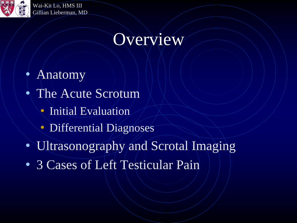
Overview
• Anatomy• The Acute Scrotum
• Initial Evaluation• Differential Diagnoses
• Ultrasonography and Scrotal Imaging• 3 Cases of Left Testicular Pain
Wai-Kit Lo, HMS IIIGillian Lieberman, MD
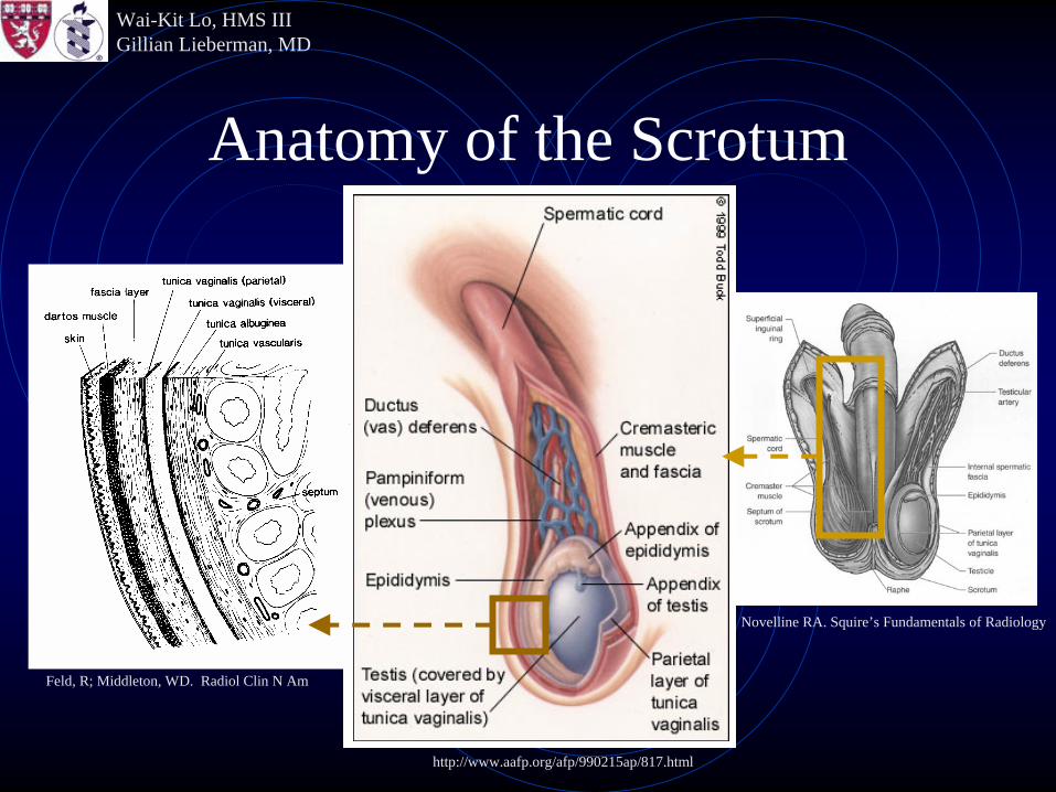
Anatomy of the Scrotum
Novelline RA. Squire’s Fundamentals of Radiology
http://www.aafp.org/afp/990215ap/817.html
Feld, R; Middleton, WD. Radiol Clin N Am
Wai-Kit Lo, HMS IIIGillian Lieberman, MD
Anatomy of the Scrotum
• Anatomic structures include• Testis• Epididymis• Vas deferens• Venous plexus• Testicular artery• Appendix of epididymis (remnant from embryogenesis)• Appendix of testis (remnant from embryogenesis)
Wai-Kit Lo, HMS IIIGillian Lieberman, MD
Anatomy of the Scrotum
• Layers include• (Testis: Seminiferous tubules)• Tunica albuginea• Visceral layer of Tunica vaginalis• Parietal layer of Tunica vaginalis• Fascia• Dartos muscle• Skin
Wai-Kit Lo, HMS IIIGillian Lieberman, MD
The Acute Scrotum
• The Acute Scrotum refers to the acute onset of scrotal pain with or without swelling, often seen in an emergent setting.
Wai-Kit Lo, HMS IIIGillian Lieberman, MD
The Acute Scrotum
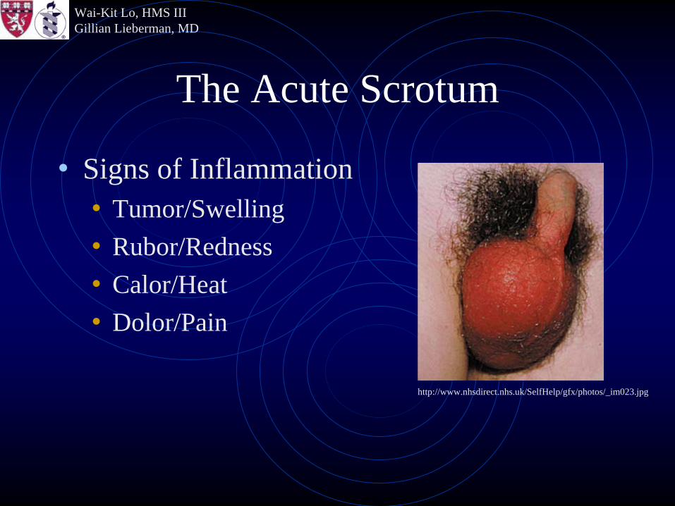
• Signs of Inflammation• Tumor/Swelling• Rubor/Redness• Calor/Heat• Dolor/Pain
Warning!Graphicimageahead!
http://www.nhsdirect.nhs.uk/SelfHelp/gfx/photos/_im023.jpg
Wai-Kit Lo, HMS IIIGillian Lieberman, MD
The Acute Scrotum
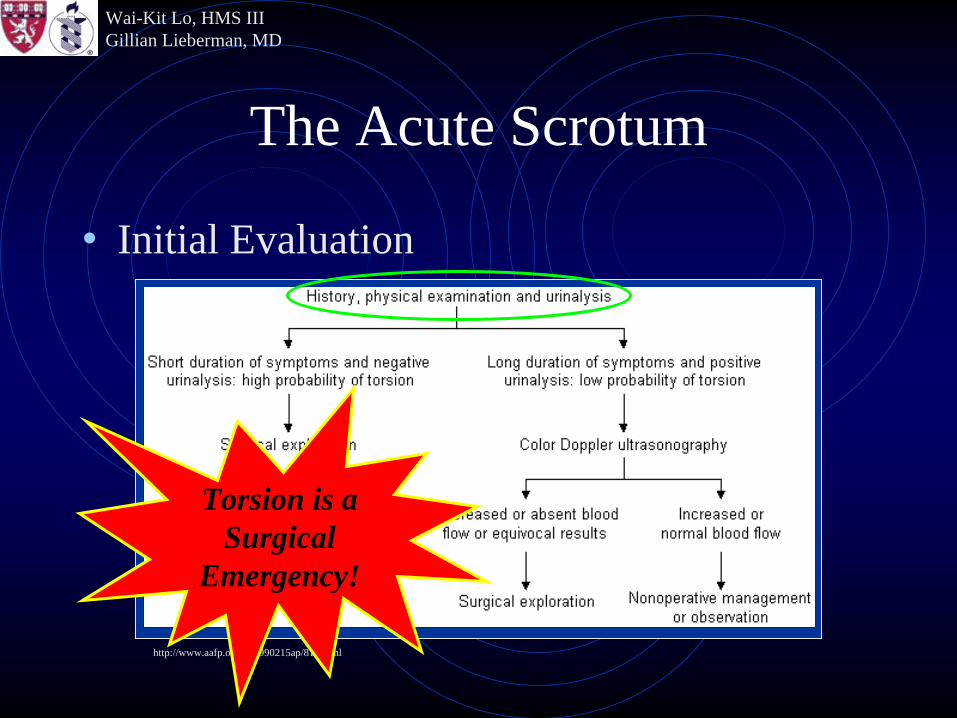
• Initial Evaluation
http://www.aafp.org/afp/990215ap/817.html
Torsion is aSurgical
Emergency!
Wai-Kit Lo, HMS IIIGillian Lieberman, MD
The Acute Scrotum
• Initial Evaluation• A pertinent history, physical examination, and
urinalysis are often sufficient to make a diagnosis.
• Imaging can be used to clarify diagnoses.
Wai-Kit Lo, HMS IIIGillian Lieberman, MD
The Acute Scrotum
• Differential Diagnoses• Separated into 4 general groups for ease of
memorization.
Wai-Kit Lo, HMS IIIGillian Lieberman, MD
The Acute Scrotum
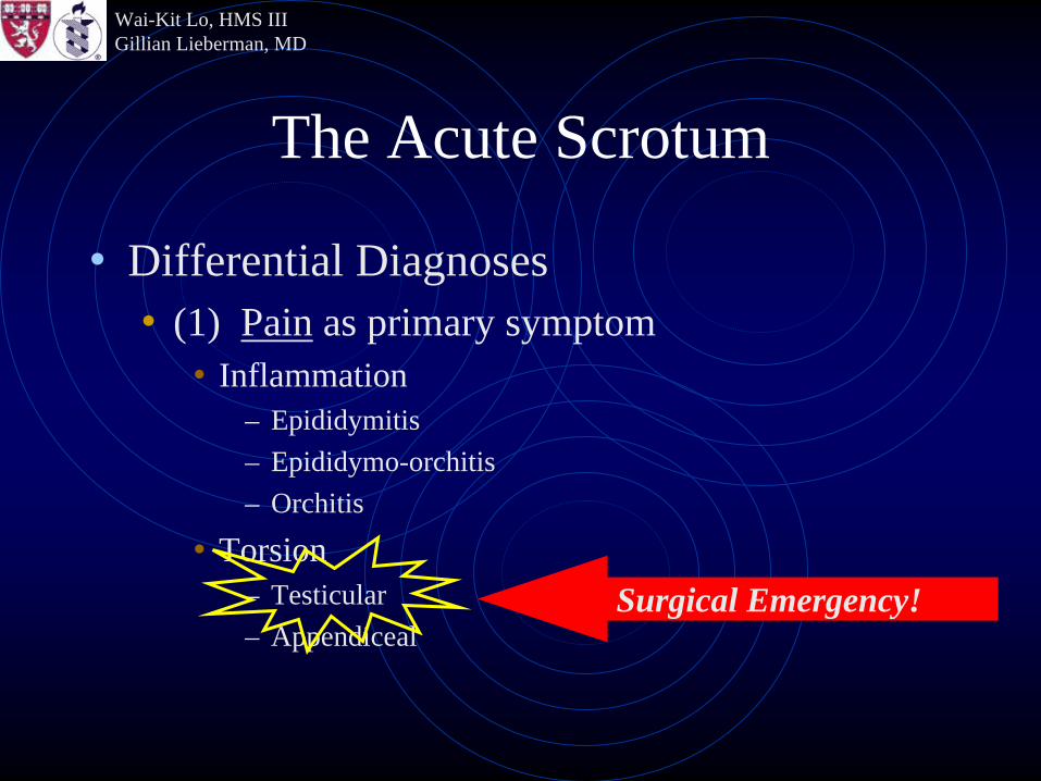
• Differential Diagnoses• (1) Pain as primary symptom
• Inflammation– Epididymitis– Epididymo-orchitis– Orchitis
• Torsion– Testicular– Appendiceal
Surgical Emergency!
Wai-Kit Lo, HMS IIIGillian Lieberman, MD
The Acute Scrotum
• Differential Diagnoses• (2) Pain with history of trauma
• Contusion• Rupture• Hematoma
Also a Surgical Emergency!
Wai-Kit Lo, HMS IIIGillian Lieberman, MD
The Acute Scrotum
• Differential Diagnoses• (3) Palpable mass, classically painless
• Intratesticular (usually malignant)– Seminoma (40-50%)– Mixed (40%)– Germ cell tumor– Teratoma– Choriocarcinoma– Metastases (kidney, prostate, lung, pancreas, bladder, thyroid,
melanoma, GI)– Non-Germ Cell (Sertoli, Leydig, Mesenchymal)
• Extratesticular (usually benign)– Adenomatoid Tumor of the Epididymis (30%)– -omas: Leiomyoma, Fibroma, Lipoma
• Varicocele• Cyst/Abscess
Wai-Kit Lo, HMS IIIGillian Lieberman, MD
The Acute Scrotum
• Differential Diagnoses• (4) Visible swelling, usually painless (unless
co-presenting with painful pathology as stated above)
• Fluid collection– Hydrocele– Hematocele– Pyelocele
• Scrotal hernia
Wai-Kit Lo, HMS IIIGillian Lieberman, MD
Imaging the Scrotum
• Ultrasound in the Emergent Situation• Fast and relatively inexpensive compared to CT, MRI• Blood flow
• Torsion (decreased flow) v. Inflammation (increased flow)• Fluid Collection (avascular) v. Soft Tissue (vascular)
• Anatomic deformities• Trauma• Tumor
• Characterization of fluid collections• Real-time peristaltic bowel movements
• Hernia
Wai-Kit Lo, HMS IIIGillian Lieberman, MD
Imaging the Scrotum
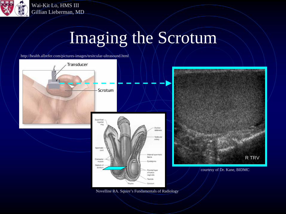
• The following slide shows the technique of scrotal ultrasound. The transducer is placed on top of the scrotum, which rests on the patient or is supported by the physician doing the study.
Wai-Kit Lo, HMS IIIGillian Lieberman, MD
http://health.allrefer.com/pictures-images/tesitcular-ultrasound.html
Imaging the Scrotum
Novelline RA. Squire’s Fundamentals of Radiology
courtesy of Dr. Kane, BIDMC
R TRV
Wai-Kit Lo, HMS IIIGillian Lieberman, MD
Imaging the Scrotum
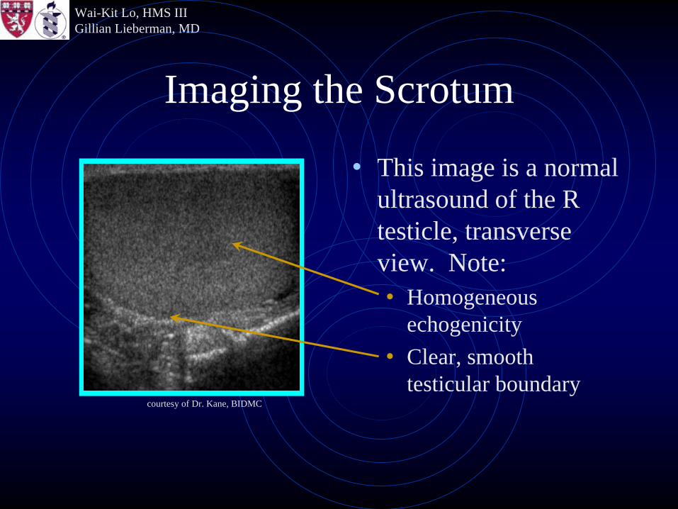
• This image is a normal ultrasound of the R testicle, transverse view. Note:• Homogeneous
echogenicity• Clear, smooth
testicular boundarycourtesy of Dr. Kane, BIDMC
Wai-Kit Lo, HMS IIIGillian Lieberman, MD
Meet the Patients
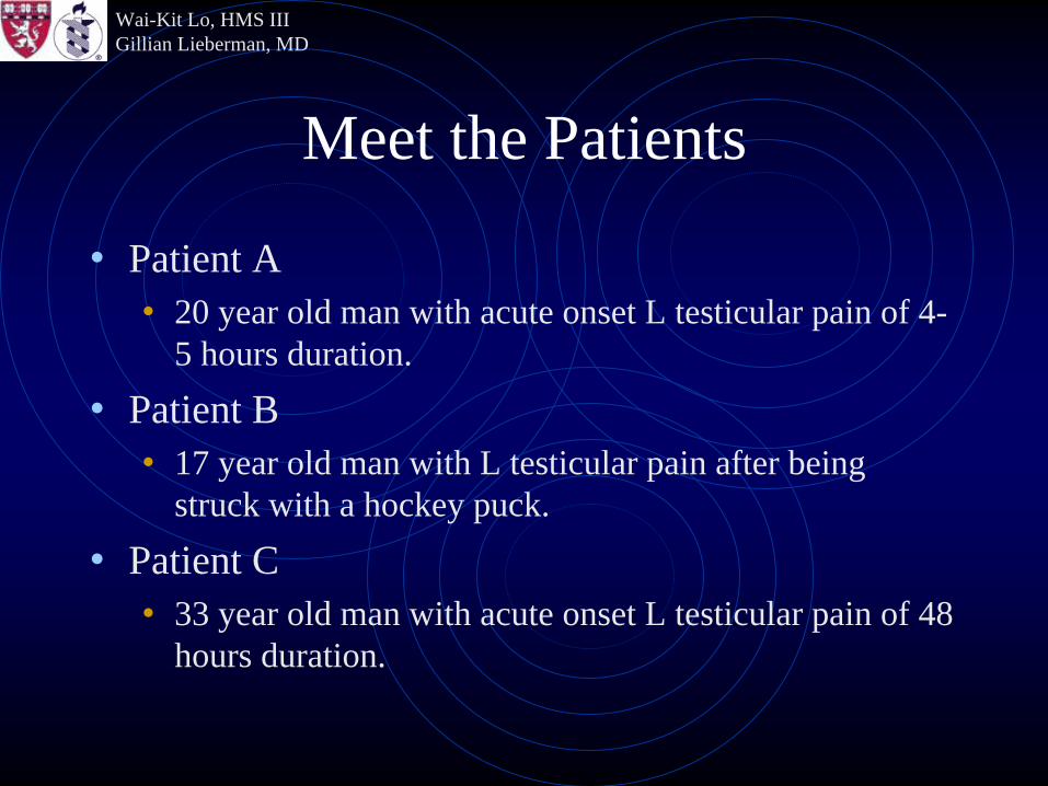
• Patient A• 20 year old man with acute onset L testicular pain of 4-
5 hours duration.
• Patient B• 17 year old man with L testicular pain after being
struck with a hockey puck.
• Patient C• 33 year old man with acute onset L testicular pain of 48
hours duration.
Wai-Kit Lo, HMS IIIGillian Lieberman, MD
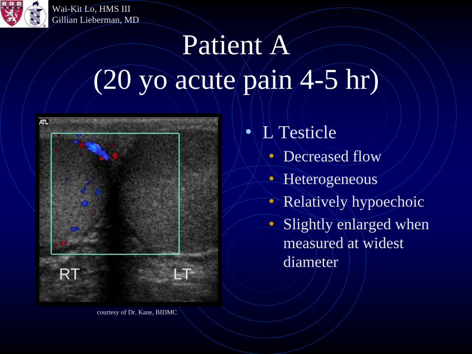
Patient A (20 yo acute pain 4-5 hr)
• L Testicle• Decreased flow• Heterogeneous• Relatively hypoechoic• Slightly enlarged when
measured at widest diameter
courtesy of Dr. Kane, BIDMC
LTRT
Wai-Kit Lo, HMS IIIGillian Lieberman, MD
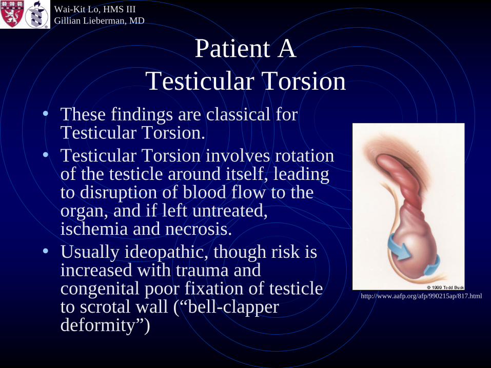
Patient A Testicular Torsion
• These findings are classical for Testicular Torsion.
• Testicular Torsion involves rotation of the testicle around itself, leading to disruption of blood flow to the organ, and if left untreated, ischemia and necrosis.
• Usually ideopathic, though risk is increased with trauma and congenital poor fixation of testicle to scrotal wall (“bell-clapper deformity”)
http://www.aafp.org/afp/990215ap/817.html
Wai-Kit Lo, HMS IIIGillian Lieberman, MD
Patient A Testicular Torsion
• Doppler Ultrasound has sensitivity of 86-100% and specificity of 100% in 2 different studies.
• Clinical suspicion often sufficient for diagnosis: short duration of symptoms in a peri-adolescent
• Testicular salvage rate in surgery: 80-100% within 5 hours of onset of pain 70% within 7-12 hours 20% after 12 hours
• Thus, important to catch early and send to OR
Wai-Kit Lo, HMS IIIGillian Lieberman, MD
Patient A Resolution
• The patient went to surgery 5-6 hours after onset of pain; surgeons were unable to reestablish blood flow in the ischemic L testicle, and the patient underwent L orchiectomy.
• Be Careful: Spontaneous detorsion increases blood flow, which can mimic hyperemia of inflammation!
!
Wai-Kit Lo, HMS IIIGillian Lieberman, MD
Patient A Spontaneous Detorsion
• Manual manipulation during clinical examination can lead to spontaneous detorsion of torsed testicle. In this instance, restoration of vascular patency to previously ischemic organ leads to increased flow, which can mimic hyperemic conditions such as orchitis.
!
Wai-Kit Lo, HMS IIIGillian Lieberman, MD
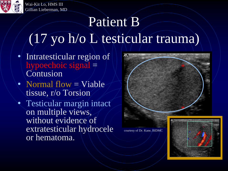
Patient B (17 yo h/o L testicular trauma)
• Intratesticular region of hypoechoic signal = Contusion
• Normal flow = Viable tissue, r/o Torsion
• Testicular margin intact on multiple views, without evidence of extratesticular hydrocele or hematoma.
*
*
*courtesy of Dr. Kane, BIDMC
Wai-Kit Lo, HMS IIIGillian Lieberman, MD
Patient B Testicular Trauma
• This is a routine evaluation of Testicular Trauma using Ultrasonography.
• Risk of testicular rupture due to blunt trauma is 50%, caused by severe crushing injury against pubic bone.
• Rupture of Tunica Albuginea leads to extravasation of the Seminiferous Tubules, causing ischemia or infection.
Wai-Kit Lo, HMS IIIGillian Lieberman, MD
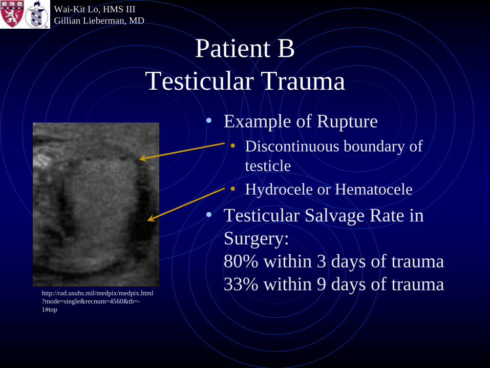
Patient B Testicular Trauma
• Example of Rupture• Discontinuous boundary of
testicle• Hydrocele or Hematocele
• Testicular Salvage Rate in Surgery: 80% within 3 days of trauma 33% within 9 days of trauma
http://rad.usuhs.mil/medpix/medpix.html ?mode=single&recnum=4560&th=- 1#top
Wai-Kit Lo, HMS IIIGillian Lieberman, MD
Patient B Resolution
• Radiology was comfortable with negative findings (regular testicular margins, absent fluid collection). Patient was discharged with pain medication.
• Be Careful: Ultrasound can miss minor tears in tunica albuginea! Nondiagnostic or suspicious ultrasounds should be followed with MRI or surgical exploration.
!
Wai-Kit Lo, HMS IIIGillian Lieberman, MD
Patient B Nondiagnostic Imaging
• If there continues to be doubt, remember that surgical exploration with negative findings is much more acceptable for the patient than missed rupture leading to ischemia, infection, and ultimately, infertility.
!

Wai-Kit Lo, HMS IIIGillian Lieberman, MD
Patient C (33 yo acute pain 48 hr)
• Heterogeneous mass in the enlarged L Testicle• Hydrocele (nonspecific inflammation)• Hypovascular
courtesy of Dr. Kane, BIDMC
Wai-Kit Lo, HMS IIIGillian Lieberman, MD
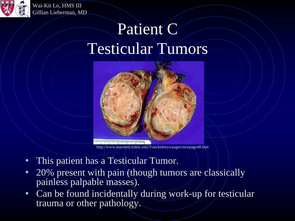
Patient C Testicular Tumors
• This patient has a Testicular Tumor.• 20% present with pain (though tumors are classically
painless palpable masses).• Can be found incidentally during work-up for testicular
trauma or other pathology.
http://www.macmed.ttuhsc.edu/Tran/kidneys/pages/newpage48.htm
Wai-Kit Lo, HMS IIIGillian Lieberman, MD
Patient C Testicular Tumor
• Usually hypervascular, as tumor growth requires increased blood flow; this also means tumors must be distinguished from inflammation (orchitis).
• Requires surgery/pathology workup for staging and diagnosis.
• May be associated with increased levels of HCG.
Wai-Kit Lo, HMS IIIGillian Lieberman, MD
Patient C Resolution
• Surgeons resected the L testicle. Pathology identified a Mixed Germ Cell tumor with clean margins. Patient has been free of tumor for 15 months, with follow-up every 2-4 months (watchful waiting). There is no single protocol for testicular tumor; the patient’s preferences should be taken into account when deciding course of treatment.
Wai-Kit Lo, HMS IIIGillian Lieberman, MD
Patient C Vascularity and Tumors
• Be Careful: Vascularity is related to Tumor Size; so small tumors (<1.5 cm) can appear hypovascular, rather than hypervascular, on Doppler Ultrasound. As you can see, however, even large tumors do not always follow the rules.
!
Wai-Kit Lo, HMS IIIGillian Lieberman, MD
Summary• Ultrasound imaging can help differentiate
among diagnoses in the setting of Scrotal Pain: We saw 3 cases of left testicular pain with different diagnoses based on imaging and history.
• Doppler ultrasound is used to distinguish between Torsion, a low-flow state which is a surgical emergency, and Inflammation, a high-flow state which is medically treated.

Wai-Kit Lo, HMS IIIGillian Lieberman, MD
Summary
• Ultrasound can be used in the setting of Trauma to evaluate damage to scrotal contents, but should be followed with MRI if nondiagnostic.
• Masses can be identified with ultrasound even when clinical suspicion is low; surgery/pathology is required for further work-up. Rule-out inflammation first.
Wai-Kit Lo, HMS IIIGillian Lieberman, MD
References• Corrales, JG; Corbel, L; Cipolla, B; Staerman, F; Darnault, P; Guille,
F; Lobel, B. Accuracy of Ultrasound Diagnosis After Blunt Testicular Trauma. J Urology 1993; 150(6): 1834-6.
• Dogra, VS; Gottlieb, RH; Oka, M; Rubens, DJ. Sonography of the scrotum. Radiology 2003; 227(1): 18-36.
• Feld, R; and Middleton, WD. Recent Advances in Sonography of the Testis and Scrotum. Radiol Clin N Am 1992; 30(5): 1033-51.
• Geraghty, MJ; Lee, FT; Bernstein, SA; Gilchrist, K; Pozniak, MA; Yandow, DJ. Sonography of testicular tumors and tumor-like conditions: A radiologic-pathologic correlation. CRC Cr Rev Diagn Im 1998; 39(1): 1-63.
• Kinkade, S. Testicular Cancer. Am Fam Physician 1999; 59(9): 2539- 44.
• Mulhall, JP; Gabram, SGA; Jacobs, LM. Emergency Management of Blunt Testicular Trauma. Acad Emerg Med 1995; 2(7): 639-43.
Wai-Kit Lo, HMS IIIGillian Lieberman, MD
References• Novelline, RA. Squire’s Fundamentals of Radiology, 6th Ed.
Cambridge: Harvard, 2004.• Pavlica, P; Barozzi, L. Imaging of the Acute Scrotum. Eur Radiol
2001; 11(2): 220-8.• Serra, AF; Hricak, H; Coakley, FV; Kim, BY; Dudley, A; Morey, A;
Tschumper, B; Caroll, PR. Inconclusive clinical and ultrasound evaluation of the scrotum: Impact of magnetic resonance imaging on patient management and cost. Urology 1998; 51(6): 1018-21.
• Weber, DM; Rosslein, R; Fliegel, C. Color Doppler sonography in the diagnosis of acute scrotum in boys. Eur J Pediatr Surg 2000; 10(4): 235-41.
• Wilberg, DM; Schaerfe, CW; Stern, WD; Strohmaier, WL; Bichler, KH. Evaluation of the Acute Scrotum by Color-coded Doppler Ultrasonography. J Urology 1993; 149(6): 1475-7.

Wai-Kit Lo, HMS IIIGillian Lieberman, MD
Special Thanks
• Robert Kane, MD• Gillian Lieberman, MD• Pamela Lepkowski• Larry Barbaras, our webmaster
Wai-Kit Lo, HMS IIIGillian Lieberman, MD
The End