Sonographic Evaluation of Pelvic Masses
42
Sonographic Evaluation of Pelvic Masses Aboubakr Elnashar Benha university Hospital, Egypt ABOUBAKR ELNASHAR
-
Upload
aboubakr-elnashar -
Category
Health & Medicine
-
view
280 -
download
1
Transcript of Sonographic Evaluation of Pelvic Masses

Sonographic Evaluation of Pelvic
Masses
Aboubakr Elnashar
Benha university Hospital, Egypt
ABOUBAKR ELNASHAR

Contents1.Parametrs of pelvic US
2.Transvaginal sonography of pelvic masses
3.Sonographic differential diagnosis
1.RuLes
2.Benign or malignant
3.Type
Summary
ABOUBAKR ELNASHAR

1. Parameters of pelvic US .1. Confirmation
presence or absence
2. Determination of 1. Size
2. consistency
3. contour .
4. Origin
5. anatomic relationship
3. Determine
abnormalities associated with malignant
disease: ascites or metastatic lesions.
4. Guidance for 1. Aspiration
2. biopsy
ABOUBAKR ELNASHAR

TVS and TA
TVS:
used as an adjunct to TAS.
evaluation of the uterus and adnexa.
tumor composition
location
its limited field of view and unusual image
orientation}
ABOUBAKR ELNASHAR

Color Doppler sonography (CDS)
helpful in distinguishing
benign from malignant ovarian masses
evaluation of adnexal torsion
an adjunct to morphologic assessment of
ovarian lesions.
ABOUBAKR ELNASHAR

2. DD between benign and malignant
ABOUBAKR ELNASHAR

Morphologic scoring by TVS.
Each of four parameters as assessed
Malignancies tended to have high scores (over 9).
ABOUBAKR ELNASHAR

Simple ultrasound rules: 2012
5 ultrasonic features to predict a malignant tumour
(M features):
Irregular solid tumour (M1),
Ascites (M2),
At least four papillary structures (M3),
Irregular multilocular solid tumour with a largest
diameter of at least 100 mm (M4)
Very high colour content on colour Doppler
examination (M5).
ABOUBAKR ELNASHAR
5 ultrasonic features to predict a benign tumour (B
features):
Unilocular cyst (B1),
Presence of solid components for which the largest
solid component is <7 mm in largest diameter (B2)
Acoustic shadows (B3)
Smooth multilocular tumour (B4)
No detectable blood flow on Doppler examination
(B5).
ABOUBAKR ELNASHAR

3. Type of mass
ABOUBAKR ELNASHAR

Purely cystic:
more likely to be benign
Complex cyst :
more likely to be malignant.
Purely solid:
more likely to be benign.
ABOUBAKR ELNASHAR

ovaryuterus
Unilocular, thin-walled, anechoic
I. CYSTIC
Simple Cystic
ABOUBAKR ELNASHAR

Unilocular
Thin-walled
Anechoic
Follicular cyst ABOUBAKR ELNASHAR

Simple cysts
Corpus luteal or follicular cyst
Haemorrhagic cysts
ABOUBAKR ELNASHAR

Massively enlarged ovariesThin-walled septationAscites may be present
OHSS
ABOUBAKR ELNASHAR

Hydrosalpinx
Tubular-shaped
structure
Anechoic content
Incomplete septum
ABOUBAKR ELNASHAR

Low-level echo cysts1. Endometrioma 95%2. Hemorrhagic cyst 50%3. Teratoma 18%4. Malignant Neoplasm 12%(Patel et al, 1999.)
ABOUBAKR ELNASHAR

Anechoic with lacelike internal echoes within cyst
Hemorrhagic C.
Corpus Luteum
ABOUBAKR ELNASHAR

Low-level echo cysts + Characteristic Features Endometrioma
Hyperechoic wall foci (in 35%)
Hemorrhagic cyst :Lacelike internal echoes (in 40%)
TeratomaRegional bright echoes ( in 97% )
ABOUBAKR ELNASHAR

Diffuse ‘ground glass’ pattern
{old blood}Endometrioma
ABOUBAKR ELNASHAR

smooth-walled ovarian cyst. Same patient after 5 w.
complete regression of the
physiologic cyst.
ABOUBAKR ELNASHAR
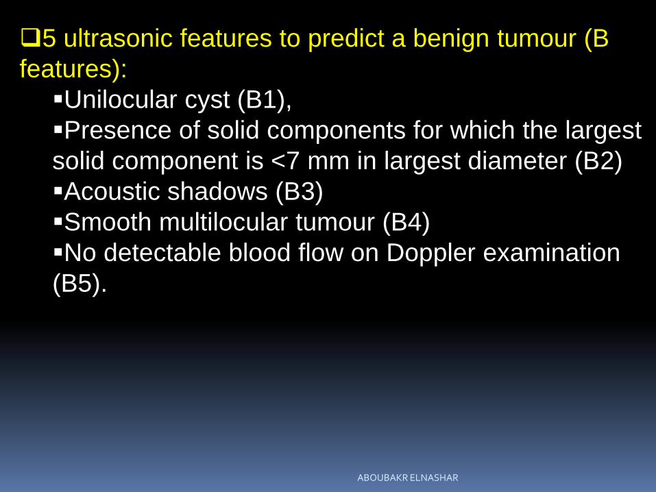
Completely cystic ovarian
masses in peri-menopausal
woman.
most small cystic ovarian
masses in pre- and
perimenopausal women are
physiologic
Completely cystic ovarian
masses in post menopausal
woman.
most postmenopausal cysts
are inclusion cysts.
ABOUBAKR ELNASHAR

Septated cystic masses. Mucinous cystadenoma.
cystic mass containing
multiple thin internal
septations
Mucinous cystadenoma.
septated mass with
echogenic material (*) in
upper loculated area.
The echogenic material was
mucin
ABOUBAKR ELNASHAR

Mucinous cystadenocarcinoma
Malignancy was suspected
due to thickened septation
(arrow)
Malignant teratoma.
Papillary projections (arrow)
ABOUBAKR ELNASHAR

1-Dermoid Cyst
The commonest 36%
2-Endometriotic cyst 5%
3-Malignant Cyst 1-3%
II. COMPLEX CYST
ABOUBAKR ELNASHAR
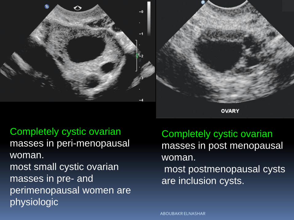
Dermoid
Complex mass solid and
cystic (fat, bone)
Fill in Pattern
ABOUBAKR ELNASHAR

Echogenic mural nodule in cystic mass.
Papillary serous Cystadenoma
Few small papillae
ABOUBAKR ELNASHAR

Mucinous Cystadenocarcinoma
Solid areas Many papillary. P
ABOUBAKR ELNASHAR

COMPLEX
Dermoid cyst.
Transverse sonogram of
complex predominantly cystic
with calcific focus (arrow)
arising from tooth
luteal cyst
with fluid surrounding
adhesion.
ABOUBAKR ELNASHAR

Mucinous cystadenoma
Sagittal and axial transvaginal sonogram:
multiloculated septated cystic mass with focal wall
thickening. This represented a with 1 locule containing thick
mucinous material
ABOUBAKR ELNASHAR

Dermoid cyst
layer of echogenic sebum
(*).
Hemorrhagic ovarian cyst
irregular solid area
corresponding to
displaced hemorrhagic
ovarian tissue surrounding
area of hemorrhage.ABOUBAKR ELNASHAR

Ovarian cystadenocarcinoma
irregular solid areas. Magnified transverse TAS of
cul-de-sac hemorrhage
(arrow) resulting from
ruptured ectopic pregnancy.
ABOUBAKR ELNASHAR
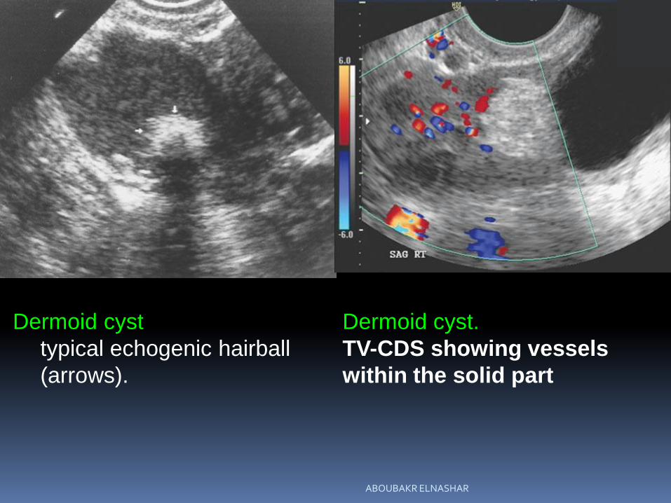
Dermoid cyst
typical echogenic hairball
(arrows).
Dermoid cyst.
TV-CDS showing vessels
within the solid part
ABOUBAKR ELNASHAR

Torsed right ovary.
(TAS)
solid mass (arrow) in cul-
de-sac
Transverse TAS of same
patient as in showing that left
ovary (straight arrow) is normal
in size and adjacent to torsed
right ovary (curved arrow).
ABOUBAKR ELNASHAR

Interligamentous fibroid (*)
appearing as solid pelvic
mass.
Transabdominal sonogram of
predominantly solid undifferentiated
ovarian neoplasm (arrow) containing a few
cystic areas.
ABOUBAKR ELNASHAR

Cystadenofibroma
TAS
solid pelvic mass
with calcifications (arrow) in
elderly patient.
Longitudinal TAS of pelvic
kidney (arrow). Pelvocalyceal
system accounts for central
echogenicity.
ABOUBAKR ELNASHAR

Transverse TAS showing
solid left-adnexal mass
(between +’s), which
represented hemorrhagic
corpus luteum cyst.
Longitudinal TAS of solid
teratoma with calcified areas.
ABOUBAKR ELNASHAR

Magnified TAS of solid mass (between +’s) representing
hemorrhagic corpus luteum cyst.
Sagittal and transverse
ABOUBAKR ELNASHAR

TAS: 5 × 7 cm solid mass
associated with ascites.
This was ovarian cancer.
TVS
large solid tumor representing
a dysgerminoma
ABOUBAKR ELNASHAR

Adnexal (ovarian) torsion
TVS: enlarged right ovary (between
+’s) with mildly echogenic area
resulting from internal hemorrhage
Cul-de-sac fluid adjacent to left side
of uterus in same patient
Two days later, the ovary (arrow) has
enlarged secondary to retorsion. On
TAS, enlarged size of ovary relative to
uterus can be better appreciated.ABOUBAKR ELNASHAR
SUMMARY
Although the sonographic features of a pelvic mass
may not allow a specific diagnosis, clinically useful
information can usually be obtained.
TVS is a useful adjunct to TAS because it adds specificity in determining intraversus extraovarianmasses and endometrial and myometrial disorders. TVS affords an accurate means for evaluation of the
ovaries and is particularly useful in obese,
postmenopausal women in whom the incidence of
ovarian carcinoma is especially high.
Although not always specific, sonographic assessment
of tumor morphology can lead to accurate diagnoses.
ABOUBAKR ELNASHAR

Thank you
ABOUBAKR ELNASHAR