Sonochemical synthesis of zinc oxide nano particles · Zinc Oxide (ZnO) is a thoroughly studied n-...
6
Research Article 2017, 2(5), 299-303 Advanced Materials Proceedings Copyright © 2017 VBRI Press 299 Sonochemical synthesis of zinc oxide nano particles Nandini Sharma, Ranjana Jha * Research Lab for Energy Systems, Department of Physics, Netaji Subhas Institute of Technology, University of Delhi, Dwarka, New Delhi-110078, India * Corresponding author: E-mail: [email protected]; Tel: (+91) 9654573775 Received: 30 March 2016, Revised: 30 September 2016 and Accepted: 14 April 2017 DOI: 10.5185/amp/2017.504 www.vbripress.com/amp Abstract Zinc oxide nanoparticles were synthesized utilizing a green and simple sonochemical route. The synthesized ZnO nanoparticles were characterized for the analysis of structural and optical properties. Characterization with XRD and TEM shows that the synthesized particles were uniformly distributed, crystalline in nature with spherical shape and narrow size distribution of particles (48-50 nm). UV-Vis and PL spectra shows optical band gap to be 3.5 eV and ZnO sample possess fewer defect states. ZnO nanoparticles synthesized shows good optical properties and was observed to be a promising candidate to be used in thin film solar cells. Copyright © 2017 VBRI Press. Keywords: Zinc oxide, nanoparticles, sonochemical, acoustic cavitation, green route. Introduction Zinc Oxide (ZnO) is a thoroughly studied n- type wide band gap (3.37eV) semiconductor with a large binding energy of 60 meV at room temperature and chemically stable [1][2][3]. The zinc oxide nano particles are widely used in comparison to the bulk [4] for various applications as it exhibits various interesting physical properties like more surface active sites [5]. There are several chemical methods reported by researchers to synthesize metal oxide nanoparticles [6]. Oxidation process [7], polymerization method[8], hydrothermal [9], solvothermal[10], sol–gel combustion [11] , precipitation and sol–gel synthesis are the various methods[12][13] that are used to grow ZnO nanostructures. These methods employ complicated protocols and conditions like high temperature, long reaction time. Recently, Sonochemical method [14][15] was used to synthesize various types of nanomaterials with unusual properties. In this process, ultrasound is applied to the reactant solution to initiate the chemical reactions. Sonochemistry originates from acoustic cavitation phenomena, that is, the formation, growth and implosive collapse of bubbles inside the solution[16]. Ultrasound waves produced by horn or bath type sonicator is induced directly or indirectly into the prepared solution[17]. Using horn type sonicator is an energy efficient method, as less reaction time is required [18]. A series of longitudinal waves are generated into the reaction solution by virtue of the presence of the ultrasound waves. Cavitation bubbles are developed when the rarefaction cycles are greater than the compressive forces inside the liquid molecules. Mechanical and chemical energies are generated by implosions inside the reaction mixture when the bubbles are collapsing as a result of cavitation. Reports are available on the sono-chemical synthesis of various nanomaterials[19][20][21][22][23][24][25] with good optical and structural properties. Manickam [26] in his paper has mentioned the advantages of this facile route over the other chemical routes. The reduced reaction time, moderate reaction conditions (low temperature) and consumption of non-toxic solvent are the main features of Sonochemical route. Almost all the chemical reactions like oxidation, reduction, dissolution, decomposition, and hydrolysis are induced under ultrasonic radiation[27]. The reaction activity of the reactants increases considerably by the presence of more free radicals generated inside the system. Sonochemical synthesis of Transition metal oxides nanoparticles [28] and the effect of solvent ratio on their size, morphology was studied [6][29]. In 2011[30], ZnO of crystal size 57.5 nm was prepared sonochemically but sodium hydroxide was used as an alkali material. In 2013, hierarchical ZnO nanostructures have been synthesized using base like sodium hydroxide and ammonia water in Sonochemical process [31] . Nanostructures of ZnO with controlled morphology have been synthesized by Sonochemical method using a cationic surfactant[32][33]. A simple Sonochemical route was demonstrated for the shape-selective preparation of ZnO nanorods, nanocups, nanodisks, nanoflowers, and nanospheres depending on the concentration of precursor chemicals, the kind of hydroxide anion-generating agents, the ultra-sonication time, and the use of a capping agent [34]. Bang and Suslick reviewed applications of ultrasound in the synthesis of nanomaterials [35]. To the best of our
Transcript of Sonochemical synthesis of zinc oxide nano particles · Zinc Oxide (ZnO) is a thoroughly studied n-...

Research Article 2017, 2(5), 299-303 Advanced Materials Proceedings
Copyright © 2017 VBRI Press 299
Sonochemical synthesis of zinc oxide nano particles
Nandini Sharma, Ranjana Jha*
Research Lab for Energy Systems, Department of Physics, Netaji Subhas Institute of Technology,
University of Delhi, Dwarka, New Delhi-110078, India *Corresponding author: E-mail: [email protected]; Tel: (+91) 9654573775
Received: 30 March 2016, Revised: 30 September 2016 and Accepted: 14 April 2017
DOI: 10.5185/amp/2017.504
www.vbripress.com/amp
Abstract
Zinc oxide nanoparticles were synthesized utilizing a green and simple sonochemical route. The synthesized ZnO nanoparticles
were characterized for the analysis of structural and optical properties. Characterization with XRD and TEM shows that the
synthesized particles were uniformly distributed, crystalline in nature with spherical shape and narrow size distribution of particles
(48-50 nm). UV-Vis and PL spectra shows optical band gap to be 3.5 eV and ZnO sample possess fewer defect states. ZnO
nanoparticles synthesized shows good optical properties and was observed to be a promising candidate to be used in thin film solar
cells. Copyright © 2017 VBRI Press.
Keywords: Zinc oxide, nanoparticles, sonochemical, acoustic cavitation, green route.
Introduction
Zinc Oxide (ZnO) is a thoroughly studied n- type wide band
gap (3.37eV) semiconductor with a large binding energy of
60 meV at room temperature and chemically stable [1][2][3].
The zinc oxide nano particles are widely used in comparison
to the bulk [4] for various applications as it exhibits various
interesting physical properties like more surface active sites
[5]. There are several chemical methods reported by
researchers to synthesize metal oxide nanoparticles [6].
Oxidation process [7], polymerization method[8],
hydrothermal [9], solvothermal[10], sol–gel combustion[11],
precipitation and sol–gel synthesis are the various
methods[12][13] that are used to grow ZnO nanostructures.
These methods employ complicated protocols and
conditions like high temperature, long reaction time.
Recently, Sonochemical method [14][15] was used to
synthesize various types of nanomaterials with unusual
properties. In this process, ultrasound is applied to the
reactant solution to initiate the chemical reactions.
Sonochemistry originates from acoustic cavitation
phenomena, that is, the formation, growth and implosive
collapse of bubbles inside the solution[16]. Ultrasound
waves produced by horn or bath type sonicator is induced
directly or indirectly into the prepared solution[17]. Using
horn type sonicator is an energy efficient method, as less
reaction time is required [18]. A series of longitudinal waves
are generated into the reaction solution by virtue of the
presence of the ultrasound waves. Cavitation bubbles are
developed when the rarefaction cycles are greater than the
compressive forces inside the liquid molecules. Mechanical
and chemical energies are generated by implosions inside the
reaction mixture when the bubbles are collapsing as a result
of cavitation.
Reports are available on the sono-chemical synthesis of
various nanomaterials[19][20][21][22][23][24][25] with
good optical and structural properties. Manickam [26] in his
paper has mentioned the advantages of this facile route over
the other chemical routes. The reduced reaction time,
moderate reaction conditions (low temperature) and
consumption of non-toxic solvent are the main features of
Sonochemical route. Almost all the chemical reactions like
oxidation, reduction, dissolution, decomposition, and
hydrolysis are induced under ultrasonic radiation[27]. The
reaction activity of the reactants increases considerably by
the presence of more free radicals generated inside the
system.
Sonochemical synthesis of Transition metal oxides
nanoparticles [28] and the effect of solvent ratio on their size,
morphology was studied [6][29]. In 2011[30], ZnO of crystal
size 57.5 nm was prepared sonochemically but sodium
hydroxide was used as an alkali material. In 2013,
hierarchical ZnO nanostructures have been synthesized
using base like sodium hydroxide and ammonia water in
Sonochemical process [31] .
Nanostructures of ZnO with controlled morphology
have been synthesized by Sonochemical method using a
cationic surfactant[32][33]. A simple Sonochemical route
was demonstrated for the shape-selective preparation of ZnO
nanorods, nanocups, nanodisks, nanoflowers, and
nanospheres depending on the concentration of precursor
chemicals, the kind of hydroxide anion-generating agents,
the ultra-sonication time, and the use of a capping agent [34].
Bang and Suslick reviewed applications of ultrasound in the
synthesis of nanomaterials [35]. To the best of our

Research Article 2017, 2(5), 299-303 Advanced Materials Proceedings
Copyright © 2017 VBRI Press 300
knowledge, sonochemical growth of good quality spherical
ZnO nanoparticles (well separated, no agglomeration, low
defect states, crystallinity with no impurity phase) without
making use of any template or surfactant has not been
reported previously.
Here, we report the green and template-free
sonochemical method to synthesize the ZnO nanoparticles.
The obtained nanoparticles possess uniform size distribution
with controlled morphology and reduced surface defect sites.
The experiment was carried out at very low temperature
(55◦C), devoid of surfactant or toxic chemical. The pH was
maintained at 7 value throughout the experiment. The size,
crystallinity, morphology and optical properties of the
synthesized nanoparticles were studied using different
characterization techniques.
Fig. 1: XRD graph of ZnO nanoparticles and WH analysis of XRD data
Experimental
Zinc oxide nanoparticles were synthesized by ultra-
sonication of aqueous – alcohol (composed of methanol,
ethanol and distilled water) solution of Zinc acetate in the
absence of alkali, by maintaining the neutral pH, i.e., 7 and
at temperature 55oC.
Materials
All the reagents used in the present study, were of analytical
grade. Zinc acetate (Dihydrate) 99.5 %, from Loba Chemie,
Laboratory reagents and Fine chemicals (Molecular weight
219.50), ethanol (99.7%) from Merck, Solution for
Leishman’s stain from Loba Chemie, Laboratory reagents
and Fine chemicals (Molecular weight 32.04) and double
distilled water were used directly without any further
purification.
Synthesis of zinc oxide nanoparticles by ultrasonic method
ZnO nanoparticles were prepared via the ultrasonic method.
0.1M solution of zinc acetate dihydrate was prepared in
200ml aqueous alcohol (180 ml DI water, 10 ml ethanol and
10 ml Solution for Leishman’s stain). The solution was
stirred on magnetic stirrer for 10 minutes to obtain a clear
solution. Then, the beaker containing clear solution was kept
in a bath type sonicator (150 Watts at 20 KHz frequency, of
Transonic Company) and sonicated at 55◦C for 2 hours to
obtain a turbid solution. pH value of solution was measured
throughout the experiment using Decibel DB-1003 digital
pH meter and it varied from 6 (clear solution) to 7 (turbid
solution). Then the turbid solution was filtered using micron
filter paper to obtain the residue on filter paper. The obtained
residue was kept in hot air oven for 4 hours at 140◦C in order
to evaporate the excess solvent, ions and get dry powder. The
dry powder was again washed with a mixture of 1:1 ethanol
and methanol, followed by drying at 100◦C again under hot
air oven for 3 hours.
Characterizations/ measurements
High Resolution X-Ray Diffraction (HR-XRD)
measurements of the sample was performed on the machine
D8-Advance model of Bruker Inc. using monochromatic
CuKα line (λ=0.154056 nm) and was compared with the
available Joint Committee on Powder Diffraction Studies
(JCPDS card no.89-1397) data files to confirm the size and
phase. The sample was characterized using Transmission
electron microscopy to confirm the size and morphology.
Optical properties were measured by Ultraviolet Visible
spectroscopy (Analytik Jena Specord 250) and
Photoluminescence spectroscopy techniques. pH was
monitored using Decibel DB-1003 digital pH meter.
Results and discussion
Sono chemically synthesized Zinc oxide nanoparticles were
characterized by several analytical techniques and
instruments to study structural, optical and morphological
properties.
Structural studies
The obtained XRD pattern of the ZnO matches well with the
JCPDS card no. 89-1397. The crystallite size of the ZnO was
estimated by Debye Scherer equation[36], i.e., D=(k λ / 𝛃 cos
θ) where D is the crystallite size, λ is the wavelength of
incident X-ray (1.54 Å for CuKα ), k is a constant equal to
0.93, 𝛃 is the peak width at half maxima intensity and θ is
Research Article 2017, 2(5), 299-303 Advanced Materials Proceedings
Copyright © 2017 VBRI Press 301
the Bragg diffraction peak position. The estimated crystallite
size was 48 nm.
The observed peaks in diffraction pattern corresponds to
reflection planes (100), (002), (101), (102), (110), (103),
(200), (112), (201), (004) and (202) and indicates a wurtzite
structure for the sample under study. The lattice constants of
the prepared sample were a = b = 3.26047 Ǻ, c = 5.23650 Ǻ
which was found to be slightly larger than those of pure ZnO
(a = b = 3.249 Ǻ, c = 5.206 Ǻ).
Williamson-Hall analysis resulted in the average
distribution of crystallite size to be 39 nm as calculated from
the y-intercept of the graph, as shown in Figure 1. The
average particle size of ZnO nanoparticles evaluated from
Scherer’s formula and W-H plot analysis are inter correlated.
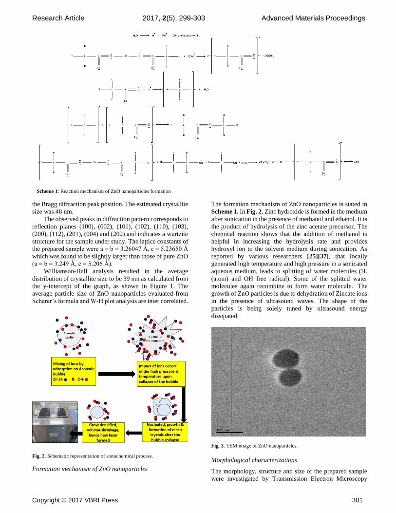
Fig. 2. Schematic representation of sonochemical process.
Formation mechanism of ZnO nanoparticles
The formation mechanism of ZnO nanoparticles is stated in
Scheme 1. In Fig. 2, Zinc hydroxide is formed in the medium
after sonication in the presence of methanol and ethanol. It is
the product of hydrolysis of the zinc acetate precursor. The
chemical reaction shows that the addition of methanol is
helpful in increasing the hydrolysis rate and provides
hydroxyl ion to the solvent medium during sonication. As
reported by various researchers [25][37], that locally
generated high temperature and high pressure in a sonicated
aqueous medium, leads to splitting of water molecules (H.
(atom) and OH free radical). Some of the splitted water
molecules again recombine to form water molecule. The
growth of ZnO particles is due to dehydration of Zincate ions
in the presence of ultrasound waves. The shape of the
particles is being solely tuned by ultrasound energy
dissipated.
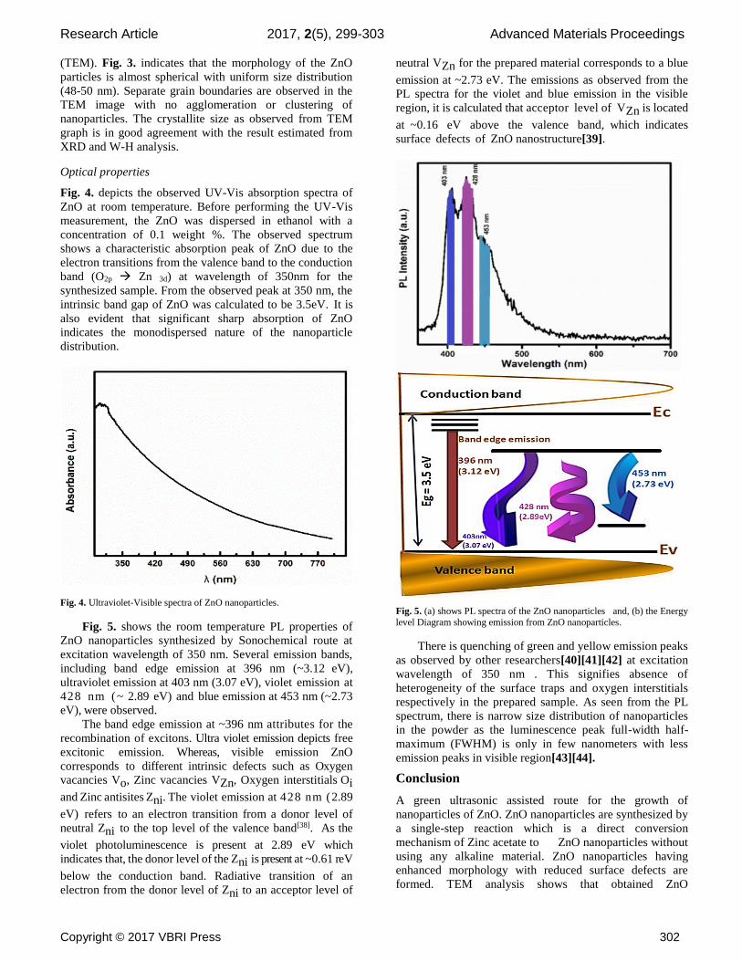
Fig. 3. TEM image of ZnO nanoparticles.
Morphological characterizations
The morphology, structure and size of the prepared sample
were investigated by Transmission Electron Microscopy
Scheme 1: Reaction mechanism of ZnO nanoparticles formation
Research Article 2017, 2(5), 299-303 Advanced Materials Proceedings
Copyright © 2017 VBRI Press 302
(TEM). Fig. 3. indicates that the morphology of the ZnO
particles is almost spherical with uniform size distribution
(48-50 nm). Separate grain boundaries are observed in the
TEM image with no agglomeration or clustering of
nanoparticles. The crystallite size as observed from TEM
graph is in good agreement with the result estimated from
XRD and W-H analysis.
Optical properties
Fig. 4. depicts the observed UV-Vis absorption spectra of
ZnO at room temperature. Before performing the UV-Vis
measurement, the ZnO was dispersed in ethanol with a
concentration of 0.1 weight %. The observed spectrum
shows a characteristic absorption peak of ZnO due to the
electron transitions from the valence band to the conduction
band (O2p Zn 3d) at wavelength of 350nm for the
synthesized sample. From the observed peak at 350 nm, the
intrinsic band gap of ZnO was calculated to be 3.5eV. It is also evident that significant sharp absorption of ZnO
indicates the monodispersed nature of the nanoparticle
distribution.
Fig. 4. Ultraviolet-Visible spectra of ZnO nanoparticles.
Fig. 5. shows the room temperature PL properties of
ZnO nanoparticles synthesized by Sonochemical route at
excitation wavelength of 350 nm. Several emission bands,
including band edge emission at 396 nm (~3.12 eV),
ultraviolet emission at 403 nm (3.07 eV), violet emission at
428 nm (~ 2.89 eV) and blue emission at 453 nm (~2.73
eV), were observed.
The band edge emission at ~396 nm attributes for the
recombination of excitons. Ultra violet emission depicts free
excitonic emission. Whereas, visible emission ZnO
corresponds to different intrinsic defects such as Oxygen
vacancies Vo, Zinc vacancies VZn, Oxygen interstitials Oi
and Zinc antisites Zni. The violet emission at 428 nm (2.89
eV) refers to an electron transition from a donor level of
neutral Zni to the top level of the valence band[38]. As the
violet photoluminescence is present at 2.89 eV which
indicates that, the donor level of the Zni is present at ~0.61 reV
below the conduction band. Radiative transition of an
electron from the donor level of Zni to an acceptor level of
neutral VZn for the prepared material corresponds to a blue
emission at ~2.73 eV. The emissions as observed from the
PL spectra for the violet and blue emission in the visible
region, it is calculated that acceptor level of VZn is located
at ~0.16 eV above the valence band, which indicates
surface defects of ZnO nanostructure[39].
Fig. 5. (a) shows PL spectra of the ZnO nanoparticles and, (b) the Energy
level Diagram showing emission from ZnO nanoparticles.
There is quenching of green and yellow emission peaks
as observed by other researchers[40][41][42] at excitation
wavelength of 350 nm . This signifies absence of
heterogeneity of the surface traps and oxygen interstitials
respectively in the prepared sample. As seen from the PL
spectrum, there is narrow size distribution of nanoparticles
in the powder as the luminescence peak full-width half-
maximum (FWHM) is only in few nanometers with less
emission peaks in visible region[43][44].
Conclusion
A green ultrasonic assisted route for the growth of
nanoparticles of ZnO. ZnO nanoparticles are synthesized by
a single-step reaction which is a direct conversion
mechanism of Zinc acetate to ZnO nanoparticles without
using any alkaline material. ZnO nanoparticles having
enhanced morphology with reduced surface defects are
formed. TEM analysis shows that obtained ZnO

Research Article 2017, 2(5), 299-303 Advanced Materials Proceedings
Copyright © 2017 VBRI Press 303
nanoparticles are having a consistent grain size of 50 nm and
uniform spherical shape. Recombination centers are reduced
from the synthesized ZnO powder as there is no
agglomeration as depicted in TEM analysis. Only two trap
state visible emission peaks (blue and violet) in PL spectra
which indicates less surface defects. Ultrasonic assisted
synthesized ZnO nanoparticles having unique properties as
discussed above are compatible with the required parameters
of photoanode for high efficiency thin film solar cells.
Acknowledgements
The authors gratefully acknowledge the financial support by
Research Lab for Energy Systems, Department of Physics, Netaji
Subhas Institute of Technology, New Delhi and Prof. Sreenivas for
providing the characterization facility at USIC.
References
1. Schmidt-Mende, L.; MacManus-Driscoll, J. L.; Materials Today 2007,
10, 40.
DOI: 10.1016/S1369-7021(07)70078-0
2. Rajalakshmi, M. ; Arora, A. K. ; Bendre, B. S.; Mahamuni, S.; Journal
of Applied Physics 2000, 87, 2445. DOI: 10.1063/1.372199
3. Wang, Z. L.; Materials Today 2004, 7, 26.
DOI: 10.1016/S1369-7021(04)00286-X 4. Kołodziejczak-Radzimska, A.; Jesionowski, T. ; Materials 2014, 7,
2833.
DOI: 10.3390/ma7042833 5. Liu, Y. ; Jian-er, Z. ; Larbot, A.; Persin, M.; Journal of Materials
Processing Technology 2007, 189, 379.
DOI: 10.1016/j.jmatprotec.2007.02.007 6. Kumar, R. V.; Diamant, Y. ; Gedanken, A.; Chemistry of Materials
2000, 12, 2301.
DOI: 10.1021/cm000166z 7. Wang, Z. H.; Geng, D. Y.; Han, Z. ; Zhang, Z. D. ; Materials Letters
2009, 63, 2533.
DOI: 10.1016/j.matlet.2009.08.044 8. Jajarmi, P.; Materials Letters 2009, 63, 2646.
DOI: 10.1016/j.matlet.2009.08.062
9. Fang, X. ; Bando, Y.; Gautam, U. K.; Zhai, T.; Zeng, H.; Xu, X.; Liao, M.; Golberg, D.; ZnO and ZnS Nanostructures: Ultraviolet-Light
Emitters, Lasers, and Sensors, 2009, 34, 190.
DOI: 10.1080/10408430903245393 10. Ehrentraut, D.; Sato, H.; Kagamitani, Y.; Sato, H.; Yoshikawa, A. ;
Fukuda, T.; Progress in Crystal Growth and Characterization of
Materials 2006, 52, 280. DOI: 10.1016/j.pcrysgrow.2006.09.002.
11. Shi, L.; Zeng, C.; Jin, Y. ; Wang, T. ; Tsubaki, N. ; Catalysis Science and Technology 2012, 2, 2569.
DOI: 10.1039/C2CY20423A
12. Baruah, S.; Dutta, J. ; Science and Technology of Advanced Materials 2008, 9.
DOI:10.1088/1468-6996/9/2/025009.
13. Xu, S.; Wang, Z. L. ; Nano Research 2011, 4, 1013. DOI: 10.1007/s12274-011-0160-7
14. Banerjee, P.; Chakrabarti, S.; Maitra, S.; Dutta, B. K.; Ultrasonics
Sonochemistry 2012, 19, 85. DOI: 10.1016/j.ultsonch.2011.05.007
15. Sharma, N.; Jha, R.; Baghel, S.; Sharma,D.; J. Alloys and Comp. 2016,
695,270. DOI: 10.1016/j.jallcom.2016.10.194.
16. Kandjani, A. E. ; Tabriz, M. F. ; Pourabbas, B.; Materials Research
Bulletin 2008, 43, 645. DOI:10.1016/j.materresbull.2007.04.005.
17. Mason, T. J.; Cintas, P.; Handbook of Green Chemistry and
Technology, 2002. 18. Xu, H.; Zeiger, B. W.; Suslick, K. S.; Chemical Society reviews 2013,
42, 2555.
DOI: 10.1039/c2cs35282f. 19. Wongpisutpaisan, N. ; Charoonsuk, P. ; Vittayakorn, N.; Pecharapa,
W.; Energy Procedia, 2011, pp. 404–409.
DOI: 10.1016/j.egypro.2011.09.044. 20. Vijayakumar, R.; Koltypin, Y.; Felner, I.; Gedanken, A.; Materials
Science and Engineering: A 2000, 286, 101.
DOI: 10.1016/S0921-5093(00)00647-X.
21. Xiong, H.-M.; Shchukin, D. G.; Möhwald, H. ; Xu, Y.; Xia, Y.Y.;
Angewandte Chemie (International ed. in English) 2009, 48, 2727.
22. Okitsu, K. ; Theoretical and Experimental Sonochemistry Involving Inorganic Systems, 2011.
DOI: 10.1007/978-90-481-3887-6.
23. Zhu, S.; Zhou, H.; Hibino, M. ; Honma, I. ; Ichihara, M.; Advanced Functional Materials 2005, 15, 381.
DOI: 10.1002/adfm.200400222.
24. Gedanken, A. ; Ultrasonics Sonochemistry 2004, 11, 47. DOI: 10.1016/j.ultsonch.2004.01.037.
25. Yadav, R. S.; Mishra, P. ; Pandey, A. C.; Ultrasonics Sonochemistry
2008, 15, 863. DOI: 10.1016/j.ultsonch.2007.11.003.
26. Manickam, S.; Theoretical and Experimental Sonochemistry
Involving Inorganic Systems, 2011, pp. 191–211. DOI: 10.1007/978-90-481-3887-6_8.
27. Bhatte, K. D.; Sawant, D. N.; Pinjari, D. V.; Pandit, A. B.; Bhanage,
B. M.; Materials Letters 2012, 77, 93. DOI: 10.1016/j.matlet.2012.03.012.
28. Roy, A.; Maitra, S.; Ghosh, S.; Chakrabarti, S.; Materials Research
Bulletin 2016, 74, 414. DOI: 10.1016/j.materresbull.2015.11.006
29. Wang, Y.; Yin, L.; Gedanken, A.; Ultrasonics Sonochemistry 2002, 9,
285. DOI: 10.1016/S1350-4177(02)00090-1
30. Askarinejad, A.; Alavi, M. A.; Morsali, A.; Iranian Journal of
Chemistry & Chemical Engineering 2011, 30, 74. 31. Khorsand Zak, A.; Majid, W. H. A.; Wang, H. Z.; Yousefi, R.;
Moradi Golsheikh, A. ; Ren, Z. F.; Ultrasonics Sonochemistry 2013,
20, 395. DOI: 10.1016/j.ultsonch.2012.07.001
32. Ghosh, S. ; Majumder, D.; Sen, A.; Roy, S.; Materials Letters 2014,
130, 215. DOI: 10.1016/j.matlet.2014.05.112
33. Stubbing, J. ; Brown, J.; Price, G. J.; Ultrasonics Sonochemistry
2016. DOI: 10.1016/j.ultsonch.2016.04.036.
34. Jung, S. H.; Oh, E.; Lee, K. H. ; Yang, Y.; Park, C. G.; Park, W.;
Jeong, S. H.; Crystal Growth and Design 2008, 8, 265. 35. Bang, J. H.; Suslick, K. S.; Advanced Materials 2010, 22, 1039.
DOI: 10.1002/adma.200904093
36. Cullity, B. D.; American Journal of Physics 1957, 25, 394. 37. Capelo-Martnez, J. L.; Ultrasound in Chemistry: Analytical
Applications, 2009. 38. Mishra, S. K.; Srivastava, R. K. ; Prakash, S. G.; Yadav, R. S. ;
Panday, A. C.; Opto-Electronics Review 2010, 18, 467.
DOI: 10.2478/s11772-010-0037-4. 39. Klason, P. ; Berseth, T. Moe ; Zhao, Q. X.; Svensson, B. G.;
Kuznetsov, A. Y.; Bergman, P. J.; Willander, M.; Solid State
Communications 2008, 145, 321. DOI: 10.1016/j.ssc.2007.10.036.
40. Kang, H. S.; Kim, J. W.; Lim, S. H.; Chang, H. W.; Kim, G. H.;
Kim, J. H.; Lee, S. Y.; Superlattices and Microstructures 2006, 39, 193.
DOI: 10.1016/j.spmi.2005.08.042.
41. Simmons, J. G.; Foreman, J. V. ; Liu, J.; Everitt, H. O.; Applied Physics Letters 2013, 103.
DOI: 10.1063/1.4829745.
42. Djuriic, A. B. ; Leung, Y. H. ; Tam, K. H.; Ding, L. ; Ge, W. K.; Chen, H. Y. ; Gwo, S.; Applied Physics Letters 2006, 88.
DOI: 10.1063/1.2182096.
43. Talam, S.; Karumuri, S. R.; Gunnam, N.; ISRN Nanotechnology 2012, 1.
DOI:10.5402/2012/372505.
44. Manoharan, D. ; Loganathan, A. ; Kurapati, V.; Nesamony, V. J.; Ultrasonics Sonochemistry 2015, 23, 174.
DOI: 10.1016/j.ultsonch

Research Article 2017, 2(5), 299-303 Advanced Materials Proceedings
99