Some terpenoid and steroid derivatives from echinoderms and...
14
Pure & AppL Chem., Vol. 58, No. 3, pp. 423—436, 1986. Printed in Great Britain. © 1986 IUPAC Some terpenoid and steroid derivatives from echinoderms and sponges Valentin A. Stonik Pacific Institute o Bioorganic Chemistry, Par East Science Centre, Academy o Sciences, V1adivostok-22, USSR Abstract Some echinoderms and sponges produce cytotoxic terpenoid or steroid derivatives probably to defend themselves from fish and pathogenic microforins. Most of these compounds proved to be glycosides or steroid suiphates and may cause a disturbance in the permeability of biological membranes. The presence of such and other cytotoxins is likely to be correlated with the chemical composition of membranes. As a result, usual sterols such as cholesterol are absent or present in small amounts in these species. Structures, distribution and properties of the active compounds and sterols are described. INTRODUCTION Echinoderms and sponges are widespread marine invertebrates usually characterized by an abundance of protective secondary metabolites toxic to fish and microorganisms. Nigrelli and Yamanouchi (ref s • 1, 2) were the first to discover the echinoderms, in particular sea cucumbers, a possible source of cytotoxic triterpene glycosides. Physiologically active steroid glycosides were found in starfishes by Yasumoto et al. (ref. 3), cytotoxic nucleosides were isolated from sponge extracts by Bergmann et al. (ref s. 4, 5). Later, the studies on cytotoxic compounds from echinoderms and sponges were activated and resulted in the structural elucidation of some new natural products and in a series of review publications (ref s. 6— 9). From 1968 and especially in the past decade our laboratory has parti- cipated in this research. The aim of the present work is to summarize some of our most significant efforts, giving attention to the structures, chemotaxonomic aspects and peculiarities of the biological action of isolated substances. TRITERPENE GLYCOSIDES FROM SEA CUCUMBERS In the animal kingdom triterpene glycosides have been found in the exclusively marine phylum Echinodermata and particularly in species of the class Holothuroidea (sea cucumbers). At the very beginning of our research the following points still remained obscure : 1) the full chemical struc- tures of these natural products, 2) the degree of variety of the glycosides from sea cucumbers, 3) the presence or absence of a relationship between the systematic positions of the animals and their glycoside content and 4) a relationship between their structures and activities. We have tried to provide answers to the above problems. It was shown that all types of isolated glycosides have the related aglycones of the holostane skeleton (ref. 10), differing from each other by the structure of the side chain, the position of the endocyclic double bond and the presence of various functional groups. Triterpene glycosides are widely distributed in holothurians, and a correlation exists between the systematic position of the animals and the glycoside structures contained therein. There is a definite structural regularity in both the aglycones and carbohydrate moieties of different glycosides as well as resemblance (or identity) of such compounds from related species (refs. 11—14). 423
Transcript of Some terpenoid and steroid derivatives from echinoderms and...
Pure & AppL Chem., Vol. 58, No. 3, pp. 423—436, 1986.Printed in Great Britain.© 1986 IUPAC
Some terpenoid and steroid derivatives fromechinoderms and sponges
Valentin A. Stonik
Pacific Institute o Bioorganic Chemistry, Par EastScience Centre, Academy o Sciences, V1adivostok-22, USSR
Abstract Some echinoderms and sponges produce cytotoxicterpenoid or steroid derivatives probably to defendthemselves from fish and pathogenic microforins. Most ofthese compounds proved to be glycosides or steroidsuiphates and may cause a disturbance in the permeabilityof biological membranes. The presence of such and othercytotoxins is likely to be correlated with the chemicalcomposition of membranes. As a result, usual sterolssuch as cholesterol are absent or present in smallamounts in these species. Structures, distribution andproperties of the active compounds and sterols aredescribed.
INTRODUCTION
Echinoderms and sponges are widespread marine invertebrates usuallycharacterized by an abundance of protective secondary metabolites toxic tofish and microorganisms. Nigrelli and Yamanouchi (ref s • 1, 2) were thefirst to discover the echinoderms, in particular sea cucumbers, a possiblesource of cytotoxic triterpene glycosides. Physiologically active steroidglycosides were found in starfishes by Yasumoto et al. (ref. 3), cytotoxicnucleosides were isolated from sponge extracts by Bergmann et al.(ref s. 4, 5). Later, the studies on cytotoxic compounds from echinodermsand sponges were activated and resulted in the structural elucidation ofsome new natural products and in a series of review publications (ref s. 6—9). From 1968 and especially in the past decade our laboratory has parti-cipated in this research.
The aim of the present work is to summarize some of our most significantefforts, giving attention to the structures, chemotaxonomic aspects andpeculiarities of the biological action of isolated substances.
TRITERPENE GLYCOSIDES FROM SEA CUCUMBERS
In the animal kingdom triterpene glycosides have been found in theexclusively marine phylum Echinodermata and particularly in species of theclass Holothuroidea (sea cucumbers). At the very beginning of our researchthe following points still remained obscure : 1) the full chemical struc-tures of these natural products, 2) the degree of variety of the glycosidesfrom sea cucumbers, 3) the presence or absence of a relationship betweenthe systematic positions of the animals and their glycoside content and4) a relationship between their structures and activities. We have tried toprovide answers to the above problems.
It was shown that all types of isolated glycosides have the relatedaglycones of the holostane skeleton (ref. 10), differing from each other bythe structure of the side chain, the position of the endocyclic double bondand the presence of various functional groups. Triterpene glycosides arewidely distributed in holothurians, and a correlation exists between thesystematic position of the animals and the glycoside structures containedtherein. There is a definite structural regularity in both the aglyconesand carbohydrate moieties of different glycosides as well as resemblance(or identity) of such compounds from related species (refs. 11—14).
423
424 V.A. STONIK
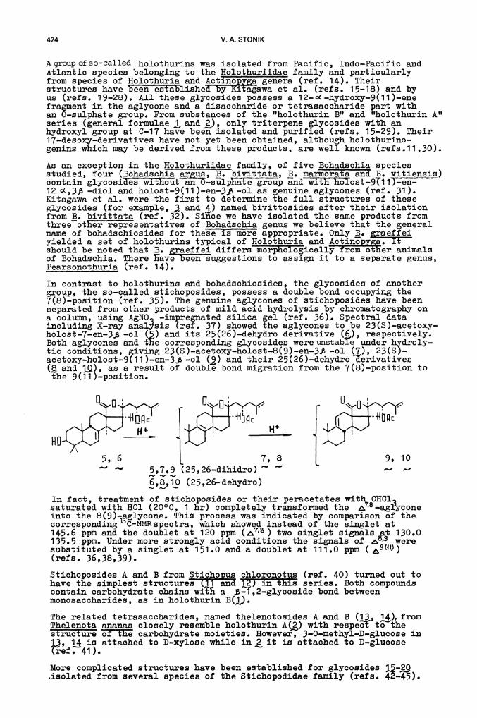
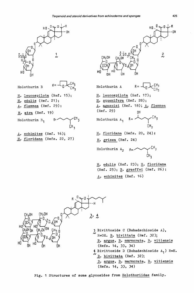
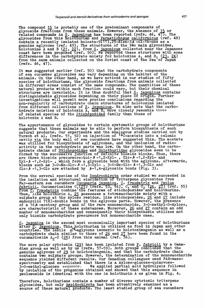
Agroupofso-called holothurins was isolated from Pacific, Indo—Pacific andAtlantic species belonging to the Holothuriidae family and particularlyfrom species of Holothuria and Actinopya genera (ref. 14). Theirstructures have in estalished by Ka.agawa et al. (reTh. 15-18) and byus (refs. 19—28). All these glycosides possess a 12...o(..hydroxy—9(11)-.enefragment in the aglycone and a disaccharide or tetrasaccharide part withan 0-sulphate group. Prom substances o± the "holothurin B" and "holothurin A"series (general formulae , and ), only triterpene glycosides with anhydroxyl group at C-17 have been isolated and purified (refs. 15.-29). Their17.-.desoxy-.derivatives have not yet been obtained, although holothurino-genins which may be derived from these products , are well known (refs . 1 1 , 30).
As an exception in the Holothuriidae family, o five Bohadschia speciesstudied, four (Bohadshia ais , B. bivittata, B. marmoraTa an1 B. vitiensis)contain glycosidés wit1iut an O...sulphat e group and with holost...9T1 1 ) -en—12 Q(,3 —diol and holost-.9(11)..en-.3,.ol as genuine aglycones (ref. 31).Kitagawa et al. were the first to determine the ±ull structures of theseglycosides (for example, and 4) named bivittosides after their isolationfrom B. bivittata (reL 32). Since we have isolated the same products fromthree other representatives of Bohadschia genus we believe that the generalname of bohadechiosides for these L mor appropriate. Only B. raeffeiyielded a set of holothurins typical of Holothuria and Actinopy&a. Itshould be noted that B. gaeffei differs morpThologically from other animalsof Bohadschia. There save been suggestions to assign it to a separate genus,Pearsonothuria (ref. 14).
In contrast to holothurins and bohadschiosides, the glycosides of anothergroup, the so—called stichoposides, possess a double bond occupying the7(8)—position (ref. 35). The genuine aglycones of stichoposides have beenseparated from other products of mild acid hydrolysis by chromatography ona column, using AgNO. —impregnated silica gel (ref. 36). Spectral dataincluding X—ray analsis (ref. 37) showed the aglycones to be 23(S)—acetoxy—holost—7—en—3, —ol (,) and its 25(26)-.dehydro derivative (i,), respectively.Both aglycones and the corresponding glycosides were unstable under hydroly-tic conditions, giving 23(S)_acetoxy—holost8(9)en—3j3 —ol (7), 23(5)—acetoxy—holost—9(1 1 )—en—3ft —ol (9) and their 25(26)—dehydro 'erivatives( and 1Q), as a result of double bond migration from the 7(8)—position tothe 9(11)—position.
In fact, treatment of stichoposides or their peracetates with CHCLsaturated with HC1 (20°C, 1 hr) completely transformed the 8—agryconeinto the 8(9)—glycone. This process was indicated by comparison of thecorresponding C-NMRspectra, which showed instead of the singlet at145.6 ppm and the doublet at 120 ppm (7) two singlet signals at 130.0135.5 ppm. Under more strongly acid conditions the signals of 8,8 weresubstituted by a singlet at 151 .0 and a doublet at 111 .0 ppm(refs. 36,38,39).
Stichoposides A and B from Stichppus chloronotus (ref. 40) turned out tohave the simplest structures Qj and i) in this series. Both compoundscontain carbohydrate chains with a $—1,2—glycoside bond betweenmonosaccharides, as in holothurin B().
The related tetrasaccharides, named thelenotosides A and B (13, 14), fromThelenota ananas closely resemble holothurin A(2) with respe' to thestructure of the carbohydrate moieties. However, 3—0—methyl—D—glucose in13, 14 is attached to D—xylose while in, it is attached to D-glucosetef. 41).
More complicated structures have been established for glycosides J—9.isolated from several species of the Stichopodidae family (refs. 42—45).
5, 6 I7, 8— 5,7,9 (25,26—dihidro)
6,8,10 (25,26-dehydro)
9, 10— d
Terpenoid and steroid derivatives from echinoderms and sponges 425
Holothurin B
H. leucospilota (Ref. 15);
H. edulis (Ref. 21);
A. flammea (Ref. 29);
H. atra (Ref. 19)
Holothurin B1 :'7LhLh3
A. echinites (Ref. 16);
H. floridana (Refs. 22, 27)
Holothurin A —J<3H. leucospilota (Ref. 17);
H. squamif era (Ref. 28);
A. gsizi (Ref. 18); A. flammea
(Ref. 29)au
HolothurinA1 [H3D
113
H. floridana (Ref s. 20, 24);
H. grisea (Ref. 24)
Holothurin A2
Eh3
H. eduli$ (Ref. 23); H. floridana
(Ref. 25); B. graeffei (Ref. 26);
A. echinites (Ref. 16)
3 Bivittoside C (Bohadschioside A),
R=OH. B. bivittata (Ref. 32);
B. argus, B. marmorata, B. vitiensis
(Refs. 14, 33, 34)4 Bivittoside D (Bohadschioside A ) R=H.
1
ft. bivittata (Ref. 32);
B. argus, B. marmorata, B. vitiensis
(Refs. 14, 33, 34)
Pig. 1 Structures of some glycosides from Holothuriidae family.
HO
OH
1
OH
HOOH
2"I
3, 4A, Pi
HO
426 V.A. STONIK
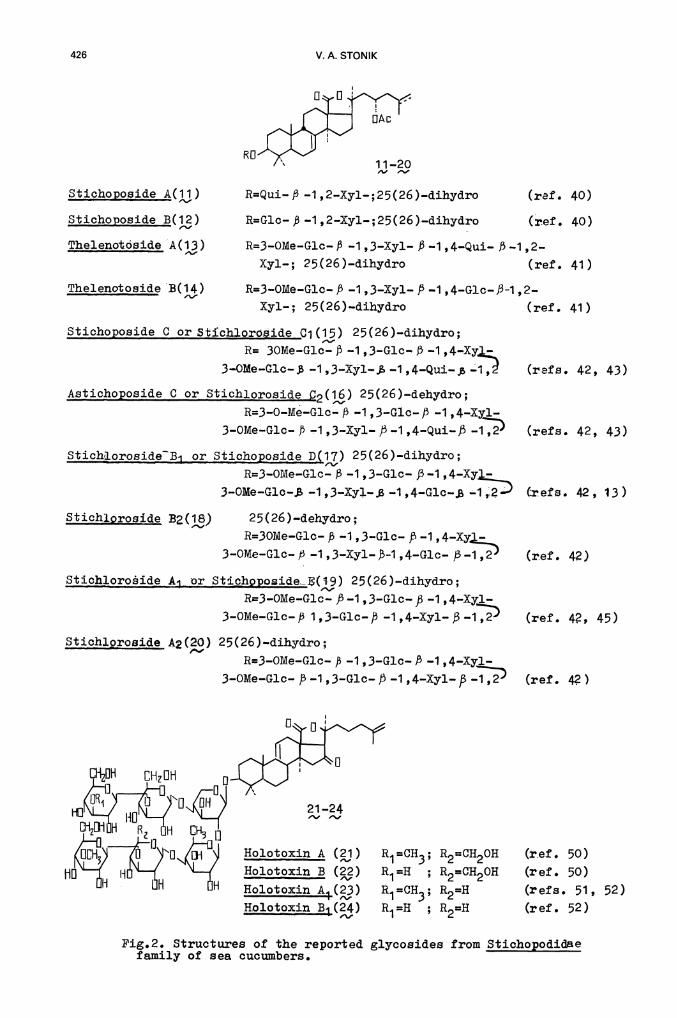
Thelenotôside A(13) R=3-OMe—G].c-. P .-1 ,3—Xyl— P .1 ,4-'Qui-' P .1 ,2..
Xy1-; 25(26 )—dihydro (ref. 41)
Thelenotoside B(14) R=3OMe-.G1c. P 1 ,3—Xyl-. /.-i ,4—Glc.-.J3-1 ,2.-.
Xyl-.; 25(26)—dihydro (ref. 41)
Stichoposide C or Stichioroside Ci (15) 25(26)-.dihydro;
R= 3OMe—Glc-. )3 —1 ,3—Glc— f3 —1 ,4—Xy
3—OMe—G1c— —1 ,3—Xy].—$ —1 ,4—Qui. —1,2! (refs. 42, 43)
Astichoppside C or Stichioroside j32(16) 25(26)—dehydro;
R=3—O—Me—Glc— B —1 ,3—G1c— —1 ,4—Xyl-.
3—OMe—Gic- 1 ,3—Xyl— )3 —1 ,4—Qui—j (refs. 42, 43)
StichLorosideB or Stichooside D(17) 25(26)—dihydro;
R=3—OMe-Glc— —1 ,3—Glc— 53—1 ,4—Xy3—OMe—G1c-, -1 ,3—Xy1... —1 ,4—G1c..$ —1 ,2 refs. 42, 13)
Stichlprosjde B2(18) 25(26)—dehydro;
R=3OMe—Glc- ,B —1 ,3—Glc— 53—i ,4—Xy —
3—OMe—Gic— 53 —1 ,3—Xy1—-i ,4—Glc— 53—i ,2 (ref. 42)
Stic1oroidA1_or Stichpposidei(i9) 25(26)—dihydro;R=3—OMe—Glc— —1 ,3—Gic— 53 —1 ,4.-Xyl-.
3...OMe..Glc...53 1,3—Glc—/3 —1,4—Xyl—)3—1,2 (ref. 42, 45)
Stichioroside A2(•20) 25(26)—dihydro;
R=3—OMe—Glc-- ft —1 ,3—Glc— P —1 ,4—Xyl—
3—OMe—Gic— 5-3 -.1 ,3.-Glc—J5 —1 ,4—Xy1— —1,2
CHzDH GHDH
21—24, ,o1m H E5H D13
Holotoxin A ()HolotoxinB (3)Ho1otoxi4(23)Holotoxin B(24)
Pig.2. Structures of the reported glycosides from Stichopodidaefamily of sea cucumbers.
11—20, ,Stichoposide A(1i) R=Qui—J3 —i ,2—Xyl—;25(26)—dihydro
Stichoposide B(12) R=Glc— 53 —i ,2—Xyl—;25(26)—dihydro
(ref. 40)
(ref. 40)
R1=CH3; R2=CH2OH (ref. 50)
R1H ; R2=CH2OH (ref. 50)
R1 =0H3;
R1H ;R2=H
R2=H
(r efs.
(ref.
51,
52)
52)
Terpenoid and steroid derivatives from echinoderms and sponges 427
The compound is probably one o the predominant components ofglycoside fractions from these animals. However, the absence of j, orrelated compounds in S. lappnicus has been reported (refs. 46, 47). Theglycosides from this olothurian and Parastichopis californicus (ref. 48)contain a recently isolated holosta.-'9(11i,2—dten—38 cl-.16.-one as agenuine aglycone (ref. 49). The structures of the two main glycosides,holotoxins A and B (j, 22), from S. jponicus collected near the Japanesecoast have been proposed'lref. 50). We reported these structures with somedifferences in the carbohydrate moiety for holotoxins A1 and B1 (,)from the same animals collected on the Soviet coast of he Sea of Japan(refs. 46, 47).
It was suggested earlier (ref. 50) that the carbohydrate componentsof sea cucumber glycosides may vary depending on the habitat of theanimals. On the other hand, as we have noticed in our studies of fiftyspecies of holothurians, the glycoside fractions from animals collectedin different areas consist of the same compounds. The quantities of thesenatural products within each fraction could vary, but their chemicalstructures are invariable. It is thus doubtful that S. jaonicus containsdistinguishable glycosides depending on their place of origin. Purtherinvestigations will permit more precise conclusions regarding thenon—regularity of carbohydrate chain structures of holotoxins isolatedfrom different collections of S. jponicus. We also note that the carbo—hydrate moieties of holotoxin A1 anUB1 more closely resemble thoseof related species of the Stiohópodidaê family than those ofholotoxin A and B.
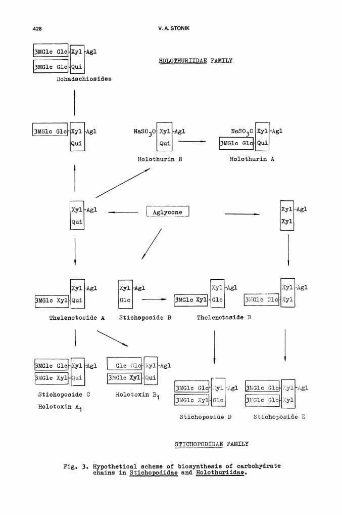
The appurtenance of glycosides to certain systematic groups of holothurianssuggests that these animals may be able to perform biosynthesis of suchnatural products. Our experiments and the analogous studies carried out byTursch et al. (refs. 51, 52) on injection of 14C—acetate into a celomiccavity xtQ the body wall of sea cucumbers have suggested this. The acetatewas utilized for biosynthesis of aglycones, and the inclusion of radio-Sactivity in the carbohydrate parts was low. On the other hand, the carbo—hydrate chains of Stichqpodidae and Holothuriidae glycosides seem to beformed by the successive junôELon of dèfiniteioside blocks. Thus, thereare three bioside precursors:Qui—.$—1,2—Xyl— , Glc-1,2—Xy1— andXyl— —1,2—Xyl- , which form a glycoside bond with the aglycone. Afterwards,blocks such as 3—O—Me—Glc—$—1,3—Xyl— , 3—O—Me—Glc—$—1,3—Glc andGlc —1,3—Glc are attached by $—1,4—glycoside bonds (Pig. 3).
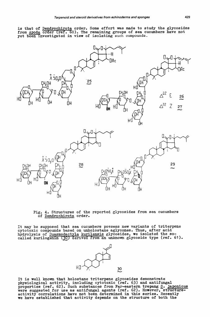
From the several species of the Dendrochirota order studied we succeeded inthe isolation and structural elucidation of triterpene glycosides fromFar-eastern sea cucumbers Cucuxnaria iaponica, C. fraudatrix and Psolusfabricii. Cucumariosides Ci(25) tfs. 53, 54), C. an 02 ' i) ref. 55)from C. fraudatrix combine tire features of stichoosides and holothurins.Thus, like holothurin A, possesses a tetrasaccharide moiety with asulphate group at the xylose residue. Like stichoposides, and includeendocyclic 7(8)—double bonds in the aglycone parts. However, the presenceof a 16.g, —acetoxy group and of the rare monosaccharide, 3—O-methyl—D—xy].ose,are characteristic of these substances. Moreover, and contain an oddnumber of monosaccharides and consequently their biosynthesis utilizes notonly bioside carbohydrate precursors but monosaccharide ones.
C. ponica is the second most economically important species of holothuriansafter aponicus. This holothurian is utilized as food in Japan and othercountries. The labile T—aglycone isomeric to holotoxinogenin as well as acarbohydrate chain similar to those of and have been found in the
cucumarioside A2—2 () from this animal (ref. 56).
The more polar cytotoxin () has been isolated from P. fabricii by a Cana-dian group as well as by us (ref s. 57—59). Both groups concIuded that thegenuine aglycone of is holotoxinogenin, and that the carbohydrate chaincontains two sulphate groups. However, the determination of the monosaccharidesequence yielded different results. Our Canadian colleagues used FAB—mass—spectrometry and established that there is a xylose—glucose—quinovose—3—0—methyl—glucose sequence. We applied partial acid hydrolysis followedby, isolation of the progenins obtained and showed that this sequence inpsolusoside is identical with the one in holothurin A as given in Pig. 4.
Therefore, holothurians contain a number of diverse cytotoxic triterpeneglycosides, but only Aspidochirota has been attentively examined as asource of these natural products. The least studied group of sea' cucumbers
428 V.A. STONIK
HOLOTHURIIDAE FAMILY
NaSO3O XylAl NaSO3O [iJAl[ GlcuiHolothurin B Holothurin A
_________i1Agl [ij-Agl _________ [i-AglL3MG1c XY}[j [j — - [ic
Thelenotoside A
13tG1c G1c Xyi -Agi
[G1c G1cXy1
Stichoposide B
STICHOPODIDAE FAMILY
Fig. 3. Hypothetical scheme of biosynthesis of carbohydratechains in Stichopodidae and Holothuriidae.
3MG1c G]J Xyl Agi
MGlc Gici QuiBohads chios ides
(3MG1c G1ciAgl
iAglL±i
Lco _1
/[iAg1LJ
X3r1 Agl
[MGJ.c Xyl
Thelenotoside BStichoposide B
MGlc Glc-1i1-Ag]. Gic G1c-Iy1-Ag1
Glc J-j [3MG10
13MG1cj1g1Stichoposide C Holotoxin B1
Holotoxin A1
Stichoposide B
Terpenoid and steroid derivatives from echinoderms and sponges 429
is that of Dendroohirota order. Some effort was made to study the glycosidesfrom poda order (ref. 60). The remaining groups of sea cucumbers have notyet beeiflnvestigated in view of isolating such compounds.
H
[H OH
HöHHH
Fig. 4. Structures of the reported glycosides from sea cucumbersof Dendrochirota order.
It may be supposed that sea cucumbers possess new variants of triterpenecytotoxic compounds based on unholostane aglycones. Thus, after acidhydrolysis of Duasmodact kurilensis glycosides, we isolated the so—called kurilogenin derived from an unknown glycoside type (ref. 61).
30
It is well known that holostane triterpene glycosides demonstratephysiological activity, including cytotoxic (ref. 63) and antifungalproperties (ref. 62). Such substances from Far—eastern trepang S. japonicuswere suggested for use as antifungal agents (ref. 62). However, structure—activity correlations have not been determined in this series. Recentlywe have established that activity depends on the structure of both the
X5125
[ 26.
7 27
28 29
430 VA. STONIK
aglycone and carbohydrate parts of the glycosides (refs. 64, 65). Thus,25(26)-.dihydroholotoxin A possesses strong antifungal action, but thisproperty is absent in 3—su].phoxy...25(26)—dihydroho1otoxinogenin havingsimilar polarity. Comparison of the activity o stichoposides havingidentical aglycones but dirferent carbohydrate moieties showed: 1) thestronger action of tetrasaccharides than that of hexasaccharides andespecially o disaccharides ; 2) the high activity of quinovose.-containingglycosides in the series o analogous products (ref. 65). Physiologicalaction of holothurins is a function of the side chain structure of theiraglycones. The compounds with open side chains are often more active.
The antifungal properties and other aspects of the physiological activityof these metabolites oC sea cucumbers are probably dependent on theglycoside's ability to form complexes with membrane sterols. As a con—sequence, these natural products may serve as instruments or studying therole of sterols in biological membranes (ref. 66). The ability of lowconcentrations of these substances to hinder the development of fertilizedeggs of sea urchins shows the probable participation of sea cucumberglycosides in interspecies interactions.
STEROID GLYCOSIDES FROM ECHINODERMS
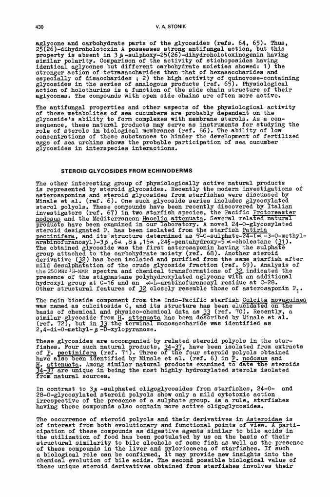
The other interesting group of physiologically active natural productsis represented by steroid glycosides. Recently the modern investigations ofasterosaponins and steroid glycosides from starfishes were discussed byMinale et al. (ref. 6). One such glycoside series includes glycosylatedsterol polyols. These compounds have been recently discovered by Italianinvestigators (ref. 67) in two starfish species, the Pacific Protoreasternodosus and the Mediterranean Hacelia attenuata. Several related naturaloducts have been examined in our laboratory. A novel 24—0—glycosylatedsteroid designated P1 has been isolated from the starfish Patinapectinifera, and its structure determined as 5-O—sulphate—24—(o&3—O—methyl-'arabinofuranosyl)—3 j , ,8 ,1 5o ,24S—pentahydroxy—5 o(—cholestane (31).The obtained glycoside was the first asterosaponin having the sulphategroup attached to the carbohydrate moiety (ref. 68). Another steroidderivative (32) has been isolated and purified from the same starfish aftermild desulphation of the crude glycoside fraction (ref. 69). Analysis ofthe 250MHz 'H-NMR spectra and chemical transformations of . indicated thepresence of the stigmastane polyhydroxylated aglycone with an additionalhydroxyl group at C-16 and an —L—arabinofuranosyl residue at O...28.Other structural features of closely resemble those of asterosaponin P1.
The main bioside component from the Indo—Pacific starfish Culcita novaguineawas named as culcitoside C, and its structure has been elucidated on thebasis of chemical and physico—chemical data as (ref. 70). Recently, asimilar glycoside from H. attenuata has been described by Minale et al.(ref. 72), but in the terminal monosaccharide was identified as2 ,4—di—O-methyl— -D-xylopyranose.
These glycosides are accompanied by related steroid polyols in the star—fishes. Four such natural products, have been isolated from extractsof P. pectillifera (ref. 71). Three of the four steroid polyols obtainedhave also beeThentified by Minale et al. (ref. 6) in P. nodosus andH. attenuaj. Among similar natural products examined to date the steroids
are unique in being the most highly hydroxylated sterols isolatedfrom natural sources.
In contrast to 3 —sulphated oligoglycosides from starfishes, 24—0— and28—0—glycosylated steroid polyols show only a mild cytotoxic actionirrespective of the presence of a sulphate group. As a rule, starfisheshaving these compounds also contain more active oligoglycosides.
The occurrence of steroid polyols and their derivatives in Asteroidae isof interest from both evolutionary and functional points of view. A parti-cipation of these compounds as digestive agents similar to bile acids inthe utilization of food has been postulated by us on the basis of theirstructural similarity to bile alcohols of some fish as well as the presenceof these compounds in the liver and pyloniccaeca of starfishes. If sucha biological role can be confirmed, it may provide new insights into thechemical evolution of bile acids. The second possible biological value ofthese unique steroid derivatives obtained from starfishes involves their
() R1=SO3Na, R2=CH3, R3=H x=(CH2)0
() R1=R=H, R3=OH, x=(CH2)2
22(23)—dehydro, R1=H, CH3, OH2, C2H5
R2=CH(CH3)2
22(23)—dihydro, R1=H, OH3, OH2, 02H5
R2=CH(0H3)2
HO -
OH
31—32OH
OH
i-il]
Terpenoid and steroid derivatives from echinoderms and sponges 431
HO
R1 =R2=R3=H, R4= K-OHR1=R2=H, R3=OH, R4= K—OHR1=R3=OH, R2=H, R4= K—OH
R1=R3=OH, R2=SO3Na, R4= ft—OH
33
HO-F3
()()()()34—37
38 39
432 V.A. STONIK
Thrxnation as products of sterol catabolism in these animals, necessary toexpel any excess o sterols.
The diversity o± steroid glycosides in echinoderms does not limit itse]i toasterosaponins. We have studied .xylosides (general Thrmulae 38 and 9)from sea cucumbers (reTh. 73, 74). The aglycone compounds of these natural.products turned out to be stanols , aY , and A -sterols . However,-xylosides as well as 5sterol sulphates isolated from echinoderms(ref. 75) are not cytotoxins.
These natural compounds, being architecturally membrane constituents, areassociated with the membrane phospholipid bilayers, and contribute to thestabilization of the bilayer phase (refs. 77, 76).
SULPHATED POLYHYDROXYLATED STEROIDS FROM SPONGES
Steroids with two or tZtree sulphated hydroxyl groups in the conventionalC1 tetracyclic nucleus have been isolated from tropical species ofHèiichondriidae sponges in the last years. Halistanol sulphate (9)describeUby Japanese scientists (ref. 78), sokotrasterol sulphate (j) andthe desuiphated derivative (4) obtained by us (refs. 79,80) all possesscytotoxic action and induce srong foaming in aqueous solutions, similarto triterpene and steroid oligoglycosides from echinoderms.
a5U3U
Na5O3U0 Na5U3U
Na593U Na59 UiUNa40 41 3
A')
The physiological activity of the sulphated steroids from sponges as well asof the triterpene glycosides from sea cucumbers is associated with theformation of pores on the biological membranes. In contrast withholothurins, sponge metabolites do not decrease their membranolyCic abilityin the presence of cholesterol. At the same time, a diminished action oncell membranes was observed for halistanol sulphate or sokotrasterolsulphate when bovine serum albumin was added to the corresponding medium.It may be suggested that some cytotoxic effects of these substances arisefrom their interactions with protein membrane components.
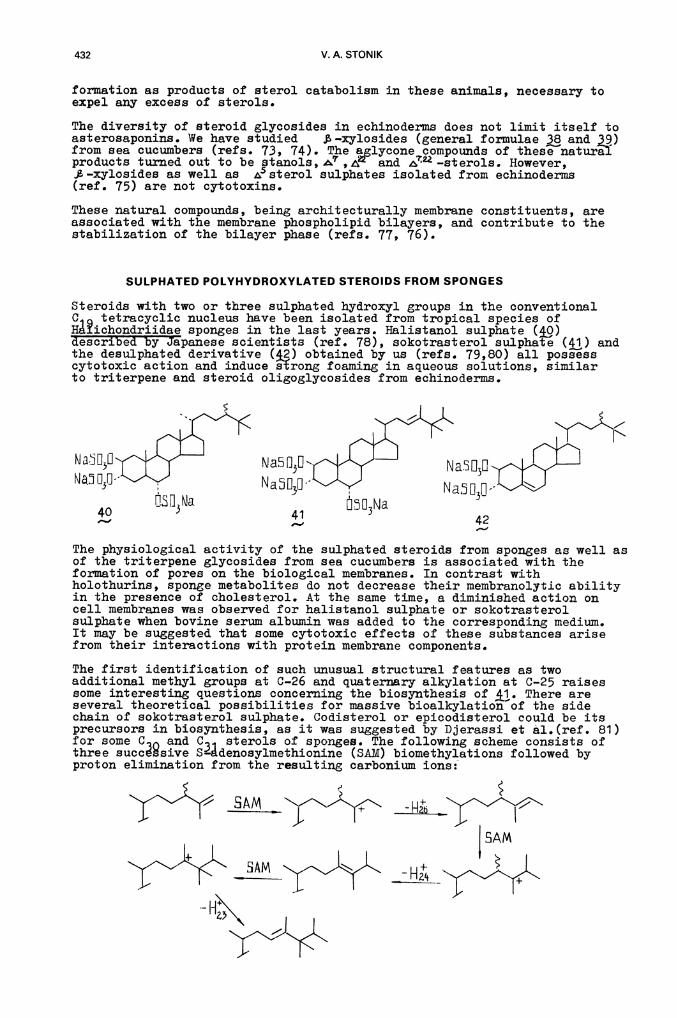
The first identification of such unusual structural features as twoadditional methyl groups at C-.26 and quaternary alkylation at C-25 raisessome interesting questions concerning the biosynthesis of j,. There areseveral theoretical possibilities for massive bioalkylation of the sidechain of sokotrasterol sulphate. Codisterol or epicodisterol could be itsprecursors in biosynthesis, as it was suggested by Djerassi et al.(ref. 81)for some C. and C1 sterols of sponges. The following scheme consists ofthree succsive Sádenosylmethionine (SAM) biomethylations followed byproton elimination from the resulting carbonium ions:
SAM
Terpenoid and steroid derivatives from echinoderms and sponges 433
CORRELATION BETWEEN THE PRESENCE OF CYTOTOXINS AND THE
STEROL COMPOSITION OF BIOMEMBRANES
It is known that the presence of cytotoxic agents in plants and animals issometimes bound with biochemical alterations as compared with the relatednon-toxic species. Such alterations o sterol compositions of biomembranesmay be seen in echinoderms and sponges containing either glycosides orpolysuiphated steroids.
Echinoderms and sponges are probably characterized by the most availableset of sterols, even compared with all other terrestrial orjnarineinvertebrates. Indeed,besides A sterol, stanols, , , ,2't(28) sterolsexist in sea cucumbers and starfishes aa membrane constituents (ref. 82).In other echinoderms having no cytotoxic glycosides, cholesterol is thepredominant sterol. Moreover, sea cucumbers show very low contents of freesterols and high contents of sterol sulphates and ,8-.xylosides (ref. 77).It should be noted that such steroid derivatives as stanols, —sterols,%$-.xylosides or sterol suiphates form complexes with cytotoxic glycosidesfrom echinoderms with greater difficulty than cholesterol does or else theydor not form complexes at all (ref. 77).
Therefore, the stability of echinoderm cells to the action of their owncytotoxins results from : 1) the low concentration of free sterols inbiomembraies and 2) the absence of sterols being as sensitive to glycosidesas are is—derivatives in these membranes.
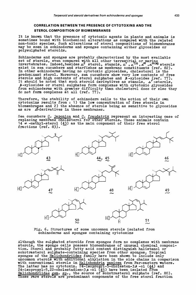
Sea cucumbers . iaponica and . fraudatrix represent an interesting case ofreplacing membrane c1bIesterol for other sterols. These animals contain14 o( —methyl—sterol (43) as the main component of their free sterolfractions (ref. 83).
ND
Pig. 6. Structures of some uncommon sterols isolated fromechinoderms and sponges containing cytotoxins
Although the sulphated steroids from sponges form no complexes with membranesterols, the sponge cells possess biomembranes of unusual chemical composi-tion. Sterol and probably fatty acid content distinguish halistanol orsokotrasterol sulphate—containing species from other sponges. Tropicalsponges of the Halichondriidae family have been shown to include onlyuncommon sterols wth additional alkylation in the side chains in comparisonwith conventional sterols in Halichondria anicea from Par—eastern waters.The latter has no cytotoxin. —ol (j) and24—isopropyl—5,22—cholestadien—3 —ol () have been isolated fromHalichondriidae . ., the source of sokotrasterol sulphate (ref. .80).These rare steroIare predominant components of the free sterol fraction
47 48
50
434 V. A. STONIK
Trom the sponge. Cholesterol and other conventional sterols have not beenidentified in this animal. Halichondria contains halistanol sulphateand a novel uncommon sterol, 25—methy124ethy1.-5,22—cholestadien-.3j3 —ol
(&€) as a sole sterol component. A series o unique sterols has been roundin the extracts o Halichondria containing the same cytotoxin. Allnatural products mentione (47—5TJ possess side chains with uncommonalkylation patterns as compared with conventional sterols (re±. 84, 85).
It is probable that biosynthesis o± cytotoxic sulphated polyhydroxylatedsteroids and the presence of uncommon sterols are connected with specificmicroflora of the tropical Halichondriidae species.
Acknowledgement The author thanks Prof. G.B. Elyakov for fruitfuldiscussions.
REFERENCES
1. R.F. Nigrelli, Zoologca, 37, 89.-90 (1952).2. T. Yamanouchi, Bull. Seo Marine Biol. Lab., , 184—202 (1952).3. T. Yasumoto, T. Watanabe aifY. HaskLmoto, Bu)l. Jap. Soc. Scient. Fish.,
30, 357—364 (1964).4. w: Bergrriann and D.C. Burke, J. Org. Chem., 21, 226—228 (1956).5. W. Bergmann and R.J. Feenky, J. Am. ChéIIi Soc., 20, 15O1—1507 (1955).6 • L. Minale, C. Pizza , R. Riccio an P. Zollo, Pure and Appl. Chem. ,
1935—195O (1982).7. Y. Hashimoto, Marine Toxins and Other Bioactive Marine Metabolites,
pp. 266—288, Japan Scientific Societies Press, Tokyo (1979).8. L. Minale, G. Cimino, S. De Ste±ano and G. Sodano, Fortschritte Chem.
Or . Naturst., , 1—27 (1976).9. . . S'ieuer, Chemistry of Marine Natural Products, pp 36—44,
Academic Press, New York c19T3).10. G. Habermehi and G. Volkwein, Toxicon, 9, 319—326 (1971).11 • G.B. Elyakov, V.A. Stonik, B.V. Leviña, V.P. Slanke, T.A. Kuznetsova
and V.5. Levin, Coma. Biochem. Physiol., 44, 325—336 (1973).12. G.B. Elyakov, }üzneta, VA. SlonIF!' V.5.Levin andR. Allores,
Corn. Biochem. Physiol., 52, 413—417 (19755.13. V.A. 1aTLtsev, A.I. Kalinovsky and G.B. Elyakov, Khim.
Prir. Soedin., 200—203 (1982).14. T.S. Levin, V.1. Kalinin and V.A. Stonik, Biologia moija, 33—38
(1984).15. I. Kitagawa, T. Nishino, T. Matsuno, H. Akatsu and Y. Kyogoku,
Tetrahedron Lett., 985—988 (1978).16.1. Kagawa, T. Inamoto, M. Fushida, S. Okada, M. Kobayashi, T. Nishino
and Y. Kyogoku, Chem. Pharm. Bull. (Tokyo), 28, 1651—1653 (1980).17. I. Kitagawa, T. ishino aY. Kyogoku,etredron Lett., 1419—1422
(1979).18. I. Kitagawa, M. Kobayashi and Y. Kyogoku, Chem. Pharm. Bull. (Tokyo),
Q, 2045—2050 (1982).19. VA. Stonik, A.D. Chuinak, V.V. Isakov, N.I. Belogortseva, V.Ia. Chirva
and G.B. Elyakov, Khim. Prir. Soedin., 522—527 (1979).20. G.K. Oleynikova, T.A. uznetsova, A.I. Kalinovsky, V.A. Stonik and
G,B, Elyakov, Khim. Prir. Soedin., 101—103 (1981).21. V.1. Kalinin, VA. Stonik, LA. Avilov and G.B. Elyakov, Khim.Prir.
Soedin., 403—404 (1981).22. B. Elyakov, N.I. Kalinovskaya, A.I. Kalinovsky, V.A. Stonik and
T.A. Kuznetsova, Khim. Prir. Soedin., 323—327 (1982).23. V.1. Kalinin and V.A. Stonik, Khim. Prir. Soedin., 215—219 (1982).24. G.K. Oleynikova, T.A. Kuznetsova, N.S. Ivailova, A.I. Kalinovsky,
N.V. Rovnih and G.B. Elyakov, Khim. Prir. Soedin., 464-469 (1982).25. G.K. Oleynikova, T.A. Kuznetsovi,.V. Rbvnih, A.I. Kalinovsky and
G.B. Elyakov, Khim. Prir. Soedin., 527—528 (1982).26. V.1. Kalinin anfV'A. tonik, Khim. Prir. Soedin., 789 (1982).27. T.A. Kuznetsova, N.I. Kalinovskaya, A.I. Kalinovsky, G.K. Oleynikova,
N.V. Rovnih and G.B. Elyakov, jim. Prir. Soedin., 482—484 (1982).28. N.S • Ivanova, 0.P. Smetanina and7L'.A. Kuznetsova, Khim. Prir. Soedin.,
448—451 (1984).29. 5. Bbatnagar, A. Ahond, B. Dudoaet, C. Poupat and P. Potier, Fourth
International Symposium on Marie Natural Products, Tenerife, Spain,P—b, (1982).
Terpenoid and steroid derivatives from echinoderms and sponges 435
30. J.D. Chanley, T. Mezzetti and H. Sobotka, Tetrahedron, 22, 1857-1864(1966).
31. V.A. Stonik, V.P. Sharypov, T.A. Kuznetsova, A.I. Kuznetsova andG.B. Elyakov, Khim. Prir. Soedin., 790 (1982).
32. I. Kitagawa, I1ayashi, Tt. 1Iri and Y. Kyogoku, Chem. Pharm. Bull.(Tokyo, 29, 282—285 (1981).
33. A.I. Ialinovsky, 1.1. Maltsev, A.S. Antonov and V.A. Stonik,Bioorg. Khim., 10, 1655—1663 (1984).
34. A. Clastres, A.lhond, C. Poupat and A. Intes, Experientia, 34,973.-974 (1978).
35. V.A. Stonik, V.P. Sharypov, A.I. Kalinovsky and G.B. Elyakov,Doki. Akad. Nauk SSSR, 245, 1'133-.1134 (1979).
36. G.B. Elya1ov, V.A, StonT1 Sh.Sh. Afiatullov, A.I. Kalinovsky,V.P. Sharypov and L.Ja. Korotkih, Doki. Akad. Nauk SSSR., 259,1367—1369 (1981).
37. S.G. Ilyin, V.P. Sharypov, V.A. Stonik, G.V. Malinovskaya, N.I. Uvarovaand G.B. Elyakov, Structure and properties oi° polyrnetallic andmetalloranic compounds, Abst±aets, Chernogolovica, UpSR, 145 (1981).
38. A.L 1(alinovsky, V.P Sharypov, V.A. Stonik A.K. Dzizenko andG. B. Elyakov, Bioop. Khirn. , 6, 86—89 (1980S.
39. A.I. Kalinovsky, V.?. Sharypov, Sh.Sh. Afiatullov, T.A. Kuznetsova,V.A. Stonik and G.B. Elyakov, Bioorg. Khim., 9, 1558-1563 (1983).
40. V.P. Sharypov, A.D. Churnak, V.A. Stonik and G.B. Elyakov, Khim. Prir.Soedin., 181—184 (1981).
41. V.A. Stonik, 1.1. Maltsev and G.E. Elyakov, Khim. Prir. Soedin.,624—627 (1982).
42. I. Kitagawa, M. Kobayashi, T. Inamoto, T. Yasuzawa, Y. Kyogoku andM. Kido, Chem. Pharm. Bull. (Tolçyo), j, 1189-1192 (1981).
43. V.A. Stonik, LI. Maltsev, A.I. ICalinovaky, K. Konde and G.B. Elyakov,Khim. Prir. Soedin., 194—199 (1982).
44. V.Atonik, 1.1. Maltsev, A.I. Kalinovsky and G.B. Elyakov,Pirat international conference on chemistry and biotechnology ofbioIiafiy afive nat ural products, 3, 326-39 (1981).
45. 1.1. MaltsevV.A. Stonik and A.IXalThovsky, Khim. Prir. Soedin.,308—312 (1983).
46. G.B. Elyakov, 1.1. Maltsev, A.I. Kalinovsky and V.A. Stonik,Bioorg. Khim., 9, 280—281 (1983).
47. 1.1. V.A. Stonik, A.I. Kalinovaky and G.B. Elyakov,Biochem. Physiol., 78B, 421—426 (1984).
48. Y.M. Sheikh, C. Djerassi, J. C. S. Chem. Comm., 1057—1058 (1976).49. V.P. Sharypov, N.I. Kalinovikaya, V.A. Stonfk and G.B. Elyakov,
Khim. Prir. Soedin., 845—846 (1980).50. I. Kitagawa, H. imanaka, M. Kobayashi T. Kishino, I. Yosioka and
T. Sugawara, Chem._Phprm. Bull. (Toky, 26, 3722—3731 (1978).51. G.B. Blyakov, V.A. Stonik, E.V. Levina anV.S. Levin, Comp. Biochem.
Physiol., 52B, 321—323 (1975).52. A. KeIecom7. Dalcize and B. Tursch, Tetrahedron, 32, 2353—2358 (1976).53. Sh.Sh. Afiatullov, V.A. Stonik and G.. flya1cov, Khim. Prir. Soedin.,
56—64 (1983).54. Sh.Sh. Afiatullov, V.A. Stonik and G.B. Elyakov, Khim. Prir. Soedin.,
59—64 (1983).55. Sh.Sh. Afiatullov, A.I. Kalinovsky, V.A. Stonik and G.B. Elyakov,
Khim. Prir. Soedin., (in press).56. S.X. Avilov, L.Ja. Tiechenko and V.A. Stonik, IQiim. Prir. Soedin.,
799—800 (1984).57. P.—X. Garneau, J.-L. Simard, 0. Harvey, J.W. Ap Simon. and M. Girard,
Can. J. Chem., 61, 1465—1471 (1983).58. V.A. Xa1ixitn, VT. Stepanov and V.A. Stonik, Khim. Prir. Soedin.,
789—790 (1983).59. V.A. Kalinin, A.I. Kalinovsky and V.A. Stonik, Khim. Prir. Soedin.,
212—217 (1985).60. T.A. Kuznetsova, N.I. Kalinovskaya, A.I. Kalinovsky and G.B. Elyakov,
Khim. Prir. Soedin., (in press)..61. AI. Kalinovsky, S.A. Avilov, V.R. Stepanov and V.A. Stonik, Khirn.
Prir. Soedin., 724—727 (1983).62. Shimada, Science, 163, 1462—1464 (1969).63. M.M. Anisimov, If. G. PTcofieva, L. Ja. Korotkih, I • I. Kapustina and
V.A. Stonik, Toxicon, 18, 221—223 (1980).64. T.A. Kuztietsoira., M.M. Anisimov, A.M. Popov, S •I• Baranova,
Sh.Sh. Afiatullov, 1.1. Kapustina, S.A. Antonov and G.B. Elyakov,Comp. Biochem. Physiol., , 41—43 (1982).
65. 1.1. Maltsev, S.1. Stechova, B.B. Shentsova, M.M. Anisimov andG.B. Elyakov, Khim. Pharm. Zhurn., 54—56 (1985).
436 V. A. STONIK
66. M.M. Anisimov, E.B. Shentsova, V.V. Scheglov, Ju.N. Shumilov,V.A. Rasskazov, L.I. Strigina, N.S. Chetyrina and G.B. Elyakov,Toxicon, 207—218 (1978).
67. R. Riccio,E. Minale, C. Pizza, P. Zollo and J. Pusset, TetrahedronLett., 23, 2899—2902 (1982).
68. T Kia, A.I. Kalinovsky, E.V. Levina, V.A. Stonik and G.B. Elyakov,Tetrahedron Lett., 24, 3893—3896 (1983).
69. A.A.K[fta, A.I. Kainovsky, E.V. Levina, Ja.V. Raschkes, V.A. Stonikand G.B. Elyakov, Khim. Prir. Soedin., (in press).
70. A.A. Kicha, A.I. Kalinovsky, LV. Levina, P.V. Andriyaschenko,Khirn. Prir. Soedin., (in press).
71. LL Xicha, A. I. Kalinovsky, E.V. Levina, V.A. Stonik and G.B. Elyakov,Bioog. Khim., 9, 975—977 (1983).
72. P Riccio, L. Minale, S. Pagonis, C. Pizza and J. Pusset, Tetrahedron,38, 3615—3622 (1982).
73. B. Elyakov, N.I. Kalinovskaya, V.A. Stonik and T.A. Kuznetsova,Comp. Biochern. Physiol., 65B, 309—314 (1979).
74. G.B Elyakov, T.A. Kuznetsova, K. Konde, N.I. Kalinovskaya and0.P. Smetanina, Khim. Prir. Soedin., 799—802 (1979).
75. G.B. Elyakov, S.N. Pedoro, A.. Ohuinak, V.V. Isakov and V.A. Stonik,Comp. Biochem. Physiol., 71B, 325—328 (1982).
76. A.M. Popov, K.I. KaIinovsa, T.A. Kuznetsova, I.G. Agafonova andM.M. Anisimov, Antibiotiki, 656—659 (1983).
77. M.M. Anisimov, D.LAminin, Yu.G. Rovin, G.N. Lichatskaya, A.M. Popov,T.A. Kuznetsova, N.I. Kalinovskaya and G.B. E].yakov, Dok].. Akad. NaukSSSR, 270, 991—993 (1983).
78. ustni, S. Matsunaga and S. Konosu, Tetrahedron Lett., 22,1985—1988 (1981).
79. T.N. Makarieva, L.K. Shubina, A.I. Kalinovsky, V.A. Stonik andG.B. Elyakov, Steroids, 43, 267—292 (1983).
80. T.N. Makarieva, L.1Chu15Tna A.I. Kalinovsky and V.A. Stonik,Ichim. Prir. Soedin., 272—273 (1985).
81. 0. DjerassI, fure and Appl. Chem., 53, 873—890 (1981).82. N.I. Kalinovskaya, T.A. Xuznetsova and G.B. Elyakov, Conp. Biochem.
Physiol., 597—601 (1983).83. N.I. Xalinovskaya, A.I. Kalinovsky, T.A. Kuznetsova, V.A. Stonik and
G.B. Elyakov, Doki. Akad. Nauk SSSR, 278, 630—634 (1984).84 • L•K. Shubina, TU. Makarieva and T. A.Eonik, Khirn. Prir. Soedin.,
464—467 (1984).85. L.K. Shubina, T.N. Makarieva, A.I. Kalinovsky and V.A. Stonik,
Khim. Prir. Soedin., 232—239 (1985).