Some methods in fish immunology and protein chemistry Methods.pdf · 1 Some methods in fish...
69
1 Some methods in fish immunology and protein chemistry Bergljót Magnadóttir 2012 http://www.hi.is/gadus SAP IgG ApoLP C3 These methods have been routinely used by me for many years. Some mistakes may have been introduced in this write-up without my noticing and this method book is published here without liability.
Transcript of Some methods in fish immunology and protein chemistry Methods.pdf · 1 Some methods in fish...

1
Some methods in fish immunology
and protein chemistry
Bergljót Magnadóttir 2012 http://www.hi.is/gadus
SAP IgG
ApoLP
C3
These methods have been routinely used by me for many years.
Some mistakes may have been introduced in this write-up without my noticing
and this method book is published here without liability.

2
Content Page
1. Standard ELISA: The antibody activity of cod serum
2. Competitive ELISA for quantitative analysis of IgM in serum
3. ELISA for quantitative analysis of IgM in serum (Pilstöm’s method)
4. SCN - ELISA: Antibody affinity
5. ELISA: Antibody (anti-cod IgM) titration
6. Capture ELISA for CRP PI & PII quantity measurements
7. ELISA: Cortisol in serum – a kit from Neogen
8. Fish serum or plasma collection
9. Fish IgM purification
10. Crude preparation of (fish) IgM
11. Purification of cod pentraxins, CRP-PI and CRP-PII
12. Apolipoprotein A1 isolation from cod serum
13. Isolation of fish C3 complement component
14. C3 isolation for immunization using Macrogard
15. Production of polyclonal antibody in mouse ascites
16. Immunoglobulin isolation from mouse ascites
17. Biotinylation of Antibodies (Ig’s) and other proteins
18. Protein coupling to NHS-activated columns, ligand binding and elution
19. Proteolytic digestion of Ig’s (fish IgM)
20. Preparation of organ/larvae protein suspension
21. Sponatneaous haemolytic assay
22. Bactericidal activity
23. Anti-trypsin activity of fish serum
24. Lysozyme assay
25. Dot blot
26. Enzyme SDS-PAGE for cod larvae lysate
27. Thin (3.5%) SDS-PAGE
28. Destaining of silver stained acrylamide gels
29. Bradford method for protein assay
30. Protein concentration at 280 mµ
31. Ammonium sulphate & organic solvent protein precipitation
32. Protein isolation from gel for amino acid sequence analysis or immunization
33. Preparation of haptenated protein
34. Titration of NaCl concentration
35. Detection of sulphate in dialysis
36. Immunohistochemistry using a kit from Dako
37. Two dimensional (2D) electrophoresis
38. Glycoprotein detection by Western blotting
39. Routine bath challenge test for cod larvae/fry
40. Dehert’s stress test
41. Buffer preparations
3
5
7
9
11
13
15
18
19
21
22
24
25
26
27
28
30
31
33
35
36
37
38
39
40
41
43
44
45
47
48
50
52
53
54
55
57
60
61
63
65

3
1. Standard ELISA: The antibody activity of cod serum This protocol was developed and used at Keldur, see Magnadottir et al. 1999, CBP Part B, 122, 173-180 and
Magnadottir et al. 1998 in Methodology in Fish Diseases Research.
MATERIALS
MaxiSorp 96 well microtrays from Nunc (442404)
Coating buffer (made fresh once a week), from tablets (Sigma C3041), 0.05M carbonate-
bicarbonate buffer, pH 9.6.
Diluting buffer, PBS-tween (made fresh once a week) from tablets (Sigma P4417), containing
0.05% tween 20 (50 µl/100 ml).
Block solution (made fresh): 0.1% semi skimmed milk powder in coating buffer (10 mg/10
ml).
ELISA washing buffer, PBS-tween 20 hand made. 400 ml 5x PBS Keldur stock solution, 1600
ml H2O, 1 ml tween 20.
o Stock solution: 250g NaCl, 6.25g KCl, 89.5g Na2HPO412H2O, 6.25g KH2PO4, in 5 l
H2O.
Ethanolamine buffer, 0.1M, pH 9: 610µl ethanolamine, 250 µl conc. HCl in 100 ml H2O.
Alkaline phosphatase conjugated antibody (from DAKO), e.g. goat anti-mouse Ig’s AP
D0486.
AP substrate solution (made fresh). Use 5 mg tablets of p-nitrophenyl phosphate (Sigma 104):
o For 10 ml (1 tray): 2 tablets (10 mg), 10 ml ethanolamine buffer, 10 µl of 1M MgCl2
(2g MgCl26H2O in 10ml H2O). Kept in the dark until used.
Stop solution, 3M NaOH: 60g NaOH in 500 ml H2O.
METHOD
Coating of trays
MaxiSorp microtray is coated with 10 µg/ml of antigen in coating buffer, 100 µl/well.
Cover with tape and incubated overnight at 4°C. Can be stored for about 1 – 2 weeks at 4°C.
Can be stored for several months at –20°C but may then show slightly reduced results
compared to the original unfrozen trays.
The coating concentration of the antigen can vary and has to be checkerboard tested for each
antigen.
Bacterial or other protein antigens are usually coated in the concentration 10 µg/ml, TNP-BSA
is usually coated in the concentration 5 µg/ml and whole bacteria are commonly coated in the
concentration 5x10⁶ of sonicated cells/ml.
The ELISA
The liquid is tipped out of the tray (after thawing if frozen) and (without washing) blocked in
blocking solution, 100 µl/well for 1 h at room temperature (RT) (washing before blocking is
ok, makes no difference).
Wash the tray 2x in ELISA washing buffer: 1x wash = emptying and filling the wells 3x and
then allow to sit for at least 5 min before next wash.
4
Test serum is kept on ice while diluting. Diluted in fresh PBS-tween, vortex and then put 50
µl/well in duplicates. Buffer in place of serum is blank. Cover and incubate overnight at 4°C.
Wash 3x
Add (mouse) anti-cod IgM antibody diluted in fresh PBS-tween in predetermined
(checkerboard titration) dilution, commonly 1/2000 – 1/5000, 50 µl/well.
Incubate for 1 h at 37°C. Wash 3x.
Add goat anti mouse, alkaline phosphatase conjugated, in fresh PBS-tween in predetermined
dilution, commonly 1/1000 or 1/2000, 50µl/well.
Incubate for 1 h at 37°C. Wash 3x.
Add freshly prepared AP substrate solution, 100µl/well.
Incubate for 30 min exactly at room temperature (22°C).
Add stop solution, 3M NaOH, 50µl/well.
Read optical density at 405 nm.
Expression of activity
After subtracting the blank (should be < 0.12) the antibody activity is expressed as the mean
OD value of serum diluted 1/100.
Serial dilution of the serum and expression of the antibody activity as titre is usually not of
value when measuring cod serum antibody activity except for the natural (anti-TNP)
antibody activity when OD 405 nm >1.0.
When measuring antibody activity of the same fish over a period of extended time it is a good
practise to include a set of standard sera with known OD values for comparison
Notes
Peroxidase conjugated anti-mouse/rabbit antibody can be used in place of alkaline phosphatse
conjugated antibody with an OPD (H2O2/1,2-phenylendiamin, dihydroclorid, Dako, Dk)
substrate, H2SO4 stop solution and OD read at 492 nm.
Overnight incubation of serum is needed for cod serum. Shorter incubation e.g. 1 – 2 h at
room temperature will work for other fish serum (like salmonids or halibut).
Summary of Method 1

5
2. Competitive ELISA for quantitative analysis of IgM in serum Magnadottir & Guðmundsdottir 1992, VII, 127, 1525 and Magnadottir, 1998, Icel.Agr.Sci. 12, 47
MATERIALS
Purified cod IgM (or any other fish IgM)
Mouse anti-cod IgM (or any other anti-IgM) antibody
MaxiSorp 96 well microtrays from Nunc (442404)
Coating buffer (made fresh once a week), from tablets (Sigma C3041), 0.05M carbonate-
bicarbonate buffer, pH 9.6.
Diluting buffer, PBS-tween (PBST) made fresh once a week) from tablets (Sigma P4417),
containing 0.05% tween 20 (50 µl/100 ml).
Block solution (made fresh): 2% bovine serum albumin in coating buffer (0.2 g/10 ml).
ELISA washing buffer, PBST 20 hand made. 400 ml 5x PBS Keldur stock solution, 1600 ml
H2O, 1 ml tween 20 (see Method 1)
Ethanolamine buffer, 0.1M, pH 9: 610µl ethanolamine, 250 µl conc. HCl in 100 ml H2O.
Alkaline phosphatase conjugated antibody (from DAKO), e.g. goat anti-mouse Ig’s AP
D0486.
AP substrate solution (made fresh). Use 5 mg tablets of p-nitrophenyl phosphate (Sigma 104),
see Method 1.
Stop solution, 3M NaOH: 60g NaOH in 500 ml H2O.
METHOD
Coat the tray with purified cod (fish) IgM: 10 µg/ml in coating buffer, 100 µl/well
Incubate overnight at 4°C, wash 3x with ELISA wash buffer
Block residual sites with blocking solution, 100 µl/well, for 1 h at room temp. Wash 3x
o Each tray should contain 6 twofold serial dilutions purified cod (fish) IgM e.g.: 40, 20,
10, 5, 2.5 and 1.25 µg/ml (competing IgM), in duplicates, 50 µl/well for plotting of a
standard graph
o The test serum is diluted in PBST, (cod serum is normally tested 1/100), add in
duplicates add 50 µl/well
o The tray should also include, in duplicates: a blank – PBST replacing the competitive
IgM/serum and the anti-IgM antibody and a 100% reference: PBST replacing
competitive IgM/serum but keeping the anti-IgM antibody.
In duplicates add 50 µl/well of the standard IgM dilution and 50 µl/well of serum dilutions
To all wells (except blank) add 50 µl/well of mouse-anti-cod (fish) IgM diluted in PBST to
give optimum reaction (see Method 5 for anti-IgM titrations)
Incubate for 1 h at 37°C. Wash 3x
Prepare anti-mouse-AP conjugated antibody (1/2000), add 50µl/well.
Incubate for 1 h at 37°C. Wash 3x
Add freshly prepared AP substrate solution, 100µl/well, incubate for 30 min exactly at room
temperature (22°C).
Add stop solution, 3M NaOH, 50µl/well.
Read optical density at 405 nm

6
Plott a standard graph for each tray and extrapolate the IgM concentration of the serum from
this graph
Notes
This is a competitive ELISA, hence, the higher the reading the lower IgM concentration in the
serum.
Generally the “Pilström” method (see Method 3) is for measuring cod serum using two types
of anti-IgM antibodies: mouse anti-cod IgM and rabbit-anti-cod IgM. This is, however, a
useful method when only one type of anti-fish IgM antibody is available.
Cod serum has relatively high IgM content (>2 mg/ml) while other fish sera like salmon has
low IgM content (<1 mg/ml) and the serum dilution used has to take this into account.
Peroxidase conjugated anti-mouse/rabbit antibody can be used in place of alkaline phosphatse
conjugated antibody with an OPD (H2O2/1,2-phenylendiamin, dihydroclorid, Dako, Dk)
substrate, H2SO4 stop solution and OD read at 492 nm.
Summary of Method 2

7
3. ELISA for quantitative analysis of IgM in serum
(Pilstöm’s method) Pilström & Peterson 1991, DCI 15:143-152, Magnadottir et al. 1999, CBP, 122B:173-180
MATERIALS
MaxiSorp 96 well microtrays from Nunc (442404), buffers, blocking solution etc. as in
Method 2.
Alkaline phosphatase conjugated antibody (from DAKO), e.g. goat anti-mouse Ig’s AP
D0486.
Purified cod IgM for a standard
Rabbit anti-cod IgM purified immunoglobulin stock solution from L.P., stored at -80°C.
Mouse anti-cod IgM polyclonal antibody prepared at Keldur (or monoclonal from L.P.), stored
at -80°C.
METHOD
Using rabbit anti-cod IgM Ig solution from L.P. for coating: Stock solution is diluted to give
2.5 – 5.0 µg/ml* in coating buffer, coating with 100 µl/well
Incubated overnight at 4°C, wash 3x
Prepare IgM standard solutions in PBS-T (see note below), add 50µl/well in duplicates.
Prepare test serum dilutions in PBS-T, add 50µl/well in duplicates
Incubate at 37°C for 2 hours, wash 3x
Dilute mouse anti-cod IgM in PBS-T, 1/2000*, add 50µl/well
Incubate at 37°C for 1 hour, wash 3x
Dilute goat-anti-mouse AP conjugate in PBS-T, 1/2000, add 50µl/well
Incubate at 37°C for 1 h, wash 3x
Prepare the AP substrate solution and add 100µl/well, incubate for exactly 30 min at room
temperature
Add 3M NaOH, 50µl/well to stop the reaction
Read OD at 405 nm
Calculate mean values, draw a standard graph of IgM standards and extrapolate the serum IgM
concentration from this, choosing dilutions that give OD values that fall on the best standard
line.
*checkerboard titration is needed for exact dilutions
Summary of Method 3

8
Standard IgM dilutions
Normally standard IgM is diluted to give about 10 – 6000 ng/ml:
Example, stock IgM solution 120 µg/ml:
Standard Stock solution + PBS-T Dilution ng IgM/ml
A
B
C
D
E
F
G
H
30 µl stock + 570 µl buffer
200 µl A + 600 µl buffer
200 µl B + 600 µl buffer
300 µl C + 300 µl buffer
300 µl D + 300 µl buffer
300 µl E + 300 µl buffer
300 µl F + 300 µl buffer
300 µl G + 300 µl buffer
1/20
1/4
1/4
1/2
1/2
1/2
1/2
1/2
6000
1500
375
187,5
93,7
46,8
23,4
11.7
Serum dilutions
Normally 3 serum dilutions are tested: 1/1000, 1/10.000 and 1/50.000:
Prepare the following 5 dilutions
Serum Serum + buffer Dilution Final dilution
A
B
C*
D*
E*
10 µl serum + 90 µl buffer
10 µl A + 90 µl buffer
20 µl B + 180 µl buffer
20 µl C + 180 µl buffer
40 µl D + 160 µl buffer
1/10
1/10
1/10
1/10
1/5
1/10
1/100
1/1000*
1/10.000*
1/50.000*
*The last 3 dilutions are used in the ELISA and the reading that falls on the standard line used

9
4. SCN - ELISA: Antibody affinity Nieto et al., 1984, Mol. Immunol, 21, 537-543, Magnadottir et al. 2009 CBP 154, 309-316
MATERIALS
Standard ELISA trays (MaxiSorp, Nunc) and other material (see Method 1)
Ammonium thiocyanide, 6M solution: 45.7 g/100 ml of 0.1M phosphate buffer, pH 6.0 (see
buffer tables, Method 38)
METHOD
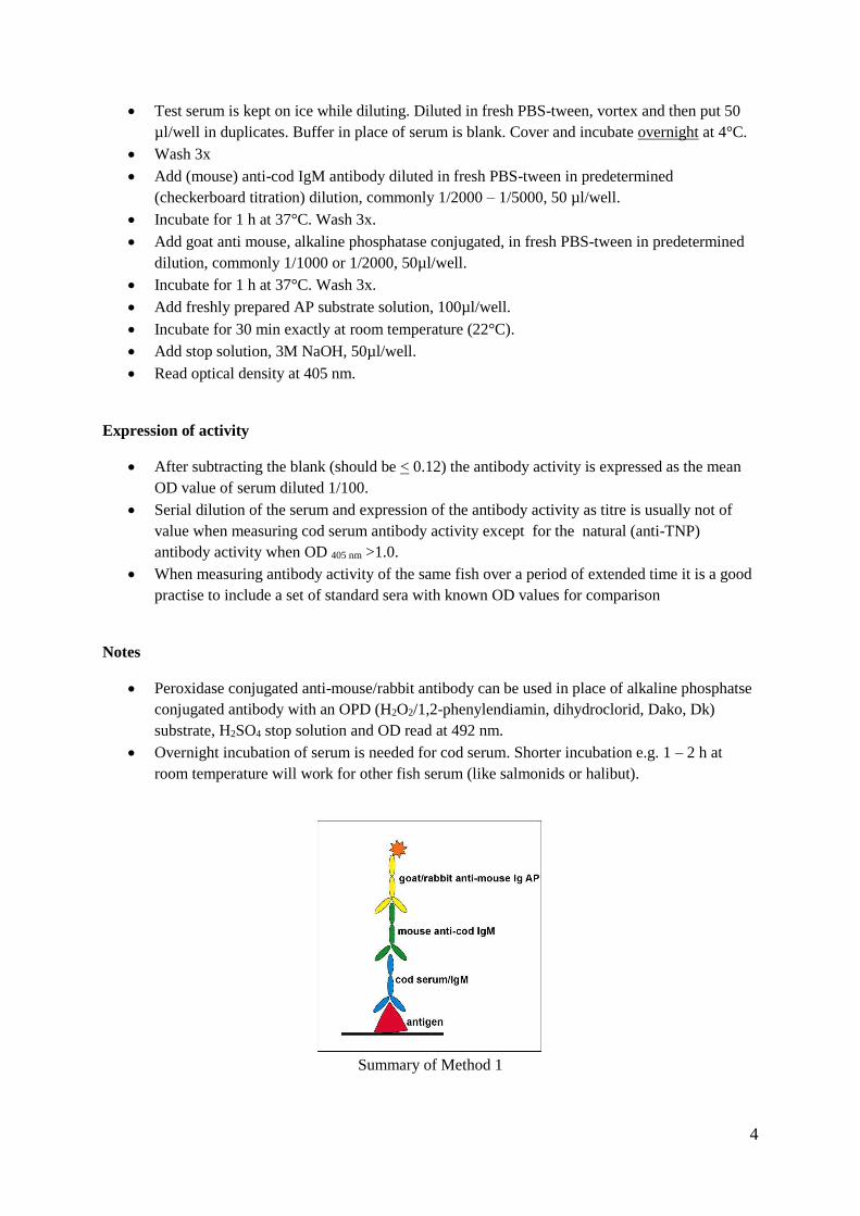
Prepare double serial dilutions of the coating antigen (here TNP-BSA) e.g. 5.0, 2.5, 1.25,
0.625 and 0.315 µg/ml in coating buffer
Put 100 µl per well per column as shown below and incubate overnight at 4°C. For blank coat
3 wells of each dilution in columns 11 and 12 as shown.
Tip out the buffer and block with 100 µl/well of 0.1% semi skimmed milk powder in coating
buffer for 1 h at room temperature
To each set of coating dilutions (total 40 wells) add 50 µl/well of serum (antibody) diluted
in PBS-T to give OD (with 5 µg/ml coating antigen) of about 1.0 – 1.5 (see Method 1).
Omit serum from the blank columns.
Incubate overnight at 4°C. Wash the tray
Prepare ammonium-SCN dilutions in 0.1M phosphate buffer, pH 6.0: 0.05, 0.1, 0.5,
1.0, 2.0, 4.0 and 6.0 M
Add 50 µl/well as shown 1 row per each dilution
Incubate for 15 min at rt.
Wash the tray and add mouse anti cod –IgM, diluted 1/2000 in PBS-T, 50 µl/well
Incubate for 1 h at 37°C
Wash the tray and add anti-mouse Ig-AP conjugate (1/2000) in PBS-T, 50 µl/well
Incubate for 1 h at 37°C
Develop colour with substrate solution, 100 µl/well for 30 min and stop the reaction
using 50 µl/well of 3 M NaOH.
Read OD at 405 nm
Calculating affinity
Blank should be about < 0.1 for all coatings.
The affinity constant K is worked out by plotting SCN molarity versus absorbance and K is
the molaritiy of SCN that elutes 50% of the antibody for each Ag coating concentration
The % of maximum binding in row A is calculated:e.g. 100[(Ab2.5-Ab1.25)/Ab5].
An affinity distribution patterns are then constructed by plotting the K (x-axis) and the %
binding for, in the set up below, 4 values.
10
The experimental set up for 2 serum samples per 1 tray
Antigen
NH4SCN
1
0,3
2
0,6
3
1,25
4
2,5
5
5,0
6
0,3
7
0,6
8
1,25
9
2,5
10
5,0
11
Anti-
12
gen
A
0
0,3 0,6
B
0,05
0,3 0,6
C
0,1
0,3 0,6
D
0,5
1,25 2,5
E
1,0
1,25 2,5
F
2,0
1,25 2,5
G
4,0
5 5
H
6,0
5 5
SERUM 1 SERUM 2 BLANK

11
5. ELISA: Antibody (anti-cod IgM) titration
MATERIALS
MaxiSorp 96 well microtrays from Nunc (442404)
Purified fish (cod) IgM about 0.5 – 1 mg/ml
Coating buffer (made fresh once a week), from tablets (Sigma C3041), 0.05M carbonate-
bicarbonate buffer, pH 9.6.
Diluting buffer, PBS-T (made fresh once a week) from tablets (Sigma P4417), containing
0.05% tween 20 (50 µl/100 ml).
Block solution (made fresh): 2% bovine serum albumin in coating buffer (0.2g /10 ml).
ELISA washing buffer, PBS-tween 20 hand made. 400 ml 5x PBS Keldur stock solution, 1600
ml H2O, 1 ml tween 20, see Method 1.
Ethanolamine buffer, 0.1M, pH 9: 610µl ethanolamine, 250 µl conc. HCl in 100 ml H2O.
Alkaline phosphatase conjugated antibody (from DAKO), e.g. goat anti-mouse Ig’s AP
D0486.
AP substrate solution (made fresh). Use 5 mg tablets of p-nitrophenyl phosphate (Sigma 104),
see Method 1.
Stop solution, 3M NaOH: 60g NaOH in 500 ml H2O.
METHOD
Coat the tray with purified IgM: 10 µg/ml in coating buffer, 100 µl/well
Incubate overnight at 4°C, wash 3x with ELISA wash buffer
Block residual sites with blocking solution, 100 µl/well, for 1 h at room temp. Wash 3x
Prepare serial dilutions of the antibody (serum/ascites) in PBS-T
In duplicates add 50 µl/well of each dilution
Incubate for 1 h at 37°C. Wash 3x
Prepare anti-mouse/rabbit-AP conjugated antibody (1/2000), add 50µl/well and incubate for 1
h at 37°C. Wash 3x
Add freshly prepared AP substrate solution, 100µl/well, incubate for 30 min exactly at room
temperature (22°C).
Add stop solution, 3M NaOH, 50µl/well.
Read optical density at 405 nm
After subtracting the blank (buffer in place of serum) a titration graph is plotted of OD values
versus dilutions. Titre is the dilution that gives approximately double - treble the blank value
(see example).
Notes
0.1% semi-skimmed milkpowder in coating buffer can also be used for blocking
Peroxidase conjugated anti-mouse/rabbit antibody can be used in place of alkaline phosphatse
conjugated antibody with an OPD (H2O2/1,2-phenylendiamin, dihydroclorid, Dako, Dk)
substrate, H2SO4 stop solution and OD read at 492 nm.

12
Figure: The titration curve of mouse-anti-cod IgM ascites, showing the optimum dilution for e.g.
competitive ELISA (Method 2) and the titre
Summary of Method 5

13
6. Capture ELISA for CRP PI & PII quantity measurements Magnadottir et al., Icel.Agric.Sci. 2010. 23, 23-35.
MATERIALS
MaxiSorp 96 well microtrays from Nunc (442404)
Coating buffer (made fresh once a week), from tablets (Sigma C3041), 0.05M
carbonate-bicarbonate buffer, pH 9.6.
Diluting buffer, PBS-tween (made fresh once a week) from tablets (Sigma P4417),
containing 0.05% tween 20 (50 µl/100 ml).
Block solution (made fresh): 0.1% semi skimmed milk powder in coating buffer (10
mg/10 ml).
ELISA washing buffer, PBS-tween 20 hand made. 400 ml 5x PBS Keldur stock
solution, 1600 ml H2O, 1 ml tween 20, see Method 1.
Ethanolamine buffer, 0.1M, pH 9: 610µl ethanolamine, 250 µl conc. HCl in 100 ml
H2O.
AP substrate solution (made fresh), see Method 1.
Mouse anti CRP-PI and CRP-PII immunoglobulins (Ig’s) (see Method 16), used for
coating the tray.
The same as above biotinylated for the detection (see Method 17 for the biotinylation).
Streptavidin-alkaline phosphatase (AP) labelled (from DAKO, Dk)
Purified CRP-PI and CRP-PII proteins for standards (see Method 11)
METHOD
Coat 96 well microtray, MaxiSopr, with 10 µg/ml, 100 µl per well of
o a) mouse anti CRP-PI Ig’s (e.g. 569 µg CRP-PI/ml: 26.4 µl + 14.97 ml
coating buffer)
o b) mouse anti CRP-PII Ig’s (e.g. 340 µg CRP-PII/ml: 440 µl + 14.56 ml
coating buffer)
Incubate overnight at 4°C
Tip out the coating solution and block with 100 µl per well of 0.1% semi skimmed
milk powder in coating buffer and leave for 1 h at room temperature
Wash 3x in PBS-t (phosphate buffered saline containing 0.05% tween 20)
Prepare standard solutions and serum dilutions:
o Standard:
CRP-PI purified, 240 µg/ml prepare 8 five fold dilutions starting with
30 µl + 120 µl PBS-t, mix and transfer 30 µl of this to 120 µl PBS-t etc.
total 8 tubes
CRP-PII purified, 960 µg/ml, as above but starting with 7.5 µl of this +
142.5 (1/20) µl PBS-t, then transfer 30 µl of this to 120 µl PBS-t etc.
total 8 tubes
o Serum: Prepare 2 dilutions: 1/500 and 1/2000:
2 µl serum in 1000 µl and transfer from this 200 µl + 600 µl PBS-t
14
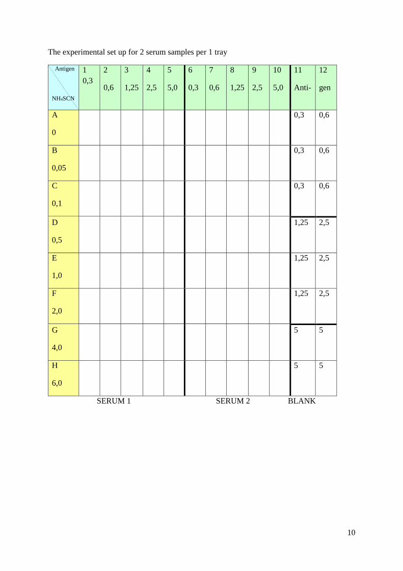
On each coated, blocked and washed tray put 1 column blank (PBS-t), 2 columns 8
standard solutions and the rest serum samples diluted 1/2000 in duplicates, in all cases
50 µl per well and incubate for 2 h at room temperature.
Wash 3 x in PBS-t
Prepare anti-CRP PI and PII biotinylated:
o anti-CRP-PI-biotin: 515 µg/ml diluted 1/100 (from 23.4.08): 80 µl + 7.92 ml
PBS-t
o anti-CRP-PII-biotin: 343 µg/ml diluted 1/100: 80 µl + 7.92 ml PBS-t
On the whole tray put 50 µl per well, of a) or b) above (depending on the parameter to
be measured), and incubate for 1 h at 37°C
Wash 3 x in PBS-t
Prepare Streptavidin-AP 1/1000: 8 µl + 8 ml PBS-t for each set of tests (1 ½ tray), 50
µl per well and incubate for 1 h at 37°C.
Wash 3 x in PBS-t
Add AP substrate solution (made fresh), 100 µl per well and incubate for 30 min at
room temperature
Add stop solution, 3M NaOH, 50µl/well and read optical density at 405 nm.
Plot a standard graph and interpolate the serum values and calculate the CRP-PI and
CRP-PII concentration in serum.
Comments:
This is not a very “robust” method, preferably 3 serum dilutions should be tested in duplicates
or/and all the serum samples to be tested in an experiment should be tested in one go.
However, this is an “expensive” method so normally serum is tested 1/2000 and if the value
does not fall on the standard line the test is repeated for this serum either more or less diluted.
Summary of Method 6
15
7. ELISA: Cortisol in serum – a kit from Neogen Neogen Cortisol ELISA kit, Magnadottir et al. 2010, Icel Agric Sci 23, 23-35
MATERIALS
A kit from Neogen Corp.Ky, USA
HCl
Ethyl ether
Liq. nitrogen
Speed-Vac
Vortex
METHOD
Preparation of sample
In a glass test tube (or an eppendorf 1.5 ml test tube) mix together 100 µl of serum/plasma
and 1 ml ethylene ether (FUME CUBOARD)
Vortex for 30 sec and allow phases to separate (can be speeded up by centrifugation at 400
rpm for 2 min
Collect the upper organic phase into a clean eppendorf tube, cool on ice or freeze in liq.
nitrogen
Evaporate in the SpeedVac until dry
Dissolve the residue in 100 µl of diluted extraction buffer from the kit
Take 10 µl of this and add to 990 µl of diluted extraction buffer (1/100)
Assay in duplicates 50 µl of this sample
Preparation of standards (from the kit)
A: Stock Cortisol solution 1 µg/ml
B: 20 µl of A and 0.980 µl of EIA buffer from the kit, mix (=20 ng/ml)
C: 200 µl of B and 1.8 ml EIA buffer, mix (=2 ng/ml)
D: 200 µl of C and 1.8 ml EIA buffer, mix (=0.2ng/ml)
16
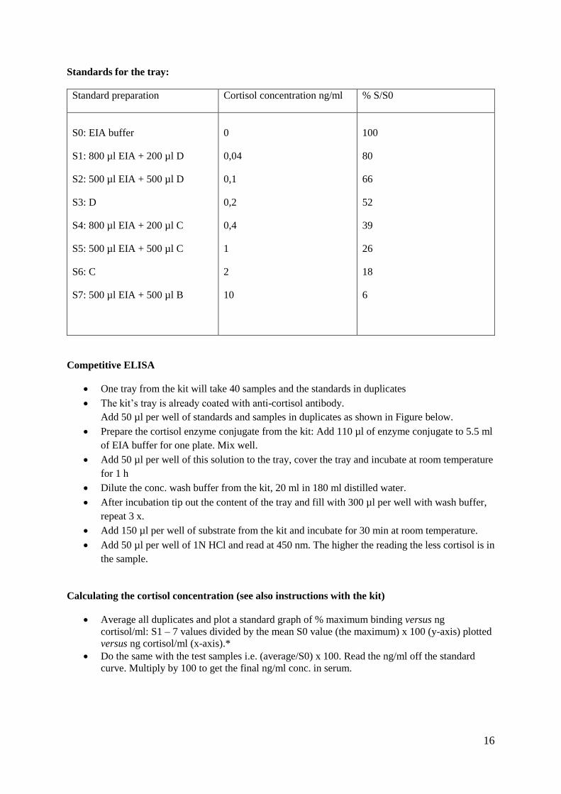
Standards for the tray:
Standard preparation Cortisol concentration ng/ml % S/S0
S0: EIA buffer
S1: 800 µl EIA + 200 µl D
S2: 500 µl EIA + 500 µl D
S3: D
S4: 800 µl EIA + 200 µl C
S5: 500 µl EIA + 500 µl C
S6: C
S7: 500 µl EIA + 500 µl B
0
0,04
0,1
0,2
0,4
1
2
10
100
80
66
52
39
26
18
6
Competitive ELISA
One tray from the kit will take 40 samples and the standards in duplicates
The kit’s tray is already coated with anti-cortisol antibody.
Add 50 µl per well of standards and samples in duplicates as shown in Figure below.
Prepare the cortisol enzyme conjugate from the kit: Add 110 µl of enzyme conjugate to 5.5 ml
of EIA buffer for one plate. Mix well.
Add 50 µl per well of this solution to the tray, cover the tray and incubate at room temperature
for 1 h
Dilute the conc. wash buffer from the kit, 20 ml in 180 ml distilled water.
After incubation tip out the content of the tray and fill with 300 µl per well with wash buffer,
repeat 3 x.
Add 150 µl per well of substrate from the kit and incubate for 30 min at room temperature.
Add 50 µl per well of 1N HCl and read at 450 nm. The higher the reading the less cortisol is in
the sample.
Calculating the cortisol concentration (see also instructions with the kit)
Average all duplicates and plot a standard graph of % maximum binding versus ng
cortisol/ml: S1 – 7 values divided by the mean S0 value (the maximum) x 100 (y-axis) plotted
versus ng cortisol/ml (x-axis).*
Do the same with the test samples i.e. (average/S0) x 100. Read the ng/ml off the standard
curve. Multiply by 100 to get the final ng/ml conc. in serum.

17
Figure showing a typical experimental setup: S: standards, T: test sera
1 2 3 4 5 6 7 8 9 10 11 12
A S0 S0 T1 T1 T9 T9 T17 T17 T25 T25 T33 T33
B S1 S1 T2 T2 T10 T10 T18 T18 T26 T26 T34 T34
C S2 S2 T3 T3 T11 T11 T19 T19 T27 T27 T35 T35
D S3 S3 T4 T4 T12 T12 T20 T20 T28 T28 T36 T36
E S4 S4 T5 T5 T13 T13 T21 T21 T29 T29 T37 T37
F S5 S5 T6 T6 T14 T14 T22 T22 T30 T30 T38 T38
G S6 S6 T7 T7 T15 T15 T23 T23 T31 T31 T39 T39
H S7 S7 T8 T8 T16 T16 T24 T24 T32 T32 T40 T40
Note:
The principle of this method is the same as in Method 2, i.e. competitive ELISA.
The preparation of the serum sample is critical. Other samples, e.g. mucus may not need extraction
procedure (see the kits’s instructions). Kits may be available for serum and other extractions.

18
8. Fish serum or plasma collection
MATERIALS
Monovette syringes Serum or Plasma (EDTA or heparin coated): 1.2 ml, 4.5 ml or 9.5 ml,
from Sarstedt, Germany
Needles from Sarstedt, Germany for Monovette syringes, 20G or 21G
METHOD
Serum collection
Using Monovette Serum syringe, blood is usually collected from the caudal vessel but in some
cases (e.g. halibut) it is easier to collect blood from an aorta in the gills.
Blood is allowed to clot at room temperature for 2 hours (optional) and then overnight at 4°C
and serum collected by centrifugation at 2000 rpm for 10 min. Generally stored at -20°C.
Plasma collection
Using Monovette Plasma syringe, blood is collected as above
Centrifuged at once at 7000 xg for 10 min (can be stored on ice for a while before
centrifugation) and the plasma collected and stored at -80°C.
Notes
Clots may form in the fish serum samples during storage, these can be removed with a pincer or just
ignored.
Precipitates (fat?) may also form in serum during prolonged storage. This does not normally affect
antibody activity.

19
9. Fish IgM purification
Magnadottir, B., Icel.Agr.Sci.4, 1990, 49-54, Magnadottir, B., Icel.Agr.Sci.12, 1998, 47-59
This is a three step column isolation:
a) “Clean-up” using CM-Affi-Gel Blue and SAS precipitation,
b) Gel filtration (isolation based on size) and
c) Anion exchange chromatography (isolation based on charge).
The method has worked well for the isolation of IgM from several fish species (e.g. cod,
salmon, rainbow trout, arctic char, haddock, halibut, sea-bass, sturgeon, turbot and wolf fish)
MATERIALS
Fish serum
CM Affi-Gel Blue (BioRad) 100 ml column
Superose 6 column (HR10/30, Pharmacia)
MonoQ anion exchange column (HR5/5)
FPLC system
Buffers – filtered and de-gassed before use (see buffer preparations, Method 38):
o For CM Affi-Gel Blue: 10mM Potassium phosphate buffer pH7.25, 0.15M NaCl
o For the Superose 6 column: 0.1M Tris-HCl buffer, pH 8.0, 0.15M NaCl
o For MonoQ column: 0.02M Tris-HCl, pH 7.5,
a) containing 0.15M NaCl for equilibrating the MonoQ column and
b) containing 1M NaCl for the gradient elution from the MonoQ column
METHOD
About 20 ml of fish serum is passed through the CM Affi-Gel Blue column and the unbound,
fall through material collected (the column should retain albumin and other major non-Ig
serum proteins)
The fall through fractions are pooled and protein precipitated by 50% saturation with
ammonium sulphate (0.313 g/ml – see Method 31). Stirred gently for 2 h at room temp.
Collect the precipitate by centrifugation at 10 - 15.000 rpm for 30 min and dissolve in
minimum volume of saline (0.15M NaCl).
Dialyse in saline until free of sulphate (see Method 35), finally in the Superose 6 buffer.
Measure the protein using Bradford’s method or the extinction coefficient E=13.7 (see
Methods 29/30) and aliquote suitable amounts for Superose separation.
Put about 250 µl of crude IgM on the column containing about 0.5 – 1 mg crude IgM.
Fractions are tested in dot blot or Western blot with anti-fish IgM and to obtain the purest
isolation check also by SDS-PAGE and pool fractions that contain IgM only.

20
Further separation on anion exchange MonoQ column is generally advisable and will also
show possible isotypes of IgM if present (e.g. salmon, haddock and sturgeon IgM)
Sample of about 250 µg IgM in 250 µl of MonoQ equilibration buffer is put on the MonoQ
column and eluted with a gradient which upper limit is 1M NaCl.
The final IgM preparation is best kept in PBS in aliquots at -20°C or -80°C, 20 – 50% glycerol
or sucrose can be added to improve stability.

21
10. Crude preparation of (fish) IgM N.M.S. dos Santos et al. FSI 1997, 7, 176
MATERIALS
Dextran sulphate: 10% solution in H2O
CaCl2H2O 1M: 0.147 g in 10 ml
TBS: 20 mM Tris, 50 mM NaCl, pH adjusted to pH 7.6 (with HCl)
Ammonium sulphate
METHOD
To 5 ml of fish serum add 100 µl of the dextran solution and 500 µl of the calcium chloride
solution
Stirr on ice for 30 min
Centrifuge for 15 min at 4°C at 10.000 rpm (Sorval)
Collect the supernatant and dialyse in TBS at 4°C overnight (2 – 3 changes)
Measure the volume and add 0.243 g ammonium sulphate per ml (about 40% saturation, see
Method 31)
Stirr on ice for 4 h
Centrifuge at 10.000 rpm (Sorval) for 30 min at 4°C
Collect the precipitate and dissolve in TBS
Dialyse in TBS until free of sulphate (see Method 35)
Check purity in SDS-PAGE
Note
This method saves time if you want to isolate IgM from a great quantity of serum but it will need
further purification e.g. on anti-IgM coupled column (or see Method 18).

22
11. Purification of cod pentraxins, CRP-PI and CRP-PII From Lund & Olafsen DCI 1998, 22, 185-194 and Gisladottir et al. FSI 2009, 26, 210-219
MATERIALS
Columns
Phosphoryl- agarose (PC) from Pierce Chemicals, Thermo Fisher Scientific, IL, USA. About
10 ml are required and will be packed into XK16 column from Amersham.
MonoQ column HR5/5 from Amersham Pharmacia Biotech, GE Healthcare, Dk.
PD buffer exchange column from Amersham Pharmacia Biotech, GE Healthcare, Dk.
Buffers (see also Method 38)
PBS from tablets (Sigma P4417), containing 0.05% sodium azide.
PC equilibrium buffer, Tris buffered saline + Ca (TBS-Ca): 50 mM Tris-HCl buffer, pH 8.0,
containing 150 mM NaCl (TBS) and 2 mM CaCl2.
PC elution buffers:
1) 50 mM Tris-HCl buffer, pH 8.0, containing 150 mM NaCl (TBS) and 10 mM EDTA
2) 50 mM Tris-HCl buffer, pH 8.0, containing 150 mM NaCl (TBS) and 30 mM
phosphorylcholine.
MonoQ start buffer 1: 20 mM Tris-HCl, pH 7.5
MonoQ elution/gradient buffer 2: 20 mM Tris-HCl, pH 7.5 containing 1 M NaCl
METHOD
Affinity Chromatography
Approximately 10 ml of Phosphorylcholine (PC)-agarose from Pierce Chemicals (U.S.A.) is
used. This is stored in a 50 ml centrifuge tube at 4°C in PBS containing sodium azide.
The PC-agarose is equilibrated with several changes of excess TBS-Ca buffer (PC equilibrium
buffer) in the centrifuge tube.
The gel is mixed with 10 ml of TBS-Ca buffer and 10 ml of pooled and filtered cod serum and
gently rotated overnight at 4°C.
The gel is allowed to settle and the supernatant discarded (can be kept for IgM isolation) and
the gel washed twice with the TBS-Ca buffer.
The gel is packed onto a chromatographic column (XK16, Amersham Pharmacia Biotech,
Sweden), connected to the ÄKTA FPLC system from Amersham Pharmacia Biotech and
washed in TBS-Ca buffer until zero optical density is obtained.
Bound protein is eluted with 10 mM EDTA in TBS buffer (PC elution buffer 1) and collected.
This contains crude cod pentraxins.
The column is then cleaned with 30 mM PC in TBS (PC elution buffer 2), this elutes some as
yet unidentified proteins.
Ion Exchange Chromatography – separation of CRP-PI and CRP-PII
MonoQ anion ion exchange column is used. The column is equilibrated in MonoQ start buffer.

23
The crude pentraxin is equilibrated in the start buffer using PD buffer exchange column or
dialysis and the protein concentration measured using Bradford’s method (see Method 29).
A sample is loaded onto the MonoQ column and eluted first with start buffer and then
followed by a salt gradient of 0 to 1 M NaCl in the start buffer (the MonoQ elution buffer).
The salt elution should distinguish 2 peaks, the first is CRP-PI and the second CRP-PII.
The quantity is measured using the Bradford method and the purity is verified by SDS-PAGE
and the specificity by Western blotting using specific anti-CRP-PI and CRP-PII antibodies.
The tris buffer recipes:
Stock-Tris buffer: 6g Tris and 8,7g NaCl in 600 ml distilled water, pH adjusted to 8,0 með
HCl, volume adjusted to 1 l
This stock-Tris buffer is divided into 3 parts:
o PC equilibrium buffer: 600 ml + 0,176g CaCl2
o PC elution buffer 1: 200 ml + 0.74 g EDTA
o PC elution buffer 2: 200 ml + 1,5 g phosphorylcholine chloride (PC)

24
12. Apolipoprotein A1 isolation from cod serum
Amthauer et al., Comp. Biochem. Physio. 1989, 92B, 787-793.
MATERIALS
AffiGel Blue 50 ml column
Equilibration buffer: Sodium citrate buffer, 50 mM, pH 6.5 containing 50 mM NaCl
Elution buffer 1: Sodium citrate buffer, 50 mM, pH 6.5 containing 350 mM NaCl
Elution buffer 2: Sodium citrate buffer, 50 mM, pH 6.5 containing 50 mM NaCl and
containing 2 mM 8-anilino-1-naphthalene sulfonate (ANS)
UV lamp in a darkroom
Anti-cod IgM-HiTrap column (see Method 18)
METHOD
About 2 ml of cod serum is put on the AffiGelBlue column with the equilibration buffer.
The column is washed with Elution buffer 1.
Apolipoprotein is then eluted with the Elution buffer 2 containing ANS.
Fractions are examined under a UV light and those with a high fluorescence pooled
Pooled fractions dialysed in equilibration buffer at 4°C, concentrated in centrifugal filter and
tested in SDS-PAGE and Western blotting using anti-ApoLP A1 antibody (also anti-IgM and
anti-C3 antibodies, all available at Keldur).
ApoLpA1 is the major 30 kDa band observed. However, this preparation may be contaminated
with IgM (72 kDa + 26 kDa). To remove this the ApoLP A1 preparation, after a buffer
change, is passed through an anti-cod IgM-HiTrap column (see Method 18).
Notes
Mouse-anti-cod IgM antibody columns are available at Keldur
Contamination with C3 may also be seen and an anti-cod C3 column can then be prepared from rabbit
anti-cod C3 antibody available at Keldur
Typical elution of serum on AffiGel Blue column with ANS, circle shows the fractions collected
AffigelBlue01:1_UV AffigelBlue01:1_Conc AffigelBlue01:1_Fractions AffigelBlue01:1_Inject
0
100
200
300
400
500
mAU
0 100 200 300 400 500 ml
A1 A3 A5 A7 A9 A12 B10B8 B6 B4 B2C1C5 C7 C9 C12 D10 D7 D5 D3 D1 E2 E4 E6 E8E10 F12 F9 F7 F5 F3 F1 G2 G4 G6 G8 G11 H11 H8 Waste
350 mM
NaCl
sample
ANS
25
13. Isolation of fish C3 complement component
Lange et al., FSI 2004, 16, 227-239; Dodds et al. DCI, 1998,22, 207 - 216
MATERIALS
Fish plasma* isolated by centrifugation at 700 xg for 10 min. from blood, collected into
EDTA coated syringes from Monovette, kept on ice. Will need about 100 ml of serum. Make
5 mM with EDTA and store at 4°C until used.
Polyethylene glycol (PEG 3350)
Start buffer: 20 mM Tris-HCl, pH 7.4, 50 mM EACA (aminocaproic acid), 5 mM EDTA, 50
mM NaCl and 0.1mM protease inhibitor (Boehringer-Mannheim pefablock or others)
Start buffer made 500 mM with NaCl for the gradient elution
Q-Sepharose HP column (1.6 x 20 cm, Amersham)
Superose 6 column (HR10/30)
Superose 6 elution buffer: 20mM Tris-HCl, pH 6.8, 150 mM NaCl, 5mM EDTA, 0.1M
pefablock
MonoQ column 0.5 x 5 cm)
MonoQ elution buffer: gradient from 20 mM Tris-HCl, pH 7.4, 5 mM EDTA, 50 mM NaCl
and 0.1mM protease inhibitor (Boehringer-Mannheim pefablock) to the same buffer with 500
mM NaCl.
METHOD
Add PEG slowly to the plasma solution to give 5% PEG, using 15% PEG in the starting
buffer, stir gently at 4°C for 45 min.
Centrifuge at 20.000 xg for 20 min.
Collect the supernatant.
Put on the Q-Sepharose column in the start buffer and elute bound proteins with the gradient
elution buffer
Collect fractions and test in Western blotting using anti-fish C3 antibody (if not available see
original references for detecting C3)**
Further purification on Superose 6 and MonoQ will be needed to obtain pure C3
Test the final purity in SDS-PAGE***
Notes
*Serum can also be used but plasma is better.
**Monospecific rabbit anti-cod C3 is available at Keldur
***Two bands should appear under reducing conditions: the approx. 115 kDa α-chain and the 74 kDa
β-chain.

26
14. C3 isolation for immunization using Macrogard Jarl Bögwald personal communication
MATERIALS
Macrogard (MG, dietary yeast beta-1.3/1.6-glucan) from Biotec Pharmacon, Norway
Phosphate buffered saline (PBS)
Gelatin-Veronal buffer (GVB) from Sigma (G6514) containing Ca2+ and Mg2+
Sodium dodecyl sulphate (SDS)
METHOD
Approximately 2 ml of 2.5% suspension of MG is washed twice in 20 ml of PBS, centrifuge
between the washing at 400 xg (1350 rpm) for 10 min and discard the supernatant
The pellet is resuspended in 15 ml of the GVB buffer, hence, containing approximately 3.33
mg MG ml-1
Add 10 ml of fish serum and rotate at 14°C for 60 min
Centrifuge at 400 xg for 10 min
Wash the pellet 3x in PBS containing 0.1% SDS
Wash the pellet 3x in PBS
Resuspend the pellet in 2.5 ml PBS, hence, containing 20 mg coated MG ml-1
This suspension is used to immunize rabbits or mice with about 5 mg MG per rabbit (less per
mouse (acites))
Comment
Macrogard is expected to bind C3b/C3bi.
This has been tested on cod and halibut serum for producing mouse anti-C3 antibody in ascites (see
Method 15). The resulting antibody was not mono-specific against C3 but was still useful for detecting
C3 in column fractions by Western blotting etc.

27
15. Production of polyclonal antibody in mouse ascites
Overkamp et al., J.Immunoassay 1988, 9, 51-68
MATERIALS
Use 2 – 3 mice for each antigen, Balb/c mice have normally been used at Keldur but other
types can be used
Freund’s complete and incomplete adjuvant (FCA/FIA)
Pristane (Sigma)
METHOD
The protein antigen (e.g. fish IgM) has to be as pure as possible. The purity of approximately
1 – 10 µg has to be checked in SDS-PAGE using silver staining (coomassie blue is not
sensitive enough)
100 µl of the protein containing approximately 1 mg/ml is mixed with 100 µl PBS and 1.8 ml
FCA (or FIA) and homogenized (using an syringe and as fine a needle as possible)
200 µl of this is injected intra-peritoneal into 2 – 3 mice on day 0 (approx. 10 µg
protein/mouse).
On day 14 this injection is repeated with a fresh protein/adjuvant preparation and the mice
receive 500 µl of pristane i.p. as well
On day 21 and day 28 the injection of antigen is repeated as required. Ascites fluid often starts
to form before day 28 in which case the last injection is omitted
When the swelling of the abdomen is suitable the mouse is sacrificed and the ascites collected
with a bulb pipette.
The ascites is centrifuged, aliquoted and stored at -20 or -80°C.
Notes
Many labs do not allow this method due to the pain induced in the mice, hence, use as few mice as
possible and collect the ascites as soon as it forms
FCI is usually needed, at least in the first injection
The antibody response is dose related but higher dosage does not necessarily result in higher antibody
titre, 5 – 50 µg of fish IgM per mouse per injection is usually used
Make sure the antigen solution does not contain Tris or any other harmful component (e.g.
azide) – this has to be dialysed out before injection
28
16. Immunoglobulin isolation from mouse ascites/rabbit serum Based on Midi and Mini Proteus kit from Pro-Chem Inc., GA, USA (see leaflet for more informations)
MATERIALS
Proteus Mini or Midi kit from Pro-Chem Inc.
Binding buffer A: 0.1M sodium phosphate, 0.15M, pH 7.4 (see buffers, Method 38)
Elution buffer B2: 0.2M Glycine/HCl pH 2.5
Neutralization buffer C: 1M Tris-HCl, pH 9.0
METHOD
1. Mini plug
Load the Mini plug into the barrel of the Proteus spin column.
Equilibrate the column with 650 µl of binding buffer A by centrifuging the spin column at
1800 g (4 – 5000 rpm) for 1 min and repeat once.
Filter 1 ml of sample through 0.2 µm syringe filter and then dilute 1:1 in binding buffer A (or
dilute before filtration if too viscous).
Add 650 µl of sample to the spin column
Centrifuge at 640 g (2.6 – 3000 rpm) for 6 min (increase the time if any sample remains above
the plug.
Wash the spin column 3 x with 650 µl of binding buffer A, spin for 1 min at 1800 g.
Elute bound Ig’s with 500 µl of elution buffer B2 directly into a fresh centrifuge tube
containing 65 µl of neutralization buffer C, spin for 1 min at 1800 g. Mix the eluate
immediately.
Repeat the elution step and pool the two eluates (total about 1.1 ml).
Change the buffer of the sample from glycine/tris to PBS using PD column and then
concentrate in a centrifugation filter.
Estimate protein by measuring OD at 280 nm using the coefficient E1%=13.7 i.e. 1 mg/ml will
give a reading of 1.37 (see Method 30).
Make 0.05 – 0.2% with sodium azide and store at 4°C or make 10 – 50% with glycerol and
store in aliquots at –20°C.
Regenerate the column
Wash the Mini plug twice with 650 µl of elution buffer B2 and then twice with 650 µl of
binding buffer A, centrifuging at 1800 g for 1 min between each wash.
Store in binding buffer A with 0.1% sodium azide at 2 – 8°C until further use.
29
2. Midi plug
Load the Midi plug into the barrel of the Proteus spin column.
Equilibrate the column with 10 ml of binding buffer A by centrifuging the spin column at 500
g for 3 min.
Filter 12 - 15 ml of sample through 1.2 µm syringe filter and then through 0.2 µm filter, dilute
1:1 in binding buffer A (or dilute before filtration if too viscous).
Add 20 ml of sample to the spin column
Centrifuge at 150 g for 30 min (increase the time if any sample remains above the plug.
Wash the spin column with 10 ml of binding buffer A for 3 min at 500 g.
Elute bound Ig’s with 10 ml of elution buffer B2 directly into a fresh centrifuge tube
containing 1.3 ml of neutralization buffer C, for 3 min at 500 g. Mix the eluate immediately.
Repeat the elution step and pool the two eluates (total about 22 ml).
Change the buffer of the sample from glycine/tris to PBS using centrifugation filter, spinning
and adding PBS to the concentrated top layer 1 – 2 x.
Estimate protein by measuring OD at 280 nm using the coefficient E1%=13.7 i.e. 1 mg/ml will
give a reading of 1.37 (see Method 30).
Make 0.05 – 0.2% with sodium azide and store at 4°C or make 10 – 50% with glycerol and
store in aliquots at –20°C.
Regenerate the column
Wash the Midi plug with 10 ml of elution buffer B2 and then 10 ml of binding buffer A,
centrifuging at 500 g for 3 min between each wash.
Store in binding buffer A with 0.1% sodium azide at 2 – 8°C until further use.

30
17. Biotinylation of Antibodies (Ig’s) and other proteins Based on a protocol from the kit’s producer (Amersham, ECL RPN 2202/03)
MATERIALS
Biotinylation reagent from the kit (Amersham, ECL RPN 2202/03) – equilibrated at room
temperature before use
Bicarbonate buffer from the kit diluted 1/20: 40 mM, pH 8.6
Sephadex G-25 column from the kit equilibrated with 5 ml PBS containing 1% BSA and then
with 20 ml PBS
Purified immunoglobulin from the antibody solution (e.g. mouse ascites or rabbit serum) using
the Proteus kit (see Method 16).
METHOD
Estimate the Ig concentration using e.g. the OD280 absorbance method (see Method 30) and
prepare a 1 mg/ml solution in the bicarbonate buffer, 2 – 2.5 ml is a suitable volume
For each mg of Ig in the antibody solution add 40 µl of the biotinylation reagent and incubate
for 1 h at room temperature with constant agitation.
At the same time prepare the G-25 column as described above
After 1 h apply the biotinylation solution to the column, let it enter the column and then elute
with 5 ml of PBS. Collect 0.5 - 1 ml fractions and monitor at 280 nm and pool the peak
fractions.
The biotinylated antibody is ready for use. Best to store at 4°C in the presence of 0.1% sodium
azide.
Note
More than one biotinylation kits have been tested and this kit from Amersham has worked best.

31
18. Protein coupling to NHS-activated columns,
ligand binding and elution Based on protocol that comes with the columns (Amersham)
These columns have mainly been used for coupling mouse anti- cod IgM Ig’s (isolated
using the Proteus kit (see Method 16)) and then used for the purification of fish IgM from
serum or crude preparations; also for the decontamination of other samples (e.g. cod ApoLP
A1).
MATERIALS
Columns
NHS- activated HiTrap columns, 1 or 5 ml, from Amersham Biosciences
Buffers
Coupling buffer, 0.2M NaHCO3, 0.5M NaCl, pH 8.3: 8.4 g NHCO3 and 14.6 g NaCl in 3-400
ml H2O, pH adjusted to 8.3, volume adjusted to 500 ml
Washing and deactivating buffers, 0.5 M ethanolamine, 0.5M NaCl, pH 8.3: 15 ml
ethanolamine in 3 – 400 ml H2O add 14.6 g NaCl, adjust pH to 8.3, adjust volume to 500 ml.
Acetate buffer, 0.1 M acetate, 0.5M NaCl, pH 4.0 (see Method 37)
Storage buffer, 0.05 M Na phosphate buffer, pH 7, 0.05% sodium azide (Buffers ÐÐÐ)
Sample and column buffer, PBS, prepared from tablets from Sigma
Elution buffer, 0.1M glycine, pH 2.3: 3.75 g glycine in 250 ml H2O, pH adjusted to 2.3 with
HCl, volume adjusted to 500 ml
Neutralizing buffer, 1M Tris: 1.12 g Tris in 10 ml H2O
1 mM HCl: 10 µl conc. HCl in 100 ml
METHOD
A) Coupling proteins to a 1 ml NHS-activated HiTrap column
The protein is equilibrated in the coupling buffer e.g. by dialysis og by buffer change on PD-
10 or NPD column. The concentration should be 0.5 – 10 mg/ml in 1 ml
Remove the cap of the column and add a drop of ice cold 1 mM HCl.
Remove the bottom end when ready
Using a 1 or 2 ml syringe wash the column with 3x2 ml of 1 mM HCl – do not exceed flow
rate of ca. 2 drops/sec
Inject 1 ml of the protein
Seal the column and let stand for 15 – 20 min at room temperature or 4 h at 4°C
Wash and deactivate the column as follows:
o 3x2 ml ethanolamine buffer
o 3x2 ml acetate buffer
o 3x2 ml ethanolamine buffer, let stand for 15 – 30 min
o 3x2 ml acetate buffer
o 3x2 ml ethanolamine buffer
o 3x2 ml acetate buffer
o Finally inject storing buffer or PBS

32
B) Coupling proteins to a 5 ml NHS-activated HiTrap column
Same reagents and general protocols as above but using a 5 ml column
The protein is equilibrated in the coupling buffer e.g. by dialysis og by buffer change on PD-
10 or NPD column. The concentration should be 0.5 – 10 mg/ml in 5 ml
Remove the cap of the column and add a drop of ice cold 1 mM HCl.
Remove the bottom end when ready
Using a 5 or 10 ml syringe wash the column with 3x10 ml of 1 mM HCl – do not exceed flow
rate of ca. 2 drops/sec
Inject 5 ml of the protein
Seal the column and let stand for 15 – 20 min at room temperature or 4 h at 4°C
Wash and deactivate the column as follows:
o 3x10 ml ethanolamine buffer
o 3x10 ml acetate buffer
o 3x10 ml ethanolamine buffer, let stand for 15 – 30 min
o 3x10 ml acetate buffer
o 3x10 ml ethanolamine buffer
o 3x10 ml acetate buffer
o Finally inject storing buffer or PBS
C) Binding to ligand and elution of affinity bound protein
The FPLC system is used and the programme for 1 or 5 ml HiTrap NHS affinity
column.
Equilibrium buffer is PBS, elution buffer 0.1M glycine pH 2.3, neutralizing eluted bound
fractions with a suitable amount of 1M Tris.
Test the amount of Tris needed to neutralize the fraction size of glycine-HCl using pH strips is
the safest but potassium chromate* titration (yellow to orange) can also be used.
Note
Five 5 ml columns coupled with mouse anti-cod IgM immunoglobulins are available at Keldur,
stored in the lab refrigerator.
*Potassium chromate:

33
19. Proteolytic digestion of Ig’s (fish IgM) Magnadottir et al. 1996 FSI 6:185-198, protocols from Sterogen Biochemicals
MATERIALS
Trypsin-actigel from Sterogen Biochemicals, USA
Trypsin buffer: 0.1N Tris-HCl buffer, pH 8.0, 10mM CaCl2
Pepsin-actigel from Sterogen Biochemicals, USA
Pepsin buffer: 20mM sodium acetate buffer, pH 5.5 – 6.0
Papain-actigel from Sterogen Biochemicals, USA
Papain buffer: 20 mM phosphate buffer, pH 6.2 containing 1mM dithiothreitol and 5 mM
EDTA
Glass filter connected to a water suction pump
METHOD
1. Trypsin digestion
On a glass filter trypsin-gel containing about 1 mg enzyme per ml gel was washed several
times with the trypsin buffer and finally suction dried, 1 ml of suction dry trypsin actigel
weighs about 570 mg
The immunoglobulin solution, approximately 1 mg/ml, is equilibrated by dialysis or PD
column in the trypsin buffer
50 mg of the suction dry gel (= 85 µg trypsin) are mixed with 75 µl of the trypsin buffer
and 50µl of the Ig solution
Control omitting the trypsin gel is included
Incubated overnight (> 16 h) at 45°C with gentle or occasional shaking
Centrifuge the sample at 200xg for 10 min and collect the supernatant
Check digestion by SDS-PAGE
2. Pepsin digestion
On a glass filter pepsin-gel containing about 3 - 4 mg enzyme per ml gel was washed
several times with the pepsin buffer and finally suction dried
The immunoglobulin solution, approximately 1 mg/ml, is equilibrated by dialysis or PD
column in the pepsin buffer
10 mg of the suction dry gel (= 50 - 70 µg pepsin) are mixed with 75 µl of the trypsin
buffer and 50µl of the Ig solution
Control omitting the pepsin gel is included
Incubated for 1 h at 37°C with gentle or occasional shaking
Centrifuge the sample at 200xg for 10 min and collect the supernatant
Check digestion by SDS-PAGE

34
3. Papain digestion
On a glass filter pepsin-gel containing about 2 mg enzyme per ml gel was washed several
times with the papain buffer and finally suction dried
The immunoglobulin solution, approximately 1 mg/ml, is equilibrated by dialysis or PD
column in the papain buffer
10 mg of the suction dry gel (= 34 µg papain) are mixed with 75 µl of the trypsin buffer
and 50µl of the Ig solution
Control omitting the papain gel is included
Incubated for 1 h at 37°C with gentle or occasional shaking
Centrifuge the sample at 200xg for 10 min and collect the supernatant
Check digestion by SDS-PAGE
Note
Digestion conditions i.e. pH, enzyme concentration and incubation temperature will vary depending
on the Ig-species (or protein) being studied
35
20. Preparation of organ/larvae protein suspension Magnadottir et al., Comparative Biochemistry and Physiolog PartB 2004, 139:217-224
MATERIALS
Triton X-100 lysis buffer prepared by adding 0.5 ml of Triton X-100 to 100 ml of Tris-saline
buffer (ice cold):
Tris saline buffer, 50 mM Tris-HCl, pH 7.6: 0.6 g tris in <100 ml, pH adjusted with HCl, add to
this 1.75 g NaCl (0.3M) and adjust the volume to 100 ml.
As needed add 100 µl protease inhibitor cocktail (from Sigma) per ml of lysis buffer
METHOD
Organ or larvae samples are pressed few times through a tea strainer with ca. 1 ml of the lysis
buffer containing the protease inhibitor using syringe piston and a petri dish (done on ice),.
The suspension was frozen at - 80°C
Thawed out again and centrifuged at 15.000 rpm for 5 min (at 4°C)
Supernatant collected
Protein estimated at OD 280 nm and/or using Bradford’s method
Used at once or stored in aliquots at -80°C.
Note
If lipids are a problem this may work:
In a glass tube or separation funnel:
To 1 ml of supernatant add 3.75 ml of methanol:chloroform (2:1)
Shake well, let stand for 5 min, shake again.
Centrifuge for few min or let stand until separated into layers.
Collect the upper water/methanol phase which should contain proteins free of lipids.
Also available is Lipoclean lipid removal kit from Behring.
Extra step to reduce protein – protein bonds (used befor e.g. immunoprecipitaion):
Add 10% Na-DOC to give a final concentration of 0.2% Na-DOC and 10% SDS to give final
concentration of 0.2%, all kept at room temperature before mixing.

36
21. Sponatneaous haemolytic assay
(the alternative pathway) Magnadottir 2000, FSI 10: 731-735, Lange et al. 2001, FSI 11:523-535.
MATERIALS
Gelatin-Veronal buffer (GVB) from Sigma (G6514) containing Ca2+ and Mg2+ (expensive!)
Sheep blood stored 1:1 in Alsever‘s solution (from Eggert)
Saline, 0.85% NaCl in H2O
EDTA (ethylene diamine tetra acetic acid)
EGTA (ethylene glycol tetra scetic acid)
96 well round bottom micro tray from Nunc (CFT-trays)
96 well flat bottom, non-absorbent tray from Nunc (269620)
METHOD
Sheep red blood cells (RBC) are washed 3x in saline and then diluted in GVB to give 1%
suspension.
The RBC suspension is standardised as follows: Add 100 µl of the 1% suspension to 3.4 ml
H2O and check OD at 414 nm using H2O as a blank. The OD should be about 0.720. Add to
the 1% suspension more washed RBC or more GVB to obtain this value (approx.).
The standardized 1% RBC suspension is diluted 1:1 in GVB to give 0.5% RBC suspension
Prepare twofold serial dilutions of the fish serum in GVB and put 100 µl per well in duplicates
for each dilution on the CFT-tray. Include on the tray (in duplicates) 1) 100% lyses control of
100 µl distilled water and 2) Blank or 0% lyses control of 100 µl GVB.
Add 50 µl of the 0.5% RBS suspension to each well.
Cover the tray and incubate with gentle shaking at room temperature* for 60 min. Knock the
tray gently from time to time to suspend the RBC.
Centrifuge the tray for 10 min at 750 xg. Remove carefully 125 µl of the supernatant and
transfer to a 96 well flat bottom tray and read OD at 405 nm.
Subtract the blank value and calculate the % haemolysis with reference to the 100% lyses
control. Plot a graph of % haemolyses versus log dilution and calculate the SH50 (ACH50)
value i.e. the serum dilution that gives 50% lyses,
Note
Further analysis of the HA activity:
EDTA blocks all HA activity i.e. by the alternative, classical and lectin pathway. Use 1 – 10 mM
EDTA in the GVB.
EGTA blocks the HA activity of the classical pathway only. Use 1 – 10 mM EGTA in GVB.
*Check also the effects of variable incubation temperature for each fish species

37
22. Bactericidal activity Budino et al. 2006 CBP 145, 108-113
MATERIALS
Bacterial culture of choice
Flat-bottomed, non-absorbent 96 well microtiter plates (Nunc nr. 269620)
3-(4,5-dimethylthiazol-2-yl)-2,5-diphenyltetrazolium bromide (MTT) (Sigma M5655)
METHOD
Bacteria were cultivated to approx. 109 CFU/ml, washed twice in saline and diluted to
107, 5x10⁷ and 108 CFU/ml in culture broth.
Using flat-bottomed, non-absorbent 96 well microtiter plates 4 wells were used per
bacterial dilution.
Add to each well 33 µl of fresh serum, 133 µl of each bacterial dilution, mix
Use PBS instead of serum for control
Incubate for 6.5 h at 20°C
Add to each well 86 µl MTT (2 mg/ml), mixed and incubated for 15 min at 20°C.
MTT reduction (by live bacteria) was measured at 630 nm.
Bactericidal index was determined as the mean absorbance of 4 wells versus
absorbance of control.
Note
Have tested this method once with the results that cod serum seemed to feed the bacteria
rather than kill!

38
. 23. Anti-trypsin activity of fish serum Ellis, Tech.Fish Immunol. 1999 p. 95-96, Magnadottir et al. 1999, CPB122B: 173 - 180
MATERIALS
Phosphate buffer, 0.1M, pH 7.0: 5.44 g KH2PO4, 43 g Na2HPO4,12H2O (or 17 g Na2HPO4) in
1 L. Store at 4°C.
Azocasein, 2%: 1 g azocasein (Sigma A-2765) in 50 ml of H2O. Divide into 8 ml aliquotes
and store at -20°C.
TCA, 10%: 5 g trichloro acetic acid in 50 ml of H2O. Store at 4°C (fume cupboard)
NaOH, 1N: 4 g NaOH in 100 ml of H2O. Store at 4°C (fume cupboard).
Trypsin: 5 mg trypsin (Sigma T-7409, 1000 – 2000 BAEE per mg solid) per ml H2O (ice
cold). Estimate how much is needed for the whole experiment (or more) make up this amount
and divide into 1 ml aliquotes and store at -80°C.
Non-absorbent microtrays (Nunc nr. 269620).
METHOD
Suitable test size for one 96 well microtray: 29 serum samples (tested singly), 2 standards and
1 blank all in triplicates and this needs 1 ml trypsin solution and 8 ml of azocasein solution.
Take all reagents to room temperature but keep the trypsin solution on ice until needed.
Serum sample: In an 1.5 ml eppendorf test tube mix 20 µl of serum and 20 µl of trypsin
(make 2 sets if there is enough serum)
100% control: Mix 20 µl of phosphate buffer and 20 µl of trypsin solution (make 2 sets)
Blank: 40 µl phosphate buffer.
Once the trypsin has been added allow to stand at room temperature for 10 min
Add 200 µl phosphate buffer and 250 µl azocasein to each tube, vortex and let stand at room
temperature for 60 min.
Add 500 µl TCA (in fume cupboard), vortex and let stand at room temperature for 30 min.
Centrifuge at 8000 rpm for 5 – 10 min.
Using 96 well non-absorbent microtray, add 100 µl of NaOH to each well and to this add 100
µl of the supernatant in duplicates or triplicates
Read OD at 450nm
Express the anti-trypsin activity: Calculate the mean values and for each serum sample
calculate the % of the 100% control value. The higher the serum anti-trypsin activity the lower
this value.
To express the anti-trypsin activity in reverse i.e. % inhibition: Subtract this value from 100
i.e. the higher the % value the stronger the anti-trypsin activity.

39
24. Lysozyme assay Ellis 1990, Techniques in Fish Immunology, Lange et al. 2001 FSI 11, 523-535
MATERIALS
96 well non-absorbent microtray (Nunc nr. 269620)
Phosphate buffer, 0.05M, pH 6.2*
Micrococcus lysodeikticus (Sigma, USA), 0.4 mg ml-1, fresh suspension
Hen Egg White lysozyme (ICN, USA)
METHOD
Test serum is diluted 1/10** in the phosphate buffer and 100 µl per well in duplicates put on
the microtray
For a positive control replace serum with hen egg white lysozyme in serial dilutions starting
with 1.6 µg ml-1 in phosphate buffer
For a negative control replace the serum with phosphate buffer
Add 100 µl of the micrococcus suspension and incubate at room temperature***
Read the OD at 540 nm at 0 and 15 min (and up to 60 min if required) and plot the decrease in
OD versus time
The reduction by 0.001 min-1 is 1 lysozyme unit.
Notes:
*The optimum pH has to be checked for each species (here sea bass)
**Suitable serum dilution has to be pre-determined (here sea bass)
***The optimum incubation temperature has to be checked for each species (here sea bass)
Unlike many fish serum cod serum shows no or very low lysozyme activity while cod mucus shows
lysozyme activity.
40
25. Dot blot From Schleicher & Schuell & various testing of cod serum
MATERIALS
96 well Dot-blot instrument from Schleicher & Schuell (Germany)
NC paper (water mixable e.d. Hybond ECL from Amersham, (not PDV film)
Filterpaper
TBS-T buffer: 0.5M Tris, 150 mM NaCl pH 7.2, 0.1% tween 20
Blocking solution: 1% semi skimmed milk powder in TBS-T
Primary & Secondary antibody diluted in TBS-T
ECL developing kit (Amersham Pharmacia Biotech)
METHOD
a) Using the Schleicher & Schuell instrument
Prepare standard solutions of the protein of interest e.g. CRP and of the test material (e.g. fish
serum) in TBS-T
Wet the NC paper and the filter paper in TBS-T, place the filter paper on the platform and the
NC paper on top, close the cassette and connect to the suction pump. Put 30 µl of TBS-T per
well and suck dry, then disconnect the pump
Dot 40 µl of standard or test samples per well in duplicates, suck dry, then disconnect the
pump
Remove the NC paper from the cassette and allow to dry before placing in the blocking
solution overnight at 4°C or at room temperature for 1 h
Incubate the NC paper in the primary antibody solution e.g. mouse-anti-CRP antibody for 1 h
at room temperature
Wash in TBS-T for 3 x 10 min at room temperature
Incubate in the secondary antibody e.g. goat anti-mouse Ig’s peroxidase labelled for 1 h at
room temperature
Wash in TBS-T as before
Develop using the ECL kit and analyse the film using a density scanner
Plot a graph of density versus standard concentration and extrapolate the concentration of the
test sample from this
Note
This can be problematic method e.g. the dots are often not of uniform density and hence difficult to
scan the density accurately. Careful direct dotting on NC paper often gives better results.
b) Direct dotting using strips of NC paper
For testing the presence of a particular protein 0.5 – 2 µl of the protein solution can be dotted directly
onto the NC paper, allow to dry before blocking and incubating in the primary and secondary
antibodies as described above
41
26. Enzyme SDS-PAGE for cod larvae lysate Magnadottir et al., 2004, CBP B 139, 217-224
MATERIALS
1.5M Tris buffer pH 8.8
0.05M Tris, pH 8.0
0.005M Ca Cl2 in 0.05M Tris buffer, pH 8.0
Triton X-100
Acrylamide-bis (Sigma)
10% gelatine, heat to dissolve
10% casein
10% collagen
10% sodium dodecyl sulphate (SDS)
10% ammonium persulphate (AMPS)
Temed (Sigma)
Useful sample buffer: 5% SDS, 2% sucrose, pheol red mixed 1:1 with the sample
1. Gelatine gel to analyse gelatinase activity
H2O 3.8 ml
1.5M pH 8.8 Tris buffer 2.5 ml
Acrylamide-bis 3.5 ml
10% gelatine, heat to dissolve 50 µl
SDS 100 µl
AMPS 50 µl
Temed 5 µl
2. Casein gel to analyse caseinase activity
H2O 3.8 ml
1.5M pH 8.8 Tris buffer 2.5 ml
Acrylamide-bis 3.5 ml
10% casein 50 µl
SDS 100 µl
AMPS 50 µl
Temed 5 µl
3. Collagen gel to analyse collagenase activity
H2O 3.8 ml
1.5M pH 8.8 Tris buffer 2.5 ml
Acrylamide-bis 3.5 ml
10% collagen 0.4 ml
SDS 100 µl
AMPS 50 µl
Temed 5 µl

42
METHOD
N.B. sample is neither heated nor reduced before being loaded onto the gel
Standard SDS PAGE is carried out in MINI PROTEAN II SYSTEM from Bip-Rad (USA)
using 4.5% stacking gel and 14% resolving substrate gel (see above) according to the
manufacturer’s procedure
Following the electrophoresis SDS is eluted from the gel by washing in 2% Triton X-100 in
0.05M Tris, pH 8.0 for 30 min at room temperature
This is followed by an incubation in the same tris buffer containing 0.005M Ca Cl2 overnight
at room temperature or for 3 h at 37°C.
Standard staining with coomassie blue and carefull destaining to detect clear bands of enzyme
activity

43
27. Thin (3.5%) SDS-PAGE
Avtalion & Mor 1992, Israeli Journal of Aquaculture 44, 93-98, Magnadottir 1998, Icel. Agr. Sci. 12, 47 - 59
MATERIALS & METHOD
All ingredients and tools for standard SDS-PAGE
Set up the gel casette and keep it and the bulb pipette and other pipettes to be used and the
sample comb warm (at 37 - 40°C) until used.
2% agarose in SDS-PAGE running buffer, dissolved in a boiling waterbath and keep at 50°C
Make up the acrylamide solution:
o 3.5 ml H2O
o 2.5 ml 1.5 M tris-HCl pH 8.8
o 0.875 ml of 40% Acrylamide
o Warm at 40 – 50°C
Add to this:
o 3 ml of 2% agarose,
o 100 µl 10% SDS,
o 50 µl 10% ammonium persulphate
o 5µl Temed
Put into the warmed cassette with the sample comb in place (no sample gel)
Allow to set and then run the samples within the same day
Silver or coomassie blue staining as for normal gels.
Note
This gel works well for analysis of large proteins or about 150 kDa - 1000 kDa

44
28. Destaining silverstained SDS-PAGE gel
MATERIALS
Solution A: 18.5 g NaCl and 18.5 g CuSO4 anhydr. in 425 ml H2O, put on a stirrer and add
conc. Ammonium hydroxide until no precipitation forms or remains
Solution B: 218 g sodium thiosulphate 5H2O in 500 ml H2O
10% acetic acid, 10 ml of conc. acetic acid in 100 ml H2O
Silver staining kit from BioRad
METHOD
Following standard silver staining the gel is washed in H2O for 30 min
Mix 20 ml of Solution A and 20 ml of Solution B and suspend the gel in this for few seconds
Wash few times with H2O
Stop the destaining with 10% acetic acid for 15 min
Wash with several changes of H2O for 60 min
Restain with the silver staining kit omitting the first step (Fixative Enhancer)
Note
This method greatly enhances the results of standard silver staining of acrylamide gels and
gives clearer background. It should be carried out if the results are to be published.

45
29. Bradford method for protein assay Bradford, Anal.Biochem 1976. 72 ,248-254
MATERIALS
Coomassie Plus Protein Assay Reagent from Pierce or Thermo Scientific stored at 4°C but
kept at room temperature before the assay is carried out.
Bovine Serum Albumin 2 mg/ml (from Pierce) is used as a standard for most assays (Bovine
Gamma Globulin can also be used if measuring the concentration of immunoglobulins)
Use a non-absorbent 96 well microtray (Nunc nr. 269620).
METHOD
Prepare the following set of standards and mix well (can be stored at –20°C):
BSA standard (2 mg/ml)
µl
PBS or H2O
µl
µg protein/sample
(i.e. in 10 µl)
µg protein/ml
0
5
10
20
40
60
80
100
95
90
80
60
40
20
0
1
2
4
8
12
16
0
100
200
400
800
1200
1600
The assay
10 µl of standard per well in duplicates or triplicates
10 µl test sample per well (fish serum normally diluted 1/100), in duplicates or triplicates
Add 300 µl (2x 150 µl) of the reagent per well* and incubate at room temperature for 30 min.
Read OD at 600 nm (or 590 nm)
Draw up a graph of mean OD values versus standard protein quantity and extrapolate the test
sample from this.
*NB: Calculate how much Bradford reagent is needed for the assay and remove this
quantity from the bottle before the analysis – discard the leftovers i.e. do not pour the
remains back into the reagent bottle
Notes
The buffer that the protein sample is in can affect the results (see figure). Tris buffer and
Triton-X-100 lysis buffer interfere seriously – see graph below!
Other methods less sensitive to Tris or lysis buffers are available as kits (e.g.from BioRad)
based on the Lowry’s method (Lowry et al. 1951, J. Biol. Chem. 193, 265) or the Bramhall

46
method (Bramhall et al, Anal. Biochem. 1969, 31.146) and can be used instead of the
Bradford kit.
Standard protein (bovine serum albumin) in distilled water, PBS, PBS-tween and tris buffer
0,000
0,200
0,400
0,600
0,800
1,000
1,200
1,400
1,600
1,800
0 100 200 400 800 1200 1600
PBS-tween
Dist. water
PBS
Tris

47
30. Protein concentration at 280 mµ Methods in Immunology and Immunochemistry vol II, appendix
MATERIALS
Protein solution, the purer the protein the more accurate is the method
Spectrophotometer Perkin-Elmer 550 S, UV-VIS and quartz cuvettes
METHOD
Reading is taken at 280 mµ subtracting a blank H2O or the buffer the protein is dissolved in
Concentration is calculated from the E [0.1% /1cm] standard for each protein: A solution of 1
mg/ml (i.e. 0.1% solutions) has an extension coefficient E = the reading at 280 mµ shown
below
Protein Optimum Mµ E [0.1% /1cm] Buffer
Fish IgM 280 1.37* PBS
Cod IgM 280 1.115** saline
BSA 279 0.667 H2O
Human IgG 280 1.43 PBS
Human IgM 280 1.185 PBS
Rabbit IgG 278 - 280 1.38 - 1.46 PBS
Chicken IgG 280 1.35 PBS
Shark IgM 280 1.279 – 1.375 saline
Pepsin 280 1.43 H2O
Trypsin 280 1.44 H2O
Papain 278 2.50 H2O
*The value commonly uses for a rough estimation of Ig concentration
**The value calculated for cod IgM according to L. Pilström (DCI, 1991, 15, 143)

48
31. Ammonium sulphate & organic solvent
protein precipitation Old QUB methods
1. 50% saturated ammonium sulphate ((NH4)2SO4) precipitation
MATERIALS
Ammonkum sulphate (sulphate) ((NH4)2SO4) solid
Saline: 0.85% NaCl in distilled water: 8.5 g NaCl in 1 l distilled water
2N NaOH
1% BaCl2: 0.5 g BaCl2 in 50 ml distilled water
Dialysis tubes
METHOD
Measure the volume of the solution* (e.g. serum) containing the protein and multiply by
0.313, e.g. 50 ml solution x 0.313 = 15.65 g ammonium sulphate to obtain 50% saturation (see
Table below)
Add this amount of ammonium sulphate gradually to the solution with mixing.
pH may be checked and adjusted to pH 7.4 with 2N NaOH.
Put on a magnetic mixer and stir gently for 2 h at room temperature or overnight at 4°C.
Centrifuge the solution at 1000 xg for 15 – 30 min, discard the supernatant, wash the
precipitate once with 50% ammonium sulphate saturated saline (see table) and repeat the
centrifugation.
Dissolve the protein precipitate in saline, approximately 1/20** of the original solution (in the
example above: 2.5 ml, see notes)
Dialyse the protein in saline until free of sulphate, generally for 24 - 48 h at 4°C with frequent
changes of saline.
Test the dialyses liquid for sulphate with the BaCl precipitation test (see Method 35). If a
cloud of precipitation is seen continue the dialysis.
When free of sulphate the sample is collected from the dialysis bag and the protein
concentration e.g. crude Ig’s is measured using Bradford’s method (Method 29).
Notes
*Applies to any liquid, see Table below for other % ammonium sulphate saturations
Ig precipitation is commonly done at 33% ammonium sulphate saturation (see Table)
**If precipitating e.g. from serum the crude Ig solution needs to be dissolved in approx. 1/5 of the
original volume (in the example above in 10 ml)

49
Table: How to make solution with different saturation of ammonium sulphate
Highlighted are the quantities used for 33% and 50% saturation
2. Protein precipitated with organic solvents
MATERIALS
Protein solutions: Should contain approx. 5 – 30 mg/ml and the salt content should not be too
high (0.05 – 0.2M)
Solvent: Ethanol or acetone
METHOD
In an ice bath (the lower the temp. the better precipitation) add the solvent slowly to the
protein solution to give final content of 20 – 30% solvent e.g. 5 ml to 10 ml of protein
solution. More solvent can be added when the solution has cooled (commonly up to 50%
solvent)
Centrifuge at < 4°C at 10.000 rpm
Dissolve the precipitated in buffer, store as before
Final concentration of ammonium sulphate % saturation
10 20 25 30 33 35 40 45 50 55 60 65 70 75 80 90 100
Grams solid ammonium sulphate to be added to 1 L of solution
56 114 144 176 196 209 243 277 313 351 390 430 472 516 561 662 767

50
32. Protein isolation from gel for amino acid sequence analysis or
immunization Magnadottir et al., 2004, CBP 139, 217-224, Lange PhD thesis 2005
MATERIALS
Standard SDS-PAGE equipment (MINI PROTEAN II system from Bio-Rad or equivalent)
and material for gel preparation
Standard Western blotting equipment and materials and PVDF (polyvinylidene difluoride)
transfer paper (nitrocellulos paper is also ok).
Coomassie blue stain (Sigma)
Red poreceau S: 0.1% (w/v) Red Ponceau S in 5% acetic acid (Amido black stain can also
be used)
METHOD
1. Protein isolated for amino acid sequence analysis
Following standard SDS-PAGE separation and standard Coomassie blue staining (see
below) the protein band of interest is excised from the gel, placed in an eppendorf
tube (can be stored at 0 - 4°C) and sent for mass spectrometric analysis as soon as
possible to:
Kevin Bailey, School of Biomedical Sciences, Queens Medical Centre, Nottingham,
UK, e-mail: [email protected]
2. Protein isolated for N-terminal sequence analysis
Following standard SDS-PAGE separation and western blot transfer on PVDF
membrane the membrane is stained using red ponceau S stain* (or amido black stain)
and the protein band of interest is excised from the membrane and sent for N-terminal
amino acid sequencing using Edman’s degradation method to:
Dr. J. d’Alayer, Protein Microsequencing and Analyses, Institut Pasteur, Paris, France,
e-mail: [email protected]
*Red Ponceau S stain (but not amido black) can be completely de-stained by washing in H2O
or buffer
3. Coomassie blue staining for proteomics
Stain the gel in 0.1% CBB in 45% methanol, 10% acetic acid for 30 min at room temp.
Destain in 45% methanol, 10% acetic acid, approx. 30 min at room temp.
Cut out the gel slice and cut at least 1 mm around the visible band if possible

51
4. Protein isolated for antibody production
Following standard SDS-PAGE separation and weak (0.01%) Coomassie blue
staining the gel is washed in distilled water for 1 h
The protein band of interest is excised from the gel and emulsified by passing cut up
gel pieces through a 1 ml syringes, starting with 18G needle, then 19G, then 20G and
finally 21G
This can be used directly for injection of mice for polyclonal antibody production or
mixed with Freund’s adjuvant (complete or incomplete, see polyclonal antibody
production in mice, Method 15, p. 23)
52
33. Preparation of haptenated protein Pilström & Petersson, DCI 1991, 15, 143 o.fl
MATERIALS
Picric acid, the chemical compound formally called 2,4,6-trinitrophenol (TNP)
Protein e.g. bovine serum albumin (BSA)
Cacodylate (Na(CH3)2 AsO2 • 3H2O) buffer, 0.28M, pH 6.9
Glycylglycine
METHOD
5 mg of 2,4,6-trinitrobenzene (TNP) picric acid in 1 ml dist.water is added to10 mg BSA in 1
ml of 0.28M cacodylate buffer, pH 6.9
Leave at room temp. for 1 h
Add 10 mg glycylglycine to each ml (i.e. here 20 mg).
Dialyse against PBS with several changes at 4°C (can instead be passed through a PD
column).
Read OD at 280 mµ (protein) and 340 mµ (TNP)
Should give about 5.25 mg/ml protein containing 16.3 TNP residues per BSA molecule
Stored in aliquots at -20°C.
Used in ELISA, coating with 5 – 10 µg/ml (protein).
Note
Haptenated proteins like TNP-LPH are also available from e.g. Sigma

53
34. Titration of NaCl concentration
Old Queen’s method
MATERIALS
5% potassium chromate* pH indicator, end-point yellow-orange
0.01M AgNO3
METHOD
To 0.1 ml sample add a drop of the indicator
Using a titration column (or pipett) add AgNO3 until end-point has been reached
Calculate the M NaCl:
ml AgNO3 x 0.1 = M NaCl
*potassium chromate:
54
35. Detection of sulphate in dialysis
Old Queen’s method
MATERIALS
1% BaCl2 solution: 0.5 g BaCl2 in 50 ml H2O
METHOD
Take 2 ml of the dialysis buffer and add 2 ml of the BaCl2 solution.
If a cloud of precitpitate forms SO4 ions are still present and dialysis is continued
Notes
This method is used when dialysing out sulphate ions after ammonium sulphate precipitation (see
Method 31).
55
36. Immunohistochemistry using a kit from Dako Dako REAL Detection system K5005, AP/Red Rabbit/Mouse
MATERIALS
All reagents to be prepare fresh
TBS buffer, pH 7.2, 0.5M Tris, 150 mM NaCl (TBS) pH 7.2: 60 g Tris, 8.7 g NaCl in ca. 500
ml H2O, pH adjusted to 7.2 with 5 N HCl, volume adjusted to 1 L
Sodium borohydrate, 1% NaBH4 in 0.1M phosphate buffer, pH 8.0: 1 g NaBH4 in 0.1 M
phosphate buffer pH 8.0 (see Section on buffers)
Blocking solution, 20% semiskimmed milk powder (smp), 3% BSA, 0.3% tween 20, 500
IU/ml heparin in TBS:
o 2 g smp,
o 0.3 g BSA,
o 30 µl tween 20 and
o 1 ml 5000 IU/ml heparin
o All in 10 ml TBS total volume
Primary antibody prepared just before use. Diluted in blocking solution e.g. 1/100, 1/200 or
1/500.
Substrate prepared just before use:
o 750 µl AP substrate – bottle F
o 30 µl Chromogen Red 1 (bottle C)
o 30 µl Chromogen Red 2 (bottle D)
o 30 µl Chromogen Red 3 (bottle E)
METHOD
Tissue (formalin fixed and paraffin wax embedded) cut sections on Super-Frost*/plus slides
incubated at 60°C for 45 min
Deparaffinise:
o Tissue clear 2 x 7 min,
o Dip in 2 alcohol baths,
o Incubate in the 3rd for 5 min,
o Dip in the 4th
Rehydrating:
o Rinse in distilled water for 2 x 5 min,
o In TBS for 5 min
Incubate in 1% NaBH4 in 0.1M phosphate buffer pH 8.0 for 20 min
Wash in TBS for 5 min
Blocked in SMP/BSA blocking solution for 20 min
Biotin-avidin blocker (Biotin Blocking System, x0590):
o Avidin 10 min,
o Biotin 10 min
Primary antibody (and control) diluted in TBS (or in block solution): Incubated in humidity at
4°C overnight

56
Wash in TBS 5 min
Incubate in secondary biotynilated antibodies:
o Solution A from the kit, ready to use, in humidity chamber at rt for 30 min
Wash in TBS 5 min
Incubate in streptavidine alkaline phosphatase
o Solution B from the kit, ready to use, in humidity chamber at rt for 30 min
Wash in TBS 5 min
Colour detection using Substrate solution from the kit (see above), rt for 5 – 10 (watch the red
colour develop)
Rinse in distilled water
Counter stain with 1% methylene green for 2 min (methyl blue may work better).
Rinse and mount with Crystal Mount Aqueous mounting medium from Sigma (C0612).
Showing red stain = antibody reaction

57
37. Two dimensional (2D) electrophoresis
From Berglind Gísladóttir’s notes from a course in proteomics in Aberdeen & MSc thesis p. 22
The method involves
1) the first dimension, isoelectrophocusing of the sample on strips with pre-determined pH
range and
2) the second dimension, standard electrophoresis of the strip on 12 – 15% SDS-PAGE gel
MATERIALS
For the first dimension
Multiphor II Electrophoresis unit (GE Healthcare BioSciences) and anode/cathode electrode
unit
DryStrip Aligner platform with a lid
Cellophane sheets with grooves, 100 per pk. From Web Scientific, cat.no. E3545-100
IPG Strips, Bio-Rad ready strips, 7 cm, pH 3 – 10, 12 per packet (cat.no. 163-2001)
[Other pH ranges are available and strips are also sold by other firms like GE Healthcare
BioSciences cat. No. 17-6001-10 (formerly Amersham)].
IEF, electrode wax strips (100 per pk.) from GE Healthcare BioSciences 18-1004-40
Ampholyte buffer: Bio-Lyte 3/10 Ampholyte, cat. No. 163-1112 (10 ml) from BioRad
IPG Reswelling solution (pH 4 – 7/3 – 10):
Urea 2.01g
Thiourea 0.76 g
CHAPS (3-((3-cholamidopropyl)-dimethylammonio)-1-propane-sulphonate) 0.2 g
DTT (dithiothreitol) 0.015 g
Add 3 ml of dH2O and stir until dissolved (makes about 5 ml), add 25 µl Ampholyte buffer
(0.2%) and a drop of bromophenol blue. Allocate 5 x 1 ml and store at -20°C.
DryStrip Cover Fluid, GE Healthcare BioSciences 17-1335-01
DTT - Equilibrium buffer, 7 ml/strip:
Stock solution:
Urea 72 g
0.5M Tris (pH 6.8) 20 ml
Glycerol 60 ml
10% SDS 40 ml
Volume made up to 200 ml, allocate and store at -20°C.
Working solution, need 7 ml per strip: add 10 mg/ml DTT, stir until dissolved.
IODO-Equilibrium buffer, 7 ml/strip using the same stock solution as above: 25 mg/ml of
iodoacetamide
IPG buffer, pH 4- 7 from GE Healthcare BioSciences, 1 ml concentrate cat.no. 17-6000-86
IPG BUFFER, Ph 3 – 10, from GE Healthcare BioSciences, 1 ml concentrate cat.no. 17-6000-
88

58
For the second dimensions
Standard SDS-PAGE equipment and gels, using a preparative sample comb and standard
running buffer.
METHOD
Sample preparation
Here: Serum (kits for protein isolation from organ samples are available, see notes)
Mix 5 µl serum with 135 µl of IPG reswelling solution and incubate for 10 min at room temperature,
centrifuge at 13.000 rpm for 5 min, use 125 µl of the supernatant.
Preparation of strips
On a Immobiline DryStrip Reswelling Tray add 125 µl of the sample to a groove. Take an IPG
strip, remove the plastic film and lay the strip with that side (which is the gel side) down on
the sample, taking care that no air bubbles remain, (the + on the strip turned in the same
direction).
Cover with DryStrip cover fluid oil (start at one end to about half way down the strip and then
add the cover fluid from the other side).
When all the samples/strips are in place, put the lid on the tray and incubate at room
temperature overnight.
First dimension electrophoresis
Oil poured over the middle of the platform of the Multiphor II unit, place the cathode/anode
unit on the platform so that the oil lies underneath and there are no air bubbles. Connect the
cathode and the anode.
Pour oil (can use used oil) on the electrode platform and put a plastic strip- tray on the
platform. To be ready, cut out two 11 cm wax strips, place on a glass surface and wet with
H2O and remove excess water with a paper towel.
Using a pincette, remove one strip at a time from the re-swelling tray, allow most of the oil to
run off onto a paper towel and then place in the strip tray on the Multiphor II unit so that gel
side turns up and the + sign towards the cathode.
Place the wetted wax strip over both ends of the strips, just touching the gel. Put the electrodes
on the wax strips, red towards the cathode, black towards the anode. Pour oil over the strips
and adjacent empty wells (use new oil but this can be re-used for underlay).
Connect the platform to running cold water for cooling.
Put the lid on and run the electrophorsis: BioRad:
200v for 1 min;
3500 v for 90 min,
3500 v for 3 h (or other settings depending on experience etc.).
Check that the blue dye is carried from the anode end to the cathode end.

59
When finished each strip is removed and put into a 15 ml covered test tube with the plastic
side leaning towards the side of the test tube. Can be stored at -20°C for few days or used at
once in standard SDS-PAGE following equilibration.
Equilibration of the strips
In the first step the strips are covered with 7 ml of the DTT equilibrium buffer and incubated
for 30 min on a rocking platform at room temperature. Pour the buffer off carefully holding
the strip or removing with a pincette while emptying the tube.
In the second step this buffer is replaced with the IODO equilibration buffer and incubated for
30 min on a rocking platform at room temperature.
Both solutions should be discarded into “chemical waste” bottles
Second dimension electrophoresis
By this time standard SDS-PAGE gels, home made or commercial, should be ready with a stacking gel
containing a standard well (may be optional) and a preparative well. Put a protein marker in the
standard well (if used). Dip the strip into dH2O and then put it sideways onto the gel in the preparative
well, with the plastic side towards the glass and overlay with running buffer, etc.
Notes
1. Cleaning the trays and equipment is important.
The reswelling tray is put on its end into a polythene bag and the oil allowed to run off and
discarded. The tray is then washed well in soap and dH2O.
The oil in the strip tray on the Multiphor II unit is poured into a bottle for reuse. The rest of
the oil is then wiped off and the tray and platform cleaned with soapy water and dH2O.
2. Kits for cleaning up serum samples (removing albumin and immunoglobulins) are available
from GE Healthcare BioSciences, VivaScience and Qiagen (Depletion columns 6), all
expensive. Using these kits has the disadvantage that one may loose important proteins and
add some artefacts.
However, fish (i.e. cod) serum is relatively “clean”.
3. Kit for preparation of organ samples: GE Healthcare BioSciences (Amersham) Cat. No. 80-
6483-37
4. So far the best results using the 2D system have been obtained by transferring the proteins
following the 2nd dimention to a NC paper (Western blotting) and immunostain with a
relevant antigen (e.g. serum vs. anti-cod CRP-PI or PII antibodies)

60
38. Glycoprotein detected by Western blotting
Cristina Columinas pers. comm.. + kit from Glycoscience
MATERIALS
Standard SDS-PAGE & Western blottin materials and equipments
NC paper
Sodium metaperiodate
Acetate buffer, 0.1M, pH 5 (see buffers, Method 38)
Biotin hydrazide
Streptavidin-HP
PBS
METHOD
Carry out normal SDS-PAGE separation of the proteins
Cool the transfer instrument, transfer buffer and NC and filter papers to 4°C
Normal transfer carried out at 4°C
Wash the blot in PBS for 10 min
In the dark, put the blot in 10 mM sodium metaperiodat in the acetate buffer for 20 min
Rinse twice in PBS and then wash for 3 x 10 min in PBS
Incubate the blott in 40 µl of 0.125 mM biotin hydrazide in 20 ml of 100 mM acetate buffer
for 1 h
Wash as above
Incubate in 20 l of streptavidin-HP in 20 ml PBS for 30 min
Wash as above
Develop with the ECL developing kit
Notes
Various kits are available for glycoanalysis of blotts e.g. from Glycoscience and Amersham

61
39. Routine bath challenge test for cod larvae/fry
FISHAID Report III, p.27 & Magnadottir et al., JFD 2006, 29, 147.
MATERIALS
Cod fry approx. 2 – 6 g
Two sets of 12 plastic 10 L buckets with lid, both bucket and lid labelled 1 – 12, mark also the
5 and 10 L level in the buckets.
Sea water and aerating equipment
Bacterial suspension of predetermined dosage (cfu/ml)
METHOD
The challenge is carried out in triplicates as shown in Figure 1.
The cod fry has been allowed to acclimatise in tanks of sea water at 8 – 10°C for few days
The cod fry are divided between 12 buckets, 20 fish per bucket, containing 5 L of sea water
Asa bacterial stock solution (e.g. 1.9 x 10⁹ cfu/ml) is prepared and dilutions in PBS prepared
and the same volume (different dosage) added to the buckets as shown (the exact dosage may
vary). Control bucket receives PBS in place of bacterial suspension. Incubated for 1 h.
Add 5 L of sea water to each bucket and incubate for further 1 h
Prepare another set of 12 buckets with fresh 10 L of sea water and transfer the fish to these.
The experiment should last about 28 days and death is monitored twice daily, bucket number
noted, dead fish measured, condition of fish noted and infection verified by bacterial isolation
from the kidney on blood agar containing salt (see Fiskjúkdómadeild!).
The fish are fed with few grains of the appropriate fish feed once a day, 2 teaspoons should do
for the 12 buckets.
Figure 1. Arrangement of buckets: The bacterial dosage is shown as cfu/ml
Routine procedure during the challenge experiment:
Each morning, starting with buckets 1 – 3 (control) and ending with buckets 10 – 12 (highest
dosage), the fish is transferred to a corresponding set of buckets containing fresh sea water (10
L) taking care not to expose the fish to air.
While this is being done the aerating equipment is placed in 500 mL flasks containing fresh
sea water, replacing old “stones” if needed.

62
The aerating equipment is put in its place, taking care this goes into the same bucket number
as before. Corresponding lid put on loosely (n.b. the cod fry may jump out!).
Used buckets and lids are washed carefully, starting with 1, ending with 12, using disinfecting
washing powder and rinsing thoroughly. They are then arranged in the right order with lids,
ready for the next day. The 500 mL flasks and all other equipment used are also thoroughly
cleaned.

63
40. Dehert’s stress test on cod larvae
Dhert P., Lavens P. Sorgeloos P. (1992). Aquaculture Europe, 17: 6 – 10.
The stress test involves testing the survival of cod larvae at high salt concentrations and can
be used to evaluate and compare the conditions and the general status of different groups of
larvae, the higher survival the lower the stress index.
The test is in two parts: a) the critical salt concentration is calculated for cod larvae of a
particular size/age and b) this concentration is used in the test proper on the same set of larvae
subjected to different treatments.
MATERIALS
100 ml glass beakers
Sea water, um 32‰
Marine salt (aquaculture quality)
Cod larvae/fry’s maintained in well aerated seawater at 7°C
METHOD
Use 100 ml beakers, 20 larvae per beaker, 3 beakers for control and 3 for each additional salt
concentration to be tested. The test is normally carried out at 7°C.
Put ca. 25 ml of well aerated seawater (32 ‰) in each container.
Fish out 20 larvae so that they are not exposed to air and avoid stressing, use white weighing
containers. Adjust the volume to 50 ml with sea water.
Allow to equilibrate for 15 min.
Prepare the double salt solutions in sea water, 200 ml of each. For example testing 35, 45, 55,
65, 75 ‰ see table below
Marine salt added to ml sea water (32 ‰)
2 x 35 ‰ 2 x 0.3 = 0.6 g / 100 ml = 1.2 g/ 200 ml
2 x 45 ‰ 2 x 1.3 = 2.6 g / 100 ml = 5.2 g/ 200 ml
2 x 55 ‰ 2 x 2.3 = 4.6 g / 100 ml = 9.2 g/ 200 ml
2 x 65 ‰ 2 x 3.3 = 6.6 g/ 100 ml = 13.2 g/ 200 ml
2 x 75 ‰ 2 x 4.3 = 8.6 g/ 100 ml = 17.2 g/ 200 ml
Add 50 ml of each salt concentration to the relevant beaker resulting in the correct final salt
concentration. Mix gently e.g. with a glass rod, trying not to disturb the larvae.
Incubate for 1 h and then count dead larvae.
64
Determine the suitable salinity for the test proper i.e. the % death has to be about 60 – 80%
within the test time, this may vary depending on the age/size of the larvae but around 70 ‰ has
commonly been used
Test proper:
Cod larvae have been subjected to some treatment e.g. immune stimulus, temperature change,
handling, change in the feeding regimes etc.
Set up (at least) 2 x 3 100 ml beakers with sea water and 2 x 3 beakers with e.g. 70 ‰ sea water.
Count 20 larvae from the control group into each of the sea water beakers and into each of the
70 ‰ beakers. Do the same with the treated larvae.
Incubate for 1 h and then count the dead larvae.
The higher the survival in 70 ‰ sea water the less stressed and better health status of the larvae.

65
41. Buffer preparations
Comprehensive description of various buffer preparations is published in Williams & Chase:
Methods in Immunology and Immunochemistry Volume II, Appendix II, p. 365 – 408.
1. For various columns and FPLC separations.
All buffers are filtered and deaerated before use. Azide is only used for storage or if the
running buffer is kept on the column for some length of time (3 – 4 days)
For washing and storing (applies to most columns):
20% ethanol: 100 ml ethanol + 400 ml H2O or
0.02% azide: 500 ml H2O + 1 ml 10% sodium azide (NaN3)
Superose 6 and Superose 12 columns
0.1M Tris-HCl, pH 8.0, 0.15M NaCl (with or without 0.01% azide):
Tris 12.1 g
NaCl 8.5 g in 500 ml H2O,
pH adjusted to 8.0 with 5N HCl
(add 1 ml of 10% NaN3 if used)
adjust the final volume to 1000 ml with H2O.
MonoQ column
Basic buffer, 20 mM Tris-HCl, pH 7.5 (0.01% azide if used):
Tris 2.42 g in 500 ml H2O
pH adjusted to 8.0 with 5N HCl
(add 1 ml of 10% NaN3 if used)
adjust the final volume to 1000 ml with H2O
Final elution buffer: 1M NaCl in basic buffer (i.e. elution from 0 – 1M NaCl)
NaCl 5.84 g in 100 ml of basic buffer

66
Basic buffer for IgM analysis: 0.15M NaCl in basic buffer (i.e. elution from 0.15M – 1M NaCl)
NaCl 4.4 g in 500 ml basic buffer
Protein G column
Basic buffer, 20 mM sodium phosphate buffer, pH 7.0 (0.02% azide if used)
For stock phosphate buffer solution and relevant pH and molarity see table below
Mix 19.5 ml of stock A and 30.5 ml stock B
(add 0.5 ml 10% azide if used)
Adjust volume to 500 ml with H2O
Elution buffer, 0.1M glycine-HCl
Glycine 3.75 g in 250 ml H2O.
Adjust pH to 2.2 – 2.4 with 1N HCl
Adjust volume to 500 with H2O.
Neutralizing 1M Tris, approx. 60µl of this solution will neutralize 1 ml of the elution buffer
Tris 1.21 g in 10 ml H2O
CM AffiGel Blue column
Stock solution A: 0.5M KH2PO4 (68.04 g/L)
Stock solution B: 0.5 M K2HPO4 (114.1 g/L)
Equilibrium buffer: 10 mM potassium (K) phosphate buffer, pH 7.25, 0.15M NaCl
Stock solution A: 5.2 ml + stock solution B: 14.8 ml + 8.5 g NaCl in 1L H2O
Elution buffers:
For gradient elution: 1.5 M NaCl in equilibrium buffer buffer (43.8 g NaCl in 500 ml)
For complete elution: 2M guanidine-HCl in H2O (95.53 g guandine-HCl in 500 ml)

67
2. Na - phosphate buffers
0.1M NaH2PO4 / Na2HPO4 phosphate buffers, pH 5.7 – 8.0
Solution A: 0.2M NaH2PO4 (27.6 g/L of NaH2PO4 H2O)
Solution B: 0.2M Na2HPO4 (53.65 g/L of Na2HPO4 7H2O or 71.64 g/L of Na2HPO4 12H2O)
pH Solution A ml/L* Solution B ml/L*
5.7
5.8
5.9
6.0
6.1
6.2
6.3
6.4
6.5
6.6
6.7
6.8
6.9
7.0
7.1
7.2
7.3
7.4
7.5
7.6
7.7
7.8
7.9
8.0
467.5
460
450
438.5
425
407.5
387.5
367.5
342.5
312.5
282.5
255
225
195
165
140
115
95
80
65
52.5
42.5
35
26.5
32.5
40
50
61.5
75
92.5
112.5
132.5
157.5
187.5
217.5
245
275
305
335
360
385
405
420
435
447.5
457.5
465
473.5
*i.e. add 500 ml H2O for 0.1 M buffer

68
3. PBS
1. Made from tablets from Sigma
2. 0.01M phosphate buffer, pH 7.0 + 8.5 g NaCl per L:
See table above: 19.5 ml Stock solution A + 30.5 ml Stock solution B and
8.5 g NaCl made up to 1L with H2O
3. ELISA washing buffer, PBS-tween 20 hand made. 400 ml 5x PBS Keldur stock solution, 1600
ml H2O, 1 ml tween 20:
Stock solution: 250g NaCl, 6.25g KCl, 89.5g Na2HPO412H2O, 6.25g
KH2PO4, in 5 l H2O.
4. K - phosphate buffer
0.1M KH2PO4 / K2HPO4 0.1M phosphate buffers, pH 5.8 – 7.7 (more soluble at 4°C than the Na
phosphate buffers
Solution A: 0.5M KH2PO4 (68.04 g/L) and Solution B: 0.5M K2HPO4 (87.09 g/L)
0.1 M
pH Solution A ml/L* Solution B ml/L*
5.8
5.9
6.0
6.1
6.2
6.3
6.4
6.5
6.6
6.7
6.8
6.9
7.0
7.1
7.2
7.3
7.4
184
180
175.4
170
163
155
147
137
125
113
102
90
78
66
56
46
38
16
20
24.6
30
37
45
53
63
75
87
98
110
122
134
144
154
162

69
7.5
7.6
7.7
32
26
21
168
174
179
*i.e. and add 800 ml H2O for 0.1M buffer
5. Acetate buffers
0.1M Acetate buffer, pH 3.6 – 5.6
Stock solution A: 0.2M acetic acid (11.55 ml glacial acetic acid/L) and Stock solution B: 0.2M sodium
acetate (27.2 g/L of Na acetate 3H2O)
pH Stock solution A ml/L* Stock solution B ml/L*
3.6
3.8
4.0
4.2
4.4
4.6
4.8
5.0
5.2
5.4
5.5
5.6
436
440
410
368
305
255
200
148
105
88
68
48
37
60
90
132
195
245
300
352
395
412
432
542
*i.e. add 500 ml H2O for 0.1M buffer

![Fish and Shellfish Immunologywebsite60s.com/upload/files/fish-shellfish-immunology... · 2019. 11. 4. · sistance and synergism with other antimicrobial substances [10]. ... poultry,](https://static.fdocuments.net/doc/165x107/6138a8970ad5d20676496482/fish-and-shellish-2019-11-4-sistance-and-synergism-with-other-antimicrobial.jpg)