Evoked potential and behavioral outcomes for experimental ...
Somatosensory evoked potentials
and intraoperative neuromonitoring
Nabil J. Azar, M.D. Vanderbilt University Medical Center

Disclosure
I have no financial relationships to disclose that are relative to the content of my
presentation.
SSEP- Definition
• Presynaptic and postsynaptic responses recorded over the limbs, spine, and scalp following the stimulation of peripheral nerves, trunks or cutaneous nerve.
• Evaluate the integrity of the somatosensory pathways by recording obtained from all levels of the nervous system: peripheral nerve, spinal cord, brain.
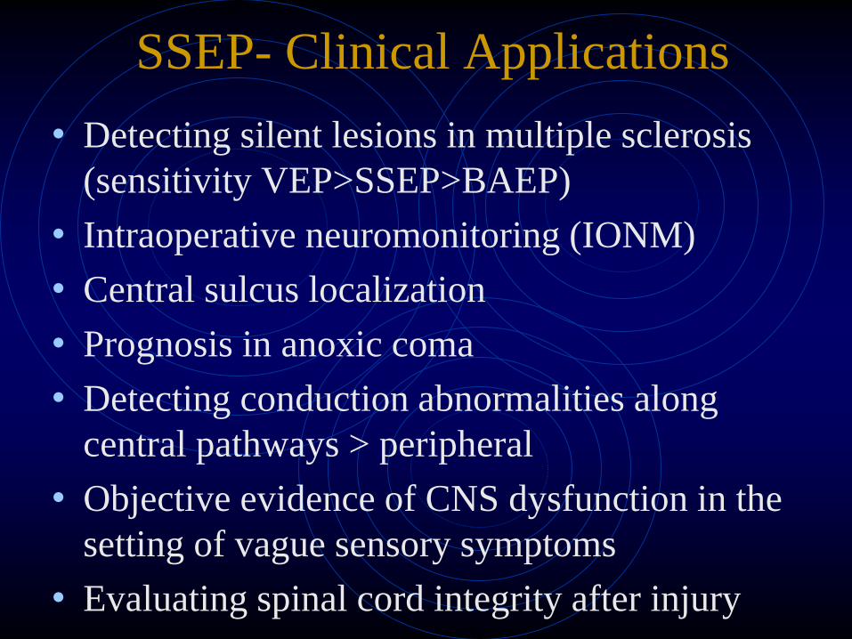
SSEP- Clinical Applications • Detecting silent lesions in multiple sclerosis
(sensitivity VEP>SSEP>BAEP) • Intraoperative neuromonitoring (IONM) • Central sulcus localization • Prognosis in anoxic coma • Detecting conduction abnormalities along
central pathways > peripheral • Objective evidence of CNS dysfunction in the
setting of vague sensory symptoms • Evaluating spinal cord integrity after injury
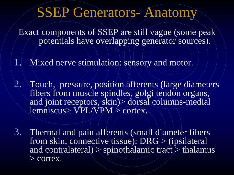
SSEP Generators- Anatomy Exact components of SSEP are still vague (some peak
potentials have overlapping generator sources).
1. Mixed nerve stimulation: sensory and motor.
2. Touch, pressure, position afferents (large diameters fibers from muscle spindles, golgi tendon organs, and joint receptors, skin)> dorsal columns-medial lemniscus> VPL/VPM > cortex.
3. Thermal and pain afferents (small diameter fibers from skin, connective tissue): DRG > (ipsilateral and contralateral) > spinothalamic tract > thalamus > cortex.
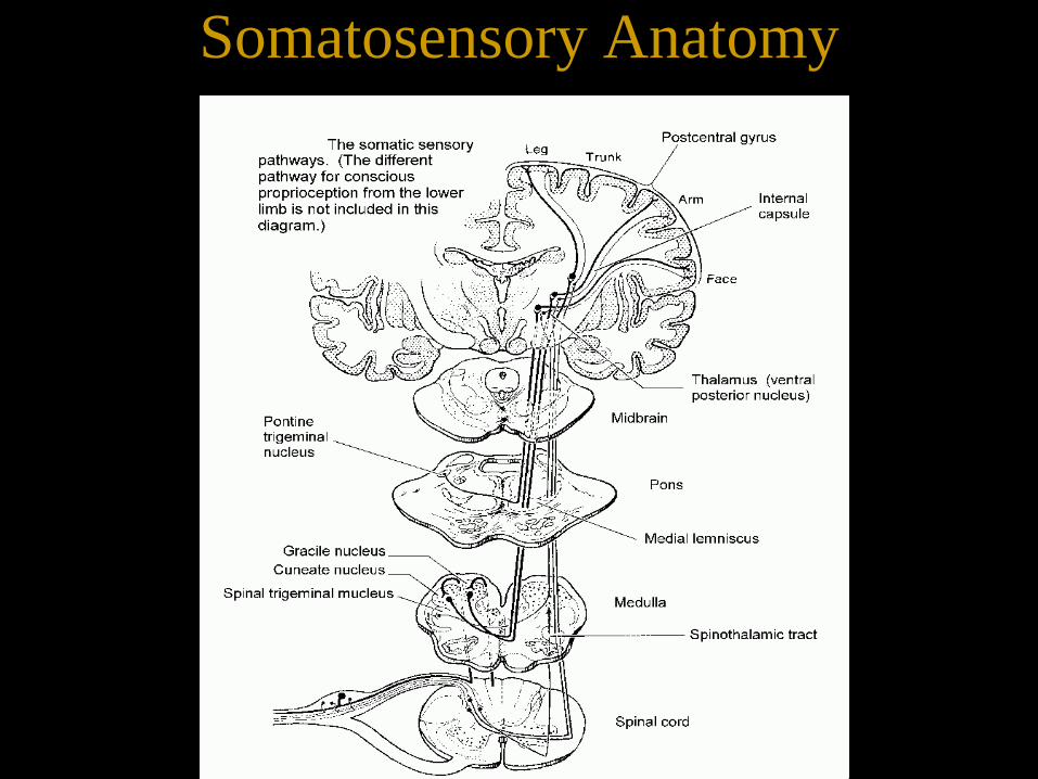
Somatosensory Anatomy
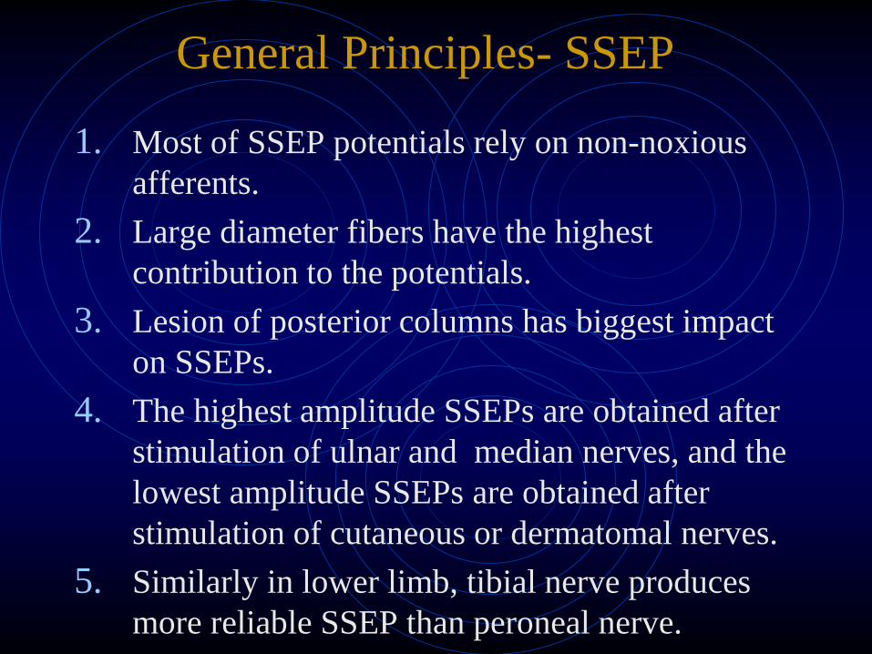
General Principles- SSEP
1. Most of SSEP potentials rely on non-noxious afferents.
2. Large diameter fibers have the highest contribution to the potentials.
3. Lesion of posterior columns has biggest impact on SSEPs.
4. The highest amplitude SSEPs are obtained after stimulation of ulnar and median nerves, and the lowest amplitude SSEPs are obtained after stimulation of cutaneous or dermatomal nerves.
5. Similarly in lower limb, tibial nerve produces more reliable SSEP than peroneal nerve.
General principles- SSEP
• The latencies of central SSEPs are a function of body height and limb length.
• The conduction velocity decreases and the latency of spinal and cortical responses is prolonged with low limb temperature. (33° for UE and 30° for LE).
• Latencies are prolonged as function of age (>60 years). Also children reach adult parameter ranges by age 8.
• Stimulation: unilateral (localization)>bilateral. Bilateral stimulation is used to increase amplitude of
potentials. (used when absent or poorly formed spinal or tibial scalp potentials).
SSEPs- subject variables • Age • Sex • Relaxed state • Sedated state • Height • Limb temperature • Neuropathy • Body habitus

SSEP- Artifacts
1. Muscle artifact: especially neck and paraspinal muscle contraction: can be controlled by relaxing the patient, using reclining chair or bed (Use sedation with diazepam if needed).
2. Electric artifact: Stimulus artifacts and 60 Hz: Use of stimulus –isolation device and fast-recovery amplifier (low impedance electrodes and lower intensities).
SSEP- Standard settings (ACNS) • Stimulus intensity: sufficient to cause a slight muscle twitch, higher used in
atrophied limbs (higher intensity is painful and recruit motor >sensory responses).
• Stimulus rate: 2-5 Hz for upper extremity stimulation and 1-2 Hz for lower extremity stimulation (rates greater than 10 Hz may cause an increase in latency and decrease in amplitude).
• Filter setting: 30-3000 Hz. (Not recommended to use the 60 Hz notch filter because SSEP in this range contains important physiologic information).
• Pulse width : 100-300 µs • Analysis time: 40 ms for UE and 60 ms for LE.
• Amplifier sensitivity: 10 µV for the spine and scalp potentials and 20 µV for
peripheral potentials.
• Number averaged: 500-2000 for reproducible wave forms (depends on noise).
• Electrode impedance: <5000 Ω.
SSEP- Limitation and averaging
• SSEP amplitude is low compared to noise of motor activity, movement artifact, ECG, EEG and electromagnetic activity in the environment.
• Averaging summates activity that is time locked to the stimulus trigger while gradually subtracting random background noise.
• Most artifacts can be eliminated by averages
rejecting sweeps that contain waveforms exceeding fixed maximal amplitudes.
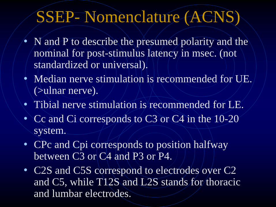
SSEP- Nomenclature (ACNS) • N and P to describe the presumed polarity and the
nominal for post-stimulus latency in msec. (not standardized or universal).
• Median nerve stimulation is recommended for UE. (>ulnar nerve).
• Tibial nerve stimulation is recommended for LE. • Cc and Ci corresponds to C3 or C4 in the 10-20
system. • CPc and Cpi corresponds to position halfway
between C3 or C4 and P3 or P4. • C2S and C5S correspond to electrodes over C2
and C5, while T12S and L2S stands for thoracic and lumbar electrodes.
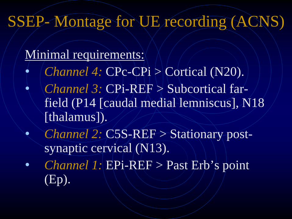
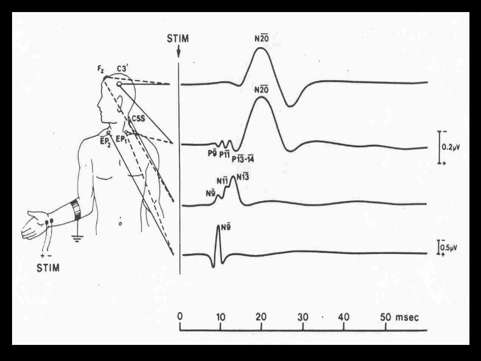
SSEP- Montage for UE recording (ACNS)
Minimal requirements: • Channel 4: CPc-CPi > Cortical (N20). • Channel 3: CPi-REF > Subcortical far-
field (P14 [caudal medial lemniscus], N18 [thalamus]).
• Channel 2: C5S-REF > Stationary post-synaptic cervical (N13).
• Channel 1: EPi-REF > Past Erb’s point (Ep).
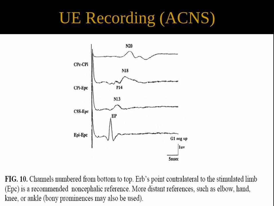
UE Recording (ACNS)
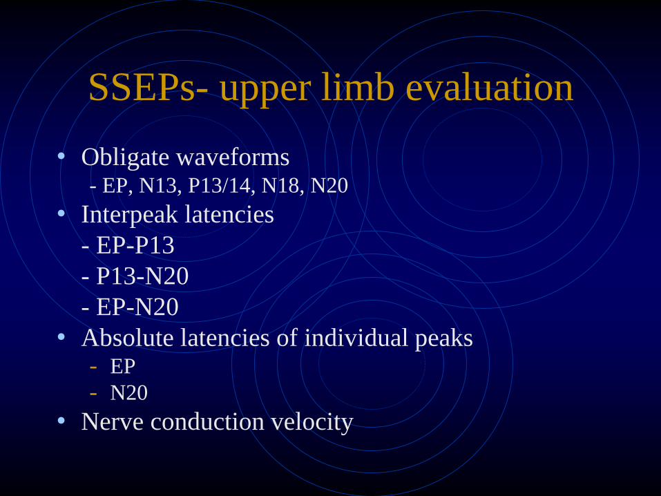
SSEPs- upper limb evaluation • Obligate waveforms
- EP, N13, P13/14, N18, N20 • Interpeak latencies - EP-P13 - P13-N20 - EP-N20 • Absolute latencies of individual peaks
- EP - N20
• Nerve conduction velocity
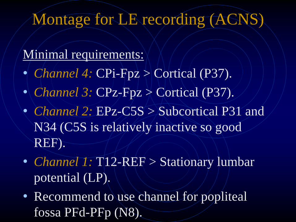
Montage for LE recording (ACNS)
Minimal requirements: • Channel 4: CPi-Fpz > Cortical (P37). • Channel 3: CPz-Fpz > Cortical (P37). • Channel 2: EPz-C5S > Subcortical P31 and
N34 (C5S is relatively inactive so good REF).
• Channel 1: T12-REF > Stationary lumbar potential (LP).
• Recommend to use channel for popliteal fossa PFd-PFp (N8).
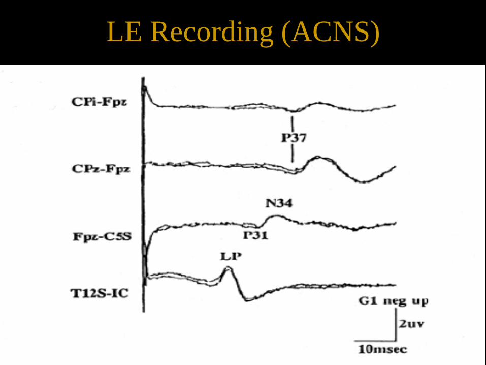
LE Recording (ACNS)
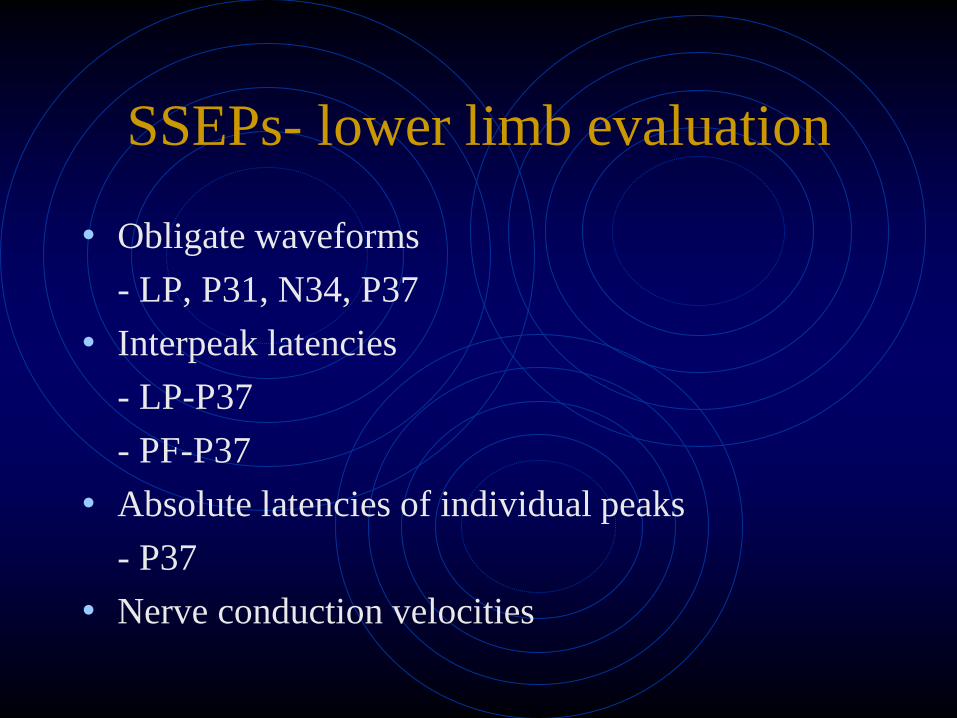
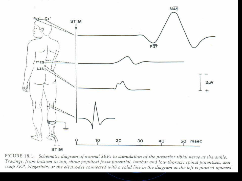
SSEPs- lower limb evaluation
• Obligate waveforms - LP, P31, N34, P37 • Interpeak latencies - LP-P37 - PF-P37 • Absolute latencies of individual peaks - P37 • Nerve conduction velocities
SSEP- Normal Values
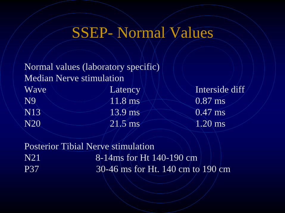
Normal values (laboratory specific) Median Nerve stimulation Wave Latency Interside diff N9 11.8 ms 0.87 ms N13 13.9 ms 0.47 ms N20 21.5 ms 1.20 ms Posterior Tibial Nerve stimulation N21 8-14ms for Ht 140-190 cm P37 30-46 ms for Ht. 140 cm to 190 cm
SSEP- Principles of interpretation
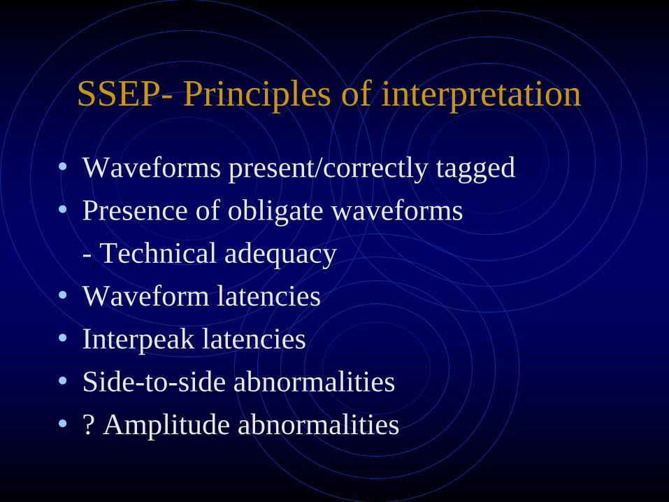
• Waveforms present/correctly tagged • Presence of obligate waveforms - Technical adequacy • Waveform latencies • Interpeak latencies • Side-to-side abnormalities • ? Amplitude abnormalities
Median SSEP- Dipole
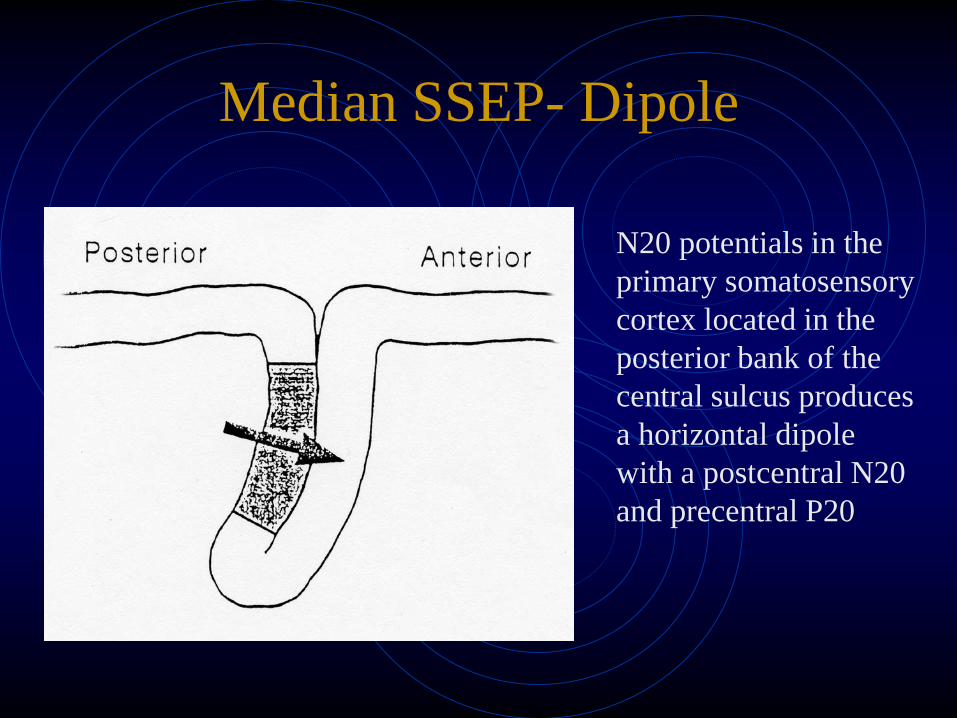
N20 potentials in the primary somatosensory cortex located in the posterior bank of the central sulcus produces a horizontal dipole with a postcentral N20 and precentral P20
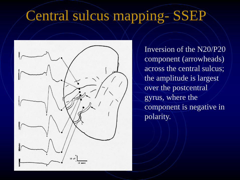
Central sulcus mapping- SSEP
Inversion of the N20/P20 component (arrowheads) across the central sulcus; the amplitude is largest over the postcentral gyrus, where the component is negative in polarity.
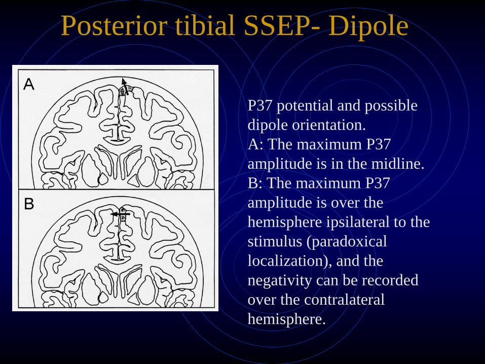
Posterior tibial SSEP- Dipole
P37 potential and possible dipole orientation. A: The maximum P37 amplitude is in the midline. B: The maximum P37 amplitude is over the hemisphere ipsilateral to the stimulus (paradoxical localization), and the negativity can be recorded over the contralateral hemisphere.
SSEP in Cardiac Arrest
• SSEPs have emerged as a reliable test for prognostication after cardiac arrest (CA).
• The short latency cortical response (N20) has been extensively evaluated in the prediction of never awakening.
• Meta-analysis from 18 studies and 1136 patients found 336 patients who had bilaterally absent N20 after cardiac arrest never recovered.
• Sensitivity for identifying patients who would never awaken was 42% with a PPV of 100%.
Chiappa, Lippincott-Raven 1997
SSEP- Brain death
• All SEPs rostral to N 13 are absent. • Presence of N13 is helpful because it establishes
that the input signal has reached the CNS.
• Can be used as a confirmatory test. • Very important tool in evaluating the patient in
barbiturate coma with suspected brain death.
Chiappa, Lippincott-Raven 1997
SSEP- Conclusions
• SSEP records volume conducted activity arising from myelinated peripheral and central axons, and synapses in the gray matter.
• Provide an objective measurement of function in large diameter myelinated sensory afferents peripherally and in proprioceptive pathway centrally.
• Changes in the amplitude and latency helps to localize lesions in the central nervous system.
• IONM use is most common. • Brain death and coma evaluation is helpful.
Intraoperative neuromonitoring (IONM)
• Patient’s own latencies preoperatively serve as baseline. • Populations norms are not applicable. • Spontaneous:
• EMG (Electromyography) • EEG (Electroencephalography)
• Evoked responses: • SSEP (Somatosensory Evoked Potentials) • MEP (Motor Evoked Potentials) • BAEP (Brainstem Auditory Evoked Potentials) • t-EMG (Triggered or Evoked EMG) • VEP (Visual Evoked Potentials)
• TCD (Transcranial doppler)
SSEP in IOM- General Principles
• Standard of care/practice (scoliosis, cervical or thoracic surgeries, vascular surgeries, brainstem tumors…).
• Always perform four-limb study. • Often include MEPs (remember vascular
anatomy). • Ulnar nerve stimulation preferred over median. • Patient’s own latencies preoperatively serve as
baseline (post anesthesia). • Populations normal are not applicable. • Warning criteria: 10 % increase in latency or 50 %
decrease in amplitude.
Waveform changes- Possible Mechanisms
• Ischemia and anoxia • Acute trauma • Temperature • Arterial CO2 • Hypotension • Anesthesia • Technical issues
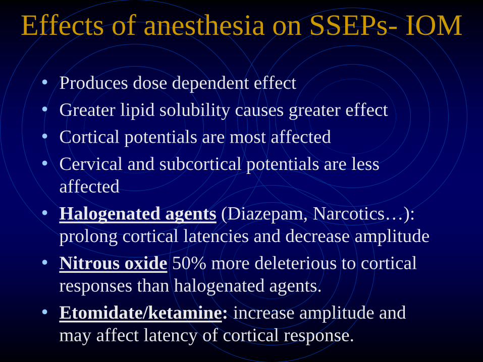
Effects of anesthesia on SSEPs- IOM
• Produces dose dependent effect • Greater lipid solubility causes greater effect • Cortical potentials are most affected • Cervical and subcortical potentials are less
affected • Halogenated agents (Diazepam, Narcotics…):
prolong cortical latencies and decrease amplitude • Nitrous oxide 50% more deleterious to cortical
responses than halogenated agents. • Etomidate/ketamine: increase amplitude and
may affect latency of cortical response.
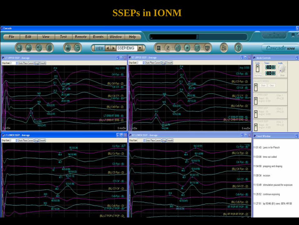
SSEPs in IONM
Intra-operative BAEP
• BAEPs are elicited by auditory stimulation and represent activity generated by the CN VIII and brainstem.
• BAEPs are useful for posterior fossa surgeries: • Trigeminal neuralgia. • Vestibular schwannoma resections. • Vertebrobasilar aneurysms. • Vascular malformation repairs. • Microvascular cranial nerve decompressions. • Skull based (especially posterior fossa), CPA tumor
resections.

Intra-operative BAEP
• Operative procedures in or adjacent to the CP angle carries an increased risk to the auditory nerve and the brainstem.
• Other CNs (usually V & VII) are monitored via EMG in conjunction with BAEPs.
• Intra-operative BAEP loss & postoperative deafness is due to interruption of the blood supply or traumatic transection of the auditory nerve or cochlea.
Intra-operative BAEP • Latency increases of 0.5 to 1ms are borderline. • Latency increases of 1 to 1.5ms require
notification of the surgeon (alternatively a 10% increase could be used as criteria for warning the surgeon).
• Amplitude decrement of 50%. • Interpeak latencies can also be helpful but this
will vary with the patient according to the baseline value; in general small changes from baseline can be significant. • I-III: 2.1ms • III-V: 1.9ms • I-V: 4.0ms
Intra-operative BAEP
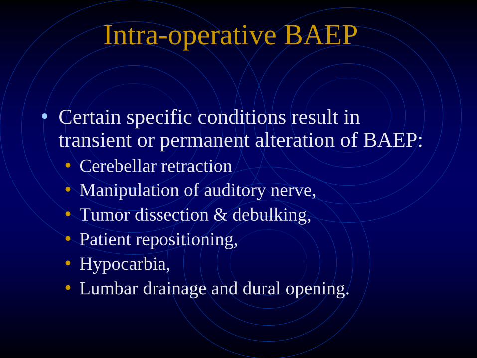
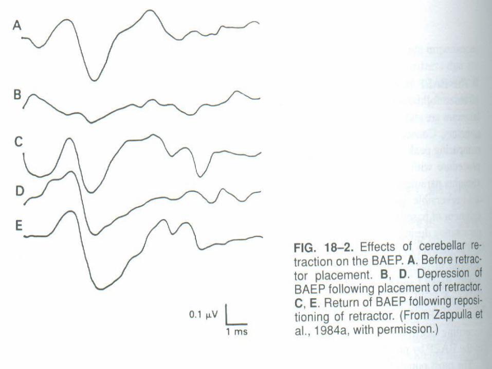
• Certain specific conditions result in transient or permanent alteration of BAEP: • Cerebellar retraction • Manipulation of auditory nerve, • Tumor dissection & debulking, • Patient repositioning, • Hypocarbia, • Lumbar drainage and dural opening.
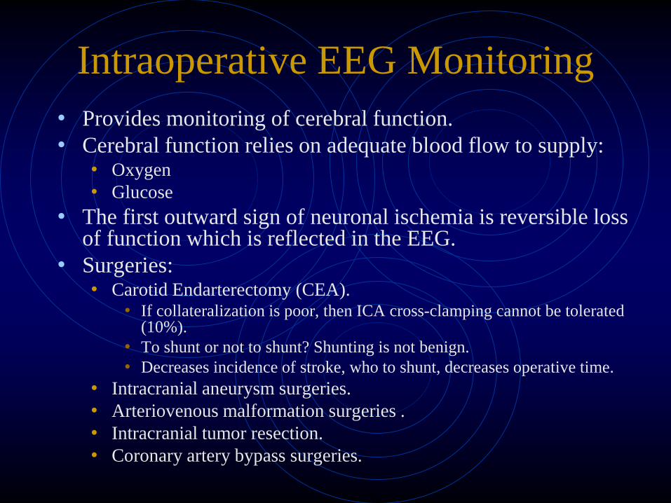
Intraoperative EEG Monitoring • Provides monitoring of cerebral function. • Cerebral function relies on adequate blood flow to supply:
• Oxygen • Glucose
• The first outward sign of neuronal ischemia is reversible loss of function which is reflected in the EEG.
• Surgeries: • Carotid Endarterectomy (CEA).
• If collateralization is poor, then ICA cross-clamping cannot be tolerated (10%).
• To shunt or not to shunt? Shunting is not benign. • Decreases incidence of stroke, who to shunt, decreases operative time.
• Intracranial aneurysm surgeries. • Arteriovenous malformation surgeries . • Intracranial tumor resection. • Coronary artery bypass surgeries.
Intraoperative EEG Interpretation
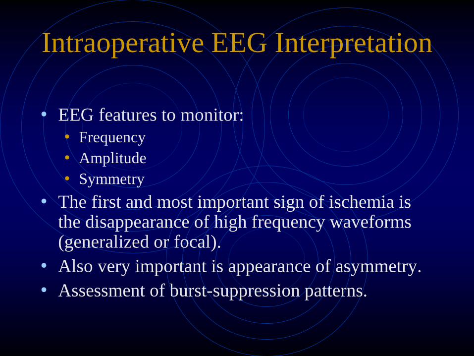
• EEG features to monitor: • Frequency • Amplitude • Symmetry
• The first and most important sign of ischemia is the disappearance of high frequency waveforms (generalized or focal).
• Also very important is appearance of asymmetry. • Assessment of burst-suppression patterns.
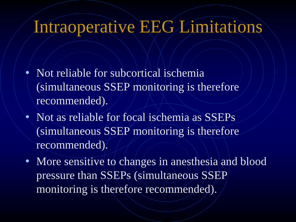
Intraoperative EEG Limitations
• Not reliable for subcortical ischemia (simultaneous SSEP monitoring is therefore recommended).
• Not as reliable for focal ischemia as SSEPs (simultaneous SSEP monitoring is therefore recommended).
• More sensitive to changes in anesthesia and blood pressure than SSEPs (simultaneous SSEP monitoring is therefore recommended).
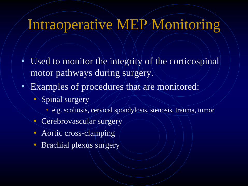
Intraoperative MEP Monitoring
• Used to monitor the integrity of the corticospinal motor pathways during surgery.
• Examples of procedures that are monitored: • Spinal surgery
• e.g. scoliosis, cervical spondylosis, stenosis, trauma, tumor
• Cerebrovascular surgery • Aortic cross-clamping • Brachial plexus surgery
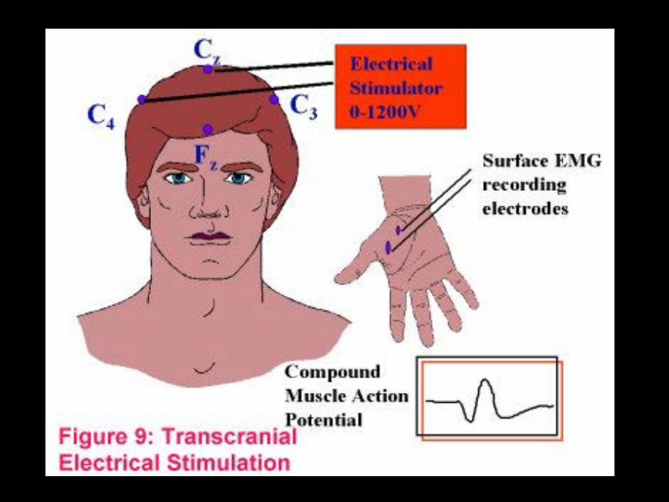
TCMEP: Anesthesia approach
• Total Intravenous Anesthesia (TIVA). • Usually include opioids with ketamine,
etomidate, or closely-titrated propofol. • Neuromuscular blockade
• 3-4/4 twitches is preferred
EMG indications in IONM
• Posterior fossa surgery - Microvascular decompression for hemifacial spasm or trigeminal neuralgia; Surgical resection of the sensory branch of the trigeminal nerve for treatment of cluster headache
• Spinal surgery – especially helps with proper pedicle screw placement with cervical or lumbar fusion; and surgery below the conus.
Operative factors affecting EMG
• Paralytic agents will dampen or altogether silence the EMG response. • Train of Four (TOF) is used to assess muscle relaxation (a train of
four electrical stimuli is delivered to a designated muscle and the number of twitches is recorded).
• 4 twitches: no significant relaxation • 0 twitches: paralysis • 3-4 twitches: ideal for monitoring (reduces muscle tension that creates
a noisy background)
• Inhalation or intravenous anesthetic agents will not have a significant effect on EMG.
• Blood pressure and temperature changes will not have a significant effect on EMG.
• Chronically compressed nerves may have enhanced sensitivity to neuromuscular blockade.
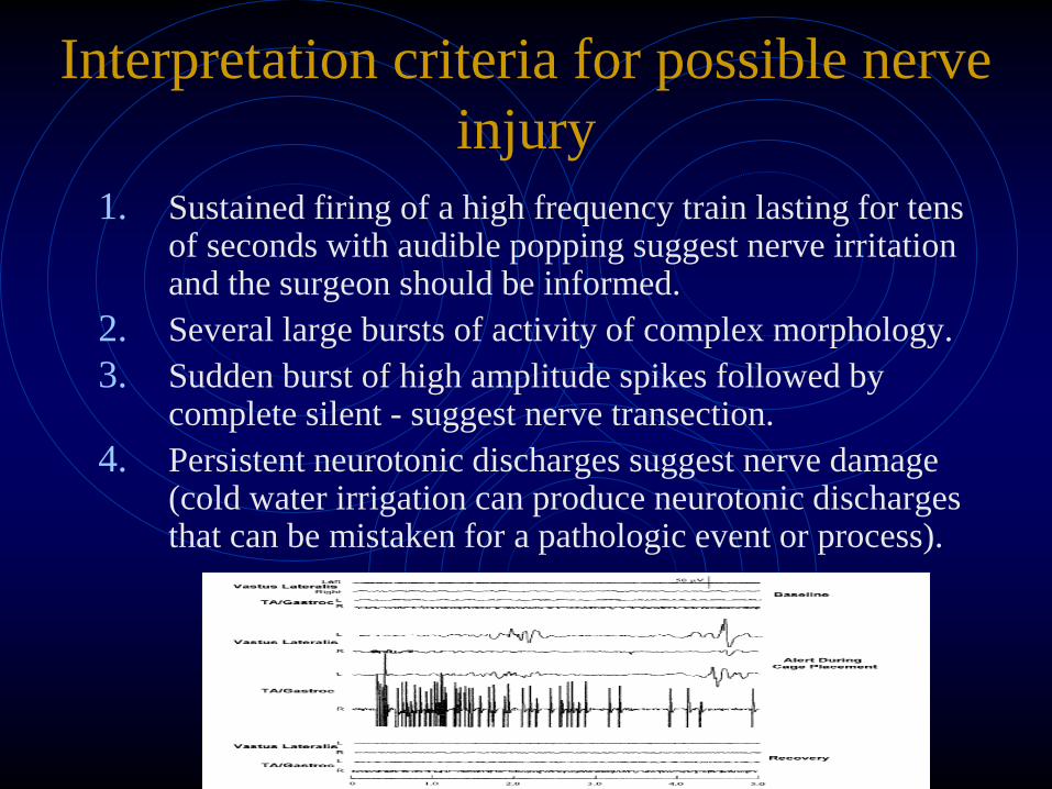
Interpretation criteria for possible nerve injury
1. Sustained firing of a high frequency train lasting for tens of seconds with audible popping suggest nerve irritation and the surgeon should be informed.
2. Several large bursts of activity of complex morphology. 3. Sudden burst of high amplitude spikes followed by
complete silent - suggest nerve transection. 4. Persistent neurotonic discharges suggest nerve damage
(cold water irrigation can produce neurotonic discharges that can be mistaken for a pathologic event or process).
Question 1:
• SSEPs test the integrity of: • A- Corticospinal tract • B- Spinothalamic tract • C- Posterior columns * • D- Spinocerebellar tract
Question 2:
• The N11-13 complex corresponds to: • A- Brachial plexus • B- Cauda equina • C- Cervicomedullary junction * • D- Thalamus
Question 3:
• The first sign of ischemia on EEG during IONM is : • A- Decrease in amplitude • B- Loss of high frequency waveforms * • C- Loss of occipital rhythm • D- All of the above
Question 4:
• With median nerve stimulation, which of the following may produce a false negative cortical potentials: • A- Isoflurane • B- Nitrous oxide • C- Etomidate * • D- Desflurane