Solving the Enigma of Frozen Shoulder Luise …...Solving the Enigma of Frozen Shoulder Luise...
89
Solving the Enigma of Frozen Shoulder Luise Hollmann Sydney School of Medicine The University of Sydney 2017 A thesis submitted in fulfilment of the requirements for the degree of Master of Philosophy
Transcript of Solving the Enigma of Frozen Shoulder Luise …...Solving the Enigma of Frozen Shoulder Luise...

Solving the Enigma of Frozen Shoulder
Luise Hollmann
Sydney School of Medicine
The University of Sydney
2017
A thesis submitted in fulfilment of the requirements for the degree of Master of Philosophy

ii
AUTHOR DECLARATION
This is to certify that to the best of my knowledge, the content of this thesis is my own work. This
thesis has not been submitted for any degree or other purposes.
I certify that the intellectual content of this thesis is the product of my own work and that all the
assistance received in preparing this thesis and sources have been acknowledged.
Luise Hollmann

iii
ABSTRACT
Frozen shoulder is a common shoulder condition affecting 2-5% of the population. It is characterised
by the spontaneous onset of pain, stiffness and range of motion (ROM) loss at the shoulder. The
exact pathophysiology of frozen shoulder is unclear. However, it is commonly believed that a
combination of capsular contracture and fibrosis of the rotator cuff interval, the subscapular recess
and the coracohumeral ligament lead to global movement restriction of the glenohumeral joint.
There is no gold standard clinical test for frozen shoulder. Frozen shoulder is therefore a diagnosis of
exclusion and relies on the accurate assessment of active and passive ROM. The generally accepted
diagnostic criteria for frozen shoulder are active as well as passive movement restriction in at least
two planes of shoulder range of motion, one being external rotation. However, the accuracy of
active and passive ROM assessment has not been tested in people with frozen shoulder. Further, the
evidence of the effectiveness of treatments for frozen shoulder that aim to stretch the presumed
tight shoulder structures has been questioned recently. The overall aim of this thesis was to analyse
the effectiveness of stretch-based treatments for frozen shoulder and to investigate if capsular
contracture is responsible for movement loss in frozen shoulder.
Chapter 2 of this thesis contains a systematic review of stretch based treatments for frozen
shoulder. The aim of the review was to analyse the current evidence regarding the effectiveness of
interventions that aim to stretch the tissues of the shoulder region or release the presumed capsular
fibrosis. The findings of six high quality randomised controlled clinical trials were reported and
discussed. The RCTs included in the study evaluated the effectiveness of manipulation under general
anaesthetic (MUA), manual therapy, distension and stretching & strengthening exercises on pain,
ROM and function in frozen shoulder. Overall, it was found that mobilisation combined with
stretching may result in small gains in passive ROM in the short term compared to stretching and
strengthening exercises. Physiotherapy after capsular distension consisting of manual therapy and
stretching and strengthening exercise provides no additional benefit in terms of pain, function, or
iv
quality of life over sham-ultrasound, but may result in improved active ROM in the short term.
However, these improvements may not be clinically significant. Distension, regardless of the
medium used to distend the glenohumeral capsule, had no benefit with respect to pain, disability or
shoulder abduction and flexion ROM over cortisone injection alone in the short term. Distension
with hyaluronic acid lead to a small increase in passive external rotation ROM compared to a
glenohumeral corticosteroid injection. MUA did not confers any additional benefit over a home
exercise program in terms of pain, function and ROM in people with frozen shoulder
Chapter 3 contains a cohort study investigating external rotation ROM in healthy shoulders. The
effects of sex, handedness, shoulder and body position on active and passive ROM were investigated
in twenty healthy participants. The results indicate that passive external rotation ROM was
significantly greater than active ROM in people with healthy shoulders. Both active and passive
shoulder external rotation ROM were greater when the arm was abducted at 90 degrees compared
to lower positions of abduction. There was no difference in active or passive external rotation ROM
between dominant and non-dominant shoulders. Female subjects demonstrated significantly more
passive external rotation ROM than males. This study also found that measuring external rotation
ROM with the arm by the side yields similar results to external rotation ROM measured in side-lying
in 45 degrees of abduction. The latter is not commonly utilised in clinical practice but was the
position required for external rotation ROM measurement for the study in Chapter 4 as dictated by
the participant position in preparation for shoulder surgery.
Finally, Chapter 4 contains a case series of five subjects with global restriction of active and passive
shoulder movement of greater than 50% of normal ROM in external rotation and at least one other
plane of movement. This study demonstrates that capsular contracture is not a major contributor to
movement restriction in all patients who exhibit classical clinical features of frozen shoulder.
Although all five cases presented with painful, global restriction of passive shoulder movement, four
subjects demonstrated significantly greater abduction range of motion (ROM) and three

v
demonstrated significantly greater external rotation ROM under anaesthesia. These findings
highlight the need to reconsider the diagnostic process used for frozen shoulder as well as our
understanding of the pathology of frozen shoulder and offers an explanation for why treatments
aimed at stretching tight passive structures have not proven to be more effective.

vi
Publications and presentations from this thesis
The following paper has been submitted for publication to the New England Journal of Medicine:
‘Capsular contracture is not a major contributor to range of motion loss in some patients with frozen
shoulder.’
Hollmann L., Halaki M., Haber M., Ginn K.
The following manuscript has been prepared for submission to the British Journal of Sports
Medicine:
‘Are stretching techniques effective in the treatment of frozen shoulder? A systematic review’
Hollmann L., Halaki M., Ginn K.
At the time of submitting this thesis the following abstracts had been published and oral
presentations given:
Hollmann, L., Halaki, M., Haber, M., Herbert, R., Dalton, S., & Ginn, K. (2015). Determining the
contribution of active stiffness to reduced range of motion in frozen shoulder. Physiotherapy, 101,
e585.
World Congress of Physiotherapy, Singapore 2015 – Platform Presentation: Determining the
contribution of active stiffness to reduced range of motion in frozen shoulder. (Presented by L
Hollmann)
Shoulder and Elbow Physiotherapists Australasia, Sydney 2015 – 5x5 Presentation: Determining
the contribution of active stiffness to reduced range of motion in frozen shoulder. (Presented by L
Hollmann)
International Congress of Shoulder and Elbow Therapists, Edinburgh 2016: Contribution of ‘Active
Stiffness’ to the Clinical Presentation Known as Frozen Shoulder. (Presented by K Ginn)
European Society for Shoulder and Elbow Rehabilitation, Gothenburg 2016: ‘Frozen Shoulder – Is it
really frozen.’ (Presented by L Hollmann)

vii
Table of Contents
Chapter 1
Chapter 2
Chapter 3
Chapter 4
Chapter 4.1
Chapter 4.2
Chapter 5
Author Declaration
Abstract
Publications and presentations from this thesis
List of Tables
List of Figures
Introduction………………………………………………………………………………………………..
Literature Review………………………………………………………………………………………..
Introduction………………………………………………………………………………………………..
Methods……………………………………………………………………………………………………..
Results………………………………………………………………………………………………………..
Discussion……………………………………………………………………………………………………
Shoulder External Rotation Range of Motion in the Healthy Shoulder………
Introduction…………………………………………………………………………………………………
Methods………………………………………………………………………………………………………
Results ………………………………………………………………………………………………………..
Discussion……………………………………………………………………………………………………
Active Stiffness in Frozen Shoulder…………………………………………………………….
Introduction………………………………………………………………………………………………...
Methods……………………………………………………………………………………………………...
Results ………………………………………………………………………………………………………..
Discussion……………………………………………………………………………………………………
Active Stiffness in Frozen Shoulder - Additional Methods……………………………
Active Stiffness in Frozen Shoulder – Additional Results……………………………..
Conclusion………………………………………………………………………………………………….
ii
iii
vi
ix
ix
1
9
10
11
15
21
25
25
28
32
35
40
42
43
44
47
49
56
58

viii
Appendix
Summary of Projects……………………………………………………………………………………
Summary of Main Findings………………………………………………………………………….
Clinical Implications…………………………………………………………………………………….
Directions for Future Research…………………………………………………………………….
A………………………………………………………………………………………………………………….
B………………………………………………………………………………………………………………….
58
59
60
61
68
78

ix
List of Tables
Table 3.1: Effect of sex, body position and handedness on active and passive external
rotation ROM……………………………………………………………………………………………………………….… 34
Table 4.2.1: Participant demographic information, SPADI and PCS Scores……………………………………….56
Table 4.2.2: Active ROM measures…………………………………………………………………………………………………. 57
Table 4.2.3: Passive Abduction and External Rotation ROM pre and post anaesthesia in degrees.…. 57
List of Figures
Figure 3.1: Measurement of Active Shoulder External Rotation in sitting…………………………………..….. 29
Figure 3.2: Measurement of Active Shoulder External Rotation in supine………………………………..……. 30
Figure 3.3: Active shoulder external rotation range of motion……………………………………………………….. 32
Figure 3.4: Passive shoulder external rotation range of motion…………………………………………………….. 33
Figure 4.1.1: Passive Shoulder abduction ROM measurement……………………………………………………….. 51
Figure 4.1.2: Hand behind back ROM measurement………………………………………………………………………. 53

1
Chapter 1 - Introduction
Frozen shoulder, also termed adhesive capsulitis, is a common shoulder condition affecting 2-5% of
the population (A. S. Neviaser & Hannafin, 2010). It is characterised by the spontaneous onset of
pain, progressive stiffness and range of motion loss at the shoulder accompanied by significant
disability (R. Buchbinder et al., 2007; Kelley et al., 2013). People who experience the condition
usually complain of pain near the deltoid insertion, severe night pain, pain with sudden movements,
and severe movement restriction (Lewis, 2015; Reeves, 1975). Frozen shoulder can be described as
either primary, if the aetiology is unknown, or secondary, when it can be attributed to another cause
such as trauma or surgery to the affected shoulder (Kelley et al., 2013). For the treating clinician,
frozen shoulder presents a challenge both in terms of diagnosis and treatment.
Who gets frozen shoulder?
In China and Japan frozen shoulder is referred to as “50 year old” shoulder as a result of the
condition primarily affecting people in the 5th decade of life (Lewis, 2015). There is no certainty
about whether men or women are more commonly affected (Lewis, 2015). Frozen shoulder has a
strong association with diabetes mellitus, occurring in 20% to 36% of the diabetic population (T. D.
Bunker & Anthony, 1995). Arthroscopic observations have revealed an appearance of the shoulder
capsule similar to that found in the finger flexor tendons of patients with Duputren’s disease and
reports suggest an association between the two conditions with Duputren’s contracture being eight
times more common in people with frozen shoulder compared to the normal population (T. D.
Bunker & Anthony, 1995; Smith, Devaraj, & Bunker, 2001). Risk factors for frozen shoulder also
include hypothyroidism, family history and genetic predisposition (Lewis, 2015).
Natural History
Frozen shoulder is generally described as a self-limiting condition; i.e. it resolves spontaneously even
without treatment. The literature frequently describes frozen shoulder as passing through three

2
distinct stages; the freezing stage (most painful, gradually increasing stiffness), the frozen stage
(characterised by severe stiffness and movement restriction) and the thawing (resolution) stage
where pain and stiffness gradually diminish until a full resolution of symptoms is reached (Reeves,
1975). However, a recent systematic review found no studies that demonstrated a recovery pattern
consistent with the theoretical pain, stiff and thawing recovery phases as suggested by Reeves
(Wong et al., 2016). The average duration of symptoms is 30.1 months (Reeves, 1975). However, it
has been reported that up to 50% experience ongoing pain and stiffness seven years post onset of
symptoms (Shaffer, Tibone, & Kerlan, 1992). Whilst Reeves was the first document the different
stages of frozen shoulder, this belief is still commonly held by many practitioners today even though
evidence of distinct phases may be lacking. (Lewis, 2015; Shaffer et al., 1992; Wong et al., 2016)
Historical Perspective
Frozen shoulder was first described by Duplay in 1872 and termed peri-arthritis. The key symptoms
as described earlier, i.e. gradual onset of shoulder stiffness, severe pain, especially at night, and
restriction in active and passive range of movement of the shoulder, were initially attributed to
inflammation of the subacromial and subdeltoid bursae (Codman, 1911). However, other
investigators favoured the opinion that the joint capsule was the site of the pathological changes in
frozen shoulder. In 1945, Neviaser was the first to described fibrosis and adhesions in the shoulder
capsule in 10 cases and suggested the term ‘adhesive capsulitis’ as a more accurate description of
the condition (J. S. Neviaser, 1945). Observational studies conducted since then concur with these
findings and it is generally accepted that the shoulder capsule is the cause of symptoms in frozen
shoulder (G. Hand, Athanasou, Matthews, & Carr, 2007; T. D. Bunker & Anthony, 1995) .
Pathophysiology
The exact pathophysiology of frozen shoulder remains unclear. Acute inflammation as originally
suggested, does not appear to occur in frozen shoulder (T. D. Bunker & Anthony, 1995; B. J.
3
Lundberg, 1969). However, Hand et al. described the presence of chronic inflammatory cells,
including mast cells and macrophages (G. Hand, Athanasou, Matthews, & Carr, 2007). Other studies
have noted hypervascularity of the shoulder capsule and the presence of fibroblasts (cells associated
with initiating inflammation) and myofibroblasts (cells associated with contractile scar tissue) within
the coracoacromial ligament (T. D. Bunker & Anthony, 1995; Bo J Lundberg, 1969; Simmonds, 1949).
The presence of these cells may explain the process of pain and adhesions described in frozen
shoulder.
Arthroscopic evaluations of the shoulder have revealed the presence of fibrosis particularly in the
rotator cuff interval (the space between the tendons of subscapularis, supraspinatus, and the base
of the coracoid process), the subscapular recess and the coracohumeral ligament (Uitvlugt, Detrisac,
Johnson, Austin, & Johnson, 1993; A. M. Wiley, 1991). In summary, it is suspected that a process of
chronic inflammation leads to scarring and fibrosis of the capsule and it is believed that this process
is responsible for pain and range of motion loss in patients with frozen shoulder. It is unclear,
however if or how the fibrosis and scarring of the glenohumeral capsule resolves in people who
experience a resolution of their frozen shoulder symptoms.
Diagnosis
There is no gold standard clinical test for frozen shoulder. The generally accepted diagnostic criteria
for frozen shoulder are active as well as passive movement restriction in at least two planes of
shoulder range of motion, one being external rotation. A plain X-ray is needed to exclude other
pathologies, such as glenohumeral osteoarthritis, locked dislocations and tumours, that can present
with active and passive movement restriction and therefore, mimic frozen shoulder. The diagnosis
therefore, relies on accurate clinical assessment of active and passive range of motion and the
exclusion of other pathologies.
4
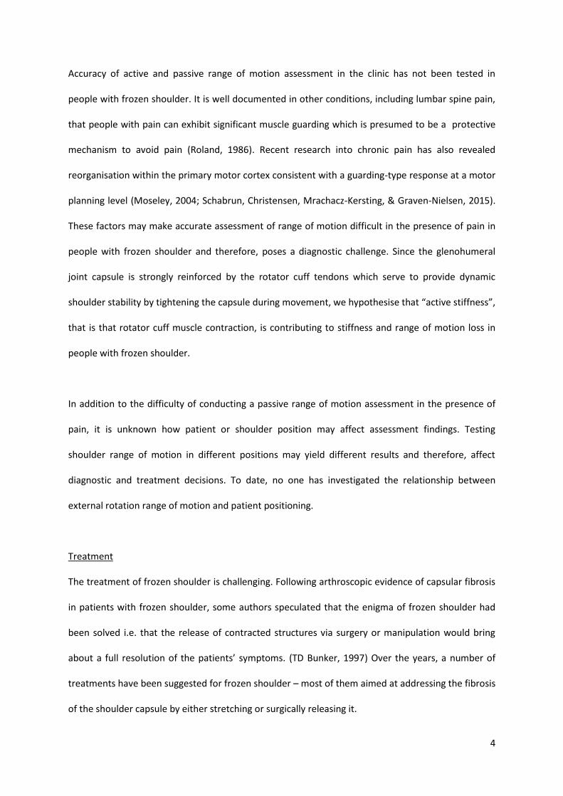
Accuracy of active and passive range of motion assessment in the clinic has not been tested in
people with frozen shoulder. It is well documented in other conditions, including lumbar spine pain,
that people with pain can exhibit significant muscle guarding which is presumed to be a protective
mechanism to avoid pain (Roland, 1986). Recent research into chronic pain has also revealed
reorganisation within the primary motor cortex consistent with a guarding-type response at a motor
planning level (Moseley, 2004; Schabrun, Christensen, Mrachacz-Kersting, & Graven-Nielsen, 2015).
These factors may make accurate assessment of range of motion difficult in the presence of pain in
people with frozen shoulder and therefore, poses a diagnostic challenge. Since the glenohumeral
joint capsule is strongly reinforced by the rotator cuff tendons which serve to provide dynamic
shoulder stability by tightening the capsule during movement, we hypothesise that “active stiffness”,
that is that rotator cuff muscle contraction, is contributing to stiffness and range of motion loss in
people with frozen shoulder.
In addition to the difficulty of conducting a passive range of motion assessment in the presence of
pain, it is unknown how patient or shoulder position may affect assessment findings. Testing
shoulder range of motion in different positions may yield different results and therefore, affect
diagnostic and treatment decisions. To date, no one has investigated the relationship between
external rotation range of motion and patient positioning.
Treatment
The treatment of frozen shoulder is challenging. Following arthroscopic evidence of capsular fibrosis
in patients with frozen shoulder, some authors speculated that the enigma of frozen shoulder had
been solved i.e. that the release of contracted structures via surgery or manipulation would bring
about a full resolution of the patients’ symptoms. (TD Bunker, 1997) Over the years, a number of
treatments have been suggested for frozen shoulder – most of them aimed at addressing the fibrosis
of the shoulder capsule by either stretching or surgically releasing it.

5
Treatments for frozen shoulder generally aim to reduce pain and to restore range of motion. The
most commonly prescribed treatments for frozen shoulder in Australia include physiotherapy
(stretching exercises and/or manual techniques to the glenohumeral joint with the goal of
lengthening muscle or soft tissues), joint distension (a technique where a combination of saline and
a corticosteroid is injected into the shoulder joint until the shoulder capsule ruptures), manipulation
under general anaesthetic (a forceful manipulation of the glenohumeral joint by an orthopaedic
surgeon), surgery to release the capsule (an arthroscopic operation in which fibrotic tissue cut and
removed); and corticosteroid injections into the glenohumeral joint with the aim of reducing
inflammation and pain. There is conflicting evidence as to the effectiveness of these treatments for
frozen shoulder (Favejee, Huisstede, & Koes, 2011; Lewis, 2015).
In summary, many aspects of the pathophysiology, diagnosis and treatment of frozen shoulder are
poorly understood. Specifically, it is unclear whether passive range of motion measurements, the
definitive diagnostic tests for frozen shoulder, are an accurate representation of the shoulder joint’s
available range of motion, thus challenging the validity of these tests.. Further, it is unknown how
effective the most commonly used treatments for frozen shoulder are in terms of reducing pain, and
improving shoulder range of motion and function. This thesis aims to aid in our understanding of
diagnosis and treatment in frozen shoulder by addressing these knowledge gaps. Our aim is:
(1) To investigate whether current evidence supports the efficacy of treatments for frozen
shoulder aimed at stretching the shoulder capsule or tissues in relieving pain, improving range of
motion or reducing disability in patients with frozen shoulder.
(2) To establish normal active and passive shoulder external rotation range of motion in people
without shoulder pain taking into account the effect of patient and shoulder position.

6
(3) To investigate whether active stiffness (muscle guarding) contributes to range of motion loss
in people with frozen shoulder.
The effectiveness of stretch-based treatments for frozen shoulder will be investigated in a
systematic review. Shoulder external rotation range of motion in subjects without shoulder pain will
be investigated in a cross-sectional study. The effect of sex, handedness and patient position will be
investigated. Finally, a further study will investigate the contribution of active stiffness to movement
restriction in people with frozen shoulder. Passive abduction and external rotation range of motion
will be compared before and after general anaesthesia to establish whether passive range of motion
is affected by pain or muscle guarding in the awake patient.

7
References
Buchbinder, R., Youd, J. M., Green, S., Stein, A., Forbes, A., Harris, A., . . . Wright, W. J. L. (2007).
Efficacy and cost-effectiveness of physiotherapy following glenohumeral joint distension for
adhesive capsulitis: A Randomized trial. Arthritis & Rheumatism-Arthritis Care & Research,
57(6), 1027-1037.
Bunker, T. (1997). Frozen shoulder: unravelling the enigma. Annals of the Royal College of Surgeons
of England, 79(3), 210.
Bunker, T. D., & Anthony, P. P. (1995). The pathology of frozen shoulder. A Dupuytren-like disease.
Journal of Bone and Joint Surgery - Series B, 77(5), 677-683.
Codman, E. A. (1911). On stiff and painful shoulders. The Boston Medical and Surgical Journal,
164(22), 613-620.
Favejee, M., Huisstede, B., & Koes, B. (2011). Frozen shoulder: the effectiveness of conservative and
surgical interventions—systematic review. British journal of sports medicine, 45(1), 49-56.
Hand, G., Athanasou, N., Matthews, T., & Carr, A. (2007). The pathology of frozen shoulder. Bone &
Joint Journal, 89(7), 928-932.
Kelley, M. J., Shaffer, M. A., Kuhn, J. E., Michener, L. A., Seitz, A. L., Uhl, T. L., . . . Davenport, T.
(2013). Shoulder pain and mobility deficits: adhesive capsulitis: clinical practice guidelines
linked to the International Classification of Functioning, Disability, and Health from the
Orthopaedic Section of the American Physical Therapy Association. Journal of Orthopaedic &
Sports Physical Therapy, 43(5), A1-A31.
Lewis, J. (2015). Frozen shoulder contracture syndrome–Aetiology, diagnosis and management.
Manual therapy, 20(1), 2-9.
Lundberg, B. J. (1969). The frozen shoulder. Clinical and radiographical observations. The effect of
manipulation under general anesthesia. Structure and glycosaminoglycan content of the
joint capsule. Local bone metabolism. Acta Orthopaedica Scandinavica, Supplementum. 119,
1-59.

8
Lundberg, B. J. (1969). The Frozen Shoulder: Clinical and Radiographical Observations the Effect of
Manipulation Under General Anesthesia Structure and Glycosaminoglycan Content of the
Joint Capsule Local Bone Metabolism. Acta Orthopaedica Scandinavica, 40(sup119), 1-59.
Moseley, G. L. (2004). Why do people with complex regional pain syndrome take longer to recognize
their affected hand? Neurology, 62(12), 2182-2186.
Neviaser, A. S., & Hannafin, J. A. (2010). Adhesive Capsulitis: A Review of Current Treatment.
American Journal of Sports Medicine, 38(11), 2346-2356. doi:10.1177/0363546509348048
Neviaser, J. S. (1945). Adhesive capsulitis of the shoulder. J Bone Joint Surg Am, 27(2), 211-222.
Reeves, B. (1975). The natural history of the frozen shoulder syndrome. Scandinavian journal of
rheumatology, 4(4), 193-196.
Roland, M. (1986). A critical review of the evidence for a pain-spasm-pain cycle in spinal disorders.
Clinical Biomechanics, 1(2), 102-109.
Shaffer, B., Tibone, J., & Kerlan, R. K. (1992). Frozen shoulder. A long-term follow-up. J Bone Joint
Surg Am, 74(5), 738-746.
Simmonds, F. (1949). With Particular Reference to the “Frozen “Shoulder. J Bone Joint Surg, 31, 426-
432.
Smith, S. P., Devaraj, V. S., & Bunker, T. D. (2001). The association between frozen shoulder and
Dupuytren's disease. Journal of Shoulder and Elbow Surgery, 10(2), 149-151.
Uitvlugt, G., Detrisac, D. A., Johnson, L. L., Austin, M. D., & Johnson, C. (1993). Arthroscopic
observations before and after manipulation of frozen shoulder. Arthroscopy: The Journal of
Arthroscopic & Related Surgery, 9(2), 181-185.
Wiley, A. M. (1991). Arthroscopic appearance of frozen shoulder. Arthroscopy, 7(2), 138-143.
Wong, C., Levine, W., Deo, K., Kesting, R., Mercer, E., Schram, G., & Strang, B. (2016). Natural history
of frozen shoulder: fact or fiction? A systematic review. Physiotherapy.

9
CHAPTER 2 – Literature Review
Publication Title: Stretching a frozen shoulder: a systematic review of the evidence
This Chapter has been formatted for submission as a manuscript to the journal, British Journal of
Sports Medicine. As co-authors of the manuscript, ‘Stretching a frozen shoulder: a systematic review
of the evidence’, we confirm that Luise Hollmann has made the following contributions:
• Conception and design of the research
• Database search
• Data extraction and processing
• Data Analysis and interpretation of findings
• Writing the paper and critical appraisal of content
• Final editing of article for publication
Signature:
Associate Professor Karen Ginn
Date: 28.2.2017
Signature:
Dr Mark Halaki
Date: 28.2.2017

10
Stretching a frozen shoulder: a systematic review of the evidence
ABSTRACT
Aim to summarise the current evidence regarding the effectiveness of interventions aimed at
stretching the tissues of the shoulder region to improve range of motion (ROM), pain and function in
individuals with frozen shoulder.
Design Systematic review.
Data sources Medline via Ovid, EMBASE, CINAHL, PEDro and Scopus databases.
Eligibility criteria for selecting studies Randomised controlled trials (RCTs) investigating treatments
that stretch or release tissues of the shoulder region for frozen shoulder on pain, function or range
of motion (ROM) were included.
Results Six high-quality RCTs were included. There is moderate evidence that glenohumeral joint
mobilisation combined with stretching results in small gains in passive ROM in the short term
compared to stretching and strengthening exercises. There is no benefit of shoulder distension,
regardless of the medium used to distend the glenohumeral joint capsule, with respect to pain,
disability or shoulder abduction and flexion ROM over cortisone injection alone in the short term.
Distension with hyaluronic acid increases passive external rotation ROM compared to a
glenohumeral corticosteroid injection. Physiotherapy after capsular distension consisting of manual
therapy and stretching and strengthening exercise provides no additional benefit in terms of pain,
function, or quality of life over sham-ultrasound but results in improved active ROM in the short
term. There is moderate evidence from one RCT that manipulation under anaesthesia confers no
additional benefit over exercise in pain, function and ROM in people with frozen shoulder.
Conclusions Distension, manual therapy and stretching lead to small short-term improvements in
ROM in people with frozen shoulder but they do not appear to significantly alter the natural course
of frozen shoulder.
INTRODUCTION
Frozen shoulder is a common shoulder condition characterised by the spontaneous onset of pain,
progressive stiffness and range of motion (ROM) loss accompanied by significant disability.(R.
Buchbinder et al., 2007; Tim Bunker, 2009; Kelley et al., 2013) It affects 2-5% of the general
population and up to 36% of the diabetic population. (T. D. Bunker & Anthony, 1995; A. S. Neviaser
& Hannafin, 2010) People who experience the condition usually report pain near the deltoid
insertion, severe night pain, pain with sudden movements and severe, global movement restriction
of the glenohumeral joint.(Tim Bunker, 2009; Lewis, 2015; Reeves, 1975) Frozen shoulder is
generally described as a self-limiting condition; i.e. it resolves over time even without intervention.
However, the course of frozen shoulder is very protracted with an average duration of symptoms of
30.1 months.(Reeves, 1975) Further, it has been reported that up to 50% of patients experience
ongoing pain and stiffness seven years post onset of symptoms.(Shaffer et al., 1992)
The condition was first described by Duplay in 1872 and termed peri-arthritis.(Duplay, 1872) At the
time, it was thought that the symptoms were due to inflammation of the subacromial and
subdeltoid bursae.(Codman, 1911) As the understanding of the condition evolved, the glenohumeral
capsule was identified as the source of pathology.(Lewis, 2015)
The pathoaetiology of frozen shoulder is not fully understood. Histological and arthroscopic studies
of frozen shoulder suggest a process of chronic inflammation, fibrosis and glenohumeral joint
capsule contracture is responsible for the pain and restricted ROM characteristic of frozen

11
shoulder.(TD Bunker, 1997; G. Hand et al., 2007; J. S. Neviaser, 1945; A. Wiley, 1991) Commonly
reported arthroscopic findings include contracture of the glenohumeral capsule and the presence of
fibrosis in the rotator cuff interval, the subscapular recess and the coracohumeral ligament.(TD
Bunker, 1997; Uitvlugt et al., 1993; A. Wiley, 1991) The appearance of the glenohumeral joint
capsule has been compared to that found in the finger flexor tendons of patients with Duputren’s
disease.(T. D. Bunker & Anthony, 1995) Consequently, treatment is most commonly aimed at
stretching glenohumeral joint structures to restore shoulder ROM while managing pain.
Diagnosis of frozen shoulder is difficult. A gold standard diagnostic test for frozen shoulder does not
exist, so the diagnosis is generally made by excluding other pathology.(Lewis, 2015; Zuckerman &
Rokito, 2011) Due to lack of understanding of the exact pathophysiology of frozen shoulder, it has
been described as a “waste can diagnosis”(Tim Bunker, 2009; R. J. Neviaser & Neviaser, 1987),
applying the terminology to any stiff and painful shoulder. Diagnostic criteria are often not uniform
or not well described in studies investigating the efficacy of frozen shoulder. Overall, there appears
to be consensus that a “true” frozen shoulder is a chronic condition that is characterised by global
active and passive movement restriction and that a plain X-ray is essential to rule out other
conditions that may masquerade as frozen shoulder, including osteoarthritis, locked dislocations
fractures or avascular necrosis. (Rachelle Buchbinder, Green, & Youd, 2003; Tim Bunker, 2009;
Hanchard et al., 2012; Kelley et al., 2013; Reeves, 1975; Zuckerman & Rokito, 2011)
Treatments for frozen shoulder generally aim to reduce pain and to restore ROM. The most
commonly prescribed treatments for frozen shoulder include physiotherapy (stretching exercises
and/or manual techniques to the glenohumeral joint with the goal of lengthening muscle or soft
tissues), distension (a technique where a combination of saline and a corticosteroid is injected into
the shoulder joint until the shoulder capsule ruptures), manipulation under general anaesthetic (a
forceful manipulation of the glenohumeral joint by an orthopaedic surgeon), capsular release
surgery (an arthroscopic operation in which fibrotic tissue cut and removed); and corticosteroid
injections into the glenohumeral joint with the aim of reducing inflammation and pain.
This review aims to summarise and analyse the current evidence regarding the effectiveness of
interventions that aim to stretch the tissues of the shoulder region or release the presumed capsular
fibrosis to improve ROM, pain and function in individuals with frozen shoulder. To achieve this goal,
this review included available high quality randomised controlled clinical trials (RCTs) that applied a
consistent diagnostic criterion for frozen shoulder.
METHODS
The PRISMA statement and Cochrane Collaboration guidelines were followed for this systematic
review.
Selection of studies
RCTs that compared surgical and non-surgical interventions that aimed to stretch or lengthen tissues
of the shoulder region were included. Articles in English were accepted.
Studies which evaluated patients with a diagnosis of frozen shoulder and symptom duration greater
than 3 months were included. The clinical diagnosis of frozen shoulder needed to be confirmed by
documented active and passive range of shoulder motion restriction in at least two planes and a
normal plain radiograph.
All interventions that aimed to stretch the shoulder tissues, including the glenohumeral joint capsule
and shoulder region muscles were included. This included manual therapy (passive accessory or

12
passive physiological techniques to the glenohumeral joint to the end of available range, therapist
assisted or self-stretching, continuous passive motion, devices aimed at stretching the shoulder,
manipulation under general anaesthetic (MUA), glenohumeral joint (capsular) distension and
capsular release surgery. Interventions could be compared to a control group with no intervention,
placebo or any other intervention. The intervention could be the only treatment or an add-on
treatment. Studies that did not include a treatment component directly aimed at stretching or
lengthening shoulder region tissues were excluded.
Studies were included if at least one primary outcome measure investigated pain, ROM or shoulder
function. Studies with any length of follow up period were included.
Data Sources
The following databases were searched electronically: Medline via Ovid, EMBASE, CINAHL, PEDro
and Scopus with no restriction on the date of publication. All keywords were searched
independently and then combined using relevant Boolean terms. The following Medical Subject
Heading terms and keywords were used: (frozen shoulder OR adhesive capsulitis) AND
(physiotherapy OR physical therapy OR manual therapy OR mobilization OR manipulation OR
continuous passive motion OR stretch OR stretching OR hydrodilatation OR distension OR
hydroplasty OR capsular release OR capsulotomy OR arthrolysis OR microadhesiolysis). Reference
lists from included full-text articles and from other relevant systematic reviews were screened for
additional relevant papers.
Study selection and quality assessment
All studies identified from the database search were assessed for eligibility by title to exclude those
that were not relevant to the research question. Abstracts of the remaining studies were analysed to
determine whether the study met the inclusion criteria in regards to design, diagnosis, intervention
and outcomes. If it was unclear if the study met the inclusion criteria, full text articles were obtained.
Two independent reviewers (LH and KG) performed the selection process and a third reviewer (MH)
was consulted in cases of disagreement.
Studies that met the inclusion criteria were scored using the PEDro scale. The PEDro scale is a rating
scale designed for rating methodological quality of RCTs based on the Delphi list.(Maher,
Sherrington, Herbert, Moseley, & Elkins, 2003) It contains one item assessing external validity of a
study, eight criteria assessing internal validity and two criteria assessing sufficiency of the statistical
information reported. Each item except for the external validity item contributes one point to the
rating scale with a possible maximum score of 10 points. The PEDro scale has been shown to be a
valid and reliable measure of the methodological quality of clinical trials.(de Morton, 2009; Maher et
al., 2003)
PEDro scores of indexed articles in the PEDro database were maintained. Methodological quality
assessment of the remaining articles was conducted by two independent reviewers (LH and KG), and
inconsistencies of the rating were solved by a consensus with a third reviewer (MH). Only studies
with a high methodological quality defined as a PEDro score of 5 or higher(Cruz-Ferreira, Fernandes,
Laranjo, Bernardo, & Silva, 2011; Haik, Alburquerque-Sendín, Moreira, Pires, & Camargo, 2016) were
considered in the final summary of evidence.
Data extraction and management
The following data were extracted from selected studies using a data extraction form: participants,
interventions, types of outcome measures, frequency of the intervention, duration of follow-up, loss

13
to follow-up, outcome measures and results. The included outcome measures were categorised:
ROM (active or passive shoulder flexion, abduction and external rotation), pain (during movement or
global pain score) and function. Missing data were either requested from the authors or calculated.
Effect sizes (Cohen’s d) not reported were calculated from means and standard deviations.(Cohen,
1988) Due to heterogeneity of outcome measures used, a meta-analysis could not be conducted.

14
Figure 1: Flow Diagram for Literature Search Results

15
RESULTS
This review considered articles published before September 2016. Six studies fulfilled the inclusion
criteria and were included in this systematic review. A flow chart detailing the reasons for exclusion
can be found in Figure 1.
Quality of included studies
The quality of included studies ranged from 5 to 9 out of 10 on the PEDro scale. None of the studies
had a blinded therapist as this is not possible with the interventions studied. Subject blinding is also
challenging in the interventions studied but one study(R. Buchbinder, Green, Forbes, Hall, & Lawler,
2004) blinded subjects by injecting the shoulders of subjects in the placebo group with a low volume
contrast medium and compared them to subjects that received a shoulder distension. The remaining
five studies did not utilise subject blinding.
Four out of the six included studies incorporated concealed allocation and three out of six utilised
intention-to-treat analysis and a blinded assessor to minimise bias. Five out of six studies had
acceptable loss to follow-up (>85% of subjects initially allocated to the groups completed the study),
while one study(Kivimäki et al., 2007) only achieved adequate follow-up at 6 weeks but at 3, 6 or 12
months. Overall the quality of included studies was moderate to high (see Table 1).
Effectiveness of Interventions
Manipulation under general anaesthesia
One RCT(Kivimäki et al., 2007) investigated the effectiveness of MUA combined with a home
exercise program versus a home exercise program alone in 125 patients. The exercise intervention
consisted of two supervised sessions with a physiotherapist and a daily stretching program of
pendulum and stretching exercises performed within pain limits. The effects of the interventions on
shoulder passive ROM, pain and function (measured with the Shoulder Disability Questionnaire)
were evaluated. There was no difference in pain intensity, self-reported shoulder disability, passive
shoulder abduction, external rotation, or internal rotation ROM at 1.5, 3, 6 or 12 months post
treatment. The MUA plus exercise group did demonstrate a small but statistically significant increase
in passive shoulder flexion ROM (144°) at 3 months compared with 136° in the exercise alone group.
This RCT provides moderate evidence that MUA confers no additional benefit in pain, function and
ROM in people with frozen shoulder.(Kivimäki et al., 2007)
Glenohumeral joint distension
Three RCTs (R. Buchbinder et al., 2004; Park, Nam, Lee, Kim, & Park, 2013; Tveitå, Tariq, Sesseng,
Juel, & Bautz-Holter, 2008) investigated the effectiveness of glenohumeral joint distension in the
treatment of frozen shoulder. One RCT compared distension to a placebo intervention (arthrogram)
in 48 patients.(R. Buchbinder et al., 2004) The distension procedure, consisted of injection of 30-
90ml of 40mg methylprednisolone acetate (1 ml) plus normal saline until capsular rupture was
achieved or the patient requested termination of the procedure. The effects of the interventions on
pain measured using a VAS, active ROM and function measured with the SPADI (a questionnaire
assessing shoulder pain and function) and Problem Elicitation Technique (PET), a measure of
function, were reported. Significant improvements were seen in the distension group three weeks
post treatment in function measured with both the SPADI and PET, overall pain and active shoulder
abduction and hand behind back ROM. At 12 weeks there was a statistically significantly greater
improvement in the PET but not the SPADI score in the distension group compared to placebo. There
was no difference in overall pain or active ROM between groups at 12 weeks. Four subjects dropped

16
out of the placebo group. Statistical analysis excluding these subjects lead to a significant
improvement in pain and disability measures favouring the distension group at 12 weeks.(R.
Buchbinder et al., 2004)
Two RCTs(Park et al., 2013; Tveitå et al., 2008) compared the effectiveness of distension using either
corticosteroids or hyaluronic acid to a corticosteroid injection. In one study(Tveitå et al., 2008) 76
patients received an intra-articular glenohumeral joint injection of either 3-4 ml contrast medium, 2
ml corticosteroid (triamcinolone acetonide) and 3-4 ml local anaesthetic or the same injection plus
saline to achieve rupture of the glenohumeral joint capsule.(Tveitå et al., 2008) All patients received
a total of three injections with two week intervals between injections and the effects on active and
passive ROM, pain and function (SPADI) were measured. There were no differences in active or
passive ROM, or SPADI scores between intervention groups at follow-up 6 weeks after the final
injection.(Tveitå et al., 2008)
Park et al.(Park et al., 2013) compared a glenohumeral joint distension procedure using hyaluronic
acid to an intra-articular corticosteroid injection. One hundred subjects received either 0.5%
lidocaine (18 ml) with sodium hyaluronate (10 mg/ml; 2ml) for the distension procedure or 0.5%
lidocaine (4 ml) plus triamcinolone (40 mg/ml; 1ml) under ultrasound guidance. All subjects received
three intra-articular injections at two week intervals and a simple exercise program. The effects of
the interventions on pain (Verbal Numerical Scale, VRN), function and pain (SPADI) and passive ROM
were evaluated. No difference was found between groups with respect to pain and function, or
passive shoulder flexion or abduction ROM at 2 and 6 weeks follow-up. The study reported a
statistically significant increase in passive external ROM in the distension group at 2 and 6 weeks
follow-up.(Park et al., 2013)
From the limited evidence available, it appears that regardless of the medium used to distend the
glenohumeral capsule, there is no benefit with respect to pain, disability or shoulder abduction and
flexion ROM over cortisone injection alone in the short term.
Manual Therapy and Stretching
One RCT compared joint mobilisation and stretching to stretching alone.(Çelik & Kaya Mutlu, 2016)
Thirty subjects were randomised to receive manual therapy, specifically glenohumeral joint
mobilisations, and a home exercise program consisting of cyclic intermittent stretches and
strengthening exercises or the home exercise program only over a period of six weeks. The primary
outcome measure were the Disabilities of the Arm Shoulder Hand Score (DASH), a measure of upper
extremity pain and function and the Constant-Murley Shoulder Outcome score, a measure of pain,
ROM, strength and function. The study found a statistically significant improvement in Constant
Score in favour of the manual therapy plus home exercise group but not in DASH scores at 6 weeks
and 12 months. Further, there was a statistically significant improvement in passive shoulder
abduction and external rotation ROM at the conclusion of 6 weeks treatment and at 12 months
follow-up in the manual therapy plus home exercise group compared to the home exercise only
group. There were no differences in passive shoulder flexion or internal rotation between groups at
6 weeks or 12 months.(Çelik & Kaya Mutlu, 2016)
The final RCT included in this systematic review investigated the effectiveness of manual therapy
treatment for frozen shoulder after a glenohumeral distension procedure. A total of 156 subjects
underwent glenohumeral joint capsular distension and were then randomised to either an active
physiotherapy group or a placebo group. The effects of the interventions on shoulder function
(SPADI), active shoulder ROM, and quality of life (SF-36 and AQoL) were investigated. The active

17
physiotherapy intervention consisted of twice-weekly physiotherapy treatments over a six week
period. Treatments included manual therapy techniques at the glenohumeral joint and the
cervicothoracic spine as well as stretching and strengthening exercises for the shoulder that subjects
were instructed to complete daily. The placebo group had the same number of visits but were
treated with sham ultrasound. There was no difference in function and/or pain (SPADI questionnaire
and VAS scores) or quality of life (AQoL) between the physiotherapy and placebo groups at 6, 12 or
26 weeks. The physiotherapy group did report significantly greater participant-perceived success of
treatment at all time points and did have significantly greater improvements in active shoulder
flexion, abduction, external rotation and hand behind back ROM at 6 and 12 weeks but not at 26
weeks.(R. Buchbinder et al., 2007)
In summary, from the limited evidence available, glenohumeral joint mobilisation combined with
stretching may result in small gains in passive ROM in the short term compared to stretching &
strengthening exercises. Physiotherapy after capsular distension consisting of manual therapy and
stretching & strengthening exercise provides no additional benefit in terms of pain, function, or
quality of life over sham-ultrasound but may result in improved active ROM in the short term.
However, these improvements were small. All studies reported improvements in pain, range of
motion and function in all groups from baseline to later follow-up periods(R. Buchbinder et al., 2004;
R. Buchbinder et al., 2007; Çelik & Kaya Mutlu, 2016; Kivimäki et al., 2007; Park et al., 2013; Tveitå et
al., 2008), indicating that there is a natural tendency for frozen shoulder to improve overtime.

18
Table 1: Quality of included RCTs
Study Random Allocation
Concealed Allocation
Baseline comparability
Blind subjects
Blind therapist
Blind assessor
Adequate follow up
Intention to treat analysis
between group comparison
point estimates and variability
Pedro score
Buchbinder 2007
Yes yes Yes yes No Yes yes yes yes yes 9
Buchbinder 2004
Yes yes Yes No no Yes Yes Yes Yes Yes 8
Celik 2015
Yes Yes Yes No No No Yes No Yes Yes 6
Kivimaki 2007
Yes Yes Yes No No Yes Yes No Yes Yes 7
Park 2013
Yes no Yes No No No Yes No yes Yes 5
Tveita 2008
yes no No No no No Yes yes yes Yes 5
19
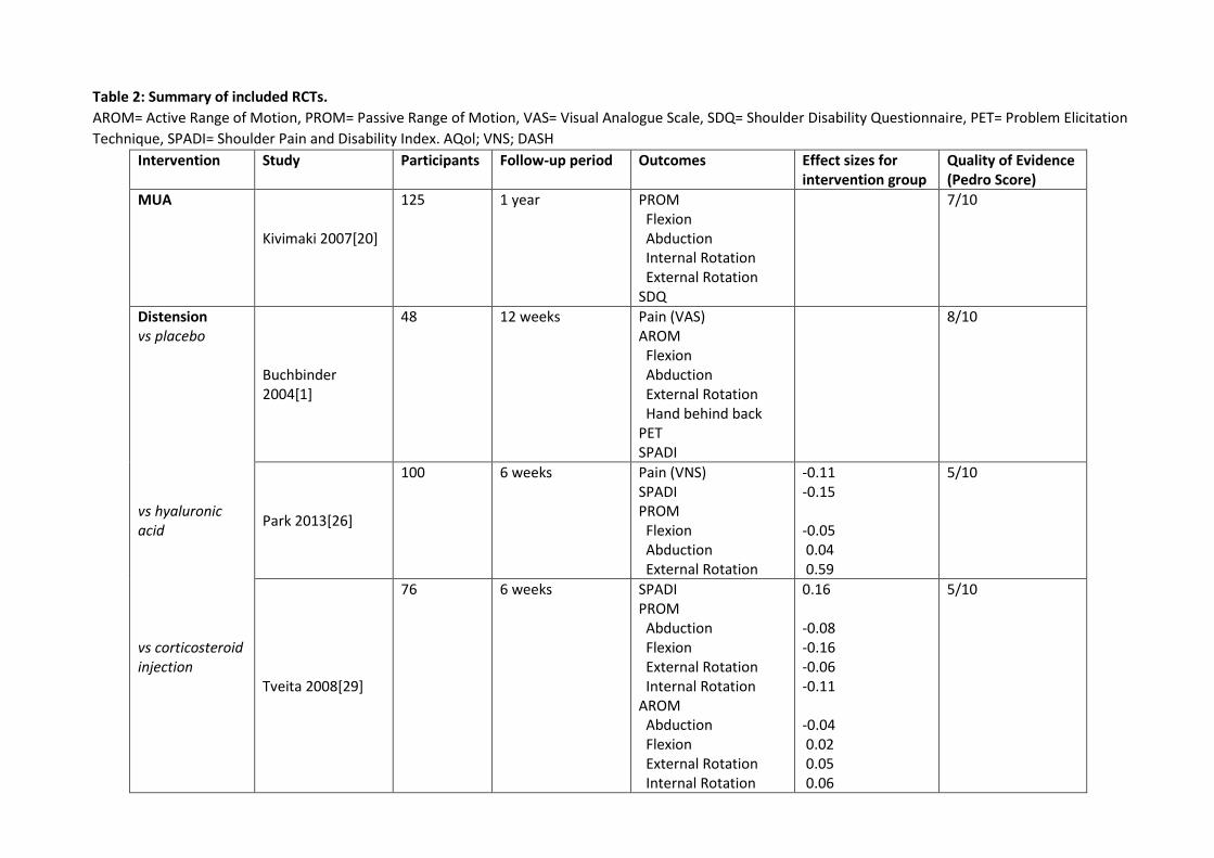
Table 2: Summary of included RCTs.
AROM= Active Range of Motion, PROM= Passive Range of Motion, VAS= Visual Analogue Scale, SDQ= Shoulder Disability Questionnaire, PET= Problem Elicitation
Technique, SPADI= Shoulder Pain and Disability Index. AQol; VNS; DASH
Intervention Study Participants Follow-up period Outcomes Effect sizes for intervention group
Quality of Evidence (Pedro Score)
MUA
Kivimaki 2007[20]
125 1 year PROM Flexion Abduction Internal Rotation External Rotation SDQ
7/10
Distension vs placebo
Buchbinder 2004[1]
48 12 weeks Pain (VAS) AROM Flexion Abduction External Rotation Hand behind back PET SPADI
8/10
vs hyaluronic acid
Park 2013[26]
100 6 weeks Pain (VNS) SPADI PROM Flexion Abduction External Rotation
-0.11 -0.15 -0.05 0.04 0.59
5/10
vs corticosteroid injection
Tveita 2008[29]
76 6 weeks SPADI PROM Abduction Flexion External Rotation Internal Rotation AROM Abduction Flexion External Rotation Internal Rotation
0.16 -0.08 -0.16 -0.06 -0.11 -0.04 0.02 0.05 0.06
5/10

20
Intervention Study Participants Follow-up period Outcomes Effect sizes for intervention group
Quality of Evidence (Pedro Score)
Manual Therapy
Buchbinder 2007(R. Buchbinder et al., 2007)
156 26 weeks AQol Flexion Abduction External Rotation Hand behind back SPADI SF-36
9/10
Celik 2016[7]
30 1 year Constant Score DASH Pain (VAS) PROM Flexion Abduction External Rotation Internal Rotation
0.35 0.03 0.13 0.16 0.44 0.29 0.13
6/10

21
DISCUSSION
Distension, MUA and manual therapy are thought to have a positive effect in the treatment of
frozen shoulder by improving glenohumeral mobility via stretching or rupturing of the joint capsule.
However, the results of the current review indicate that joint distension confers some short-term
benefit compared with placebo (arthrogram), but distension, MUA and passive mobilisation
techniques do not incur additional benefit with respect to pain & functional ability over cortisone
injection or a home-based stretching & strengthening program in the short term.
Glenohumeral capsular contracture, adhesions, coracohumeral ligament thickening and scarring of
the rotator cuff interval have been demonstrated in people diagnosed with frozen shoulder(TD
Bunker, 1997; G. Hand et al., 2007; J. S. Neviaser, 1945; Uitvlugt et al., 1993; A. Wiley, 1991)
Shoulder movement restriction associated with frozen shoulder has been attributed to these
abnormalities found within the glenohumeral capsule. In 1997, Bunker wrote: “Thus the enigma of
frozen shoulder has been unravelled. We have shown that the cause of frozen shoulder appears to
be a fibrous contracture of the rotator interval and coracohumeral ligament of the shoulder joint.
[…] Treatment must be aimed at releasing this contracture by manipulation or surgical release.(TD
Bunker, 1997)” The current available clinical trial evidence summarised in this review would not
seem to support this statement.
There may be several reasons why surgical procedures to release contractures in the glenohumeral
joint capsule have not been shown to be more effective in the treatment of frozen shoulder: The
glenohumeral joint dilatation procedure is unspecific and may not be able to release the contracted
structures, instead rupturing the weakest part of the capsule rather than necessarily the tissues
responsible for the movement restriction. It has previously been reported that most capsular
ruptures occur at the subscapularis recess or the sheath around the long head of biceps brachii, not
at the thickened capsule.(Kim et al., 2011) In addition, intra-articular corticosteroid injection, even at
low volumes, may have a distension effect on the glenohumeral joint capsule, making it more
difficult to identify between group differences in the trials which compared distension to
corticosteroid injection in this review.(J. S. Neviaser, 1945; Tveitå et al., 2008) Neviaser et al. has
reported glenohumeral joint volumes to be as low as 5 ml in some people with frozen shoulder and
injection volumes in the studies included in this review ranged from 5-10 ml in the corticosteroid
injection only groups.(R. Buchbinder et al., 2004; R. J. Neviaser & Neviaser, 1987; Park et al., 2013;
Tveitå et al., 2008) Tveita et al. in fact reported that in their trial 4 subjects belonging to the injection
group experienced capsular rupture. (Tveitå et al., 2008)
Mobilisation and physiotherapy exercises are frequently prescribed for people with frozen
shoulder.(Çelik & Kaya Mutlu, 2016; Hanchard et al., 2012) However, differing opinions exist
regarding the appropriate intensity and degree of manual therapy and stretching exercises for
people with frozen shoulder. It has been suggested that vigorous manual therapy, stretching and
exercise is counterproductive in frozen shoulder,(Diercks & Stevens, 2004; R. J. Neviaser & Neviaser,
1987) while others have favoured more vigorous techniques .(Vermeulen et al., 2000; Yang, Jan,
Chang, & Lin, 2012) The two trials included in this review investigated manual therapy and stretching
exercises that aimed to utilise end range positions but were described as being low intensity and
performed within patient comfort. It is uncertain if stretching exercises or manual therapy have a
direct effect on the contracted tissues but the results of this review indicate they may have some
benefit leading to small improvements in active ROM in the short term in patients with frozen
shoulder. These improvements may be a result of stretching of the contracted tissues of the
glenohumeral capsule. It is also possible that they are due to decreased activity/tightness in

22
surrounding shoulder muscles or as a result of increased patient confidence to move their arm
further into range.
The role of inflammation in frozen shoulder remains debatable.(G. Hand et al., 2007) Nevertheless,
results of a systematic review have shown short term benefit of intra-articular corticosteroid
injection with respect to pain and ROM over placebo and physiotherapy in patients with frozen
shoulder. (Rachelle Buchbinder et al., 2003) This suggests that it may be a reduction in pain, rather
than a change in the glenohumeral capsule, that produces the increase in ROM observed in these
patients. Therefore, it may be that the observed improvements in the distension group are partially
or entirely due to the glenohumeral corticosteroid injection rather than the distension procedure.
Some evidence exists that factors other than adhesions of the glenohumeral joint capsule may be
contributing to ROM loss in some people with frozen shoulder. In a case series, four out of five
patients diagnosed with frozen shoulder demonstrated significantly greater passive shoulder
abduction ROM when measured under general anaesthesia.(Hollmann et al., 2015) These authors
suggested that protective muscle contraction as a result of shoulder pain was a major contributing
factor to the ROM limitation in these patients making passive range of motion assessment
unreliable in patients with frozen shoulder.(Hollmann et al., 2015) If pain and muscle guarding
produce the perceived stiffness in some patients diagnosed with frozen shoulder rather than
glenohumeral capsular contracture and adhesions, this may explain why the studies included in this
review aimed at stretching/lenghtening tight passive structures at the shoulder have demonstrated
very little clinical benefit.
Conclusion
This systematic review has summarised the available evidence from six high quality RCTs for
treatments that aim to stretch or release areas of contracture or fibrosis in frozen shoulder. While
distension, manual therapy and stretching may lead to small short term improvements in ROM in
people with frozen shoulder, they do not appear to significantly alter the natural course of frozen
shoulder.

23
REFERENCES
1. Buchbinder R, Green S, Forbes A, Hall S, Lawler G. Arthrographic joint distension with saline and steroid improves function and reduces pain in patients with painful stiff shoulder: Results of a randomised, double blind, placebo controlled trial. Annals of the Rheumatic Diseases 2004; 63: 302-309
2. Buchbinder R, Green S, Youd JM. Corticosteroid injections for shoulder pain. The Cochrane Library 2003, DOI:
3. Buchbinder R, Youd JM, Green S, Stein A, Forbes A, Harris A, Bennell K, Bell S, Wright WJL. Efficacy and cost-effectiveness of physiotherapy following glenohumeral joint distension for adhesive capsulitis: A Randomized trial. Arthritis & Rheumatism-Arthritis Care & Research 2007; 57: 1027-1037
4. Bunker T. Frozen shoulder: unravelling the enigma. Annals of the Royal College of Surgeons of England 1997; 79: 210
5. Bunker T. Time for a new name for frozen shoulder—contracture of the shoulder. Shoulder & Elbow 2009; 1: 4-9
6. Bunker TD, Anthony PP. The pathology of frozen shoulder. A Dupuytren-like disease. Journal of Bone and Joint Surgery - Series B 1995; 77: 677-683
7. Çelik D, Kaya Mutlu E. Does adding mobilization to stretching improve outcomes for people with frozen shoulder? A randomized controlled clinical trial. Clinical Rehabilitation 2016; 30: 786-794
8. Codman EA. On stiff and painful shoulders. The Boston Medical and Surgical Journal 1911; 164: 613-620
9. Cohen J. Statistical power analysis for the behavioral sciences 2 edition Lawrence Erlbaum. Hillsdale, NJ 1988, DOI:
10. Cruz-Ferreira A, Fernandes J, Laranjo L, Bernardo LM, Silva A. A systematic review of the effects of pilates method of exercise in healthy people. Archives of physical medicine and rehabilitation 2011; 92: 2071-2081
11. de Morton NA. The PEDro scale is a valid measure of the methodological quality of clinical trials: a demographic study. Australian Journal of Physiotherapy 2009; 55: 129-133
12. Diercks RL, Stevens M. Gentle thawing of the frozen shoulder: A prospective study of supervised neglect versus intensive physical therapy in seventy-seven patients with frozen shoulder syndrome followed up for two years. Journal of Shoulder and Elbow Surgery 2004; 13: 499-502
13. Duplay S. De la peri-arthrite scapulo-humerale et des raideurs de l’epaule qui en sont la consequence. Arch Gen Med 1872; 20: 513-542
14. Haik M, Alburquerque-Sendín F, Moreira R, Pires E, Camargo P. Effectiveness of physical therapy treatment of clearly defined subacromial pain: a systematic review of randomised controlled trials. British journal of sports medicine 2016; 50: 1124-1134
15. Hanchard NC, Goodchild L, Thompson J, O’Brien T, Davison D, Richardson C. Evidence-based clinical guidelines for the diagnosis, assessment and physiotherapy management of contracted (frozen) shoulder: quick reference summary. Physiotherapy 2012; 98: 117-120
16. Hand G, Athanasou N, Matthews T, Carr A. The pathology of frozen shoulder. Bone & Joint Journal 2007; 89: 928-932
17. Hollmann L, Halaki M, Haber M, Herbert R, Dalton S, Ginn K. Determining the contribution of active stiffness to reduced range of motion in frozen shoulder. Physiotherapy 2015; 101: e585
18. Kelley MJ, Shaffer MA, Kuhn JE, Michener LA, Seitz AL, Uhl TL, Godges JJ, McClure PW, Altman RD, Davenport T. Shoulder pain and mobility deficits: adhesive capsulitis: clinical practice guidelines linked to the International Classification of Functioning, Disability, and

24
Health from the Orthopaedic Section of the American Physical Therapy Association. Journal of Orthopaedic & Sports Physical Therapy 2013; 43: A1-A31
19. Kim K, Lee KJ, Kim HC, Lee KJ, Kim DK, Chung SG. Capsule preservation improves short‐term outcome of hydraulic distension in painful stiff shoulder. Journal of Orthopaedic Research 2011; 29: 1688-1694
20. Kivimäki J, Pohjolainen T, Malmivaara A, Kannisto M, Guillaume J, Seitsalo S, Nissinen M. Manipulation under anesthesia with home exercises versus home exercises alone in the treatment of frozen shoulder: a randomized, controlled trial with 125 patients. Journal of Shoulder and Elbow Surgery 2007; 16: 722-726
21. Lewis J. Frozen shoulder contracture syndrome–Aetiology, diagnosis and management. Manual therapy 2015; 20: 2-9
22. Maher CG, Sherrington C, Herbert RD, Moseley AM, Elkins M. Reliability of the PEDro scale for rating quality of randomized controlled trials. Physical therapy 2003; 83: 713-721
23. Neviaser AS, Hannafin JA. Adhesive Capsulitis: A Review of Current Treatment. American Journal of Sports Medicine 2010; 38: 2346-2356
24. Neviaser JS. Adhesive capsulitis of the shoulder. J Bone Joint Surg Am 1945; 27: 211-222 25. Neviaser RJ, Neviaser TJ. The Frozen Shoulder Diagnosis and Management. Clinical
orthopaedics and related research 1987; 223: 59-64 26. Park KD, Nam H-S, Lee JK, Kim YJ, Park Y. Treatment effects of ultrasound-guided capsular
distension with hyaluronic acid in adhesive capsulitis of the shoulder. Archives of physical medicine and rehabilitation 2013; 94: 264-270
27. Reeves B. The natural history of the frozen shoulder syndrome. Scandinavian journal of rheumatology 1975; 4: 193-196
28. Shaffer B, Tibone J, Kerlan RK. Frozen shoulder. A long-term follow-up. J Bone Joint Surg Am 1992; 74: 738-746
29. Tveitå EK, Tariq R, Sesseng S, Juel NG, Bautz-Holter E. Hydrodilatation, corticosteroids and adhesive capsulitis: a randomized controlled trial. BMC musculoskeletal disorders 2008; 9: 1
30. Uitvlugt G, Detrisac DA, Johnson LL, Austin MD, Johnson C. Arthroscopic observations before and after manipulation of frozen shoulder. Arthroscopy: The Journal of Arthroscopic & Related Surgery 1993; 9: 181-185
31. Vermeulen HM, Obermann WR, Burger BJ, Kok GJ, Rozing PM, van den Ende CH. End-range mobilization techniques in adhesive capsulitis of the shoulder joint: a multiple-subject case report. Physical Therapy 2000; 80: 1204-1213
32. Wiley A. Arthroscopic appearance of frozen shoulder. Arthroscopy: The Journal of Arthroscopic & Related Surgery 1991; 7: 138-143
33. Yang J-l, Jan M-H, Chang C-w, Lin J-j. Effectiveness of the end-range mobilization and scapular mobilization approach in a subgroup of subjects with frozen shoulder syndrome: a randomized control trial. Manual therapy 2012; 17: 47-52
34. Zuckerman JD, Rokito A. Frozen shoulder: a consensus definition. Journal of Shoulder and Elbow Surgery 2011; 20: 322-325

25
Chapter 3 – External Rotation Range of Motion in Healthy Subjects
INTRODUCTION
Knowledge of normal range of motion (ROM) is essential when assessing impairments and clinical
pathology in the shoulder. The assessment of shoulder external rotation ROM in particular is of great
importance as it provides diagnostic information for several common shoulder conditions. Passive
external ROM restriction is a diagnostic feature of frozen shoulder (Tim Bunker, 2009). Excessive
active and passive external rotation ROM is thought to contribute to anterior shoulder instability
(Kuhn, Huston, Soslowsky, Shyr, & Blasier, 2005) and is also used as a measure to differentiate
between capsular tightness/joint stiffness and muscle weakness, aiding in the diagnosis of
conditions such as glenohumeral osteoarthritis. Active external rotation is commonly used as a
measure of the function of the posterior rotator cuff (Dutton, 2008). Studies have examined
shoulder ROM in healthy athletes, including elite tennis and baseball players (Baltaci, Johnson, &
Kohl III, 2001; Ellenbecker, Roetert, Bailie, Davies, & Brown, 2002; Ellenbecker, Roetert, Piorkowski,
& Schulz, 1996; Ellenbeckert, 1992). However, generalising these results to the normal population is
problematic as it is well documented that high level throwers exhibit humeral retroversion affecting
shoulder rotation ROM (Chant, Litchfield, Griffin, & Thain, 2007; R. Whiteley, Adams, Ginn, &
Nicholson, 2010; R. J. Whiteley, Ginn, Nicholson, & Adams, 2009).
Several researchers have attempted to establish normative values for shoulder external rotation
ROM in the general population and investigated factors such as sex and hand dominance on ROM.
Female subjects have been reported to have greater active and passive shoulder external rotation
ROM compared to male subjects (Allander, Björnsson, Olafsson, Sigfusson, & Thorsteinsson, 1974;
Barnes, Van Steyn, & Fischer, 2001; Kronberg, Broström, & Söderlund, 1990; Murray, Gore, Gardner,
& Mollinger, 1985). The findings of the impact of hand dominance on ROM, however, are not
consistent. Some authors reported passive external rotation ROM to be similar in dominant and

26
nondominant shoulders (Allander et al., 1974; Kronberg et al., 1990). In contrast, two studies that
investigated active and passive external rotation ROM reported conflicting findings. A study of 280
healthy subjects aged 4-70 demonstrated significantly greater active and passive external rotation
ROM in the dominant shoulder (Barnes et al., 2001; Riddle, Rothstein, & Lamb, 1987). However,
significantly less passive and active external rotation was observed in dominant shoulders compared
non-dominant shoulders in a group of 1000 male military recruits (Günal, Köse, Erdogan, Göktürk, &
Seber, 1996).
External rotation ROM is limited by different structures in different shoulder positions. Many
ligaments of the glenohumeral joint have significant roles in restraining external rotation (Kuhn et
al., 2005). A cadaveric study by Ferrari et al. reported that the coracohumeral ligament provides the
primary passive constraint to shoulder external rotation between 0 and 60 of shoulder abduction.
Between 60 and 90 of shoulder abduction, the middle glenohumeral ligament becomes the
primary passive restraint to shoulder external rotation range of motion(Ferrari, 1990). At 90 of
abduction and above, the inferior glenohumeral ligament develops the most strain, therefore
restricting external rotation ROM in these positions (O'Connell, Nuber, Mileski, & Lautenschlager,
1990). Active and passive external rotation ROM is also dependent on the function of the rotator
cuff, with infraspinatus and teres minor acting concentrically as shoulder external rotators and
subscapularis eccentrically providing a dynamic restraint to external rotation ROM in both adducted
and abducted shoulder positions (Kuhn et al., 2005; Palastanga, Field, & Soames, 2006). Kuhn et al.
also demonstrated that the long head of biceps provides a dynamic restraint to shoulder external
rotation in the abducted position (Kuhn et al., 2005). It is therefore expected that different shoulder
positions will yield different ranges of shoulder external rotation ROM as different anatomic
structures are stressed.
The impact of shoulder position when testing shoulder external rotation ROM has been a lesser
focus in previous research. Three studies measured active and passive external rotation ROM in two
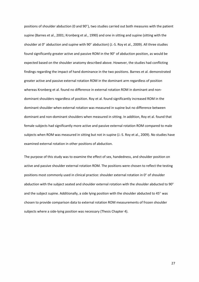
27
positions of shoulder abduction (0 and 90), two studies carried out both measures with the patient
supine (Barnes et al., 2001; Kronberg et al., 1990) and one in sitting and supine (sitting with the
shoulder at 0 abduction and supine with 90 abduction) (J.-S. Roy et al., 2009). All three studies
found significantly greater active and passive ROM in the 90 of abduction position, as would be
expected based on the shoulder anatomy described above. However, the studies had conflicting
findings regarding the impact of hand dominance in the two positions. Barnes et al. demonstrated
greater active and passive external rotation ROM in the dominant arm regardless of position
whereas Kronberg et al. found no difference in external rotation ROM in dominant and non-
dominant shoulders regardless of position. Roy et al. found significantly increased ROM in the
dominant shoulder when external rotation was measured in supine but no difference between
dominant and non-dominant shoulders when measured in sitting. In addition, Roy et al. found that
female subjects had significantly more active and passive external rotation ROM compared to male
subjects when ROM was measured in sitting but not in supine (J.-S. Roy et al., 2009). No studies have
examined external rotation in other positions of abduction.
The purpose of this study was to examine the effect of sex, handedness, and shoulder position on
active and passive shoulder external rotation ROM. The positions were chosen to reflect the testing
positions most commonly used in clinical practice: shoulder external rotation in 0 of shoulder
abduction with the subject seated and shoulder external rotation with the shoulder abducted to 90
and the subject supine. Additionally, a side lying position with the shoulder abducted to 45 was
chosen to provide comparison data to external rotation ROM measurements of frozen shoulder
subjects where a side-lying position was necessary (Thesis Chapter 4).

28
METHODS
SUBJECTS
Twenty subjects were recruited by advertisement at the University of Sydney on physical and online
notice boards. Power analysis for a dependent sample t-test was conducted in G*Power based on
the results of a study by Gunal et al. (Günal et al., 1996) To determine a sufficient sample size to
detect a 10 difference between groups, using an alpha of 0.05, a power of 0.80, an effect size (dz =
0.8), and two tails, the desired sample size is 10. Interested staff and students of the University
contacted the research team by phone or email. Potential subjects were screened for eligibility at
this time. Subjects were eligible to participate if they were over 18 years old, have not had any
shoulder pain in the past two years and have never had shoulder surgery.
Potentially eligible subjects were screened just prior to testing to ensure they did not have any
current shoulder symptoms. Subjects were asked to perform active shoulder flexion, abduction, and
external rotation in standing as well as a maximal isometric contraction of internal and external
rotation. One of the researchers, an experienced physiotherapist, then performed a passive shoulder
external rotation movement with over pressure with the subjects in supine and the arm abducted to
90. Subjects were excluded if they exhibited abnormal scapulohumeral rhythm (Lucas, 1973) or
experienced shoulder pain on any of the shoulder assessments.
ETHICS AND CONSENT
Ethics approval was obtained from The University of Sydney Human Research Ethics Committee
(Ethics protocol number: 2015/001). Following confirmation of eligibility subjects were given the
opportunity to ask questions relating to the study before giving written consent to participate
(Appendix A).

29
OUTCOME MEASURES
The primary outcome measures were active and passive external rotation ROM.
Active External Rotation Range of Motion
Active shoulder external rotation ROM was measured bilaterally with a goniometer in two positions,
seated and supine lying in a random order.
Sitting (0 shoulder abduction): The subject was seated with the arm by the side and the elbow
flexed to 90. The subject was then asked to maximally externally rotate their shoulder. The centre
of the goniometer was positioned above the axis of the glenohumeral joint. ER ROM was measured
as the angle between a line representing the sagittal plane and a line through the longitudinal axis of
the forearm, using the acromioclavicular joint and the radial styloid process as landmarks for the
alignment of the goniometer (Figure 3.1).
Figure 3.1: Measurement of Active Shoulder External Rotation in sitting.

30
Supine (90 shoulder abduction): The subject was lying supine with the shoulder abducted to 90and
the elbow flexed to 90. The centre of the goniometer was aligned with the olecranon process of the
elbow. The angle between the vertical line and the line of subjects’ forearm, along a line from the
olecranon process of the elbow to the ulna styloid process was measured (Figure 3.2).
Figure 3.2: Measurement of Active Shoulder External Rotation in supine.
Passive External Rotation Range of Motion
A custom-built arm frame was used to measure passive external rotation ROM. The frame was
instrumented with a potentiometer (Vishay Model 357, Germany) to measure the external rotation
angle and a force transducer (XTran, Model S1W 250N, Applied Measurement PTY. LTD., Australia)
attached 0.33 m from the axis of rotation was used to standardise the torque that was applied to the
subject’s arm when assessing maximal range. The angle and force signals were recorded using a 32-
bit analogue to digital converter (cDAC 9171, National Instruments, TX, USA) and LABVIEW software
at a sample rate of 100 Hz. With elbow maintained in 90 of flexion, the subject’s arm was strapped
firmly into the arm frame using velcro straps. Passive external rotation ROM was measured in sitting,

31
side lying and supine. For the seated measurement, external rotation was measured with the arm by
the side. In side-lying, the arm was abducted to 45 and supported with a cushion between the
subject’s trunk and upper arm. For the supine position, the shoulder was positioned in 90 of
abduction in the coronal plane with the upper arm supported on a treatment table.
For each movement, the subject was asked to relax their arm as much as possible. The arm was then
moved into external rotation by one of the researchers until a torque of 5 Nm was reached. The
torque target used was established from pilot testing of the force normally applied by two of the
researchers who were experienced physiotherapists when testing passive external rotation ROM.
PROCEDURE
Each subject’s age and handedness were recorded. Prior to commencing active and passive ROM
testing, subjects performed a short warm up of 5-10 repetitions of active external rotation in neutral
and in 90 shoulder abduction. This was to ensure that subjects understood how to perform the
movements without the use of compensatory strategies and to reduce the risk of injury. Both
shoulders were tested in random order.
The order of tests was block randomised for subject position using the random number generator
function in Microsoft Excel. All measurements were repeated three times. The subjects position was
stabilised by the arm frame and by using a chairs with back support to prevent compensatory
movement strategies. The average of the three trials for each active and passive ROM measurement
was used for analysis.
STATISTICAL ANALYSIS
Descriptive statistics for ROM, age and sex were calculated. Three factor Mixed-model analysis was
used to determine the effect of body position, hand dominance and sex on active and passive ROM.
Multiple pairwise comparisons with Bonferroni adjustment were used for identifying differences

32
between levels when significant effects were found using SPSS software (IBM SPSS Statistics for
Windows, Version 22.0. Armonk, NY: IBM Corp).
RESULTS
Ten female and ten male subjects entered the study. The average age of subjects was 45 years
(range 25-69). Seventeen subjects were right hand dominant. None of the subjects reported
shoulder pain or discomfort during testing and all subjects completed the study.
Active vs Passive Range of Motion
Passive ROM was significantly greater than active ROM (mean 95 vs 70, p<0.001, F(1,143)=118.65).
Active Range of Motion
Subjects had significantly greater active ROM in supine compared to sitting (mean=81 vs 59,
p<0.001, F(1,72)=67.14, Figure 3.3). There was no significant difference in active ROM between male
and female subjects (p=0.08, F(1,72)=3.16) or between the dominant and non-dominant shoulder
(p=0.46, F(1,72)=0.56). No significant interactions were found (p>0.48).
Figure 3.3: Active shoulder external rotation range of motion.
Passive Range of Motion
0
20
40
60
80
100
120
dominant side non dominant dominant side non dominant
sitting(0° shoulder abduction)
supine(90° shoulder abduction)
Act
ive
exte
rnal
ro
tati
on
ran
ge(d
egre
es)
Male Female

33
The results for passive external rotation range of motion measurements are displayed in Figure 3.4.
There was a significant effect of body position (p<0.001, F(2,107)=21.53). Subjects had significantly
greater passive ER in supine (mean=106) compared to sitting (mean=84, p<0.001) or side-lying
(mean=86, p<0.001) positions, with no difference between sitting and side lying (p=1.00). Females
had significantly more passive ROM compared to males (mean=98 vs 86, p<0.001, F(1,107)=17.13,
Table 3.1). There was no significant difference in passive ROM between the dominant and non-
dominant side (p=0.22, F(1,107)=1.51). No significant interactions were found (p>0.28).
Figure 3.4: Passive shoulder external rotation range of motion.
0
20
40
60
80
100
120
140
dominant non dominant dominant non dominant dominant non dominant
sitting(0° shoulder abduction)
side lying(45° shoulder abduction)
supine(90° shoulder abduction)
Pas
sive
Ext
ern
al R
ota
tio
n R
OM
(d
egre
es)
Male Female

34
Table 3.1: Effect of sex, body position and handedness on active and passive external rotation ROM
Mean Active
ROM
(degrees)
95% CI
(degrees)
Mean
Passive ROM
(degrees)
95% CI
(degrees)
Sex
Males 68 64-72 86 81-89
Females 73 69-76 98* 94-103
Position
Sitting (0 shoulder abduction) 59 55-63 84 79-89
Side lying (45 shoulder abduction) 86 81-91
Supine (90 shoulder abduction) 81# 78-85 106# 101-111
Handedness
Dominant 71 67-75 94 89-98
Non Dominant 69 65-73 90 86-94
*significant difference between sexes
#significant difference compared to other positions (<0.001)

35
DISCUSSION
This study investigated the effect of sex, handedness, shoulder and body position on active and
passive ROM. The main results can be summarised as follows: Passive external rotation ROM was
significantly greater than active ROM in people with healthy shoulders. Both active and passive
shoulder external rotation ROM was greater when the arm was abducted at 90 compared to lower
positions of abduction. There was no difference in active or passive external rotation ROM between
dominant and non-dominant shoulders. Female subjects demonstrated significantly more passive
external rotation ROM than males. Both active and passive external rotation ROM was greater at 90
of shoulder abduction compared to 0 abduction in both males and females and both dominant and
non-dominant sides. These findings are consistent with previous research (J.-S. Roy et al., 2009).
Anatomically, the primary passive constraint to external rotation of the shoulder between 0 and 60
of shoulder abduction is the coracohumeral ligament, between 60 and 90 of shoulder abduction is
the middle glenohumeral ligament (Ferrari, 1990) and above 90 of shoulder abduction is the
inferior glenohumeral ligament (O'Connell et al., 1990). Therefore, the testing position of ER has an
influence on which structures are assessed and their ability to act as a restraint to ER ROM.
Active ROM was greater than passive ROM in both males and females and in dominant and non-
dominant shoulders. This finding was expected and in agreement with other studies (Barnes et al.,
2001; J.-S. Roy et al., 2009). Passive ROM may be greater as it is performed by the examiner with the
subjects arm relaxed allowing the joint to be moved further into range and stretching elastic soft
tissues.
This study confirms the findings of others that have demonstrated increased passive ER ROM in
females compared to males (Allander et al., 1974; Barnes et al., 2001; Kronberg et al., 1990).
However, the lack of difference in active ER ROM between males and females conflicts previous
research which has shown females to have greater active ER ROM (Allander et al., 1974; Barnes et
al., 2001; J.-S. Roy et al., 2009). Other factors such as occupation or level of sports participation that

36
have previously hypothesised to affect shoulder ROM (Allander et al., 1974) do not appear to play a
role in the conflicting findings as, these, even though not recorded in our study, have previously
shown not to have an effect on shoulder ROM (Barnes et al., 2001). Equally, the method of
measuring ROM does not appear to be a factor as goniometry was used in all studies and has been
demonstrated to be reliable for upper limb ROM assessment, particularly when the same observer is
responsible for repeated measurements (Mayerson & Milano, 1984).
Dominant and non-dominant shoulders exhibited similar magnitudes of passive ROM in this study,
supporting the findings of Allander et al. and Kronberg et al. and but conflicting the findings of
studies by Barnes et al. and Gunal et al. that found significant, but opposite, differences in active and
passive external rotation ROM between dominant and non-dominant shoulders (Allander et al.,
1974; Barnes et al., 2001; Kronberg et al., 1990). It is unclear what causes these conflicting findings.
Normal shoulder external rotation ROM with the shoulder abducted at 45 has not previously been
investigated. While healthy subjects exhibit significantly greater passive ROM in 90 degrees of
shoulder compared to 0 abduction, there is no significant difference in external rotation ROM
between 45 abduction and 0 abduction. This result might be expected as the coracohumeral
ligament is the primary restraint between 0 and 60 abduction, therefore the primary structure
limiting shoulder external rotation ROM in the 0 and 45 abducted positions. As external rotation
ROM measures from both positions yield similar results, they can be used interchangeably in clinical
practice.
The results of this study indicate that the position of side-lying with 45 of shoulder abduction in the
operating theatre in the frozen shoulder study (as required in preparation for the shoulder surgery)
can be used as it yields similar results to shoulder external rotation measured with the arm by the
side which is the typical position used by clinicians to assess external rotation ROM limitations in
people with frozen shoulder.

37
REFERENCES
Allander, E., Björnsson, O., Olafsson, O., Sigfusson, N., & Thorsteinsson, J. (1974). Normal range of
joint movements in shoulder, hip, wrist and thumb with special reference to side: a
comparison between two populations. International Journal of Epidemiology, 3(3), 253-261.
Baltaci, G., Johnson, R., & Kohl III, H. (2001). Shoulder range of motion characteristics in collegiate
baseball players. Journal of sports medicine and physical fitness, 41(2), 236.
Barnes, C. J., Van Steyn, S. J., & Fischer, R. A. (2001). The effects of age, sex, and shoulder dominance
on range of motion of the shoulder. Journal of Shoulder and Elbow Surgery, 10(3), 242-246.
Bunker, T. (2009). Time for a new name for frozen shoulder—contracture of the shoulder. Shoulder
& Elbow, 1(1), 4-9.
Chant, C. B., Litchfield, R., Griffin, S., & Thain, L. M. (2007). Humeral head retroversion in competitive
baseball players and its relationship to glenohumeral rotation range of motion. Journal of
Orthopaedic & Sports Physical Therapy, 37(9), 514-520.
Dutton, M. (2008). The shoulder complex. Dutton M. Orthopaedic Examination, Evaluation, and
Intervention. 2nd ed. New York, NY: McGraw Hill Companies, 523-524.
Ellenbecker, T. S., Roetert, E. P., Bailie, D. S., Davies, G. J., & Brown, S. W. (2002). Glenohumeral joint
total rotation range of motion in elite tennis players and baseball pitchers. Medicine and
Science in Sports and Exercise, 34(12), 2052-2056.
Ellenbecker, T. S., Roetert, E. P., Piorkowski, P. A., & Schulz, D. A. (1996). Glenohumeral joint internal
and external rotation range of motion in elite junior tennis players. Journal of Orthopaedic &
Sports Physical Therapy, 24(6), 336-341.
Ellenbeckert, T. S. (1992). Shoulder internal and external rotation strength and range of motion of
highly skilled junior tennis players. Isokinetics and Exercise Science, 2(2), 65-72.
Ferrari, D. A. (1990). Capsular ligaments of the shoulder anatomical and functional study of the
anterior superior capsule. The American journal of sports medicine, 18(1), 20-24.
38
Günal, I., Köse, n., Erdogan, o., Göktürk, e., & Seber, s. (1996). Normal range of motion of the joints
of the upper extremity in male subjects, with special reference to side. J Bone Joint Surg Am,
78(9), 1401-1404.
Kronberg, M., Broström, L.-Å., & Söderlund, V. (1990). Retroversion of the Hurneral Head in the
Normal Shoulder and Its Relationship to the Normal Range of Motion. Clinical orthopaedics
and related research, 253, 113-117.
Kuhn, J. E., Huston, L. J., Soslowsky, L. J., Shyr, Y., & Blasier, R. B. (2005). External rotation of the
glenohumeral joint: ligament restraints and muscle effects in the neutral and abducted
positions. Journal of Shoulder and Elbow Surgery, 14(1), S39-S48.
Murray, M., Gore, D., Gardner, G., & Mollinger, L. (1985). Shoulder motion and muscle strength of
normal men and women in two age groups. Clinical orthopaedics and related research, 192,
268-273.
O'Connell, P. W., Nuber, G. W., Mileski, R. A., & Lautenschlager, E. (1990). The contribution of the
glenohumeral ligaments to anterior stability of the shoulder joint. The American journal of
sports medicine, 18(6), 579-584.
Palastanga, N., Field, D., & Soames, R. (2006). Anatomy and human movement: structure and
function (Vol. 20056): Elsevier Health Sciences.
Riddle, D. L., Rothstein, J. M., & Lamb, R. L. (1987). Goniometric reliability in a clinical setting.
Physical therapy, 67(5), 668-673.
Roy, J.-S., MacDermid, J. C., Boyd, K. U., Faber, K. J., Drosdowech, D., & Athwal, G. S. (2009).
Rotational strength, range of motion, and function in people with unaffected shoulders from
various stages of life. BMC Sports Science, Medicine and Rehabilitation, 1(1), 4.
Whiteley, R., Adams, R., Ginn, K., & Nicholson, L. (2010). Playing level achieved, throwing history,
and humeral torsion in Masters baseball players. Journal of sports sciences, 28(11), 1223-
1232.

39
Whiteley, R. J., Ginn, K. A., Nicholson, L. L., & Adams, R. D. (2009). Sports participation and humeral
torsion. Journal of Orthopaedic & Sports Physical Therapy, 39(4), 256-263.

40
CHAPTER 4 – Active Stiffness in Frozen Shoulder
Publication Title: Capsular contracture is not a major contributor to range of motion loss in some
patients with frozen shoulder.
As co-authors of the paper, ‘Capsular contracture is not a major contributor to range of motion loss
in some patients with frozen shoulder.’, we confirm that Luise Hollmann has made the following
contributions:
• Conception and design of the research
• Ethics Application
• Data collection
• Data processing
• Analysis and interpretation of findings
• Writing the paper and critical appraisal of content
• Final editing of article for publication
Signature:
Associate Professor Karen Ginn
Date: 28.2.2017
Signature:
Dr Mark Halaki
Date: 28.2.2017

41

42
Capsular contracture is not a major contributor to range of motion loss in some patients with frozen shoulder.
ABSTRACT
This case series of five subjects with frozen shoulder demonstrates that capsular contracture is not a
major contributor to movement restriction in all patients who exhibit classical clinical features of
frozen shoulder. Although all five cases presented with painful, global restriction of passive shoulder
movement, four subjects demonstrated significantly greater abduction range of motion (ROM) and
three demonstrated significantly greater external rotation ROM under anaesthesia. These findings
highlight the need to reconsider our understanding of the pathology of frozen shoulder and offer an
explanation for why treatment aimed at stretching tight passive structures has not proven to be
more effective.
INTRODUCTION
Frozen shoulder has puzzled the medical community since it was first described in the late 19th century. It occurs in 5–10% of the general population and up to 29% of the diabetic population.(Balci, Balci, & Tüzüner, 1999; Pal, Anderson, Dick, & Griffiths, 1986; Walker‐Bone, Palmer, Reading, Coggon, & Cooper, 2004) It is characterized by spontaneous onset of pain with progressive, marked active and passive stiffness at the glenohumeral joint(Bo J Lundberg, 1969; Nash & Hazleman, 1989) usually resulting in gross loss of function.(Reeves, 1975) The condition is described as self-limiting with gradual return of painfree shoulder function after an average of 1-3 years.(C. Hand, Clipsham, Rees, & Carr, 2008; Reeves, 1975)
There is no definitive diagnostic test for frozen shoulder and diagnosis is based on physical examination following exclusion of osteoarthritis, locked dislocations, fractures or avascular necrosis as the cause of symptoms.(Tim Bunker, 2009) A clinical diagnosis of frozen shoulder is made if the patient has painful restriction of active and passive motion in at least two planes of shoulder movement one being external rotation.(Tim Bunker, 2009; Zuckerman & Rokito, 2011)
The pathoaetiology of frozen shoulder is not fully understood. Histological and arthroscopic studies of frozen shoulder suggest a process of chronic inflammation, fibrosis and glenohumeral joint capsule contracture is responsible for the pain and restricted range of movement (ROM) characteristic of frozen shoulder.(TD Bunker, 1997; G. Hand et al., 2007; J. S. Neviaser, 1945; Uitvlugt et al., 1993; A. Wiley, 1991) Consequently, treatment is most commonly aimed at stretching glenohumeral joint structures to restore shoulder ROM while managing pain.
Frozen shoulder is considered notoriously difficult to treat and there is no consensus regarding optimal management.(Lewis, 2015) Evidence suggests that corticosteroid injection confers significant short term benefit.(Rachelle Buchbinder et al., 2003) However there is little evidence to support the effectiveness of treatments aimed at lengthening the glenohumeral joint capsule. Physiotherapy aimed at increasing ROM is only slightly more effective than placebo injection in the short term(Carette et al., 2003) ; efficacy of arthroscopic capsular release is not supported by evidence from randomised control trials;(Lewis, 2015) and capsular hydrodilatation or distension is no more effective than corticosteroid injection alone.(Tveitå et al., 2008)
If frozen shoulder is caused primarily by capsular adhesions, it is surprising that treatment directed at presumed glenohumeral joint capsuloligamentous contracture has not proven to be more

43
successful. In addition, if the cause of movement restriction in frozen shoulder is primarily due to capsular fibrosis how can the spontaneous recovery of ROM, commonly seen with frozen shoulder after a protracted period of symptoms, be explained?
We hypothesise that factors other than adhesions of the glenohumeral joint capsule may be contributing to ROM loss in people with frozen shoulder. Protective muscle spasm, or guarding, is a common motor strategy in response to other painful musculoskeletal dysfunction, including back and neck pain, which results in decreased ROM. At the glenohumeral joint the capsule is strongly reinforced by the rotator cuff tendons and one of the normal functions of the rotator cuff muscles is to contribute to glenohumeral joint stability by tightening the capsule during movement. Given this intimate relationship between rotator cuff muscles and the glenohumeral joint capsule, it is possible that rotator cuff muscle contraction in response to pain is contributing to the movement restriction associated with frozen shoulder.(Roland, 1986) Relaxation of these muscles as pain subsides could explain why substantial improvement in shoulder ROM can be expected in most cases of frozen shoulder. Therefore, the aim of this case series was to test whether there is muscle guarding (active stiffness) component to movement restriction in patients with frozen shoulder by comparing passive shoulder ROM before and after the administration of general anaesthesia.
METHODS
PARTICIPANTS
The participating orthopaedic surgeon identified patients scheduled to undergo capsular release surgery for frozen shoulder. All potential participants had an X-ray and MRI to ensure the absence of shoulder joint osteoarthritis, fractures or dislocations. On the day of the surgery, the potential participants were screened for eligibility to participate. Patients were eligible if their shoulder pain was associated with global restriction of active and passive shoulder movement which was greater than 50% normal ROM in external rotation and at least one other plane of movement.
Patients were excluded if they had a history of trauma to the affected shoulder within the four weeks prior to surgery, previous surgery on the affected shoulder, shoulder pain referred from the vertebral column (defined as shoulder pain exacerbated during neck movement and/or palpation of the cervicothoracic vertebral column) or concurrent inflammatory or neurological disease involving the affected shoulder.
OUTCOME MEASURES
To minimise the time participants spent under general anaesthetic prior to surgery, only two passive shoulder ROM outcome measurements were compared before and after general anaesthesia: external rotation ROM because passive restriction in this direction is one of the diagnostic criteria for frozen shoulder(Tim Bunker, 2009); and abduction ROM because it could readily be measured in the side-lying position required by the orthopaedic surgeon. A portable custom built arm frame instrumented with a potentiometer (Vishay Model 357, Germany) and a force transducer (XTran, Model S1W 250N, Applied Measurement PTY. LTD., Australia) was constructed to standardise the force applied to each participant’s ’s arm to achieve maximum passive external rotation ROM. The participant’s arm was strapped into the arm frame which maintained the elbow in 90 degrees of flexion and shoulder in 45 degrees abduction. Both shoulder external rotation angle and torque applied were recorded at a sample rate of 100 Hz using a 32-bit analogue to digital converter (cDAC 9171, National Instruments, TX, USA) and LABVIEW software. Abduction ROM was measured by passively moving the participant’s’ arm through range. Maximal achieved abduction ROM was measured from a digital photograph by measuring the angle at the intersection of a horizontal reference line and a line between the acromion process and the lateral epicondyle. (Figures 1-3).

44
PROCEDURE
On the day of the scheduled surgery, researchers met potentially eligible patients and described the aim and design of the study. For patients who were willing to participate, active and passive movements of shoulder flexion, abduction, external rotation with arm by the side and hand-behind-back were conducted on the affected shoulder to confirm eligibility. As is common clinical practice, these movements were performed with the patient standing and measured with a goniometer. Following confirmation that all inclusion and exclusion criteria were met, participants signed a consent form and demographic data were collected, including age, handedness and duration of symptoms. To determine the impact of frozen shoulder on their quality of life, participants completed the Shoulder Pain and Disability Index (SPADI).
With the participant in side-lying, passive shoulder abduction and external rotation ROM were measured three times in random order. Each participant was asked to relax their affected shoulder while the investigator slowly moved the arm through full available range. Movement was stopped when resistance to the movement was felt or severe pain prevented further movement.
Each participant then entered the operating theatre and was administered general anaesthesia. The researchers then entered the operating theatre and repeated the passive shoulder ROM measures. For external rotation ROM measurements which were not limited by severe pain preanaesthesia, the same maximum force achieved during the pre-anaesthetic condition was applied. For subjects who experienced severe pain which limited ER ROM preanaesthesia force was applied gradually until resistance to movement was felt. Following completion of passive external rotation and abduction ROM measurements, the participant was left under the care of the treating shoulder surgeon.
RESULTS
Three females and two males volunteered to participate in the study. Participants ranged from 51 to 64 years of age, with symptoms ranging from 6 to 30 months. None of the subjects were diabetic and the non-dominant shoulder was affected in three subjects. SPADI scores ranged from 67 to 87, indicating moderate to high levels of pain and disability.
Passive abduction ROM increased following anaesthesia in all subjects, with increases ranging from 53° to 111° (Figures 1-3). For one subject, passive external rotation ROM measured in the arm frame in a side-lying position at 45° abduction was within normal limits (75°) initially and did not increase following general anaesthesia (Figure 2). For the four subjects who demonstrated passive external rotation ROM restriction initially, the ROM increased in three subjects (by 20°, 27° and 44°) following anaesthesia (Figure 1) and did not change in one subject (Figure 3).

45
Figure 1: Passive abduction and external rotation range of motion pre and post
general anaesthetic. Subjects that exhibited significantly greater passive abduction
and external rotation ROM under general anaesthesia compared to awake.
Subject 1
Exte
rnal
Ro
tati
on
|
Ab
du
ctio
n
PostPre
47˚ 152˚
-4
-2
0
2
4
6
8
-20 0 20 40 60 80Ap
plie
d T
orq
ue
(N
m)
External Rotation angle (degrees)
Pre Post
Subject 2
PostPre
Exte
rnal
Ro
tati
on
|
A
bd
uct
ion
70˚ 153˚
-2
0
2
4
6
-20 0 20 40 60 80Ap
plie
d T
orq
ue
(N
m)
External Rotation angle (degrees)
Pre Post
Subject 3
Exte
rnal
Ro
tati
on
|
Ab
du
ctio
n
PostPre
53˚ 164˚
-2
0
2
4
6
8
-20 0 20 40 60 80
Ap
plie
d T
orq
ue
(N
m)
External Rotation angle (degrees)
Post Pre

46
Figure 2: Passive abduction and external rotation range of motion pre and post general
anaesthetic. Subject that exhibited significantly greater passive abduction ROM under
general anaesthesia compared to awake but demonstrated normal external rotation ROM
under both conditions.
Subject 4
Exte
rnal
Ro
tati
on
|
Ab
du
ctio
n
PostPre
90˚ 144˚
-2
0
2
4
6
8
-20 0 20 40 60 80 100
Ap
plie
d T
orq
ue
(N
m)
External Rotation angle (degrees)
Pre Post
Figure 3: Passive abduction and external rotation range of motion pre and post general
anaesthetic. Subject that exhibited significant glenohumeral stiffness awake and under
general anaesthesia
Subject 5
Exte
rnal
Ro
tati
on
|
Ab
du
ctio
n
PostPre
63˚ 116˚
-1
0
1
2
3
4
5
-20 0 20 40 60 80
Ap
plie
d T
orq
ue
(N
m)
External Rotation angle (degrees)
Pre Post

47
DISCUSSION
The accurate assessment of passive shoulder ROM is essential to determine the contribution of tight connective tissue structures to shoulder ROM deficits, as it is the critical criterion for frozen shoulder diagnosis in the clinic and guides treatment decisions. This is the first study to report passive ROM in patients diagnosed with frozen shoulder, without the confounding variables of pain and voluntary muscle contraction. All five subjects demonstrated an increase in passive shoulder abduction ROM following anaesthesia (Figures 1-3). As the scapula was not stabilised during measurement, these increases cannot be attributed solely to increased glenohumeral joint ROM. However, the large increases in passive abduction ROM (53° to 111°) indicate that the abduction movement restriction cannot be attributed solely to contracture of the glenohumeral joint capsule. Rather, active stiffness (muscle guarding) is likely to be a significant contributing factor.
Increases in passive shoulder external rotation ROM in this study further highlight the role of active stiffness to movement restriction in some patients diagnosed with frozen shoulder. In addition to increased abduction ROM, three of the five subjects demonstrated large increases in passive glenohumeral joint external rotation ROM under anaesthetic (Figure 1). At the same external rotation force level applied pre-anaesthetic, passive external rotation ROM increased by 20° to 44° (Figure 1).
For two subjects in this study, passive shoulder external ROM did not increase when measured during anaesthesia. However, only one of these subjects would have met the critical physical assessment criterion for a diagnosis of frozen shoulder (i.e. passive restriction in shoulder external rotation ROM) when measured under anaesthetic in the absence of pain and voluntary muscle contraction. This subject, who had the smallest increase in passive shoulder abduction ROM (53°) and had the longest duration of symptoms (30 months) in the small cohort examined, recorded passive external rotation ROM of 21°, which did not change under general anaesthesia (Figure 3). This result suggests that shortening of passive shoulder structures may be the result of protracted frozen shoulder signs and symptoms, warranting further research.
The remaining subject who did not record an increase in shoulder external rotation ROM following anaesthetic had a pre-anaesthetic external rotation ROM within normal limits. This subject satisfied the inclusion criterion of significantly restricted passive external rotation when measured with the arm by the side. However, when measured in side-lying position using the arm frame prior to anaesthesia, a passive shoulder external rotation range of 75° was recorded, and this range did not increase when repeated following general anaesthesia (Figure 2). If this subject was able to fully relax and/or experienced less pain when their arm was supported in the arm frame in the side-lying position, reduced muscle guarding could explain this result. Given that decreased passive external rotation ROM is the critical physical examination finding required to establish a diagnosis of frozen shoulder, current clinical practice appears to be inadequate to accurately assess this shoulder sign.
The results of this study have significant implications for understanding the pathophysiology, clinical assessment and treatment of frozen shoulder. In patients with painful, restricted shoulder movement, both tightness of passive structures and muscle guarding (active stiffness) have been shown to contribute to restricted ROM, with muscle guarding being the most significant factor in this small cohort. If movement restriction is largely due to muscle guarding, presumably in response to pain, then treatment aimed at stretching tight structures, which often causes pain, will be of limited benefit.
In addition, the accuracy of assessment of passive shoulder ROM in the presence of pain has been shown to be poor, drawing into question the validity of physical assessment procedures crucial to the diagnosis of frozen shoulder. As demonstrated by one subject in this study (Figure 2) a more
48
careful, thorough clinical assessment of passive ROM could identify some patients whose restricted, painful shoulder dysfunction is predominantly due to muscle guarding. However, the results of this study suggest that to definitively identify the cause of movement restriction in patients with frozen shoulder, and thus implement an appropriate care pathway, assessment of passive ROM under anaesthetic may be necessary.
REFERENCES
1. Balci N, Balci MK, Tüzüner S. Shoulder adhesive capsulitis and shoulder range of motion in type II diabetes mellitus: association with diabetic complications. Journal of Diabetes and its Complications 1999;13:135-40. 2. Walker‐Bone K, Palmer KT, Reading I, Coggon D, Cooper C. Prevalence and impact of musculoskeletal disorders of the upper limb in the general population. Arthritis Care & Research 2004;51:642-51. 3. Pal B, Anderson J, Dick W, Griffiths ID. Limitation of joint mobility and shoulder capsulitis in insulin-and non-insulin-dependent diabetes mellitus. Rheumatology 1986;25:147-51. 4. Lundberg BJ. The Frozen Shoulder: Clinical and Radiographical Observations the Effect of Manipulation Under General Anesthesia Structure and Glycosaminoglycan Content of the Joint Capsule Local Bone Metabolism. Acta Orthopaedica Scandinavica 1969;40:1-59. 5. Nash P, Hazleman BL. Frozen shoulder. Baillière's clinical rheumatology 1989;3:551-66. 6. Reeves B. The natural history of the frozen shoulder syndrome. Scandinavian journal of rheumatology 1975;4:193-6. 7. Hand C, Clipsham K, Rees JL, Carr AJ. Long-term outcome of frozen shoulder. Journal of Shoulder and Elbow Surgery 2008;17:231-6. 8. Bunker T. Time for a new name for frozen shoulder—contracture of the shoulder. Shoulder & Elbow 2009;1:4-9. 9. Zuckerman JD, Rokito A. Frozen shoulder: a consensus definition. Journal of Shoulder and Elbow Surgery 2011;20:322-5. 10. Bunker T. Frozen shoulder: unravelling the enigma. Annals of the Royal College of Surgeons of England 1997;79:210. 11. Hand G, Athanasou N, Matthews T, Carr A. The pathology of frozen shoulder. Bone & Joint Journal 2007;89:928-32. 12. Neviaser JS. Adhesive capsulitis of the shoulder. J Bone Joint Surg Am 1945;27:211-22. 13. Uitvlugt G, Detrisac DA, Johnson LL, Austin MD, Johnson C. Arthroscopic observations before and after manipulation of frozen shoulder. Arthroscopy: The Journal of Arthroscopic & Related Surgery 1993;9:181-5. 14. Wiley A. Arthroscopic appearance of frozen shoulder. Arthroscopy: The Journal of Arthroscopic & Related Surgery 1991;7:138-43. 15. Lewis J. Frozen shoulder contracture syndrome–Aetiology, diagnosis and management. Manual therapy 2015;20:2-9. 16. Buchbinder R, Green S, Youd JM. Corticosteroid injections for shoulder pain. The Cochrane Library 2003. 17. Carette S, Moffet FN, Tardif J, et al. Intraarticular corticosteroids, supervised physiotherapy, or a combination of the two in the treatment of adhesive capsulitis of the shoulder. Arthritis and Rheumatism 2003;48:829-38. 18. Tveitå EK, Tariq R, Sesseng S, Juel NG, Bautz-Holter E. Hydrodilatation, corticosteroids and adhesive capsulitis: a randomized controlled trial. BMC musculoskeletal disorders 2008;9:1. 19. Roland M. A critical review of the evidence for a pain-spasm-pain cycle in spinal disorders. Clinical Biomechanics 1986;1:102-9.

49
Chapter 4.1 Active Stiffness in Frozen Shoulder – Additional Methods
SUBJECTS
Active and passive movements of flexion, abduction, external rotation with arm by the side and
hand behind back were conducted on the affected shoulder. As is common clinical practice these
movements were performed with the patient standing and measured with a goniometer. The neck
was screened as a potential source of symptoms by asking the patient to perform active neck
flexion, extension, rotation and lateral flexion as well as palpation of the cervicothoracic spine.
Patients were eligible to participate if they had a diagnosis of unilateral frozen shoulder defined by
pain over the shoulder joint and/or into the proximal arm exacerbated by active shoulder movement
and if their shoulder pain was associated with global restriction of active and passive shoulder
movement which was greater than 50% normal ROM in external rotation and at least one other
plane of movement.
Patients were excluded if they had a history of trauma to the affected shoulder within the past 4
weeks, a prior history of surgery on the affected shoulder, shoulder pain referred by the vertebral
column (defined as shoulder pain exacerbated during neck movement and/or palpation of the
cervicothoracic vertebral column) or concurrent inflammatory or neurological symptoms that may
affect the shoulder.
ETHICS
Ethics approval was obtained from the Joint University of Wollongong and Illawarra Shoalhaven
Local Health District Health and Medical Human Research Ethics Committee and The University of
Sydney Human Research Ethics Committee (Ethics number: H12/434, Appendix A).
CONSENT

50
Following confirmation of eligibility to participate patients were given the opportunity to ask
questions relating to the study following which patients who agreed to participate signed a consent
form (Appendix A).
OUTCOME MEASURES
The primary outcome measures were passive shoulder abduction ROM and passive shoulder
external rotation ROM. All outcome measures were completed with the patient in side lying as this
was the position required by the orthopaedic surgeon in preparation for the surgical procedure. All
measures were recorded while the patient was awake and compared to the same measures
recorded with the patient under general anaesthesia.
To avoid prolonging the time that each participant spent under general anaesthetic prior to their
surgical procedure, only two passive ROM measurements were chosen. External rotation was chosen
because passive restriction in this direction is one of the diagnostic criteria for frozen shoulder(R.
Buchbinder et al., 2004; TD Bunker, 1997) Shoulder abduction was chosen because it could readily
be measured in the sidelying position in the operating theatre.
A portable custom built arm frame instrumented with a potentiometer (Vishay Model 357,
Germany) and a force transducer (XTran, Model S1W 250N, Applied Measurement PTY. LTD.,
Australia) was constructed to standardise the force applied to each participant’s ’s arm to achieve
maximum passive external rotation ROM. The participant’s arm was strapped into the arm frame
which maintained the elbow in 90 degrees of flexion and shoulder in 45 degrees abduction. Both
shoulder external rotation angle and torque applied were recorded at a sample rate of 100 Hz using
a 32-bit analogue to digital converter (cDAC 9171, National Instruments, TX, USA) and LABVIEW
software.
To measure passive abduction ROM, the arm was moved to maximal shoulder abduction by one of
the researchers. The movement was stopped when the researcher felt that resistance to the

51
movement was felt. Maximal achieved abduction ROM was measured from a digital photograph by
measuring the angle at the intersection of a horizontal reference line and a line between the
acromion process and the lateral epicondyle (Figure 4.1.1). A software program, PhiMatrix Golden
Ration Design and Analysis software (http://www.phimatrix.com/download-golden-ratio-design/)
was used to draw lines between markers & measure abduction angle.
Figure 4.1.1: Passive Shoulder abduction ROM measurement
PROCEDURES
For all eligible participants enrolled in the study, a more comprehensive patient interview and
physical examination were conducted to build a better clinical picture of each frozen shoulder case.
Each participants’ date of birth, handedness and complete history of shoulder pain, including details
relating to the onset of shoulder pain, the duration of symptoms, pain levels at rest, pain levels at
night, current use of pain medication and details of any previous episodes of shoulder pain were
recorded. All participants completed the SPADI (Shoulder Pain and Disability Index) and PCS (Pain
Catastrophizing Scale) questionnaires (Appendix B).

52
The SPADI was chosen as it is a commonly used clinical tool to assess a patient’s current shoulder
pain and disability. It contains a total of 13 items, including a 5-item pain subscale and a 8-item
disability subscale. Each subscale is summed and transformed to a score out of 100. A mean is taken
of the two subscales to give a total score out of 100 with a higher score indicating greater shoulder
disability. The SPADI has demonstrated good construct validity, high internal consistency and
correlates well with other region-specific shoulder questionnaires (Paul et al., 2004; Roy,
MacDermid, & Woodhouse, 2009).
The PCS is widely used instruments for measuring catastrophic thinking related to pain and is used
extensively in clinical practice and in research. It was developed to quantify an individual's pain
experience. Pain catastrophizing is the tendency to magnify the threat value of a pain stimulus and
has been found to be a significant factor in other chronic conditions, particularly in patients with
nonspecific low back pain. The PCS has been validated and has shown to have adequate to excellent
internal consistency (Osman et al., 1997; Sullivan, Bishop, & Pivik, 1995). The three subscale scores
of the PCS assess rumination, magnification and helplessness and are added to a total score of 0-52
with a score of ≥30 indicating a clinically relevant level of catastrophizing (Sullivan et al., 1995).
To build a full clinical picture of each subject’s overall shoulder movement, active shoulder flexion,
abduction and external rotation on the affected side were then recorded using digital photography.
Small white marker stickers were attached to anatomical landmarks on the participants’ spine,
shoulder and arm to allow accurate ROM measurements to be taken from the digital photographs.
Hand behind back movement was measured with a tape measure. The order of tests was
randomised using the Microsoft Excel random number generator function. The patient was
instructed to move their arm as far as they could and, due to the painful nature of frozen shoulder,
participants only performed each active movement once.
ROM measures to provide a clinical picture of each patient’s movement restriction:

53
Hand behind back movement was recorded with digital photography and measured with a tape
measure. The patient was asked to move their hand behind their back and move it upwards as far as
possible along their spine. The distance from the radial styloid process of the forearm to the spinous
process of T1 was measured with a tape measure. Measurements were recorded for the affected
and unaffected side (Figure 4.1.2).
Figure 4.1.2: Hand behind back ROM measurement
To measure active shoulder abduction ROM, a photograph was taken from behind the patient. The
angle of shoulder abduction ROM was measured between a vertical line and a line from markers on
the acromion process of the shoulder and the lateral epicondyle of the elbow.
Active shoulder flexion ROM was measured by taking a photograph taken side on to the patient’s
affected shoulder. The angle of shoulder flexion ROM was measured at the intersection of a straight
line drawn from the markers 6cm below the lateral angle of the acromion process and the olecranon

54
process and a straight line drawn from the end of the 12th rib and the marker 6cm below the lateral
angle of the acromion process.
Active shoulder external rotation ROM was measured by taking a photograph was taken from above
the patient. A long ruler was placed on the floor to indicate the neutral (starting) position of the
patients arm at 0 degrees of rotation. The angle was measured between this straight line and
another line drawn from the markers on the lateral angle of the acromion process and the radial
styloid process.
Following these, passive abduction and external rotation ROM were measured as described in the
outcomes section. Each measurement was repeated three times. During these measurements, the
subject was asked to relax their affected shoulder and arm as much as possible while the
investigator slowly moved the arm through full available range.
The subject then entered the operating theatre and was anaesthetised and positioned in side lying in
preparation for surgery. The researchers entered the operating theatre once the subject was fully
anesthetised to repeat the passive abduction and passive external rotation ROM measures as
described earlier. At the conclusion of these tests, the patient was left under the care of the treating
shoulder surgeon.

55
REFERENCES
1. Buchbinder R, Green S, Forbes A, Hall S, Lawler G. Arthrographic joint distension with saline
and steroid improves function and reduces pain in patients with painful stiff shoulder:
Results of a randomised, double blind, placebo controlled trial. Annals of the Rheumatic
Diseases 2004; 63: 302-309
2. Bunker T. Frozen shoulder: unravelling the enigma. Annals of the Royal College of Surgeons
of England 1997; 79: 210
3. Osman A, Barrios FX, Kopper BA, Hauptmann W, Jones J, O'neill E. Factor structure,
reliability, and validity of the Pain Catastrophizing Scale. Journal of behavioral medicine
1997; 20: 589-605
4. Paul A, Lewis M, Shadforth M, Croft P, Van Der Windt D, Hay E. A comparison of four
shoulder-specific questionnaires in primary care. Annals of the rheumatic diseases 2004; 63:
1293-1299
5. Roy JS, MacDermid JC, Woodhouse LJ. Measuring shoulder function: a systematic review of
four questionnaires. Arthritis Care & Research 2009; 61: 623-632
6. Sullivan MJ, Bishop SR, Pivik J. The pain catastrophizing scale: development and validation.
Psychological assessment 1995; 7: 524

56
Chapter 4.2 Active Stiffness in Frozen Shoulder – Additional Results
Three females and two males volunteered to participate in the study. Participants ranged from 51 to
64 years of age, with symptoms ranging from 6 to 30 months (Table 4.2.1). None of the subjects
were diabetic and the non-dominant shoulder was affected in three subjects. SPADI scores ranged
from 67 to 87, indicating moderate to high levels of pain and disability in all participants. PCS Scores
ranged from 0 to 49, with two subjects (subject 1 and 4) indicating very low levels of pain
catastrophizing while the others (subject 2,3 and 5) exhibited very high levels of catastrophizing
(Table 4.2.1).
Table 4.2.1: Participant demographic information, SPADI and PCS Scores
Subject 1 Subject 2 Subject 3 Subject 4 Subject 5
Sex Male Female Female Male Female Age (years) 64 53 51 59 57 Affected Side D ND ND ND D Symptom duration (mths) 9 18 6 6 30 SPADI Score 67 87 79 67 76 PCS Score 11 49 40 0 39
D= Dominant, ND=Non-dominant
All subjects demonstrated global restriction of active and passive ROM (Tables 4.2.2 and 4.2.3). The
degree of passive abduction was variable between subjects but passive abduction ROM increased
following anaesthesia in all subjects, with increases ranging from 53° to 111° (Table 4.2.3). For one
subject, passive external rotation ROM measured in the arm frame in a side-lying position at 45°
abduction was within normal limits (75°) initially and did not increase following general anaesthesia
(Table 4.2.3). For the four subjects who demonstrated passive external rotation ROM restriction
initially, the ROM increased in three subjects (by 20°, 27° and 44°) following anaesthesia and did not

57
change in one subject (Table 4.2.3). The subject with the smallest increase in passive abduction and
passive external rotation under anaesthesia (subject 5) also exhibited the most significant
movement restriction actively. All subjects reported pain with active and passive ROM testing,
ranging from 3 to 10/10 on a numerical rating scale.
Table 4.2.2: Active ROM measures
Active Movement Subject 1 Subject 2 Subject 3 Subject 4 Subject 5
Flexion (°) 86 83 78 107 40 External Rotation (°) 20 19 21 17 7 Hand behind back deficit (cm) 17 27 30 13 30
Table 4.2.3: Passive Abduction and External Rotation ROM pre and post anaesthesia in degrees.
Pre Post Change Pre vs Post
Abduction Subject 1 47 152 105 Subject 2 70 153 83 Subject 3 53 164 111 Subject 4 90 144 54 Subject 5 63 116 53 External Rotation Subject 1 20 64 44 Subject 2 26 46 20 Subject 3 16 43 27 Subject 4 75 74 1 Subject 5 19 21 2

58
CHAPTER 5 – CONCLUSIONS
SUMMARY OF PROJECTS
The aim of this thesis was to further the understanding of frozen shoulder by
(1) Investigating whether current evidence supports the efficacy of treatments for frozen shoulder
aimed at stretching the shoulder capsule or tissues in relieving pain, improving range of motion or
reducing disability in patients with frozen shoulder.
(2) Establishing the effect of body and shoulder position on active and passive shoulder external
rotation ROM.
(3) Investigating whether active stiffness contributes to range of motion loss in people with frozen
shoulder.
The following chapters addressed each of these research questions:
In Chapter 2, a systematic review of RCTs investigating stretch-based treatments for frozen shoulder
was conducted. The results from six high quality trials that included participants based on the
currently accepted diagnostic criteria for frozen shoulder were included. The treatments
investigated in these studies were: Manipulation under general anaesthetic (MUA), distension,
manual therapy and exercise. The effects of these treatments on range of motion, pain and function
were summarized.
In Chapter 3, shoulder external rotation range of motion was investigated in 20 subjects (40
shoulders). The effect of sex, handedness, body and shoulder position on ROM were discussed and
compared to the findings of previous research.
Finally, in chapter 4, the contribution of active stiffness to movement restriction in people with
frozen shoulder was investigated. In a case series, passive shoulder ROM of five participants with a

59
diagnosis of frozen shoulder was assessed pre and post anaesthesia to establish whether passive
range of motion is affected by pain or muscle guarding in the awake patient.
SUMMARY OF MAIN FINDINGS
Chapter 2 – Literature Review
Although the initial search of electronic databases yielded a large number results, including seventy-
four papers that appeared to be eligible for inclusion in this review after screening their abstracts,
many (fifty-five) were either not randomised, of low methodological quality (PEDro score <5) or did
not state a clear diagnostic inclusion criteria for study participants with frozen shoulder. Considering
that frozen shoulder is a common condition that is often addressed with a combination of
physiotherapy and medical interventions, it is surprising that high quality evidence investigating the
effectiveness of these treatments is scarce.
The results of the included RCTs suggest that joint distension confers some short-term benefit
compared with a placebo intervention, but distension, MUA and manual therapy do not incur
additional benefit with respect to pain and functional ability over cortisone injection or a home-
based stretching and strengthening program in the short term. While these treatments may lead to
small short term improvements in ROM in people with frozen shoulder, they do not appear to
significantly alter the natural course of frozen shoulder.
Chapter 3 – External Rotation Range of Motion in Healthy Subjects
This study confirmed the findings of others that demonstrated that
• Active and passive shoulder external rotation ROM is greater when the arm is abducted at
90 degrees compared to lower positions of abduction, most likely as a result of the
coracohumeral ligament not acting as a restraint to shoulder external rotation ROM above
60 degrees of shoulder abduction

60
• Passive external rotation ROM is significantly greater than active ROM in people with
healthy shoulders.
This study investigated some issues that have not previously been investigated:
• Passive external rotation measured with the arm by the side or with the arm abducted to 45
degrees yields similar results, indicating that these positions may be used interchangeably in
clinical practice.
Chapter 4 – Active Stiffness in Frozen Shoulder
Passive abduction and external rotation ROM was measured in participants with frozen shoulder pre
and post anaesthesia. All five participants in this case series demonstrated an increase in passive
shoulder abduction ROM following anaesthesia and four out of five shoulder demonstrated a
significant increase in passive external rotation ROM. The large increases in ROM indicate that
movement restriction cannot be attributed solely to contracture of the glenohumeral joint capsule.
Active stiffness or muscle guarding appears to be a significant contributing factor to movement
restriction in frozen shoulder. However, as these findings are from a small case series, further
research is required to establish whether these findings can be generalised to a larger population
with frozen shoulder
CLINICAL IMPLICATIONS
• MUA, distension, and manual therapy, although commonly prescribed for people with
frozen shoulder, do not significantly alter the natural history of frozen shoulder. However,
the evidence in this area is limited as only a small number of RCTs have investigated the
effectiveness of these treatments. Practitioners should be aware that these treatments may
offer a small short term benefit but not a cure for frozen shoulder. Costs, risks and benefits
of the treatments should be considered before recommending these interventions to
patients. Further research is required in this area.

61
• Passive ROM measurements in patients with frozen shoulder are affected by pain and/or
muscle guarding. Conclusions about the actual available ROM cannot be drawn from passive
ROM assessment performed in a clinical setting with the patient conscious in patients with
painful, restricted shoulder motion.
• Assessment of passive ROM under anaesthesia may be necessary to establish true available
shoulder range of motion.
• If movement restriction in people with frozen shoulder is largely due to muscle guarding in
response to pain, treatments aimed at stretching tight structures, which often causes pain,
will be of limited benefit. Consideration should be given to treatments that help alleviate
pain and restore pain-free shoulder function.
• The position of side lying with 45 degrees of shoulder abduction yields similar results to
shoulder external rotation measured with the arm by the side and can be used if typical
measurement position used clinically with the arm by the side is not possible or not desired.
• The effect of hand dominance on shoulder ROM is conflicting, therefore comparing ROM to
the uninjured side in unilateral shoulder conditions is not recommended.
DIRECTIONS FOR FUTURE RESEARCH
This thesis demonstrates that capsular contracture is not solely responsible for ROM in people with
frozen shoulder and it is hypothesised that ROM loss is due primarily to muscle activity of the rotator
cuff, secondary to pain that leads to tightening of the shoulder capsule. Further research should
focus on
1) whether findings from this case series are representative of people with frozen shoulder in
general, and
2) undertaking specific measures of muscle activity to analyse whether this hypothesis is
correct.

62
The review of the literature demonstrates that there is no universal agreement on diagnostic criteria
for frozen shoulder. Several authors have expressed concern that frozen shoulder has become a
catch-all term for all cases presenting with shoulder pain and stiffness (Lewis, 2015; R. J. Neviaser &
Neviaser, 1987; Zuckerman & Rokito, 2011). Future research should be directed at developing
reliable diagnostic criteria for frozen shoulder. However, since passive ROM measurements are
affected by the presence of pain, passive ROM assessment may not be a reliable diagnostic test for
frozen shoulder.
The review of the literature highlights that the understanding of the pathophysiology underlying
frozen shoulder is poor. While the histological processes within the tissues of frozen shoulder and
the arthroscopic appearance of the shoulder capsule are frequently described, the role of pain and
inflammation and how this pathological process relates to symptoms is unclear. Recent literature in
the area of chronic pain reported reorganisation within the primary motor cortex consistent with a
guarding-type response at a motor planning level and an upregulation of defensive reflexes
(Moseley & Butler, 2015; Wallwork, Grabherr, O’Connell, Catley, & Moseley, 2017). Further
consideration should be given to changes within the primary sensorimotor cortex (central
sensitisation) and resulting muscle function as a guarding or protective response in frozen shoulder.
Commonly used treatment targeting stiffness or capsular contracture result in only small short term
benefits. An effective treatment for frozen shoulder remains elusive. Novel treatment approaches
addressing pain and muscle function should be considered for patients with frozen shoulder.
Participants in all studies included in the systematic review showed improvements in ROM and pain
from baseline to later follow-up periods, confirming that frozen shoulder has a tendency to improve
over time. Therefore, it is essential to include a control group in all studies investigating treatments
for frozen shoulder over time.
To assist clinicians in the assessment of shoulder conditions, further research should also focus on
normative values of shoulder ROM in people with healthy shoulders. There is conflicting evidence on

63
the effect of handedness on ROM (Allander et al., 1974; Barnes et al., 2001; Kronberg et al., 1990;
Riddle et al., 1987). Further evidence is needed to clarify the effect of handedness on range of
motion.

64
REFERENCES
Allander, E., Björnsson, O., Olafsson, O., Sigfusson, N., & Thorsteinsson, J. (1974). Normal range of joint movements in shoulder, hip, wrist and thumb with special reference to side: a comparison between two populations. International Journal of Epidemiology, 3(3), 253-261.
Balci, N., Balci, M. K., & Tüzüner, S. (1999). Shoulder adhesive capsulitis and shoulder range of motion in type II diabetes mellitus: association with diabetic complications. Journal of Diabetes and its Complications, 13(3), 135-140.
Baltaci, G., Johnson, R., & Kohl III, H. (2001). Shoulder range of motion characteristics in collegiate baseball players. Journal of sports medicine and physical fitness, 41(2), 236.
Barnes, C. J., Van Steyn, S. J., & Fischer, R. A. (2001). The effects of age, sex, and shoulder dominance on range of motion of the shoulder. Journal of Shoulder and Elbow Surgery, 10(3), 242-246.
Buchbinder, R., Green, S., Forbes, A., Hall, S., & Lawler, G. (2004). Arthrographic joint distension with saline and steroid improves function and reduces pain in patients with painful stiff shoulder: Results of a randomised, double blind, placebo controlled trial. Annals of the Rheumatic Diseases, 63(3), 302-309. doi:10.1136/ard.2002.004655
Buchbinder, R., Green, S., & Youd, J. M. (2003). Corticosteroid injections for shoulder pain. The Cochrane Library.
Buchbinder, R., Youd, J. M., Green, S., Stein, A., Forbes, A., Harris, A., . . . Wright, W. J. L. (2007). Efficacy and cost-effectiveness of physiotherapy following glenohumeral joint distension for adhesive capsulitis: A Randomized trial. Arthritis & Rheumatism-Arthritis Care & Research, 57(6), 1027-1037. doi:10.1002/art.22892
Bunker, T. (1997). Frozen shoulder: unravelling the enigma. Annals of the Royal College of Surgeons of England, 79(3), 210.
Bunker, T. (2009). Time for a new name for frozen shoulder—contracture of the shoulder. Shoulder & Elbow, 1(1), 4-9.
Bunker, T. D., & Anthony, P. P. (1995). The pathology of frozen shoulder. A Dupuytren-like disease. Journal of Bone and Joint Surgery - Series B, 77(5), 677-683.
Carette, S., Moffet, F. N., Tardif, J., Bessette, L., Morin, F., Fremont, P., . . . Blanchette, C. (2003). Intraarticular corticosteroids, supervised physiotherapy, or a combination of the two in the treatment of adhesive capsulitis of the shoulder. Arthritis and Rheumatism, 48(3), 829-838. doi:10.1002/art.10954
Çelik, D., & Kaya Mutlu, E. (2016). Does adding mobilization to stretching improve outcomes for people with frozen shoulder? A randomized controlled clinical trial. Clinical Rehabilitation, 30(8), 786-794. doi:10.1177/0269215515597294
Chant, C. B., Litchfield, R., Griffin, S., & Thain, L. M. (2007). Humeral head retroversion in competitive baseball players and its relationship to glenohumeral rotation range of motion. Journal of Orthopaedic & Sports Physical Therapy, 37(9), 514-520.
Codman, E. A. (1911). On stiff and painful shoulders. The Boston Medical and Surgical Journal, 164(22), 613-620.
Cohen, J. (1988). Statistical power analysis for the behavioral sciences 2 edition Lawrence Erlbaum. Hillsdale, NJ.
Cruz-Ferreira, A., Fernandes, J., Laranjo, L., Bernardo, L. M., & Silva, A. (2011). A systematic review of the effects of pilates method of exercise in healthy people. Archives of physical medicine and rehabilitation, 92(12), 2071-2081.
de Morton, N. A. (2009). The PEDro scale is a valid measure of the methodological quality of clinical trials: a demographic study. Australian Journal of Physiotherapy, 55(2), 129-133.
Diercks, R. L., & Stevens, M. (2004). Gentle thawing of the frozen shoulder: A prospective study of supervised neglect versus intensive physical therapy in seventy-seven patients with frozen shoulder syndrome followed up for two years. Journal of Shoulder and Elbow Surgery, 13(5), 499-502.

65
Duplay, S. (1872). De la peri-arthrite scapulo-humerale et des raideurs de l’epaule qui en sont la consequence. Arch Gen Med, 20, 513-542.
Dutton, M. (2008). The shoulder complex. Dutton M. Orthopaedic Examination, Evaluation, and Intervention. 2nd ed. New York, NY: McGraw Hill Companies, 523-524.
Ellenbecker, T. S., Roetert, E. P., Bailie, D. S., Davies, G. J., & Brown, S. W. (2002). Glenohumeral joint total rotation range of motion in elite tennis players and baseball pitchers. Medicine and Science in Sports and Exercise, 34(12), 2052-2056.
Ellenbecker, T. S., Roetert, E. P., Piorkowski, P. A., & Schulz, D. A. (1996). Glenohumeral joint internal and external rotation range of motion in elite junior tennis players. Journal of Orthopaedic & Sports Physical Therapy, 24(6), 336-341.
Ellenbeckert, T. S. (1992). Shoulder internal and external rotation strength and range of motion of highly skilled junior tennis players. Isokinetics and Exercise Science, 2(2), 65-72.
Favejee, M., Huisstede, B., & Koes, B. (2011). Frozen shoulder: the effectiveness of conservative and surgical interventions—systematic review. British journal of sports medicine, 45(1), 49-56.
Ferrari, D. A. (1990). Capsular ligaments of the shoulder anatomical and functional study of the anterior superior capsule. The American journal of sports medicine, 18(1), 20-24.
Günal, I., Köse, n., Erdogan, o., Göktürk, e., & Seber, s. (1996). Normal range of motion of the joints of the upper extremity in male subjects, with special reference to side. J Bone Joint Surg Am, 78(9), 1401-1404.
Haik, M., Alburquerque-Sendín, F., Moreira, R., Pires, E., & Camargo, P. (2016). Effectiveness of physical therapy treatment of clearly defined subacromial pain: a systematic review of randomised controlled trials. British journal of sports medicine, 50(18), 1124-1134.
Hanchard, N. C., Goodchild, L., Thompson, J., O’Brien, T., Davison, D., & Richardson, C. (2012). Evidence-based clinical guidelines for the diagnosis, assessment and physiotherapy management of contracted (frozen) shoulder: quick reference summary. Physiotherapy, 98(2), 117-120.
Hand, C., Clipsham, K., Rees, J. L., & Carr, A. J. (2008). Long-term outcome of frozen shoulder. Journal of Shoulder and Elbow Surgery, 17(2), 231-236.
Hand, G., Athanasou, N., Matthews, T., & Carr, A. (2007). The pathology of frozen shoulder. Bone & Joint Journal, 89(7), 928-932.
Hollmann, L., Halaki, M., Haber, M., Herbert, R., Dalton, S., & Ginn, K. (2015). Determining the contribution of active stiffness to reduced range of motion in frozen shoulder. Physiotherapy, 101, e585.
Kelley, M. J., Shaffer, M. A., Kuhn, J. E., Michener, L. A., Seitz, A. L., Uhl, T. L., . . . Davenport, T. (2013). Shoulder pain and mobility deficits: adhesive capsulitis: clinical practice guidelines linked to the International Classification of Functioning, Disability, and Health from the Orthopaedic Section of the American Physical Therapy Association. Journal of Orthopaedic & Sports Physical Therapy, 43(5), A1-A31.
Kim, K., Lee, K. J., Kim, H. C., Lee, K. J., Kim, D. K., & Chung, S. G. (2011). Capsule preservation improves short‐term outcome of hydraulic distension in painful stiff shoulder. Journal of Orthopaedic Research, 29(11), 1688-1694.
Kivimäki, J., Pohjolainen, T., Malmivaara, A., Kannisto, M., Guillaume, J., Seitsalo, S., & Nissinen, M. (2007). Manipulation under anesthesia with home exercises versus home exercises alone in the treatment of frozen shoulder: a randomized, controlled trial with 125 patients. Journal of Shoulder and Elbow Surgery, 16(6), 722-726.
Kronberg, M., Broström, L.-Å., & Söderlund, V. (1990). Retroversion of the Hurneral Head in the Normal Shoulder and Its Relationship to the Normal Range of Motion. Clinical orthopaedics and related research, 253, 113-117.
Kuhn, J. E., Huston, L. J., Soslowsky, L. J., Shyr, Y., & Blasier, R. B. (2005). External rotation of the glenohumeral joint: ligament restraints and muscle effects in the neutral and abducted positions. Journal of Shoulder and Elbow Surgery, 14(1), S39-S48.
66
Lewis, J. (2015). Frozen shoulder contracture syndrome–Aetiology, diagnosis and management. Manual therapy, 20(1), 2-9.
Lucas, D. B. (1973). Biomechanics of the shoulder joint. Archives of Surgery, 107(3), 425-432. Lundberg, B. J. (1969). The frozen shoulder. Clinical and radiographical observations. The effect of
manipulation under general anesthesia. Structure and glycosaminoglycan content of the joint capsule. Local bone metabolism. Acta Orthopaedica Scandinavica, Supplementum. 119, 1-59.
Lundberg, B. J. (1969). The Frozen Shoulder: Clinical and Radiographical Observations the Effect of Manipulation Under General Anesthesia Structure and Glycosaminoglycan Content of the Joint Capsule Local Bone Metabolism. Acta Orthopaedica Scandinavica, 40(sup119), 1-59.
Maher, C. G., Sherrington, C., Herbert, R. D., Moseley, A. M., & Elkins, M. (2003). Reliability of the PEDro scale for rating quality of randomized controlled trials. Physical therapy, 83(8), 713-721.
Moseley, G. L. (2004). Why do people with complex regional pain syndrome take longer to recognize their affected hand? Neurology, 62(12), 2182-2186.
Moseley, G. L., & Butler, D. S. (2015). Fifteen years of explaining pain: the past, present, and future. The Journal of Pain, 16(9), 807-813.
Murray, M., Gore, D., Gardner, G., & Mollinger, L. (1985). Shoulder motion and muscle strength of normal men and women in two age groups. Clinical orthopaedics and related research, 192, 268-273.
Nash, P., & Hazleman, B. L. (1989). Frozen shoulder. Baillière's clinical rheumatology, 3(3), 551-566. Neviaser, A. S., & Hannafin, J. A. (2010). Adhesive Capsulitis: A Review of Current Treatment.
American Journal of Sports Medicine, 38(11), 2346-2356. doi:10.1177/0363546509348048 Neviaser, J. S. (1945). Adhesive capsulitis of the shoulder. J Bone Joint Surg Am, 27(2), 211-222. Neviaser, R. J., & Neviaser, T. J. (1987). The Frozen Shoulder Diagnosis and Management. Clinical
orthopaedics and related research, 223, 59-64. O'Connell, P. W., Nuber, G. W., Mileski, R. A., & Lautenschlager, E. (1990). The contribution of the
glenohumeral ligaments to anterior stability of the shoulder joint. The American journal of sports medicine, 18(6), 579-584.
Osman, A., Barrios, F. X., Kopper, B. A., Hauptmann, W., Jones, J., & O'neill, E. (1997). Factor structure, reliability, and validity of the Pain Catastrophizing Scale. Journal of behavioral medicine, 20(6), 589-605.
Pal, B., Anderson, J., Dick, W., & Griffiths, I. D. (1986). Limitation of joint mobility and shoulder capsulitis in insulin-and non-insulin-dependent diabetes mellitus. Rheumatology, 25(2), 147-151.
Palastanga, N., Field, D., & Soames, R. (2006). Anatomy and human movement: structure and function (Vol. 20056): Elsevier Health Sciences.
Park, K. D., Nam, H.-S., Lee, J. K., Kim, Y. J., & Park, Y. (2013). Treatment effects of ultrasound-guided capsular distension with hyaluronic acid in adhesive capsulitis of the shoulder. Archives of physical medicine and rehabilitation, 94(2), 264-270.
Paul, A., Lewis, M., Shadforth, M., Croft, P., Van Der Windt, D., & Hay, E. (2004). A comparison of four shoulder-specific questionnaires in primary care. Annals of the Rheumatic Diseases, 63(10), 1293-1299.
Reeves, B. (1975). The natural history of the frozen shoulder syndrome. Scandinavian journal of rheumatology, 4(4), 193-196.
Riddle, D. L., Rothstein, J. M., & Lamb, R. L. (1987). Goniometric reliability in a clinical setting. Physical therapy, 67(5), 668-673.
Roland, M. (1986). A critical review of the evidence for a pain-spasm-pain cycle in spinal disorders. Clinical Biomechanics, 1(2), 102-109.

67
Roy, J.-S., MacDermid, J. C., Boyd, K. U., Faber, K. J., Drosdowech, D., & Athwal, G. S. (2009). Rotational strength, range of motion, and function in people with unaffected shoulders from various stages of life. BMC Sports Science, Medicine and Rehabilitation, 1(1), 4.
Roy, J. S., MacDermid, J. C., & Woodhouse, L. J. (2009). Measuring shoulder function: a systematic review of four questionnaires. Arthritis Care & Research, 61(5), 623-632.
Schabrun, S. M., Christensen, S. W., Mrachacz-Kersting, N., & Graven-Nielsen, T. (2015). Motor cortex reorganization and impaired function in the transition to sustained muscle pain. Cerebral Cortex, 26(5), 1878-1890.
Shaffer, B., Tibone, J., & Kerlan, R. K. (1992). Frozen shoulder. A long-term follow-up. J Bone Joint Surg Am, 74(5), 738-746.
Simmonds, F. (1949). With Particular Reference to the “Frozen “Shoulder. J Bone Joint Surg, 31, 426-432.
Smith, S. P., Devaraj, V. S., & Bunker, T. D. (2001). The association between frozen shoulder and Dupuytren's disease. Journal of Shoulder and Elbow Surgery, 10(2), 149-151.
Sullivan, M. J., Bishop, S. R., & Pivik, J. (1995). The pain catastrophizing scale: development and validation. Psychological assessment, 7(4), 524.
Tveitå, E. K., Tariq, R., Sesseng, S., Juel, N. G., & Bautz-Holter, E. (2008). Hydrodilatation, corticosteroids and adhesive capsulitis: a randomized controlled trial. BMC musculoskeletal disorders, 9(1), 1.
Uitvlugt, G., Detrisac, D. A., Johnson, L. L., Austin, M. D., & Johnson, C. (1993). Arthroscopic observations before and after manipulation of frozen shoulder. Arthroscopy: The Journal of Arthroscopic & Related Surgery, 9(2), 181-185.
Vermeulen, H. M., Obermann, W. R., Burger, B. J., Kok, G. J., Rozing, P. M., & van den Ende, C. H. (2000). End-range mobilization techniques in adhesive capsulitis of the shoulder joint: a multiple-subject case report. Physical therapy, 80(12), 1204-1213.
Walker‐Bone, K., Palmer, K. T., Reading, I., Coggon, D., & Cooper, C. (2004). Prevalence and impact of musculoskeletal disorders of the upper limb in the general population. Arthritis Care & Research, 51(4), 642-651.
Wallwork, S. B., Grabherr, L., O’Connell, N. E., Catley, M. J., & Moseley, G. L. (2017). Defensive reflexes in people with pain–a biomarker of the need to protect? A meta-analytical systematic review. Reviews in the Neurosciences.
Whiteley, R., Adams, R., Ginn, K., & Nicholson, L. (2010). Playing level achieved, throwing history, and humeral torsion in Masters baseball players. Journal of sports sciences, 28(11), 1223-1232.
Whiteley, R. J., Ginn, K. A., Nicholson, L. L., & Adams, R. D. (2009). Sports participation and humeral torsion. Journal of Orthopaedic & Sports Physical Therapy, 39(4), 256-263.
Wiley, A. (1991). Arthroscopic appearance of frozen shoulder. Arthroscopy: The Journal of Arthroscopic & Related Surgery, 7(2), 138-143.
Wiley, A. M. (1991). Arthroscopic appearance of frozen shoulder. Arthroscopy, 7(2), 138-143. Wong, C., Levine, W., Deo, K., Kesting, R., Mercer, E., Schram, G., & Strang, B. (2016). Natural history
of frozen shoulder: fact or fiction? A systematic review. Physiotherapy. Yang, J.-l., Jan, M.-H., Chang, C.-w., & Lin, J.-j. (2012). Effectiveness of the end-range mobilization
and scapular mobilization approach in a subgroup of subjects with frozen shoulder syndrome: a randomized control trial. Manual therapy, 17(1), 47-52.
Zuckerman, J. D., & Rokito, A. (2011). Frozen shoulder: a consensus definition. Journal of Shoulder and Elbow Surgery, 20(2), 322-325.

APPENDIX A
68

APPENDIX A
69

APPENDIX A
70
Karen Ginn’s Office Discipline of Biomedical
Science Sydney Medical School
Faculty of Medicine
ABN 15 211 513 464
KAREN GINN Associate Professor
Room S213
Discipline of Biomedical Science C42
The University of Sydney
PO Box 170
Lidcombe NSW 1825 AUSTRALIA
Telephone: +61 2 9351 9352
Facsimile: +61 2 9351 9520
Email: [email protected] Web: http://www.sydney.edu.au/
PARTICIPANT CONSENT FORM I, ...........................................................................................[PRINT NAME], give consent to my participation in the research project TITLE: MEASURES OF STIFFNESS IN THE NORMAL SHOULDER In giving my consent I acknowledge that: 1. The procedures required for the project and the time involved have been explained to
me, including any inconvenience or discomfort and their implications, and any questions I have about the project have been answered to my satisfaction.
2. I have read the Participant Information Statement and have been given the
opportunity to discuss the information and my involvement in the project with the researcher/s.
3. I understand that being in this study is completely voluntary – I am not under any
obligation to consent. 4. I understand that my involvement is strictly confidential. I understand that any
research data gathered from the results of the study may be published however no
information about me will be used in any way that is identifiable. 5. I understand that I can withdraw from the study at any time, without affecting my
relationship with the researchers or the University of Sydney now or in the future.

APPENDIX A
71
6. I consent to:
• Receiving Feedback YES NO
If you answered YES to the “Receiving Feedback” question, please provide your details i.e. mailing address, email address.
Feedback Option Address: _______________________________________________________
_______________________________________________________ Email: _______________________________________________________
............................. ................................................... Signature ............................. .................................................... Please PRINT name .................................................................................. Date

APPENDIX A
72

APPENDIX A
73

APPENDIX A
74
APPENDIX A
75
APPENDIX A
76

APPENDIX A
77

APPENDIX B
78

APPENDIX B
79

APPENDIX B
80