Solutions for Molecular Pathology
16
Molecular Pathology Leading the way in micromanipulation Laser microdissection technologies for life sciences research and clinical applications Molecular Machines & Industries Solutions for
Transcript of Solutions for Molecular Pathology

Molecular Pathology
Leading the way in micromanipulation
Laser microdissection technologies for life sciences research and clinical applications
Molecular Machines & Industries
Solutions for

From routine to most complex laser microdissection 4-5
MMI CellCut Plus: Unique features & benefits 6-7
MMI CellCut Plus: A high degree of expandability 8-9
MMI CellCut Plus: A wide range of applications 10-11
Customer report and application protocols 12-13
Customer support and high-quality consumables 14-15
2
Solutions for Molecular Pathology
Contents

3
MMI has developed a laser microdissection technology with unmatched precision and speed where sample isolation is absolute contamination free. The result is a verified sample collection with a high degree of efficiency combined with reproducibility and least sample damage. This creates unique prerequisites for successful downstream applications. Furthermore, MMI instruments are highly modular and can be mounted on numerous microscope brands.
IntroductionIn molecular pathology and life sciences research, laser microdissection is an essential technology for the isolation of cells and tissues for further downstream analysis. Laser micro-dissection enables early diagnosis of cancers and neurological disorders, and it is now established as a step towards personalized medicine.

From routine to most complex laser microdissection
MMI CellCut Plus
4
The MMI CellCut Plus was designed for the quick and precise isolation of cells and tissue and so is an essential tool for molecular pathology. A wide variety of sample types including fresh frozen, paraffin embedded, archived slides, cytospins, smears and live cells can all be used for diagnostic purposes.
The MMI CellCut Plus enables cell selection quickly and easily directly on the touch screen. The cells of interest are marked and cut automatically using a precisely focused UV-Laser. The microdissected samples are collected for downstream analysis. This results in a pure cell population for reliable analysis and diagnosis.
The MMI CellCut Plus is highly modular and can be mounted on numerous brands of microscope from entry level, mid range to high end, suitable for the most routine to the most complex research applications.
MMI CellCut Plus on Olympus IX83 motorized inverted microscope
MMI controller unit
Interactive pen display and intuitive MMI CellTools software
Setup
• Based on an inverted or upright, manual ormotorized, entry level or high end microscope
• High precision motorized scanning stage
• Electronically controlled solid-state laser(optional with high power for thick, hard, orwet tissue), laser beam delivery and transferoptics
• MMI CapHolder with MMI Single CapLift,MMI MultiSlide insert for parallel us of up to3 slides, MMI LiveCell insert for applicationswith living cells, optional with MMI MultiCapLift for the parallel use of up to 8 caps
• High-resolution MMI CellCamera
• Modular and upgradeable workstation
• MMI CellTools software for full control of thelaser, image capture, and scanning stageactions combined with MMI CellExplorersoftware for automatic cell recognition influorescence or bright field
• Interactive LED pen display

5
The section is placed on the MMI MembraneSlide, a frame slide covered with a thin membrane that is inert and has negligible auto fluorescence. Afterwards the MMI MembraneSlide is inverted and placed onto a glass slide for protection against contamination. Now the sample is sandwiched between the membrane and the glass.
The cells of interest can be selected on the display using either the mouse, by freehand or predefined geometrical shapes which can be modified. Any number of cells across the slide can be identified as targets within one screening process. The stage is moving to trace the path drawn and the laser is fixed and focused from below.
2. Easy cell selection
3. Automated laser cutting
Adhesive MMI IsolationCap
UV solid state laser
MMI MembraneSlide and glass slide
Focusing lenses
Polarization beam combiner
Phase contrast objectives
Computer-controlled movements
Software-controlled laser focus and energy
1 1
2
2
3
3
4
4
5
6
55
6 The thin laser cutting path enables a precise and gentle extraction of the selected cell at an outstanding speed. The isolated target cell is collected by lowering and lifting of the adhesive cap held from above. The sample morphology remains 100% intact.
4. Selected target
1. Sample preparation
Technology
After cutting the sample can be visualised on the cap. Lysis buffer is added and the tube inverted for approx. 10 mins. The cells are now in suspension ready for downstream processing.
Fast and easy isolation of cells from up to three slides simultaneously: the unique MMI CapLift technology provides an automated and contamination-free transfer to the adhesive lid of the downstream reaction tube
MMI CellCut Plus on Nikon Ti-E motorized inverted microscope
Expandability:
• High power laser for microdissection of hardermaterial, e. g. plants, bone (marrow), teeth,forensic tapes
• Digital remote laser microdissection• MMI CellEctor Plus for single cell sorting• MMI CellManipulator Plus optical tweezers• Range of imaging configurations
(Fluorescence, DIC, Phase contrast)• Others on request
Compatibility:
• Nikon Ti-S, Ti-E, Ni-U, Ni-E, A-1• Olympus IX53, IX73, IX83, FV1000• Zeiss Axio Observer, LSM 780• Others on request

Contamination free cutting
6
Firstly an overview of the entire sample is produced. Secondly visual inspection and selection of the target cells is performed. This step can be fully automated, using the MMI CellExplorer software. Switch to PTP mode and cells are dissected and collected on the adhesive cap in a spiral pattern starting from the centre of the cap.
The MMI CellCut Plus allows full verification and traceability of every cut. The autodocumentation feature records images showing the position of the cut and of the collection (PTP) on the cap together with the time, date and method details.
Autodocumentation
MMI CellCut Plus: Unique features & benefits
This feature ensures the sample collection after every laser cut. The isolation of the target area occurs in the same relative position as on the slide. The morphology of the sample remains 100% intact and damages of the surrounding tissue are prevented. Reproducible results are achieved easily and effectively. This is excellent for auditability and accurate quantitative results.
The sample is sandwiched between a membrane slide and a glass slide and so it is not exposed to the air. This ensures a safer working environment and better sample integrity.
Maximum sample collection
The unique MMI CapLift technology enables highest sample collection efficiency and minimal sample damage. The yield can be verified during and after isolation.
Contamination free collection
The MMI IsolationCap is only in contact with the membrane and never in direct contact with the tissue.
Positive sample inspection
MMI uses a solid state UV-laser with higher pulse rate and lower power. The result is a sharper and cleaner ultra precise cutting line of 0.3 microns and least sample damage.
The thinnest cleanest cut
PTP is a patented product feature that allows for the precise, predefined, contamination free visually controlled collection of individual cells on the cap. PTP is excellent to maximize the number of samples collected into a single reaction tube. It guarantees that the isolated samples are not compromised by subsequent cutting which is essential for auditability.
Predefined Target Positioning (PTP)
MMI MultiCap & MMI MultiSlide
The MMI MultiSlide allows for the isolation of cells from up to three slides and the MMI MultiCap for the isolation of cells into up to eight individual caps without the need to reload. This is essential for diagnostic workflows involving RNA isolation, where speed is of the essence.
7
This feature maximises the RNA recovery by minimising the use of staining. The visible position of target cells is used on a stained section to detect, mark, cut and isolate regions of interest on an unstained section.
By this feature, thick and wet tissue can be cut without using a high power laser. Refocus the laser on different z-levels whilst cutting to minimise the use of excessive laser power and so preserve your sample and maximise its usability for downstream analysis. An optional high power laser is available for working with harder to cut material such as teeth, bones, or forensic tapes.
Serial section
Z-drillMMI LiveCell Chamber
The low power laser allows an gentle cut. In specially designed MMI LiveCell Chamber even live cells can be microdissected, for unrivalled success in re-culturing and cloning.
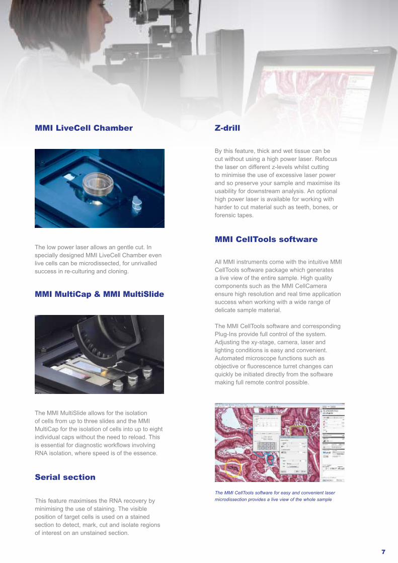
MMI CellTools software
All MMI instruments come with the intuitive MMI CellTools software package which generates a live view of the entire sample. High quality components such as the MMI CellCamera ensure high resolution and real time application success when working with a wide range of delicate sample material.
The MMI CellTools software and corresponding Plug-Ins provide full control of the system. Adjusting the xy-stage, camera, laser and lighting conditions is easy and convenient. Automated microscope functions such as objective or fluorescence turret changes can quickly be initiated directly from the software making full remote control possible.
The MMI CellTools software for easy and convenient laser microdissection provides a live view of the whole sample

Digital remote laser microdissection
MMI CellCut Plus: A high degree of expandability
8
Slide scanning
MMI CellCloud
Tissue Sampling
External Laboratory
Molecular Analyses
MMI CellCut Plus cell isolation (LMD)
Pathologistcell selectioncutting map
Slides are stored in the CaseCenter, a web based slide and case database with HIS/LIS integration possibility, and can now be shared with the remote specialist.
Laser microdissection plays an important role in modern pathology and cancer research. Now it is possible to combine the advantages of digital and molecular pathology. In collaboration with 3DHistech MMI has established the entire process from digital slide scanning to remote laser microdissection via internet based cloud technology. Transfer of the “cutting map” directly to the MMI CellCut Plus facilitates cell isolation and collection following remote digital diagnosis.
1. Scan your slides
Direct scanning to the CaseCenter, for example with the Pannoramic MIDI from 3DHistech, a 12-slide digital slide scanner which combines brightfield and fluorescence scanning in one and delivers superb image quality.
2. Share your digital slides
MMI presents the entire process from digital slide scanning to remote laser microdissection via internet based cloud technology
Workflow
3. Select cells or group of cells
The areas of interest can be found auto-matically with the image analysis software, a multi-purpose solution which runs on local and remote slides.The annotations on the digital slide serve as a cutting map for the MMI CellCut Plus.
4. Automatic cell isolation
The MMI CellCut Plus precisely cuts and isolates the cells on the imported cutting map and transfers them to the PCR tube for downstream analysis.

9
MMI CellEctor Plusfor integrated workflows
MMI’s fully modular platforms can be used either alone or in combination with other technologies or micromanipulation tools. As an example, the MMI CellCut Plus can be combined with the MMI CellEctor Plus for the detection and isolation of single cells in suspension. This creates unique flexibility for a wide range of applications and for the development of inte-grated workflows for single cell isolation.
The MMI CellEctor Plus is a capillary based single cell sorting system. The completely software manipulated 3D CellRobot controls the precise landing position of the capillary and generates a highly reproducible contact point.The high precision of the MMI CellPump regulates the acquisition and deposition in a variety of modes, giving the user complete control in manual and automated cell recognition, acquisition and deposition.
All accessories were directly developed in consultation with MMI CellEctor Plus users to reflect their exacting needs. They allow for the deposition of single cells onto reaction slides or directly into PCR tubes, IBIDI style chambers, microfluidic devices or well plates. User defined liquid handling programmes enable the development of dedicated cleaning and service cycles for capillary cleaning and maintenance.
Features & benefits
• Microscope based software controlledmovements of the capillary
• Easy to achieve accurate and precise cellisolation and deposition
• No contamination from unwanted cells• Faster and easier than manual systems• Nanoliter pump allows isolation of cells in
small volumes• Brightfield or fluorescence automated
detection of cells• Full process control of cell identification,
acquisition and deposition• Work manually or automatically as your
workflow demands• Compatibility with a wide range of accessories
and microfluidic devices
Applications
• Detection and isolation of single and rare cellsin suspension, e.g. Circulating Tumor Cells (CTCs)
• Cancer research and oncology• Detection and isolation of monoclonal antibodies• Cell sorting on a wide range of biochips• Lab-on-a-chip technologies• Stem cell research and cellular diagnostic• Single Cell PCR and gene expression analysis
Combi solution:MMI CellCut Plus and MMI CellEctor Plus (here with Olympus IX83 microscope)
For more information, please have a look into our brochure MMI Single Cell Solutions or visit our website
Designed for fast and reliable detection and isolation of single cells, stem cells and CTCs by fully controllable microscopic collection: MMI CellEctor Plus
www.molecular-machines.com/products/mmi_cellector_plus/

10
MMI CellCut Plus: A wide range of applications
The MMI CellCut Plus is an extremely versatile instrument and therefore the best choice for clinical and research microdissection. Whether isolating cells from an entire tissue, maintaining a pluripotent stem cell line, or extracting cells for forensic investigation, the MMI CellCut Plus performs efficiently and reproducibly.
The starting materials
Cryo or paraffin-preserved tissue
Single cells, smears or cytospins
Chromosomes
Others, e. g. sperms, C. elegans
Fluorescence-labeled cell components
Targeting specific cells in healthy and diseased tissues is of particular importance in elucidating the molecular mechanisms that leads to cancer and other life threatening diseases. Differences in genomic (DNA/RNA) and proteomic expression of any tissue type can be analyzed easily. For example, transverse sections of intestinal glands from colon tissues can be identified and quickly isolated for the study of specific genes and the corresponding hormonal response.
Pathological samples
Fluorescence-labeled chromosomes
The precision of the MMI CellCut Plus enables the simple location and microdissection of sub-cellular structures. For example, chromosomes with detectable fluorescence in situ hybridization (FISH) signals can be isolated for subsequent investigation.
Most stem cell lines are grown at high densities on mouse fibroblast “feeder layers”. Therefore, isolating specific pluripotent cells from the surrounding fibroblasts and differentiating cells needs to be fast and precise. The MMI CellCut Plus offers the perfect balance between speed, precision and ease of use, allowing the target stem cells to be isolated and re-cultured without any side effects, such as karyotype changes.
Cell culture in phase contrast observation
Isolation of intestinal gland from transverse tissue sections of colon (H&E stained)
Live cells

11
C. elegans are extensively used as a model organism in molecular research and develop-mental biology and can also be examined with the MMI CellCut Plus. The living organism can be placed or fixed between the membrane and a glass slide, in order to isolate and extract areas of interest as required by the individual experiment.
Others
Cancer research & Oncology Forensic analysis
The MMI CellCut Plus allows the location, cutting, isolation and collection of in excess of 300 sperm cells on a single cap, all under full audited visual control. This maximises the chances of obtaining a high quality full DNA profile that can be loaded and searched on appropriate crime databases. The MMI CellCut Plus can also be utilised for the isolation of cells from archived slides making it a powerful tool in the investigation of old unsolved cases.
Detection and isolation of sperm cells from case work smears
Isolation of living C. elegans
Detection and isolation of cancer tissues from tissues or cell cultures
The enrichment of cell populations from tumors extracted from tissue or blood samples is essential for the development of truly effective treatment programmes. The key to success in cancer treatments is fast and accurate diagnosis, rapid implementation of specific treatments and treatment effectiveness monitoring. The MMI CellCut Plus is predestinated to work with a wide range of sample types for the isolation of cancer tissue from sections facilitating fast and effective diagnosis.
The MMI CellCut Plus is widely used in traditional research, for cancer and personalized medicine
Haematological and cytological samples
Single cells, such as a plasmablast from a blood smear can be identified, cut, and isolated auto-matically. The dissected cells are now ready for downstream analysis such as molecular profiling for proteomics or genomics purposes.
Blood smear with a typical plasmablast

12
Customer report andapplication protocols
H&E Staining with the MMI H&E Staining Kit Plus
Contamination free staining: MMI Staining Kit Plus
The MMI H&E Staining Kit Plus is designed for users with a need to quickly stain only a few samples and need to ensure that the result is clean, clear of RNAse and contamination free. The staining solutions are supplied in MMI SafeStain ampoules, allowing quick handling without the need for pipetting or the preparation of staining jars.
The MMI SafeStain ampoule guarantees a uniform drop size, and ensures the solutions remain contamination free. The staining solutions have been rigorously tested for thedemanding needs of laser microdissection users. The clear staining they need is reproducibly of the samples together when viewed with the MMI IsolationCap. Each kit contains 15 MMI SafeStain ampoules, designed for 30-60 staining sessions. Ampoules are designed for the staining of 2-4 slides.
Prod. No.: 70302
Improved sample Lift-Off success rate due to quicker drying. By choosing the MMI H&E Staining Kit Plus you help to reduce the amount of water hazardous stain waste and cut your laboratory running costs. The whole workflow is illustrated in the application protocol:
Testimonial
Dr. Jonigk and his team work in the field of lung tumor diseases, fibrous lung remodeling under different circumstances, and pulmonary hyper-tension. The focus is on the combination of clinically relevant issues and the morphology of the tissue, and on the expression of DNA, RNA,and proteins in sub-compartments of the lung. With the MMI CellCut Plus laser micro-dissection system, Dr. Jongik is able to retrieve high quality samples from the starting material. This significantly improves the accuracy of his results.
Dr. med. Danny D. Jonigk, Pathological Institute, Medical School Hannover, Germany
“We appreciate the resistant MMI product quality, the professional consulting, and the competent and quick service. MMI instruments are an important basis for our in-situ analysis in cellular tissue. Laser microdissection followed by gene expression analysis is complementary for further routine methods like conventional optical microscopy (fluorescence), in-situ hybridization, and immunohistochemistry.”
MMI Bibliographies
First issue on laser microdissection in cancer research and clinical oncology, featuring technologies and references
Second issue on laser microdissection in
cancer proteomics, featuring customer
reports, technologies and references
Third issue on laser microdissection for transcriptomics, featuring customer and MMI reports, publications and references

13
Chromosome LCM
Zebrafish Application Note Tissue Transfer from Archived Samples
Smears and Cytospins
Embedding and Freezing
General Staining
FFPE Sectioning FFPE Embedding
DirectorTM Slides
Cryosectioning
Prod. No. 50301
Download the MMI Bibliographies and all protocols from www.molecular-machines.com/support/information_material
ScreenCell® Application Note
Tips for working with RNALive Cell Microdissection
Live cell dissection: MMI LiveCell Chamber
The isolation and enrichment of individual live cells for culture and differentiation experiments, or for their proteomic and genomic analysis is of increasing interest in stem and cancer cell research, and molecular pathology.
The MMI CellCut Plus in combination with the approved MMI LiveCell Chamber enables contamination free isolation of live cells in living culture. Enzyme treatments (i.e. trypsin), other potentially harmful selective reagents as well as time consuming repetitive enrichments can be avoided. The benefit is an increase in reliability, effectiveness and accuracy of your research.
1. Cell seeding and cultivation
3. Cell selection and automated laser cutting
4. Cell isolation and reculturing
2. Cell visualisation
Components of the MMI LiveCell Chamber are a membrane ring for the initial cultivation of cells, a cell culture dish to house the membrane ring with seeded cells, and a UV-permeable microdissection chamber. All components can be sterilized ready to use. The whole workflow is illustrated in the application protocol:
5. Further down-stream application
MMI LiveCell Chamber

You make a substantial investment by buyinga microdissection system and the right choice by selecting a MMI product. To ensure you always get the best from your instrument and longterm continuous use, we recommend all customers consider the benefits of service contracts, regular upgrades, application trainings and our high-quality consumables for better results.
Keep your system running smoothly, reliably and ready for use:
Choose between different service contracts over a time frame to fit your needs. They can include annual preventative maintenance by one of our service engineers and also emergency callout cover. Please contact MMI directly for availability and options in you region.
Stay up-to-date with new developments and upgrades:
We regularly provide various upgrades for all our instrument platforms, for example a more powerful workstation with interactive pen display, a new laser or camera, the latest software version with new features and benefits.
Ensure trouble-free daily operation:
We offer protocol development and instrument trainings either in-house or on-site with your own equipment.
MMI TeamViewer for remote service available by default:
The MMI TeamViewer function allows our service engineers to connect with your MMI system via remote service. This makes world-wide troubleshooting fast and effective. Even the MMI CellTools software can be updated this way. The MMI TeamViewer function operates very easily without the need for configuration by the end user, even through firewalls and proxy servers. If the TeamViewer is not yet installed on the MMI computer, you can download it from our website. Please call us after the download to facilitate the connection.
Customer support andhigh-quality consumables
To support the transfer process from a cryo section to a membrane slide. The MMI SupportSlide is designed to give rigidity to the membrane during sample preparation. It also acts as a heat sink to aid reproducibility and ensure you correctly place the sample on the correct side of the membrane.
Prod. No. 50105
The 26 mm x 76 mm metal frame is coated with a 1.5 µm thin PEN membrane. You can choose between standard MMI MembraneSlides that are packed into a slide rack (Prod. No. 50103). Or, If you need the highest standards of purity the MMI MembraneSlides are available RNAse and nucleic acid free (Prod. No. 50102). They are packaged into boxes of five for better contamination control. Every sales unit comes with 50 MMI MembraneSlides in total.
Prod. No. 50103
Prod. No. 50102 (RNAse free)
MMI MembraneSlides
14
MMI SupportSlide
MMI ServiceSlide
MMI ServiceSlide and 18-well IBIDI® Slide are used for liquid handling on the MMI CellEctor Plus.
Prod. No. 50108

For the isolation and collection of the target cells from the MMI MembraneSlide. Available in three sizes (0.2 ml, 0.5 ml and 1.5 ml) and two types (either filled with nontransparent silicone, diffused, or with transparent silicone). Diffuser caps are recommended for stained or coloured samples.
Prod. No. 50109
Diffuser caps
Transparent caps
Prod. No. 50214 (transparent) 50213 (with diffuser)
MMI 8-CapStrip (0.2 ml per cap)
Prod. No. 80105 (40 µm)
Prod. No. 50215
MMI CapStrip with 8 empty caps (0.2 ml per cap)
Recommended for first application use after instrument purchase. Contain all available consumables for MMI CellEctor Plus or MMI CellCut Plus.
Prod. No. 80103 (20 µm)
Prod. No. 80102 (10 µm)
18-well IBIDI® Slide
MMI IsolationCaps
Transparent caps are used for fluorescent applications and recommended for working with the MMI SmartCut Plus. Available in boxes of 50 pieces. MMI Capillaries
MMI Starter Kits
Used together with the MMI Multi CapLift for the collection of cells in up to 8 different caps simultaneously. A box per type contains 5 strips.
Designed for cell collection with MMI CellEctor Plus, 54 mm long, very sharp 45° beveled tip to collect even adherent cells, available in 3 sizes,10 pieces per box. Also available: MMI Oill and MMI CapillaryClean. MMI CapillaryClean keeps the capillary free of dirt and contamination and avoids blockage. Cleaning of capillaries is recommended as part of all workflows.
Prod. No. 50206 (0.2 ml) 50202 (0.5 ml) 50210 (1.5 ml)
Prod. No. 50208 (0.2 ml) 50204 (0.5 ml) 50212 (1.5 ml)
15
For the collection of cells with the MMI CellEctor Plus, 0.2 ml per cap, 5 strips per box.
If you are working with small cell populations an alternative to the MMI LiveCell Chamber is the 18-well membrane IBIDI slide. The cells are cultivated within the wells. Afterwards, the slide is turned upside down and placed on the stage. The cells can now be cut and isolated with a standard MMI IsolationCap (0.2 ml recommended). If stilled filled with liquid the MMI CellEctor Plus can, instead, be used to transfer the cells directly from the wells.
Starter Kit MMI CellCut Plus
Starter KitMMI CellEctor Plus
Prod. No. 70304
Prod. No. 70303
Prod. No. 80107MMI CapillaryClean
MMI OilProd. No. 80108

MMI Molecular Machines & Industries AGFlughofstrasse 37 | CH - 8152 Glattbrugg | Switzerlandphone +41 (0) 44 809 10 10 | fax +41 (0) 44 809 10 11E-Mail [email protected]
MMI Molecular Machines & Industries GmbHBreslauer Strasse 2 | D - 85386 Eching | Germanyphone +49 (0) 89 319 048 40 | fax +49 (0) 89 319 048 59E-Mail [email protected]
MMI Molecular Machines & Industries Inc.PO Box 348 | Haslett, MI 48840 | USAphone +1 - (603) 629 9536 | fax +1 - (321) 978 0304E-Mail [email protected]
Published by Molecular Machines & Industries AG, Glattbrugg, Switzerland | 1st edition, 2013
www.molecular-machines.com
Please note that some illustrated products or features might be unavailable in some countries. Please contact MMI or your local distributor for availability and options in your region.