Soft tissue Sarcoma - · PDF fileSoft tissue Sarcoma: Overview ... (66% deep thigh) •...
91
Soft tissue Sarcoma: Overview Kazi S. Manir MD,DNB,ECMO R.G.Kar Medical College, Kolkata
Transcript of Soft tissue Sarcoma - · PDF fileSoft tissue Sarcoma: Overview ... (66% deep thigh) •...

Soft tissue Sarcoma: Overview
Kazi S. Manir MD,DNB,ECMO
R.G.Kar Medical College, Kolkata

• Epidemiology • Pathology • Natural History • Workup • Treatment

Epidemiology • Multiple malignancies
at multiple sites • 21% Pediatric solid
tumor • 1% adult solid tumor • 87% of all sarcoma are STS • Osteosarcoma &
Chondrosarcoma : MC Bone
• “Other “ MC is Sarcoma Burningham Z et al. Clin Sarcoma Res 2012
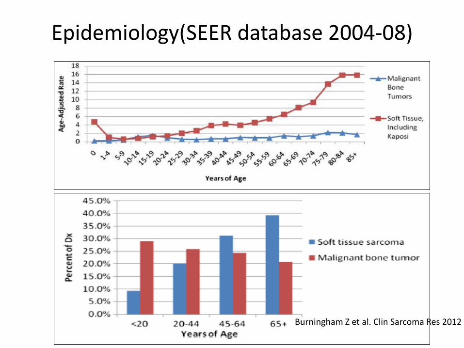
Epidemiology(SEER database 2004-08)
Burningham Z et al. Clin Sarcoma Res 2012

Epidemiology : India
• 0.9% of all cancer: Sarcoma • EWS and Osteosrcoma : MC Bone sarcoma
Ramaswamy A et al. SAJC 2016

Aetiology
• Race and geography: • Exact data lacking • Comparable incidence rates through out the
world. • EWS more common in white • STS more common in Black

Genetics
NF 1 10% EWS
TP53 (Li Fraumeni) Paediatric RMS
TP53 (Somatic) 30-60% STS
RB mutation Leiomyosarcoma
Hereditary RB Survivors OGS (500 fold risk than gen pop)
FAP (Gardner’s Syndrome) (5q21) Desmoid Tumor
Carney’s Startakis Syndrome (SDH B mutation) GIST
RAPADILINO Syndrome OGS
Rothmund Thomson Syndrome II OGS
Werner Syndrome OGS
Bloom Syndrome OGS
Diamond Blackfan Anemia OGS

• Reproductive and Obstetrics factor: • Non-significant association with: • Age of 1st childbirth (>29yrs) (OR 3.16) • Toxemia during Pregnancy (RR 2.71) • High Birth weight (>4.065gm) (OR 1.35) • Congenital Hernia (with EWS) (RR 6.67)
Burningham A et al.. Clin sarcoma Research 2012

• Infection (adult Sarcoma) • HHV 8 (Most Common): Kaposi’s Sarcoma • Both in HIV + and HIV - • HIV: Kaposi’s Sarcoma •
• Radiation (SIR :4.2) • Less in low dose Ionizing Radiation (RR 7.5 per
Gy) • STS MC secondary cancer after RT • RT in Breast, Lymphoma, GU, HNC • 16 fold increase of Angiosarcoma after Breast RT • Average time period 10yrs • Histotype: • Pleomorphic MFH (26%)> Angiosarcoma (21%) Fibrosarcoma (12%)>LMS (10%)>MPNST(9%)

• Lymphedema: • Lymphangiosarcoma • Specially after PMRT (in & outside RT field) • Filariasis

• Occupation exposure and lifestyle • Bone tumor: • Wood, cork and straw factory (OR 3.57) • Radiology worker (SIR 2.88) • Chlorophenol exposure (OR 1.79) • No significant relation with Tobacco or alcohol • Trauma anecdotal in Desmoid tumor

Distribution

Pathology • Heterogenous group of mesenchymal malignancies • HP + IHC • May have distinct genetic correlation • May have distinct clinical course with distinct outcome
• WHO subtyping (Soft Tissue Tumor): • Benign • Intermediate (locally aggressive) • Intermediate (Rarely metastasizing) • Malignant

Pathology
• Most common types: • Undifferentiated/unclassified sarcoma (pleomorphic/round
cell/spindle cell) (Pleomorphic MFH) • Liposarcoma • Leiomyosarcoma • Synovial sarcoma • Malignant peripheral nerve sheath tumor (MPNST) • Rhabdomyosarcoma • Primitive neuroectodermal tumor (PNET) /extraskeletal
Ewings • Angiosarcoma • Ephitheliodsracoma • Clear cell sarcoma • Alveolar soft part sarcoma • Solitary fibrous tumors

Myxofibrosarcoma(formerly MFH)
• Common : Malignant • Infiltrates centimeters beyond the
visible/palpable mass and when deep can invade usual barriers
• Higher rate of positive margins • Commonly in the extremities • Greater risk of local recurrence (up to 30%) • 5yr OS 60-70%

Dermato fibrosarcoma Protuberans(DFSP)
• Benign (rarely metastasizing) • + CD34 • Rare but common cutaneous form • WLE (2cm margin) TOC [5yr LR <5%] • R1/R2 LR>50% needing Adj RT • Rarely mets to lung (after high grade
transformation) • Imatinib in advanced and metastatic diseases

Lipoarsarcoma • Common 50-60yr (20% adult STS) • Any site • Common: • Thigh (24% of all extremity STS) • RP (45% of all RP STS) • Subtypes (distinct Clinico-pathology): 1. Well-differentiated(WD)/dedifferentiated LPS/Atypical
Lipomatoues Tumor (ALT) 2. Myxoid/round cell LPS 3. Pleomorphic LPS 4. Mixed

WDLS/ALT
• CDK4/MDM2/HMCA2 amplification • Has a ‘pushing’ growth pattern • Occurs Extremity muscles (most common) • Retroperitoneal (RP) • Variety of other sites • Behavior is different in limb vs. RP • Recur less frequent and late • Not develop metastases • Dedifferentiation is uncommon (0-6%) • Managed by marginal excision alone • 5yr DFS 83% SommervilleSMM, et al. ANZ J Surg. 2005;75:803. Weiss SW Am J SurgPath .1992;16:1051.

ASTRO Refresher course 2015

Myxoid/Round cell LPS
• Mean age in mid 40’s • Extremity MC (66% deep thigh) • Unusual metastatic Site: Soft tissue,RP (not
Lung) • Pure Myxoid : Low grade (5yr DFS 90%) • >5% Round cell : High Grade (5yr DFS 50%) • Exceptionally high S to Radiotherapy and
Ifosfamide/Trabectedin

Myxoid LPS -dramatic responses to radiation • McGill 50 patients, evaluated response to RT • median decrease in tumor volume: • <1% for high grade sarcomas • 13.8% non-myxoid low grade sarcomas • 82.1% myxoid liposarcomas RobergeD, et al. RadiotherOncol.2010; 97:404.
Pleomorphic LPS
• High grade aggressive rare LPS • Median age >50yrs • Resembles undifferentiated/unclassified
variety • Upper extremity < Lower extremity • Lung metastasis >50% cases • Responsive to Ifosfamide/Gemcite

Leiomyosarcoma
• Vascular Smooth muscle origin (SMA/Vimentin +ve)
• Middle age • Any site: RP>Pelvis>uterine body • Vascular outflow obstruction common • Surgery Primary treatment • RP lesion are large/high grade • Recurrence risk >50%

Malignant peripheral nerve sheath
tumors(MPNST) • Originate from peripheral nerves
• 50% occur in patients with NF type I • S-100 +ve (High Grade less S100+ ) • Most common in the extremities, trunk, H&N • NF1 associated worse outcome than sporadic • WLE +/- RT : TOC • NACT (RR 20%) : Ifosfamide/Doxorubicin • Sorafenib : Investigational
–

Angiosarocma (including lymphangiosarcoma)
• Uncommon • Commonly associated with Lympoedema
(Stewart Treves Syndrome) and RT (Breast Ca Commonly)
• Arise in skin/subcutaneous tissue –most typically of the breast or H&N
• One of most common sarcomas seen after RT • Chemo-responsive sarcoma-taxanes and anthra • LR recurrences common (Median Survival 3yr)

Synovial sarcomas • Young adult 15-35Yrs • Originally thought to arise from the synovium
of joints but actual origin is unknown • t(X;18)(p11.2;q11.2) • 2 types: monophaisc and biphasic • 80% extremity (LE>UE) • Histology that is more responsive to
chemotherapy

Alveolar soft part Sarcoma
• Rare ,F>M,20years • t(17-X)(p11.2;q25) [ASPSCR1-TFE3 fusion
protein] • LE>UE • Initially slow growing, Low local rec after Sx • Poor prognosis after metastasis • Investigational: MET-I (crizo),Antiangiogenic
(Sunitinib/Avastin)

Epitheloid Sarcoma
• Distal type (Hand & feet) and Proximal type (thigh, buttock) (Distal UE MC)
• Young adult • Deep fascial spread (wide margin) • >20% LN metastasis (LND if N+) • Poor Prognosis (5yr OS 63%) • Moderately S to CT/RT (Proximal more
resistant and aggressive)

Desmoid tumor(Fibromatosis)
• 10-25Years, rare, • Locally aggressive, non-metastatasing • Mutation in CTBNN-1 gene that code beta-
catenin • Common site abdominal wall>extremity • Incidence increased after Pregnancy • Local Recurrence (variable) : 15% in 5yr • Surgery : TOC

Indian Data
Ramaswamy A et al. 2016
Clinical Presentation
Extremity • Enlarging painless mass • Pain • Functional limitations • Symptoms associated with compression of
local structures
Clinical Presentation
Retroperitoneal • Abdominal mass –often incidentally found • Pain • Gastrointestinal: early satiety, obstruction,
bleeding • Lymphedema, neurologic or musculoskeletal
sx

Clinical Presentation
Rare • Fevers/leukocytosis • Paraneoplastic hypoglycemia
(leiomyosarcoma) • Symptoms from distant metastases

Patterns of spread
Extremity • Along longitudinal tissue planes –within the compartment • If involves nerves/vessels, can track along • Compresses/distorts adjacent soft tissue • Tumor can be well beyond the mass
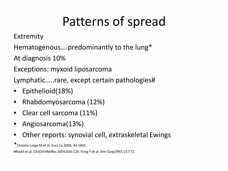
Patterns of spread Extremity Hematogenous….predominantly to the lung* At diagnosis 10% Exceptions: myxoid liposarcoma Lymphatic…..rare, except certain pathologies# • Epithelioid(18%) • Rhabdomyosarcoma (12%) • Clear cell sarcoma (11%) • Angiosarcoma(13%) • Other reports: synovial cell, extraskeletal Ewings *Christie-Large M et al. EurJ Ca 2008; 44:1841.
#RiadS et al. ClinOrthRelRes 2004;426:129. Fong Y et al. Ann Surg1993; 217:72.
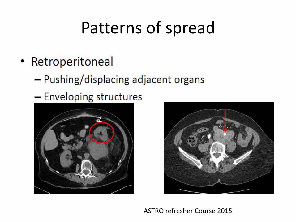
Patterns of spread
ASTRO refresher Course 2015

Evaluation
• MRI : IOC • Standard X ray to : rule out bone tumor bone erosion & risk of # if any calcification if any • CT scan in RP tumors (yield is equal to MR)

Imaging • MRI (+/- Dynamic contrast enhancement) may
help in assessing prognosis & response to ChT. • DCE MRI may act as surrogate for VEGF. • Whole body MRI – alternative to PETCT in
children for whole body staging. • FDG PET in the initial staging can lead to tt
optimisation particularly in EWS due to the superiority of FDG PET in detecting bone lesions.
• FDG PET- potential non-invasive surrogate for ChT response.
Guideline of Bone and STS, Puri A,Laskar S ,2011
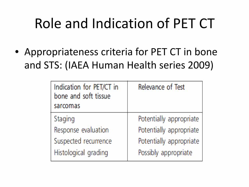
Role and Indication of PET CT
• Appropriateness criteria for PET CT in bone and STS: (IAEA Human Health series 2009)

Role of FNAC
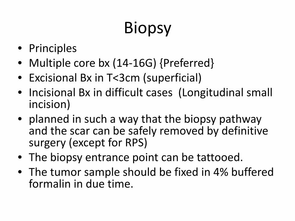
Biopsy • Principles • Multiple core bx (14-16G) {Preferred} • Excisional Bx in T<3cm (superficial) • Incisional Bx in difficult cases (Longitudinal small
incision) • planned in such a way that the biopsy pathway
and the scar can be safely removed by definitive surgery (except for RPS)
• The biopsy entrance point can be tattooed. • The tumor sample should be fixed in 4% buffered
formalin in due time.

IHC EWS/PNET MIC2/CDD99
FLi 1
OGS Nil
Cartilage Tumor ?S-100/? SOX2
Synovial Sarcoma CK,BCL2,Mic2 TFE 3 (New)
RMS MyoD,Desmin,Myoglobin
LMS SMA,Calponin Desmin,Myoglobin
Alveolar Soft Part Sarcoma TLE 3 (New)
Chordoma Brachyury (New)

Staging

Grading Some tumor types not typically graded • MPNST • Epithelioid • Clear cell sarcoma • Angiosarcoma • Extraskeletal myxoid
chondrosarcoma • Synovial sarcoma • all felt to be high grade
NCI grading: 1. Histology 2. Location 3. Tumor Necrosis

management
• Surgical principles: • R0 ( >1cm) margin is the goal • If possibility of R1/R2 (neurovascular proximity: Call Radiation Oncologist Put surgical clips • Remove biopsy scar (tattooed) • May need removal of adventia or perinureum (NV
abutted) • Dissection thru the uncontaminated normal tissue
planes. • Drain (suction /closed) site should be near to incision
site(Scope of ReEx in future Rec
Basic Principle
• All patients to be considered for organ conservation.
• All patients to be evaluated preoperatively for feasibility of Intraoperative Brachytherapy.
• R2 Sx should be considered for revision excision

Unplanned excision: common phenomena • Typically smaller • Typically subcutaneous (mistaken for lipomas) • Often low grade • Residual disease24-74%
.
Re excision to be done Inappropriate skin incision
PORT use higher Similar local control with primary radical surgery Higher rate of aggressive surgery: poor functional
outcome
AlamandaVK, et al. J SurgOncol2012;205:662-667. Fiore M, et al. Ann SurgOncol2006;13(1):110-117
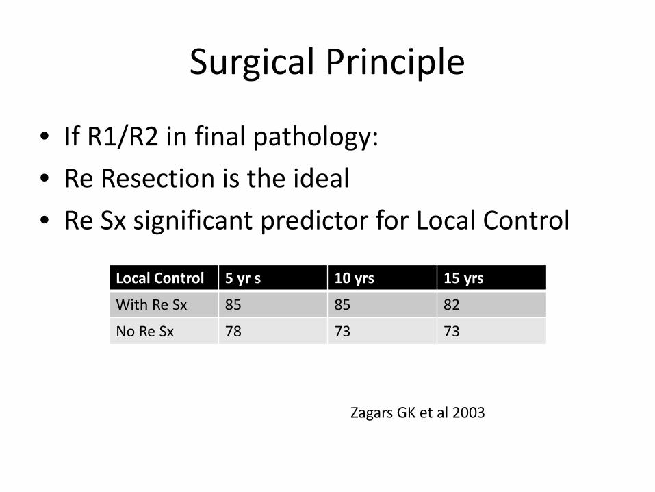
Surgical Principle
• If R1/R2 in final pathology: • Re Resection is the ideal • Re Sx significant predictor for Local Control
Local Control 5 yr s 10 yrs 15 yrs
With Re Sx 85 85 82
No Re Sx 78 73 73
Zagars GK et al 2003
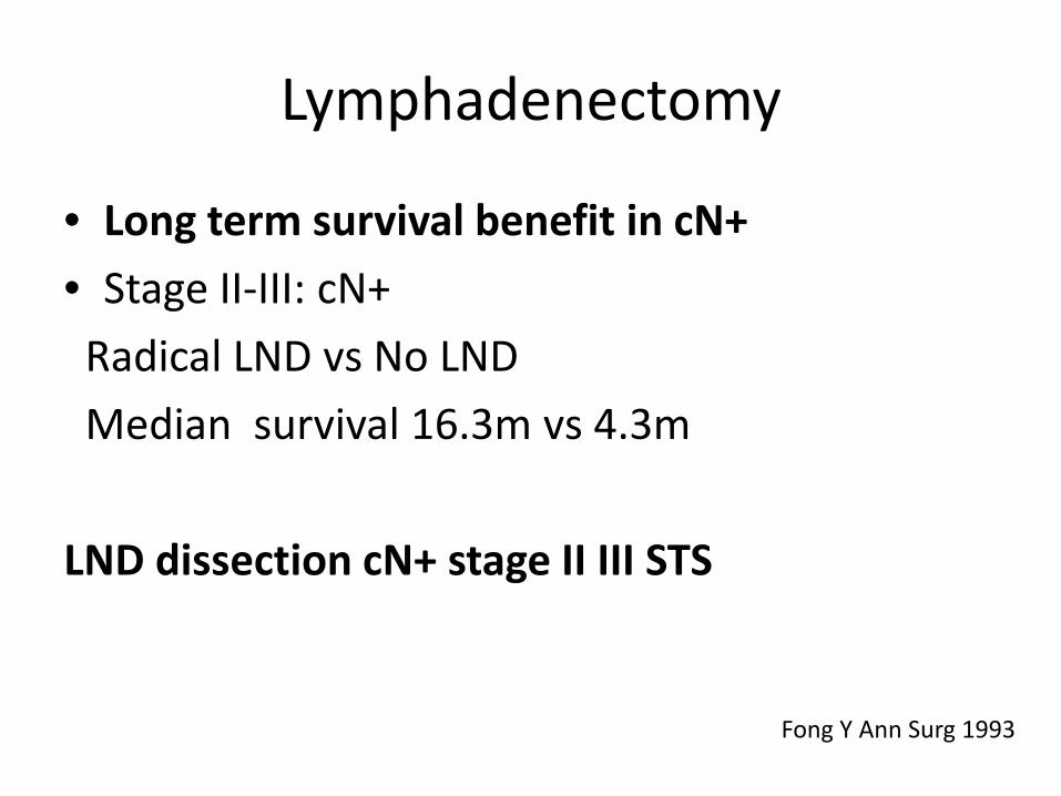
Lymphadenectomy
• Long term survival benefit in cN+ • Stage II-III: cN+ Radical LND vs No LND Median survival 16.3m vs 4.3m LND dissection cN+ stage II III STS
Fong Y Ann Surg 1993
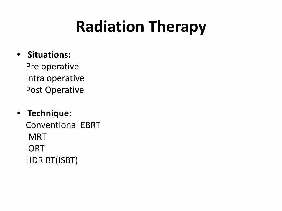
Radiation Therapy • Situations: Pre operative Intra operative Post Operative • Technique: Conventional EBRT IMRT IORT HDR BT(ISBT)

PORT: evidences
• Indications: (any) • R1/R2 • Deep seated tumor • High Grade • T>5cm Limb salvage Sx + PORT: similar LC/OS with amputation Sx + PORT vs Sx alone: PORT increases LC not OS
Rosenburg SA et al Ann Surg 1982 Yang JC et al. J Clin Oncol 1998
Pisters PW et al J Clin Oncol 1996

PORT : R0 situation • High grade: >T1 (>2cm): PORT must T1 : no RCT: Radical ISBT > No adjuvant RT • Low Grade: PORT may be avoided in (all must be present) superficial <5cm R0
Yang JC et al. J Clin Oncol 1998

PORT : ISBT vs EBRT
• No RCTs on Radical BT or EBRT • Similar LC rates BT with/out EBRT • Lesser Soft tissue complications and lesser
hospital stay • Radical BT can be done in appropriate patients
if Intestinal needles can encompass whole tumor bed
Laskar S et al. Annals of Surg Onc 2007
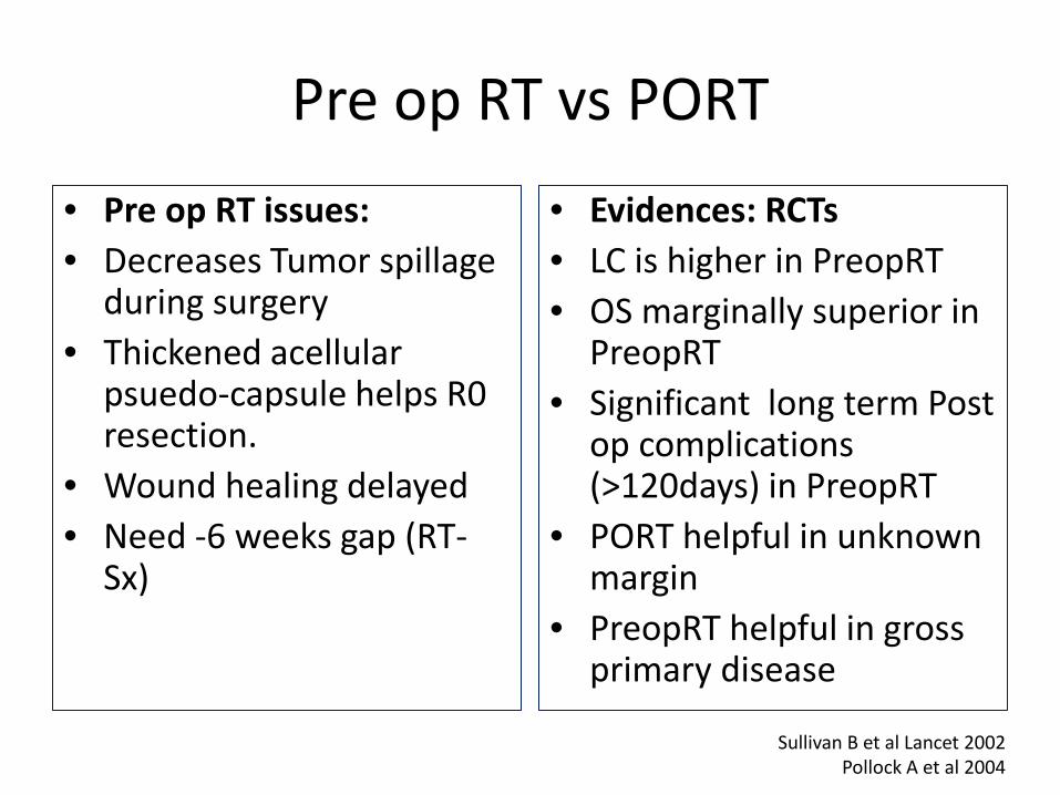
Pre op RT vs PORT • Pre op RT issues: • Decreases Tumor spillage
during surgery • Thickened acellular
psuedo-capsule helps R0 resection.
• Wound healing delayed • Need -6 weeks gap (RT-
Sx)
• Evidences: RCTs • LC is higher in PreopRT • OS marginally superior in
PreopRT • Significant long term Post
op complications (>120days) in PreopRT
• PORT helpful in unknown margin
• PreopRT helpful in gross primary disease
Sullivan B et al Lancet 2002 Pollock A et al 2004

Surgery RT interval
• Only one evidence: • A delay between surgery and the start of RT of
>30 days was associated with a decreased 10- year LC rate (76% vs. 83%, p = 0.07). • May be due to an imbalance in the
distribution of other prognostic factors • SX RT interval is not a potential prognostic
factor for LC Ballo MT et al. IJROBP 2004
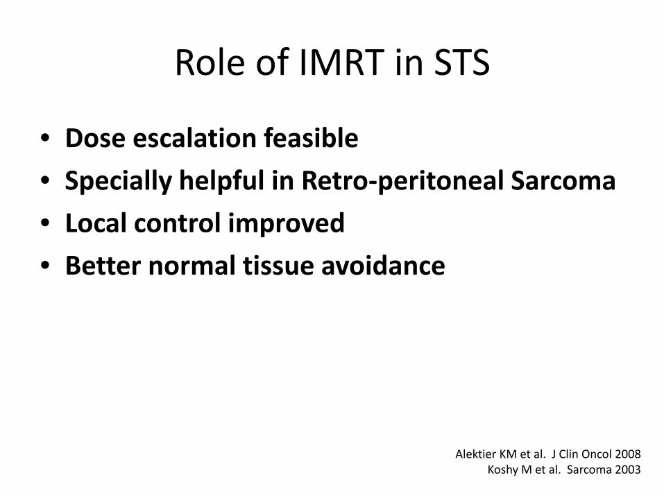
Role of IMRT in STS
• Dose escalation feasible • Specially helpful in Retro-peritoneal Sarcoma • Local control improved • Better normal tissue avoidance
Alektier KM et al. J Clin Oncol 2008 Koshy M et al. Sarcoma 2003
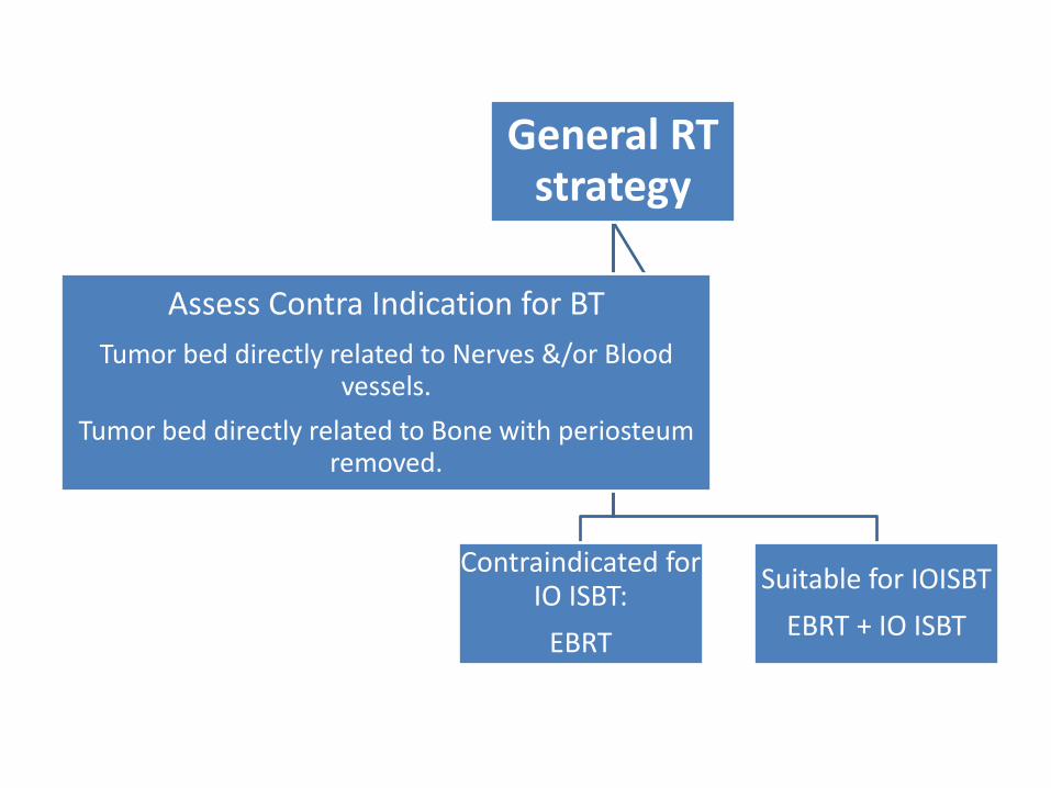
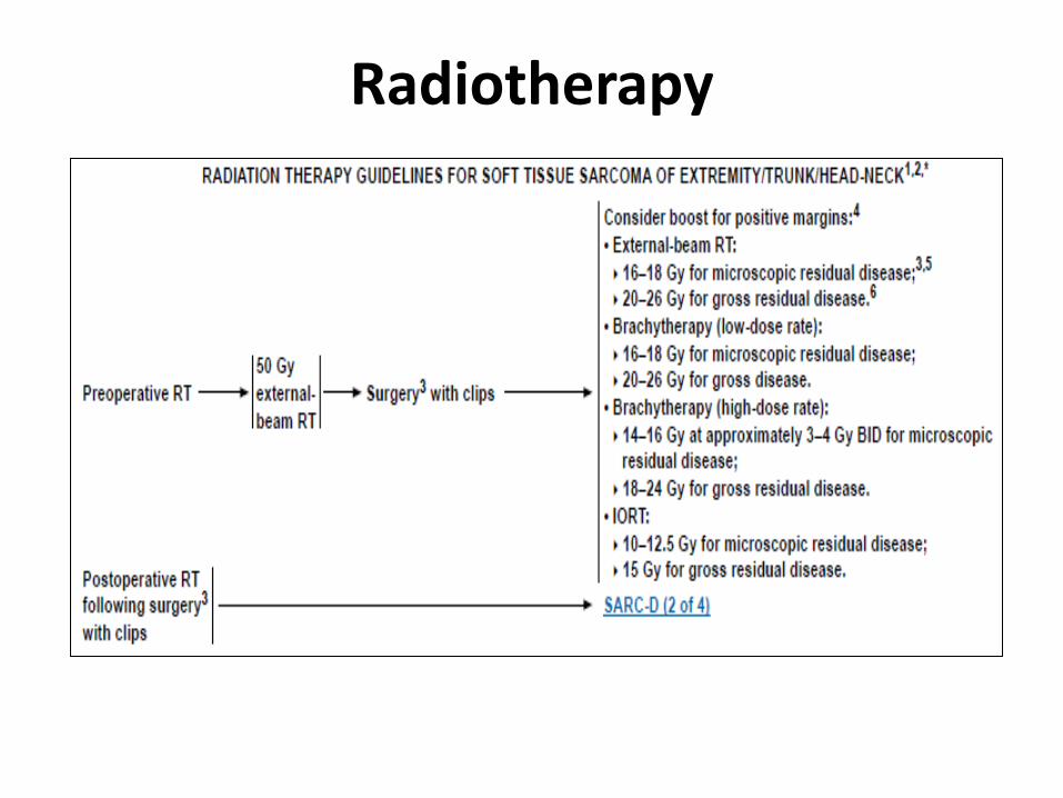
General RT strategy
Contraindicated for IO ISBT:
EBRT
Suitable for IOISBT EBRT + IO ISBT
Assess Contra Indication for BT Tumor bed directly related to Nerves &/or Blood
vessels. Tumor bed directly related to Bone with periosteum
removed.
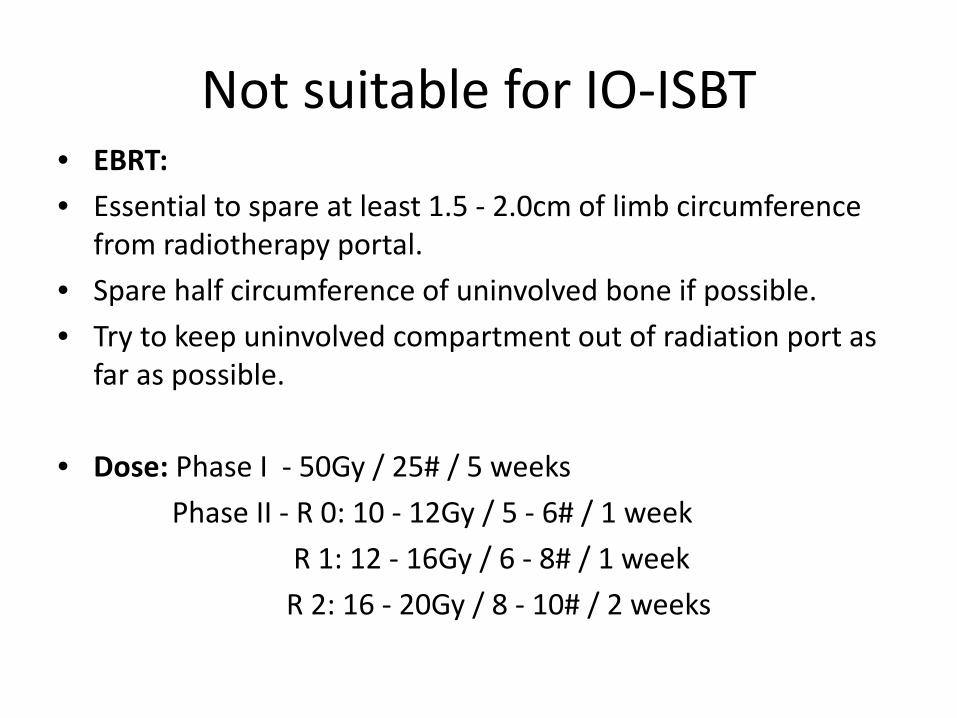
Not suitable for IO-ISBT • EBRT: • Essential to spare at least 1.5 - 2.0cm of limb circumference
from radiotherapy portal. • Spare half circumference of uninvolved bone if possible. • Try to keep uninvolved compartment out of radiation port as
far as possible. • Dose: Phase I - 50Gy / 25# / 5 weeks Phase II - R 0: 10 - 12Gy / 5 - 6# / 1 week R 1: 12 - 16Gy / 6 - 8# / 1 week R 2: 16 - 20Gy / 8 - 10# / 2 weeks

Suitable for IO ISBT • Silver clips placed after excision of tumor to delineate the tumor bed. • Brachytherapy catheters inserted uniformly to cover the entire tumor bed
with 1.5 - 2.0 cm margin. • Simulation and dosimetry to be done on 4-5th postoperative day. • Dose prescription for brachytherapy - 0.5cm on either side of the implant
plane. • Brachytherapy Dose: LDR - 25 - 30Gy @ 45 - 50cGy / hr • HDR - 21Gy / 7# @ 3Gy / # (2# / day with 6hrs gap) • Ext. Radiotherapy: • Radiotherapy to be started 3 weeks after completion of Brachytherapy • Planning Target Volume: Gross tumor volume + 6 - 8cm margin • Dose: After LDR Brachytherapy - 46 - 50Gy / 23 - 25# / 5 weeks After HDR Brachytherapy - 46 - 50Gy / 23 - 25# / 5 weeks

EBRT: Technicalities: extremity

EBRT: Technicalities: extremity

EBRT: Technicalities: extremity

Post op RT Target CTV: shrinking field technique Initial volume • Surgical bed reconstructed from pre-op imaging • Fusion of pre-op MRI with postop planning CT • Further evaluation based on postoperative
changes, operative and pathology report, surgical clips
• Expand volume 1.5 cm radially/4 cm longitudinally(TMH: grade II/III 6-8cm)
Haas RLM, et al. IntJ RadiatOncolBiolPhys. 2012.; 84(3):572-580.

Post op RT Target
• Boost volume • Same as initial volume except in the
longitudinal • Use GTV reconstructed with 2 cm (TMH:3cm)
margins • Other issue: scar/drain site to be included • Low risk situations, drain site could be omitted
Haas RLM, et al. IntJ Radiat OncolBiolPhys. 2012.; 84(3):572-580.

Post op RT Target
ASTRO refresher course 2015

RT field and Local control
LR patients had significantly • higher grade • margin +ve • recurrent disease • more postoperative boost patients • slightly older
DickieCI, et al. IntJ RadiatOncolBiolPhys2012;82(4):1528-34.
60 LR patients vs 708 patients with no recurrence LR patients: 82% (49/60) in-
field 15% (9/60) out-of-field
3% (2/60) marginal
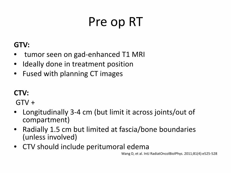
Pre op RT GTV: • tumor seen on gad-enhanced T1 MRI • Ideally done in treatment position • Fused with planning CT images
CTV: GTV + • Longitudinally 3-4 cm (but limit it across joints/out of
compartment) • Radially 1.5 cm but limited at fascia/bone boundaries
(unless involved) • CTV should include peritumoral edema
Wang D, et al. IntJ RadiatOncolBiolPhys. 2011;81(4):e525-528
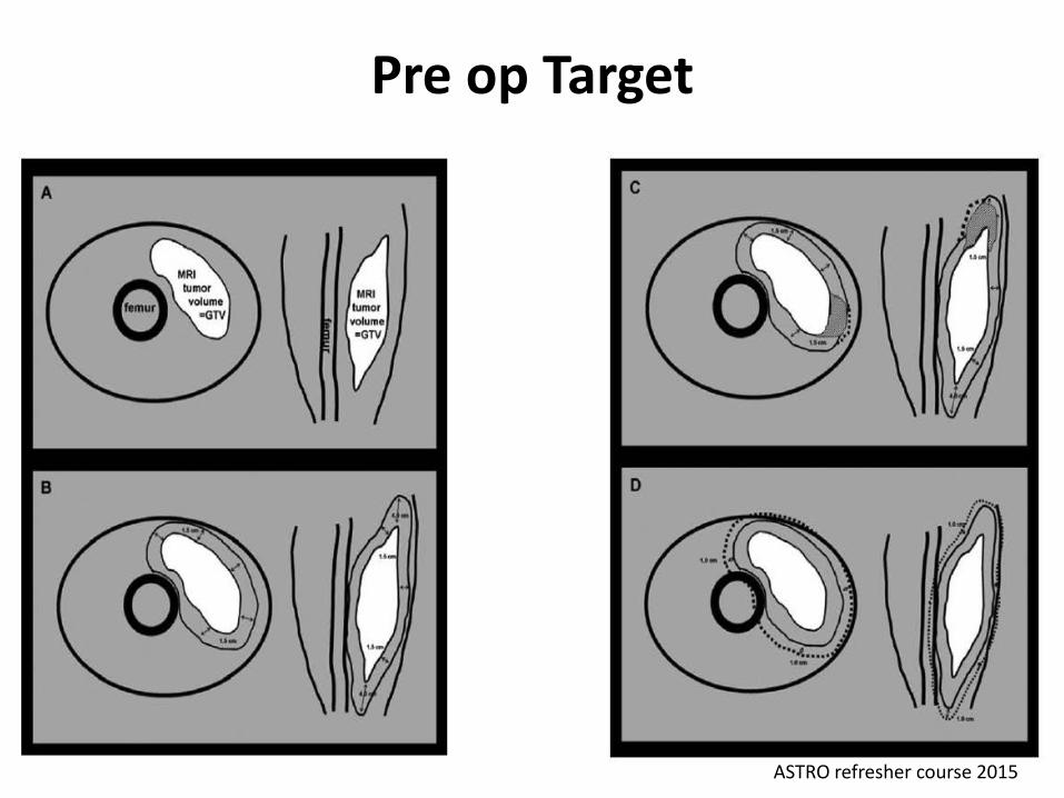
Pre op Target
ASTRO refresher course 2015
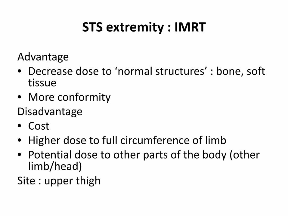
STS extremity : IMRT
Advantage • Decrease dose to ‘normal structures’ : bone, soft
tissue • More conformity Disadvantage • Cost • Higher dose to full circumference of limb • Potential dose to other parts of the body (other
limb/head) Site : upper thigh

STS extremity : IMRT
• Use of IMRT with IGRT –decrease high wound complication rate
• Minimized dose to ‘skin flaps’ as determined in conjunction with the surgeon
• PTV: 50 Gy/25 • Flaps: <20 Gy
ASTRO refresher course 2015

Head neck sarcoma
• All patients to be treated with 3 D conformal radiation therapy
• Planning Target Volume (PTV) shall vary according to exact site of disease.
• Dose: 66 - 70Gy / 33 - 35# / 6 - 7 weeks

DFSP • Extremities & Trunk: Post operative radiation to be considered if: surgical margins +ve surgical margins close recurrent tumor • Mediastinum: Post operative radiation to be considered if: surgical margins +ve surgical margins close surgical margins unknown recurrent tumor Planning Target Volume (PTV): Gross tumor volume + 3cm margin. Dose: 60 - 66Gy / 30 - 33# / 6 - 7weeks High dose Tamoxifen & Chemotherapy – Investigational

STS: Retroperitoneum Radiation issues • Volume • Patient GI stability • Ability to spare normal tissues (meet constraints) • Know kidney plans and function • Preoperative vs. postoperative

STS: Retro-peritoneum Postoperative RT • Rarely can achieve adequate dose • More gastrointestinal toxic Preoperative RT • Advantages • tumor readily identifiable • tumor displaces bowel • potential tumor reduction • Pseudo-capsule formation/margin improvement • typically a lower dose is felt to be needed

STS: Retroperitoneum
• Simulation: • Upper and lower body immobilization • Oral contrast: for upper abdominal tumors • IV contrast –to see psoas muscle invasion • 4D simulation for upper abdominal tumors • if organ motion > 1 cm consider gaiting

STS: Retro-peritoneum
GTV • Register with MRI for muscle extent • Create ITV to account for tumor motion CTV • GTV with 2-2.5 cm margin cephalo-caudal • GTV with 1.5-2 cm margin radially • Exclude: bone, kidney, liver • Include rim of adjacent bowel/air cavity (5 mm) • Include any disease extending to the inguinal
canal Baldini E et al IJROBP 2015

STS: Retro-peritoneum
• Dose escalation areas of high risk • IOERT • IO ISBT • CCB/SIB IMRT

STS: Retro-peritoneum
ASTRO Refresher course 2015
Radiotherapy


Chemotherapy
• Preoperative • Postoperative • Salvage • Metastatic setting • Targeted therapy
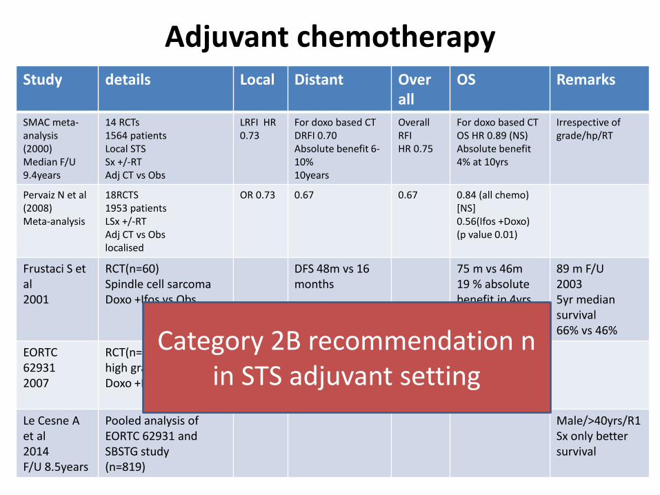
Adjuvant chemotherapy Study details Local Distant Over
all OS Remarks
SMAC meta-analysis (2000) Median F/U 9.4years
14 RCTs 1564 patients Local STS Sx +/-RT Adj CT vs Obs
LRFI HR 0.73
For doxo based CT DRFI 0.70 Absolute benefit 6-10% 10years
Overall RFI HR 0.75
For doxo based CT OS HR 0.89 (NS) Absolute benefit 4% at 10yrs
Irrespective of grade/hp/RT
Pervaiz N et al (2008) Meta-analysis
18RCTS 1953 patients LSx +/-RT Adj CT vs Obs localised
OR 0.73 0.67 0.67 0.84 (all chemo) [NS] 0.56(Ifos +Doxo) (p value 0.01)
Frustaci S et al 2001
RCT(n=60) Spindle cell sarcoma Doxo +Ifos vs Obs
DFS 48m vs 16 months
75 m vs 46m 19 % absolute benefit in 4yrs
89 m F/U 2003 5yr median survival 66% vs 46%
EORTC 62931 2007
RCT(n=351)Resected high grade Doxo +Ifos vs Obs
5yr RFS % Similar (52%)
5year OS chance 64 vs 69%
Le Cesne A et al 2014 F/U 8.5years
Pooled analysis of EORTC 62931 and SBSTG study (n=819)
Male/>40yrs/R1Sx only better survival
Category 2B recommendation n in STS adjuvant setting

Adjuvant chemotherapy

Chemotherapy in advanced stage

Chemotherapy in advanced stage
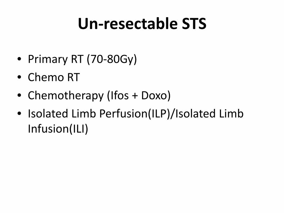
Un-resectable STS
• Primary RT (70-80Gy) • Chemo RT • Chemotherapy (Ifos + Doxo) • Isolated Limb Perfusion(ILP)/Isolated Limb
Infusion(ILI)

Limb perfusion: un-resectable extremity Sarcoma
• ILP: • TNF alfa • Melfalan • Doxirubicin • ILI less invasive
Klauser JM et al. Sarcoma 2001

Pulmonary mets: Sarcoma
• 5yr survival 59% in LN mets and 9% in lung mets Prognostic value: • Synchronous lung mets • Metastatectomy • >4 pul. nodule