60 Gall Stone Ileus - An Uncommon Cause of Intestinal Obstruction
Upload
brendan-youngCategory
view
233download
6
SMALL-BOWEL OBSTRUCTION
Ileus and other disorders of intestinal
motility
EPIDEMIOLOGY Mechanical small-bowel obstruction is
the most frequently encountered surgical disorder of the small intestine.
Although a wide range of etiologies for this condition exist, intra-abdominal adhesions related to prior abdominal surgery is the etiologic factor in up to 75% of cases of small-bowel obstruction.
More than 300,000 patients are estimated to undergo surgery to treat adhesion-induced small-bowel obstruction in the United States annually.
Less-prevalent etiologies for small-bowel obstruction include hernias and Crohn's disease.
The frequency with which obstruction related to these conditions is encountered varies according to the patient population and practice setting.
In contrast to colonic obstruction, small-bowel obstruction is uncommonly caused by neoplasms.
Fewer than 3% of cases are caused by primary small-intestinal neoplasms.
Cancer-related small-bowel obstruction is
more commonly caused by extrinsic compression or invasion by advanced malignancies arising in organs other than the small bowel
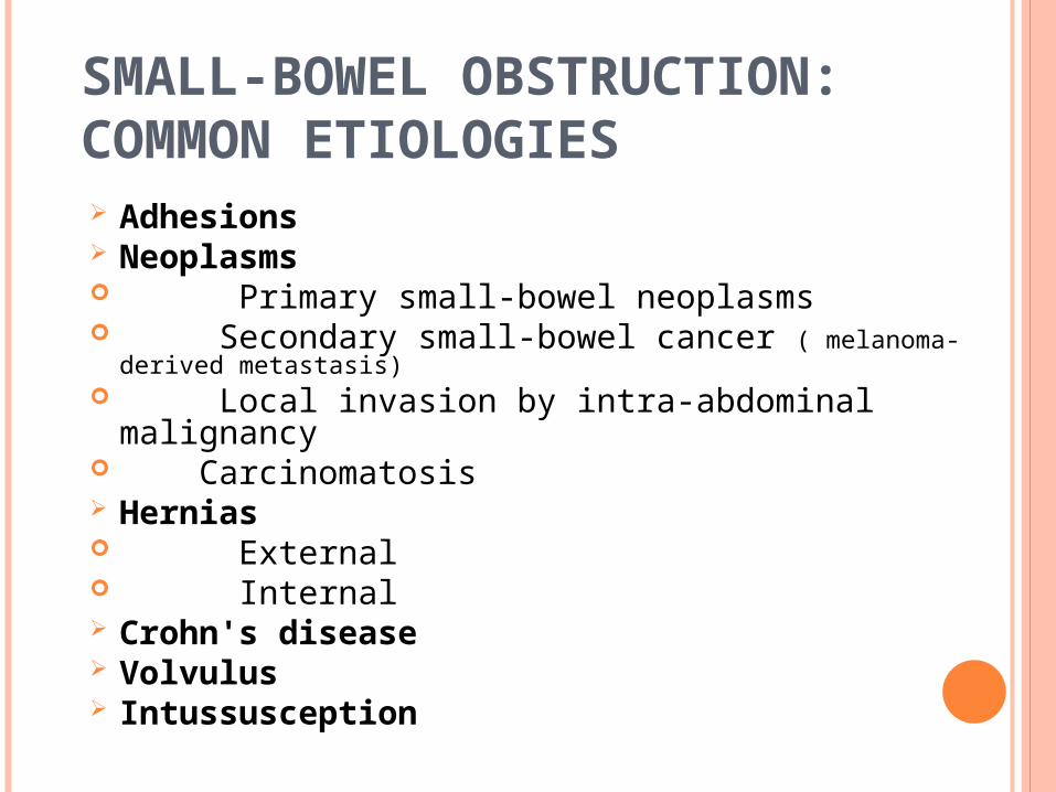
SMALL-BOWEL OBSTRUCTION: COMMON ETIOLOGIES Adhesions Neoplasms Primary small-bowel neoplasms Secondary small-bowel cancer ( melanoma-
derived metastasis) Local invasion by intra-abdominal
malignancy Carcinomatosis Hernias External Internal Crohn's disease Volvulus Intussusception
Radiation-induced stricture Postischemic stricture Foreign body Gallstone ileus Diverticulitis Meckel's diverticulum Hematoma Congenital abnormalities (e,g.. webs,
duplications, and malrotation)
Although congenital abnormalities capable of causing small-bowel obstruction usually become evident during childhood, they sometimes elude detection and are diagnosed for the first time in adult patients presenting with abdominal symptoms.
For example, intestinal malrotationand mid-gut volvulus should not be forgotten when considering the differential diagnosis of adult patients with acute or chronic symptoms of small-bowel obstruction, especially those without a history of prior abdominal surgery.
SUPERIOR MESENTERIC ARTERY SYNDROME
characterized by compression of the third portion of the duodenum by the superior mesenteric artery as it crosses over this portion of the duodenum.
This condition should be considered in young asthenic individuals who have chronic symptoms suggestive of proximal small-bowel obstruction.
PATHOPHYSIOLOGY The obstructing lesion can be
conceptualized according to its anatomic relationship to the intestinal wall as:
(1) intraluminal ( foreign bodies, gallstones, or meconium),
(2) intramural (e.g., tumors, Crohn's disease-associated inflammatory strictures,or hematomas),
(3) extrinsic (e,g., adhesions, hernias, or carcinomatosis).
With onset of obstruction, gas and fluid accumulate within the intestinal lumen proximal to the site of obstruction. Most of the gas that accumulates originates from swallowed air, although some is produced within the intestine.
The fluid consists of swallowed liquids and gastrointestinal secretions (obstruction stimulates intestinal epithelial water secretion), With ongoing gas and fluid accumulation, the bowel distends and intraluminal and intramural pressures rise.
If the intramural pressure becomes high enough, microvascular perfusion to the intestine is impaired, leading to intestinal ischemia, and, ultimately, necrosis.
This condition is termed strangulating bowel obstruction.
With partial small-bowel obstruction, only a portion of the intestinallumen is occluded, allowing passage of some gas and fluid.
The progression of pathophysiologic events described above tends to occur more slowly than with complete small-bowel obstruction, and development of strangulation is less likely.
In contrast, progression to strangulation occurs especially rapidly with closed loop obstruction in which a segment of intestine is obstructed both proximally and distally (e.g., with volvulus).
In such cases, the accumulating gas and t!uid cannot escape either proximally or distally from the obstructed segment.
CLINICAL PRESENTATION
The symptoms : colicky abdominal pain, nausea, vomiting, obstipation.
Continued passage of flatus and/or stool beyond 6 to 12 hours after onset of symptoms is characteristic of partial rather than complete obstruction.
The signs are abdominal distention, which is most pronounced if the site of obstruction is in the distal ileum, or may be absent if the site of obstruction is in the proximalsmall intestine, and hyperactive bowel sounds
LABORATORY FINDINGS
reflect intravascular volume depletion consist of hemoconcentration electrolyte abnormalities Mild leukocytosis
FEATURES OF STRANGULATED OBSTRUCTION INCLUDE: tachycardia, localized abdominal tenderness fever marked leukocytosis acidosis. Serum levels of amylase, lipase, lactate
dehydrogenase, phosphate, and potassium may be elevated.
It is important to note that these parameters lack sufficient predictive value to allow for differentiation between simple and strangulated obstruction prior to the onset of irreversible intestinal ischemia.
5 to 15% of patients who are demonstrated to have frank intestinal infarction have none of these features.
These features have an especially low prevalence in elderly patients.
As a result, strangulated obstruction is particularly treacherous in this population.
DIAGNOSIS
The diagnostic evaluation should focus on the following goals;
1. distinguishing mechanical obstruction from ileus;
2. Determining the etiology of the obstruction; discriminating partial from complete obstruction;
3. Discriminating simple from strangulating obstruction.
Important elements to obtain on history include prior abdominal operations (suggesting the presence of adhesions) and the presence of abdominal disorders (e.g., intra-abdominal cancer or inflammatory bowel disease) that may provide insights into the etiology of obstruction. Upon examination, a meticulous search for hernias (particularly in the inguinal and femoral regions) should be conducted.
The stool should be checked for gross or occult blood, the presence of which is suggestive of intestinal strangulation.
The diagnosis of small-bowel obstruction is usually confirmed with radiographic examination.
The abdominal series consists of a radiograph of the abdomen with the patient in a supine position, aradiograph of the abdomen with the patient in an upright position, and a radiograph of the chest with the patient in an upright position.
The finding most specific for small-bowel obstruction is the triad of
dilated small-bowel loops (>3 cm in diameter),
air-fluid levels seen on upright films, and a paucity of air in the colon.
The sensitivity of abdominal radiographs in the detection of small-bowel obstruction ranges from 70 to 80%.
Specificity is low, because ileus and colonic
obstruction can be associated with findings that mimic those observed with small-bowel obstruction.
False-negative findings on radiographs can result when the site of obstruction is located in the proximal small bowel and when the bowel lumen is filled with fluid but no gas, thereby preventing visualization of airfluid levels or bowel distention.
The latter situation is associated with closed-loop obstruction.
Despite these limitations, abdominal radiographs remain an important study in patients with suspected small bowel obstruction because of their widespread availability and low cost.
COMPUTED TOMOGRAPHIC (CT SCAN)
is 80 to 90% sensitive and 70 to 90% specific in the detection of small-bowel obstruction.
The findings of small-bowel obstruction include:
a discrete transition zone with dilation of bowel proximally,
decompression of bowel distally, intraluminal contrast that does not pass beyond
the transition zone, a colon containing little gas or fluid.
CT scanning may also provide evidence for the presence of closed-loop obstruction and strangulation.
Closed-loop obstruction is suggested by the presence of a V-shaped or C-shaped dilated bowel loop as-sociated with a radial distribution of mesenteric vessels converging toward a torsion point.
Strangulation is suggested by thickening of the bowel wall, pneumatosis intestinalis (air in the bowel wall), portal venous gas, mesenteric haziness, and poor uptake of intravenous contrast into the wall of the affected bowel.
CT scanning also offers a global evaluation of the abdomen and may therefore reveal the etiology of obstruction.
This feature also is important in the acute setting when intestinal obstruction represents only one of many diagnoses in patients presenting with acute abdominal conditions.
A limitation of CT scanning is its low sensitivity ( <50%) in the detection of low-grade or partial small-bowel obstruction.
A subtle transition zone may be difficult to identify in the axial images obtained during CT scanning. In such cases, contrast examinations of the small bowel, either small-bowel series (small-bowel follow through) or enteroclysis, can be helpful.
For standard small-bowel series, contrast is swallowed or instilled into the stomach through a nasogastric tube.
Abdominal radiographs are then taken serially as the contrast travels distally in the intestine.
Although barium can be used, water-soluble contrast agents, such as Gastrografin,should be used if the possibility of intestinal perforation exists.
These examinations are more labor intensive and less-rapidly performed than CT scanning, but may offer greater sensitivity in the detection of luminal and mural etiologies of obstruction, such as primary intestinal tumors.
enterocIysis: 200 to 250 mL of barium followed by 1 to 2 L of
a solution of methylcellulose in water is instilled into the proximal jejunum via a long nasoenteric catheter.
Enteroclysis is rarely performed in the acute setting, but offers greater sensitivity than small-bowel series in the detection of lesions that may be causing partial small-bowel obstruction.
The double-contrast technique used in enteroclysis permits assessment of mucosal surface detail and detection of relatively small lesions, even through overlapping small-bowel loops.
THERAPY
Small-bowel obstruction is usually associated with a marked depletion of intravascular volume caused by decreased oral intake, vomiting, and sequestration of fluid in bowel lumen and wall.
Therefore, fluid resuscitation is integral to treatment.
Isotonic fluid should be given intravenously and an indwelling bladder catheter placed to monitor urine output.
Central venous or pulmonary artery catheter monitoring may be necessary to assist with fluid management, particularly in patients with underlying cardiac disease.
Broad-spectrum antibiotics are commonly administered because of concerns that bacterial translocation may occur in the setting of small-bowel obstruction; however, there are no controlled data to support or refute this approach.
The stomach should be continuously evacuated of air and fluid using a nasogastric (NG) tube. Effective gastric decompression decreases nausea, distention, and the risk of vomiting and aspiration.
Longer nasoenteric tubes, with tips placed into the jejunum or ileum,were favored in the past, but are rarely used today.
The standard therapy for small-bowel obstruction is expeditious surgery, with the exception of specific situations described below.
The rationale for this approach is to minimize the risk for bowel strangulation, which is associated with an increased risk for morbidity and mortality.
Clinical signs and currently available laboratory tests and imaging studies do not reliably permit the distinction between patients with simple obstruction and those with strangulated obstruction prior to the onset of irreversible ischemia.
Therefore, the goal is to operate before the onset of irreversible ischemia.
The operative procedure performed varies according to the etiology of the obstruction:
For example, adhesions are lysed, tumors are resected, and hernias are reduced and repaired.
Regardless of the etiology, the affected intestine should be examined, and nonviable bowel resected.
Criteria suggesting viability are normal color, peristalsis, and marginal arterial pulsations. Usually visual inspection alone is adequate in judging viability.
In borderline cases, a Doppler probe may be used to check for pulsatile flow to the bowel, and arterial perfusion can be verified by visualizing intravenously administered fluorescein dye in the bowel wall under ultraviolet illumination.
In general, if the patient is hemodynamically stable, short lengths of bowel of questionable viability should be resected and primary anastomosis of the remaining intestine performed.
However,if the viability of a large proportion of the intestine is in question, a concerted effort to preserve intestinal tissue should be made.
In such situations, the bowel of uncertain viability should be left intact and the patient reexplored in 24 to 48 hours in a "second-look“ operation. At that time, definitive resection of nonviable bowel is completed.
Successful laparoscopic surgery for bowel obstruction is being reported with greater frequency.
Reported data suggest that up to 60% of small-bowel obstruction cases caused by adhesions may be amenable to laparoscopic therapy.
However, the presence of bowel distention and multiple adhesions can cause theseprocedures to be difficult and potentially hazardous.
Exceptions to the recommendation for expeditious surgery for intestinal obstruction include partial small-bowel obstruction, obstruction occurring in the early postoperative period, intestinal obstruction as a consequence of Crohn's disease, and carcinomatosis.
Progression to strangulation is unlikely to occur with partial small-bowel obstruction, and an attempt at nonoperative resolution is warranted.
Nonoperative management has been documented to be successful in 65 to 81% of patients with partial small-bowel obstruction.
Of those successfully treated nonoperatively,only 5 to 15% have been reported to have symptoms that were not substantially improved within 48 hours after initiation of therapy.2
Therefore, most patients with partial small obstruction whose symptoms do not improve within 48 hours after initiation of non operative therapy should undergo surgery.
Patients undergoing nonoperative therapy should be closely monitored for signs suggestive of peritonitis, the development of which would mandate urgent surgery.
The administration of hypertonic water-soluble contrast agents, such as Gastrografin used in upper GI and small bowel follow-through examinations, causes a shift of fluid into the intestinal lumen, thereby increasing the pressure gradient across the site of obstruction.
This effect may accelerate resolution of partial small-bowel obstruction; however, whether administration of watersoluble contrast agents increases the probability that an episode of bowel obstruction can be successfully managed nonoperatively remains controversial and requires further study.
Obstruction presenting in the early postoperative period has been reported to occur in 0.7% of patients undergoing laparotomy.
Patients undergoing pelvic surgery, especially colorectal procedures, have the greatest risk for developing early postoperative small-bowel obstruction.
The presence of obstruction should be considered if symptoms of intestinal obstruction occur after the initial return of bowel function or if bowel function fails to return within the expected 3 to 5 days after abdominal surgery.
Plain radiographs may demonstrate dilated loops of small intestine with air-fluid levels, but are interpreted as normal or nonspecific in up to a third of patients with early postoperative obstruction. .
CT scanning or small-bowel series is often required to make the diagnosis.
Obstruction that occurs in the early postoperative period is usually partial and only rarely is associated with strangulation.
Therefore, a period of extended nonoperative therapy consisting of bowel rest, hydration, and total parenteral nutrition (TPN) administration is usually warranted.
However, if complete obstruction is demonstrated or if signs suggestive of peritonitis are detected, expeditious reoperation should be undertaken without delay.
Intestinal obstruction in patients with Crohn's disease often responds to medical therapy and is discussed in more detail later under "Crohn's Disease."
Twenty-five to 33% of patients with a history of cancer who present with small-bowel obstruction have adhesions as the etiology of their obstruction and therefore should not be denied appropriate therapy.
Even in cases in which the obstruction is related to recurrent malignancy, palliative resection or bypass can be performed.
Patients with obvious carcinomatosis pose a difficult challenge, given their limited prognosis.
Management must be tailored to an individual patient's prognosis and desires.
OUTCOMES Prognosis is related to the etiology of
obstruction. Following laparotomy, there is a
greater than 5% lifetime incidence of small-bowel obstruction caused by adhesions. Following surgery for small-bowel obstruction caused by adhesions, the probability of
recurrent obstruction ranges from 20 to 30%.
peri operative mortality rate associated with surgery for nonstrangulating small-bowel obstruction is less than 5%, with most deaths occurring in elderly patients with significant comorbidities.
Mortality rates associated with surgery for strangulating obstruction range from 8 to 25%.

ILEUS AND OTHER DISORDERS
OF INTESTINAL MOTILITY
EPIDEMIOLOGY
Ileus and intestinal pseudo-obstruction designate clinical syndromes caused by impaired intestinal motility and are characterized by symptoms and signs of intestinal obstruction in the absence of a lesion-causing mechanical obstruction.
Ileus is a major cause of morbidity in hospitalized patients.
Postoperative ileus is the most frequently implicated cause of delayed discharge following abdominal operations
Ileus is temporary and generally reversible if the inciting factor can be corrected. In contrast, chronic intestinal pseudo-obstruction comprises a spectrum of specific disorders associated with irreversible intestinal dysmotility

ILEUS: COMMON ETIOLOGIES
Abdominal surgeryInfection
Sepsis Intra-abdominal abscess Peritonitis Pneumonia

Electrolyte abnormalities Hypokalemia Hypomagnesemia Hypermagnesemia Hyponatremia
Medications Anticholinergics Opiates Phenothiazines Calcium channel blockers Tricyclic antidepressants
Hypothyroidism Ureteral colic Retroperitoneal hemorrhage Spinal cord injury Myocardial infarction Mesenteric ischemia
Following most abdominal operations or injuries, the motility of the gastrointestinal tract is transiently impaired.
Among the proposed mechanisms responsible for this dysmotility are surgical stress-induced sympathetic reflexes, inflammatory responsemediator release, and anesthetic/analgesic effects; each of which can inhibit intestinal motility.

The return of normal motility generally follows a characteristic temporal sequence, with small intestinal motility returning to normal within the first 24 hours after laparotomy and gastric and colonic motility returning to normal by 48 hours and 3 to 5 days, respectively.
Resolution of ileus may be delayed in the presence of other factors capable of inciting ileus such as the presence of intra-abdominal abscesses or electrolyte abnormalities.
CHRONIC INTESTINAL PSEUDO-OBSTRUCTION can be caused by a large number of specific
abnormalities affecting intestinal smooth muscle, the myenteric plexus, or the extraintestinal nervous system

CHRONIC INTESTINAL PSEUDO-OBSTRUCTION: ETIOLOGIESPrimary causes Familial types Familial visceral myopathies (types I, II, and III) Familial visceral neuropathies (types I and II) Childhood visceral myopathies (types I and II) Sporadic types Visceral myopathies Visceral neuropathiesSecondary causes Smooth-muscle disorders Collagen vascular diseases (e.g., scleroderma) Muscular dystrophies (e.g., myotonic dystrophy) Amyloidosis
Neurologic disorders Chagas disease, Parkinson's disease,
spinal cord injury, Endocrine disorders Diabetes, hypothyroidism,
hypoparathyroidism Miscellaneous disorders Radiation enteritis Pharmacologic causes (e.g., phenothiazines
and tricyclic antidepressants) Viral infections

Visceral neuropathies encompass a variety of degenerative disorders of the myenteric and submucosal plexuses.
Both sporadic and familial forms of visceral myopathies and neuropathies exist.
Systemic disorders involving the smooth muscle such as progressive systemic sclerosis and progressive muscular dystrophy, and neurologic diseases such as Parkinson's disease also can be complicated by chronic intestinal pseudo-obstruction.
in addition, viral infections, such as those associated with Cytomegalovirus and Epstein-Barr virus can cause intestinal pseudo-obstruction
CLINICAL PRESENTATION
The clinical presentation of ileus resembles that of small-bowel obstruction. Inability to tolerate liquids and solids by mouth, nausea, and lack of flatus or bowel movements are the most common symptoms.
Vomiting and abdominal distention may occur.
Bowel sounds are characteristically diminished or absent, in contrast to the hyperactive bowel sounds that usually accompany mechanical small-bowel obstruction.
The clinical manifestations of chronic intestinal pseudo-obstruction include variable degrees of nausea and vomiting and abdominal pain and distention.
DIAGNOSIS
Routine postoperative ileus should be expected and requires no diagnostic evaluation.
If ileus persists beyond 3 to 5 days postoperatively or occurs in the absence of abdominal surgery, diagnostic evaluation to detect specific underlying factors capable of inciting ileus and to rule out the presence of mechanical obstruction is warranted.
Patient medication lists should be reviewed for the presence of drugs, especially opiates, known to be associated with impaired intestinal motility.
Measurement of serum electrolytes may demonstrate hypokalemia, hypocalcemia, hypomagnesemia, hypermagnesemia, or other electrolyte abnormalities commonly associated with ileus.
Abdominal radiographs are often obtained, but the distinction between ileus and mechanical obstruction may be difficult based on this test alone.
In the postoperative setting, CT scanning is the test of choice because it can demonstrate the presence of an intra-abdominal abscess or other evidence of peritoneal sepsis that may be causing ileus and can exclude the presence of complete mechanical obstruction
The diagnosis of chronic pseudo-obstruction is suggested by clinical features and confirmed by radiographic and manometric studies.
Diagnostic laparotomy or laparoscopy with full-thickness biopsy of the small intestine may be required to establish the specific underlying cause.
THE MANAGEMENT OF ILEUS CONSISTS OF :
1. limiting oral intake
2. correcting the underlying inciting factor
3. If vomiting or abdominal distention are prominent, the stomach should be decompressed using a nasogastric tube.
4. Fluid and electrolytes should be administered intravenously until ileus resolves.
5. If the duration of ileus is prolonged, TPN may be required
Although often used, the use of early ambulation, early postoperative feeding protocols, and routine nasogastric intubation have not been demonstrated to be associated with earlier resolution of postoperative ileus.
The administration of non-steroidal anti-inflammatory drugs such as ketorolac and concomitant reductions in opioid dosing have been shown to reduce the duration of ileus in most studies.
Similarly, the use of perioperative thoracic epidural anesthesia/analgesia with regimens containing local anesthetics combined with limitation or elimination of systemically administered opioids has been shown to reduce duration of post-operativeileus
Most other pharmacologic agents, including prokinetic agents, are associated with efficacy-toxicity profiles that are too unfavorable to warrant routine use.
Recently, administration of a selective opioid receptor antagonist with limited oral absorption (ADL 8-2698) was demonstrated to reduce duration of postoperative ileus in a prospective, randomized, placebo-controlled trial.
The therapy of patients with chronic intestinal pseudo-obstruction focuses on palliation of symptoms as well as fluid, electrolyte, and nutritional management. Surgery should be avoided if at all possible.

Prokinetic agents, such as metoclopramide and erythromycin, are associated with poor efficacy.
Cisapride has been associated with palliation of symptoms; however, because of cardiac toxicity and reported deaths, this agent is restricted to compassionate use.
Patients with refractory disease may require strict limitation of oral intake and long-term TPN administration.
Despite these measures, some patients will continue to have severe abdominal pain or such copious intestinal secretions that vomiting and fluid and electrolyte losses remain substantial. These patients may require a decompressive gastrostomy or an extended small-bowel resection to remove abnormal intestine.
Small-intestinal transplantation has been applied in these patients with increasing frequency; the ultimate role of this modality remains to be defined.

The End