
Case Report Slipped Distal Femoral Epiphysis in Congenital ...
Slipped Capital Femoral Epiphysis
(SCFE)
DR MUMTAZ HUSSAINSenior Registrar
DR ABDUL LATIF SAMI(Associate Professor)
Head of Department Pediatric Orthopedics
Introduction and Definition
Epidemiology and Risk Factors
Classification
Pathogenesis
Signs and Symptoms
Diagnosis
Investigations
Management Modalities
Complications
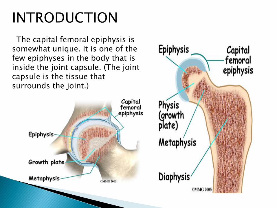
INTRODUCTION
The capital femoral epiphysis is somewhat unique. It is one of the few epiphyses in the body that is inside the joint capsule. (The joint capsule is the tissue that surrounds the joint.)

Slipped upper femoral epiphysis" term refer to slippage of the overlying epiphysis of proximal femur posteriorly and inferiorly due to weakness of the growth plate in relation to metaphysis.
Most often, it develops during periods of accelerated growth, shortly after the onset of puberty.
The femoral epiphyses maintains its relation with acetabulum ,it’s the femoral neck and shaft upward and anterior movement on epiphyses thus epiphyses displaces relatively posterior
DEFINITION

• Posterior and Medial displacement of the femoral capital epiphysis on the femoral neck through sudden or gradual deformation of the sub-capital growth plate

Neck moves LATERAL and VENTRAL creating Retroversion

2-3/100000 Most common in adolescent period with rapid growth
plate (boys aged 10-16 y, girls aged 12-14 y) Very early onset[<10yrs] and late onset[>16] should
be evaluated for endocrine disorders Male : Female is 2.4 : 1 Obesity is a risk factor because it places more shear
forces around the proximal growth plate in the hip at risk.
Bilateral slippage is common of which 2nd slip is about 12-18 months later to 1st (left hip is more common than right)
L>R, bilateral in 25-40%
1. Local trauma , obesity
2. Endocrine disorders (e.g primary or secondary hypothyroidism, adiposogenital dystrophy (hypogonadal male)
3. Deficiency or increase of androgens.
4. Acute trauma
5. Growth hormone deficiency
6. ATYPICAL SCFE associated with renal failure, radiation therapy

CLINICAL CLASSIFICATION
Acute
Chronic
Acute on chronic
FUNCTIONAL CLASSIFICATION
Stable
Unstable
MORPHOLOGICAL CLASSIFICATION
Mild
Moderate
Severe

SYMPTOMS <2WKS >2WKS
X-RAY displaced epiphyses remodelling and
no remodelling healing noted
ACUTE ON CHRONIC SLIPS
Symptoms lasting longer than 1mth and recent sudden exacerbation pain after trivial trauma
This classification scheme is unreliable because many children and parents cannot remember the exact duration of symptoms
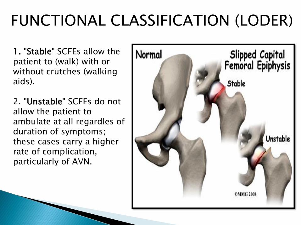
FUNCTIONAL CLASSIFICATION (LODER)
1. "Stable" SCFEs allow the patient to (walk) with or without crutches (walking aids).
2. "Unstable" SCFEs do not allow the patient to ambulate at all regardles of duration of symptoms; these cases carry a higher rate of complication, particularly of AVN.

AP VIEW-145*LATERAL VIEW-170*
FROG LEG LATERAL POSITION--Best shows posterior slippage and subtle slipping also-Normally 10*posteriorly-Increases in slippage

MORPHOLOGICAL CLASSIFICATION(Grading Severity of SCFE according to AP and Lateral X-rays)
PRESLIP-irregularity,widening,and indistinctness of physes
Grade-1 Grade-II Grade-III
Microscopically characteristic changes in PROLIFERATIVE and HYPERTROPHIC ZONES of epiphyses
◦ chondrocytes number decrease
◦ collagen fibres and Matrix are increased

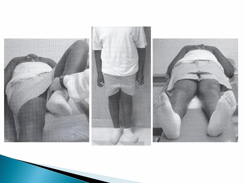
1. Pain in the groin and around the knee.2. Antalgic Limp (intermittent).3. Shortening of the affected limb (1-2 cm).4. The limb is in external rotation.[frog leg
position]5. Flexion, abduction, medial rotation are limited6. External rotation, adduction are increased.7. The presence of hip flexion contracture points
towards the possibility of chondrolysis.8. Axis deviation – pathognomonic – when hip is
flexed, the limb goes into external rotation
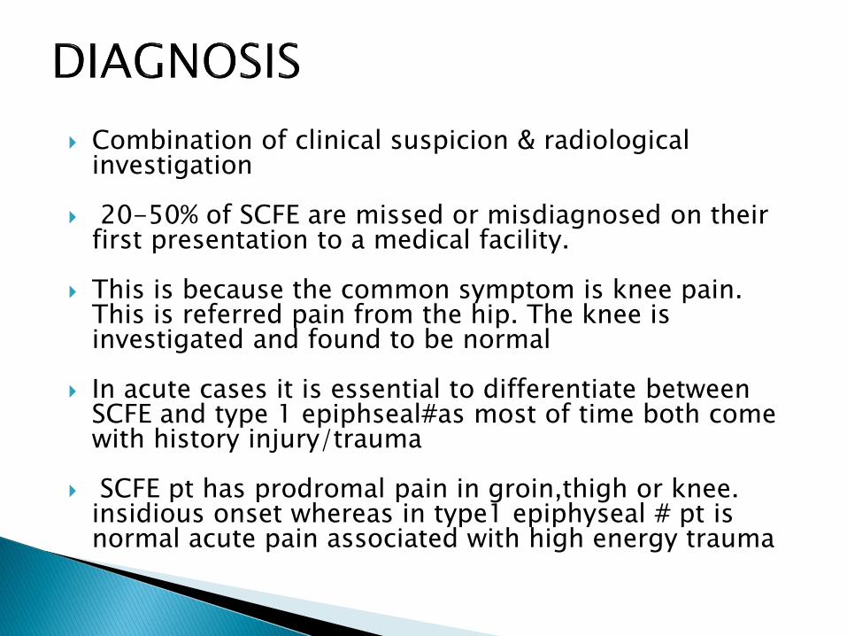
Combination of clinical suspicion & radiological investigation
20-50% of SCFE are missed or misdiagnosed on their first presentation to a medical facility.
This is because the common symptom is knee pain. This is referred pain from the hip. The knee is investigated and found to be normal
In acute cases it is essential to differentiate between SCFE and type 1 epiphseal#as most of time both come with history injury/trauma
SCFE pt has prodromal pain in groin,thigh or knee. insidious onset whereas in type1 epiphyseal # pt is normal acute pain associated with high energy trauma
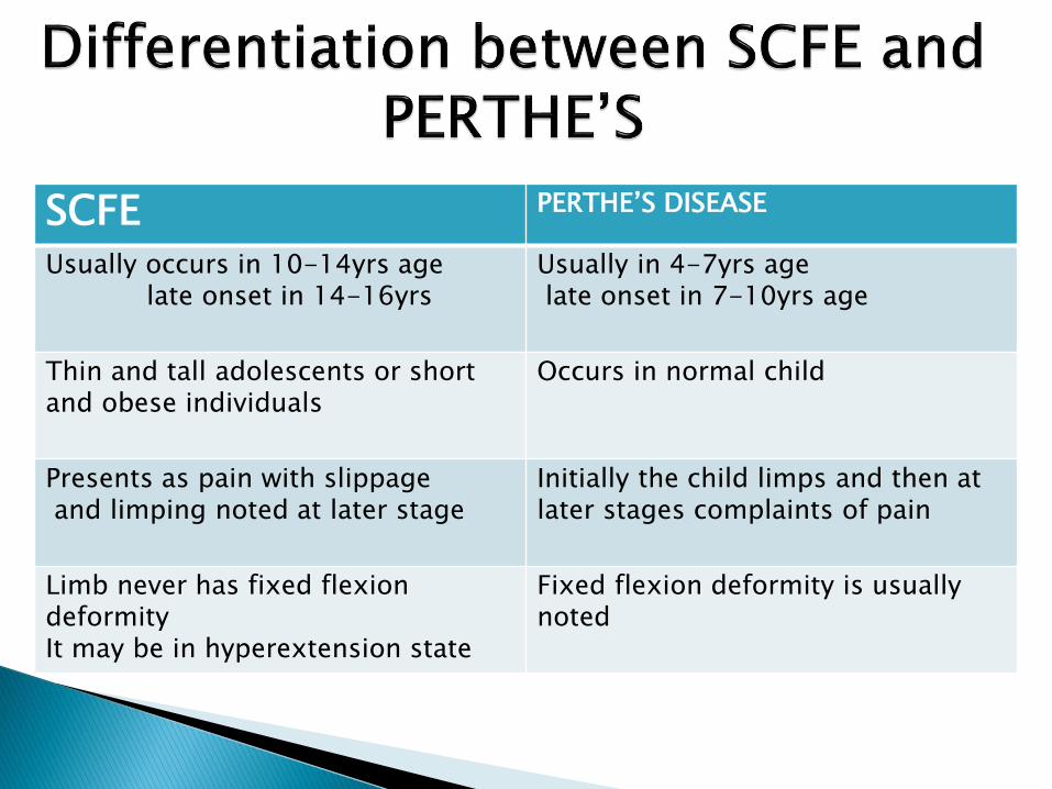
SCFE PERTHE’S DISEASE
Usually occurs in 10-14yrs agelate onset in 14-16yrs
Usually in 4-7yrs agelate onset in 7-10yrs age
Thin and tall adolescents or shortand obese individuals
Occurs in normal child
Presents as pain with slippageand limping noted at later stage
Initially the child limps and then at later stages complaints of pain
Limb never has fixed flexiondeformityIt may be in hyperextension state
Fixed flexion deformity is usually noted
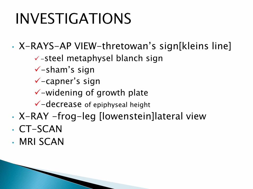
• X-RAYS-AP VIEW-thretowan’s sign[kleins line]
-steel metaphysel blanch sign
-sham’s sign
-capner’s sign
-widening of growth plate
-decrease of epiphyseal height
• X-RAY -frog-leg [lowenstein]lateral view
• CT-SCAN
• MRI SCAN

A.P. VIEW-Posterior ,inferior, And medial translation of epiphyses
LATERAL VIIEW-to measure lateral epiphysealshaft angle
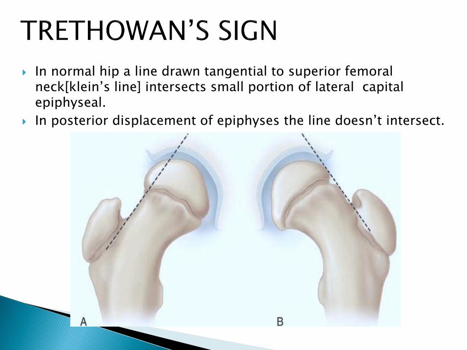
In normal hip a line drawn tangential to superior femoral neck[klein’s line] intersects small portion of lateral capital epiphyseal.
In posterior displacement of epiphyses the line doesn’t intersect.

In AP VIEW-crescent-shaped area of increased density overlying the metaphysis adjacent to the physis
This increased density is due to the overlapping of the femoral neck and the posteriorlydisplaced capital epiphysis
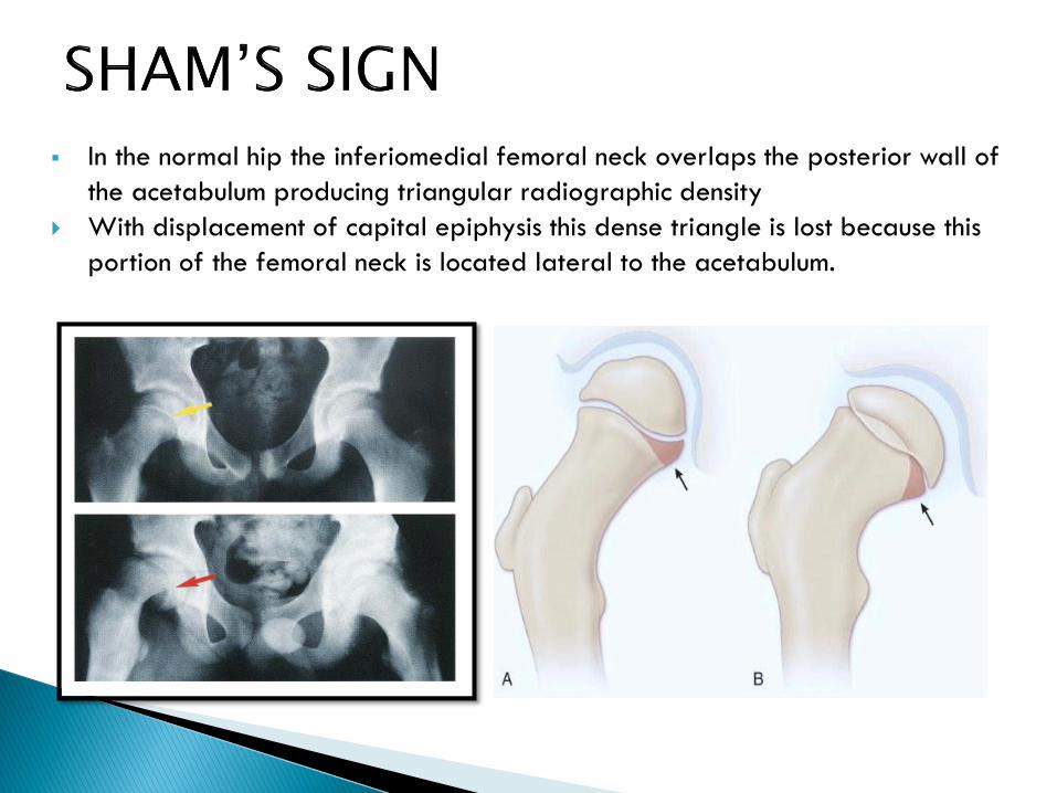
In the normal hip the inferiomedial femoral neck overlaps the posterior wall of
the acetabulum producing triangular radiographic density
With displacement of capital epiphysis this dense triangle is lost because this
portion of the femoral neck is located lateral to the acetabulum.

In pelvic AP view in the normal hip, the posterior acetabularmargin cuts across the medial corner of the upper femoral metaphysis
With slipping, the entire metaphysis is lateral to the posterior acetabular margin
Very early slips may appear to be normal in AP VIEW but may be clearly noted in lateral view
CHRONIC CASE OF SCFE X-RAY
Reactive bone formation along superolateralaspect of neck
Bone remodelling and broadening of neck resulting in PISTOL GRIP like appearance[hordons hump]


USG Early detection of early slips
Joint effusion
Step between the femoral neck and the epiphysis created by slipping
Absolute displacement of 6 mm
<2 mm of joint space is diagnostic of a slipped epiphysis
CT◦ For measurement of upper femoral neck anteversion or
true retroversion & head–neck angle
◦ To asses penetration of the hip joint by fixation devices
◦ To confirm closure of the proximal femoral physis
◦ For planing of reconstructive osteotomy of femer
MRI◦ To assess AVN
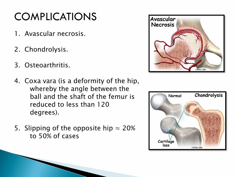
COMPLICATIONS
1. Avascular necrosis.
2. Chondrolysis.
3. Osteoarthritis.
4. Coxa vara (is a deformity of the hip, whereby the angle between the ball and the shaft of the femur is reduced to less than 120 degrees).
5. Slipping of the opposite hip ≈ 20% to 50% of cases

GOALS OF TREATMENT
Prevent further slippage so to avoid complications like osteonecrosis, chondrolysis and osteoarthritis
Stimulate early physeal closureReduction of epiphyseal displacementAny child with SCFE and open epiphyses
without stabilisation it progressesIn a patient with closed physes
functional limitationsunacceptable gaitcosmetic deformity

Conservative management-rest and traction Closed manipulative reduction Operative management
-In situ pinning
-ORIF
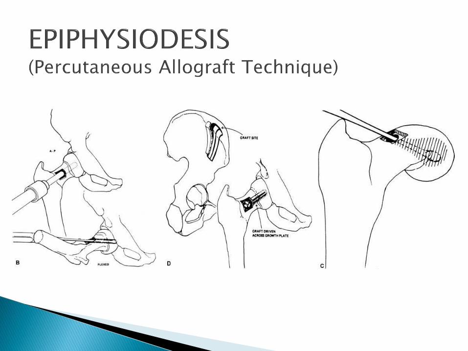
-BONE PEG Epiphysiodesis
-Osteotomy
-reconstruction by-Arthroplasty
-Arthrodesis
-Cheilectomy

Hip Spica for 12 weeks
Complications◦ Recurrent slipping after cast removal
◦ Chondrolysis (from 19% to 76%),
◦ Avascular necrosis of the femoral head (7%),
◦ Skin ulcers (16%), and
◦ Psychosocial complications

Only for acute and acute on chronic severe slips
Within 24 hours of slip High risk of ischemic necrosis of head So, manipulate only acute severe slips that
may be technically difficult or impossible to pin in situ.
Alternatively gradual reduction by skin traction and internal rotation over 3 – 4 days.
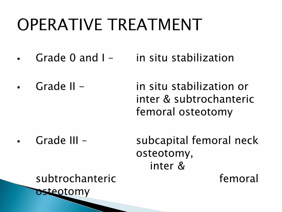
Grade 0 and I – in situ stabilization
Grade II - in situ stabilization or inter & subtrochantericfemoral osteotomy
Grade III – subcapital femoral neck osteotomy,
inter & subtrochanteric femoral osteotomy

• Percutaneous pinning
• Open pinning• when closed reduction pinning fail
• Generally 2 cannulated screws for acute (unstable) slips
• 1 screw for chronic stable slips.

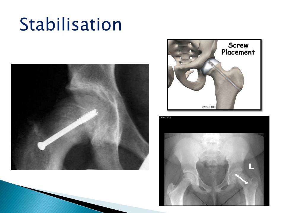
Stabilisation


Adverse affects attributed to unrecognized pin penitration :
• Joint sepsis
• Localised acetabular erosions
• Synovitis
• Post operative hip pain
• Chondrolysis
• Late degenerative osteoarthritis

Prophylactic Pinning Of The Contra-lateral Hip
•Pin if symptoms are present
•Pin if there is known metabolic/endocrine disorders
•Pin if Follow up is unreliable
Not done now a days because of associated postoperative complications:• Osteonecrosis
• Chondrolysis
• Infection
• Thigh hypesthesias
• Heterotopic ossification
• After bone peg epiphysiodesis of acute slips, spica cast immobilization may be necessary for 6 weeks or more to prevent further slipping

If there is a severe acute or chronic slip which cannot be reduced by closed methods then open reduction is indicated to prevent early degenerative joint changes
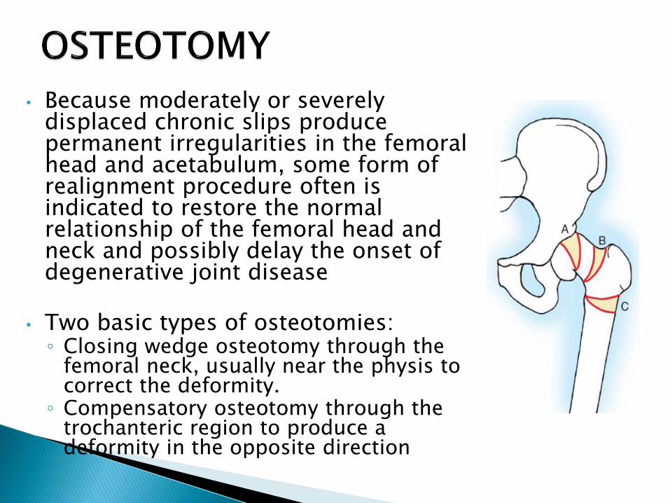
• Because moderately or severely displaced chronic slips produce permanent irregularities in the femoral head and acetabulum, some form of realignment procedure often is indicated to restore the normal relationship of the femoral head and neck and possibly delay the onset of degenerative joint disease
• Two basic types of osteotomies: ◦ Closing wedge osteotomy through the
femoral neck, usually near the physis to correct the deformity.
◦ Compensatory osteotomy through the trochanteric region to produce a deformity in the opposite direction

Four femoral neck osteotomies are described:
(1)the technique of Fish,
(2) the technique of Dunn just distal to the slip,
(3) the base of the neck technique of Kramer et al., and
(4) the technique of Abraham et al.
(5)Compensatory osteotomies in the trochanteric region and partial cheilectomy to reduce deformity also are described
(6)1 and 2 in open epiphyses
CHEILECTOMY
When a prominence on the anterosuperior aspect of the femoral neck blocks internal rotation or abduction by impinging against the acetabulum
Simple resection of the prominence removes the obstruction and improves motion

MANAGEMENT

COMPLICATIONS
SCFE Osteonecrosis
-Rare in untreated SCFE-Results from interruption of the retrograde blood supply by
•Intruption in blood suply of epiphysis due to tamponade effect of the heamarthrosis secondary to acute hemorrhage within the capsule •Increase with severity of slip•increase in acute, unstable slips•increases with forcefull repititivemanipulation, •pin placement in superior quadrant• Osteotomy of femoral neck
DIAGNOSIS: ◦ Early postoperative bone scan has excellent
sensitivity and predictive value
TREATMENT• Remove metal work
• Maintain ROM
• Realignment
• Shelf acetabuloplasty
• Arthrodesis/THR

SCFE Chondrolysis
Dissolution of articular cartilage with joint stiffness and pain
Causes:
• Persistent pin penetration• After trochantricosteotomy, open reduction, femoral neck osteotomy•Ischemia, excessive pressure
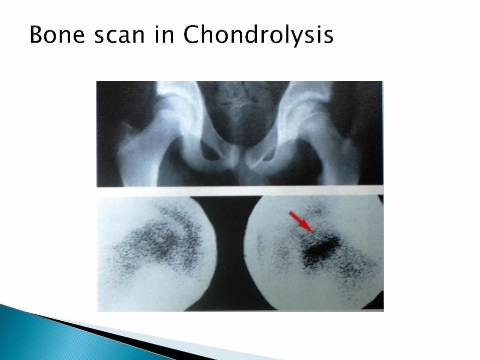
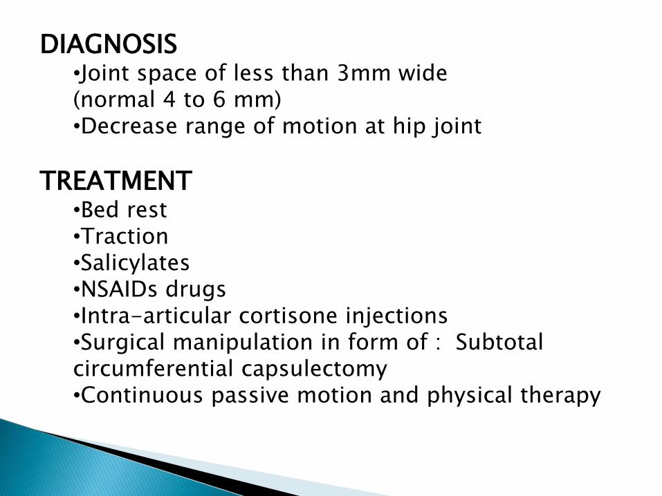
DIAGNOSIS•Joint space of less than 3mm wide (normal 4 to 6 mm)•Decrease range of motion at hip joint
TREATMENT•Bed rest•Traction•Salicylates•NSAIDs drugs•Intra-articular cortisone injections•Surgical manipulation in form of : Subtotal circumferential capsulectomy•Continuous passive motion and physical therapy


![Scoliosis BioMed Central - COnnecting REpositories · PDF fileBioMed Central Page 1 of 8 (page ... which is often missed [19]. ... Slipped capital femoral epiphysis (SCFE) is a rare](https://static.fdocuments.net/doc/165x107/5ab49deb7f8b9a7c5b8bf996/scoliosis-biomed-central-connecting-repositories-central-page-1-of-8-page-.jpg)