Slime producing, heavy metals and antibiotics resistance
12
Vol. 7(50), pp. 5697-5708, 18 December, 2013 DOI: 10.5897/AJMR12.2328 ISSN 1996-0808 ©2013 Academic Journals http://www.academicjournals.org/AJMR African Journal of Microbiology Research Full Length Research Paper Slime producing, heavy metals and antibiotics resistance in Aeromonas hydrophila isolated in Tunisia Nourhene Saidi*, Rihab Lagha, Fethi Ben Abdallah, Karima Bekir Rokbani and Amina Bakhrouf Laboratory of Analysis, Treatment, and Valorization of Environment and Products Pollutants, Department of Microbiology, Faculty of Pharmacy, Street Avicenne, Monastir, Tunisia. Accepted 20 May, 2013 Aeromonas hydrophila strains isolated from different naturally polluted environments (ten from wastewater, six from bay used for aquaculture, eight from sea coast water and six from fish) were subjected to 13 antibiotics, and to four heavy metals (Copper, Cobalt, Zinc and Mercury) by using agar diffusion and agar dilution methods, respectively. In addition, effect of heavy metals on slime production was also investigated. Results of the antibiotic resistance agreed with those of heavy metals resistance, however, treated wastewater and bay strains were much tolerant than seawater and fish bacteria. The range of metal concentrations that was tolerated in the liquid media yielded information on the tolerance levels of A. hydrophila to different tested concentrations of metals. Copper and zinc were the best tolerated metals. Mercury was the most toxic component for all bacteria. Almost all A. hydrophila produced slime and a small number of strains have changed their morphotype under the heavy metals concentration. Our results have shown that Tunisian aquatic biotopes have a significant proportion of antibiotic and heavy metal resistant to A. hydrophila. Key words: Antibiotic resistance, Aeromonas hydrophila, heavy metals and slime producing. INTRODUCTION The anthropogenic contamination of the environment with heavy metals is a serious problem. Aquaculture (Burridge et al., 2010) and agricultural practices (Han et al., 2002; Nicholson et al., 2003) contribute to this world wide pollution due to diverse applications of metals in feed additives, organic and inorganic fertilizers, pesticides, and anti-fouling products. Fish farmers frequently use phar- maceuticals (such as antibiotics) and metal containing products to prevent fouling, to feed and to treat fish in order to limit the spread of infections (Burridge et al., 2010). Therefore, bacterial communities of aquacultures are strongly exposed to the combination of heavy metals and antibiotics. The exposure to both antimicrobial substan- ces may increase the likelihood of selection and co-selec- tion of antibiotic resistance. Moreover, the high nutritional value and the relatively low cost of wastewater, excreta, and sewage sludge convert such heavy metal containing waste to valuable fish feed, especially in developing countries (WHO, 2006). In Tunisia, the persistence and proliferation of antibio- tics and heavy metals resistance in bacterial pathogens, belonging to the Aeromonas hydrophila, in aquatic envi- ronments represents a considerable public health con- cern. Subsequent measures to control the emergence and propagation of antibiotic resistance have encoun- tered limited success, and it persists in spite of the restricted use of several key antibiotics, which indicates that there are components governing the evolution, dissemination and perpetuation of these resistance sys- tems that are yet to be understood. Resistance to antibiotics can be conferred by chromo- *Corresponding author. E-mail: [email protected]. Tel: +21620871718. Fax: +21673664244.
Transcript of Slime producing, heavy metals and antibiotics resistance

Vol. 7(50), pp. 5697-5708, 18 December, 2013
DOI: 10.5897/AJMR12.2328
ISSN 1996-0808 ©2013 Academic Journals
http://www.academicjournals.org/AJMR
African Journal of Microbiology Research
Full Length Research Paper
Slime producing, heavy metals and antibiotics resistance in Aeromonas hydrophila isolated in Tunisia
Nourhene Saidi*, Rihab Lagha, Fethi Ben Abdallah, Karima Bekir Rokbani and Amina Bakhrouf
Laboratory of Analysis, Treatment, and Valorization of Environment and Products Pollutants, Department of
Microbiology, Faculty of Pharmacy, Street Avicenne, Monastir, Tunisia.
Accepted 20 May, 2013
Aeromonas hydrophila strains isolated from different naturally polluted environments (ten from wastewater, six from bay used for aquaculture, eight from sea coast water and six from fish) were subjected to 13 antibiotics, and to four heavy metals (Copper, Cobalt, Zinc and Mercury) by using agar diffusion and agar dilution methods, respectively. In addition, effect of heavy metals on slime production was also investigated. Results of the antibiotic resistance agreed with those of heavy metals resistance, however, treated wastewater and bay strains were much tolerant than seawater and fish bacteria. The range of metal concentrations that was tolerated in the liquid media yielded information on the tolerance levels of A. hydrophila to different tested concentrations of metals. Copper and zinc were the best tolerated metals. Mercury was the most toxic component for all bacteria. Almost all A. hydrophila produced slime and a small number of strains have changed their morphotype under the heavy metals concentration. Our results have shown that Tunisian aquatic biotopes have a significant proportion of antibiotic and heavy metal resistant to A. hydrophila. Key words: Antibiotic resistance, Aeromonas hydrophila, heavy metals and slime producing.
INTRODUCTION The anthropogenic contamination of the environment with heavy metals is a serious problem. Aquaculture (Burridge et al., 2010) and agricultural practices (Han et al., 2002; Nicholson et al., 2003) contribute to this world wide pollution due to diverse applications of metals in feed additives, organic and inorganic fertilizers, pesticides, and anti-fouling products. Fish farmers frequently use phar-maceuticals (such as antibiotics) and metal containing products to prevent fouling, to feed and to treat fish in order to limit the spread of infections (Burridge et al., 2010).
Therefore, bacterial communities of aquacultures are strongly exposed to the combination of heavy metals and antibiotics. The exposure to both antimicrobial substan-ces may increase the likelihood of selection and co-selec-tion of antibiotic resistance. Moreover, the high nutritional
value and the relatively low cost of wastewater, excreta, and sewage sludge convert such heavy metal containing waste to valuable fish feed, especially in developing countries (WHO, 2006).
In Tunisia, the persistence and proliferation of antibio-tics and heavy metals resistance in bacterial pathogens, belonging to the Aeromonas hydrophila, in aquatic envi-ronments represents a considerable public health con-cern. Subsequent measures to control the emergence and propagation of antibiotic resistance have encoun-tered limited success, and it persists in spite of the restricted use of several key antibiotics, which indicates that there are components governing the evolution, dissemination and perpetuation of these resistance sys-tems that are yet to be understood.
Resistance to antibiotics can be conferred by chromo-
*Corresponding author. E-mail: [email protected]. Tel: +21620871718. Fax: +21673664244.

5698 Afr. J. Microbiol. Res. somal or mobile genetic elements (for example, plasmids) and achieved using four main strategies: (i) reduction of membrane permeability to antibiotics; (ii) drug inactive-tion; (iii) rapid efflux of the antibiotic; and (iv) mutation of cellular target (s) (Krulwich et al., 2005). In addition, anti-biotic sequestration has also been suggested as a poten-tial resistance strategy (Pankaj et al., 2009). Overall, the structural and functional characteristics of antibiotic resis-tance share common themes with those of metal resis-tance (Baker-Austin et al., 2006).
Although, bacterial exposure to metals predates human history, anthropogenic-derived sources of metals repre-sent a major source of contamination in the environment. Importantly, a substantial number of reports suggest that metal contamination in natural environments could have an important role in the maintenance and proliferation of antibiotic resistance (Alonso et al., 2001; Summers, 2002). This is of particular concern considering that anthropo-genic levels of heavy metals are currently several orders of magnitude greater than levels of antibiotics (Stepanauskas et al., 2005). Unlike antibiotics, metals are not subject to degradation and can subsequently represent a long-term selection pressure (Stepanauskas et al., 2005). Thus, there are concerns regarding the potential of metal conta-mination to maintain a pool of antibiotic-resistance genes in both natural and clinical settings. In addition to metals, other toxicants are implicated in the co-selection of anti-biotic resistance, including quaternary ammonium com-pounds and antifouling agents and detergents (Sidhu et al., 2001; Chapman, 2003).
Several explanations have been proposed for the enhanced resistance of biofilm-associated cells to both metals and antibiotics (Baker-Austin et al., 2006). Both metal and antibiotic sequestration in the biofilm matrix and the presence of a small population of „persister‟ cells might be contributing factors in the time-dependent tole-rance of both planktonic cells and biofilms to high con-centrations of antimicrobial agents (Harrison et al., 2005).
In Tunisia, on the east coast of the country bordering the Mediterranean Sea is a key location for the study of antibiotic resistant bacteria and heavy metals contamina-tion in the aquatic environment. The bay is of great eco-nomic importance for fishing and aquaculture of nume-rous species of crustaceans and fish (Snoussi et al., 2006). In addition, domestic wastes, including industrial wastes are discharged into the bay and the sea.
To our knowledge, our present study is the first to determine the prevalence and resistance to antimicrobial agents and heavy metals of A. hydrophila isolated from wastewater, bay, seawater and fish. However, in this work, we focus on the current body of knowledge regar-ding (i) to characterize the A. hydrophila strains recovered from Tunisian aquatic biotopes; (ii) to determine the level of antibiotic resistance rates against widely used antibiotics in Tunisia; (iii) to determine the heavy metals resistance of the bacteria; (iv) to investigate the relationship bet-ween the antibiotics and heavy metals resistance and (v)
to determine the heavy metals effect on A. hydrophila slime producing.
MATERIALS AND METHODS
Aeromonas hydrophila strains This study includes 31 A. hydrophila strains: ten strains were isola-ted from treated wastewater (ONAS), six strains were isolated from the bay of Khenis (Aquaculture center, Monastir), eight strains were isolated from seacoast of Monastir, six strains isolated from orna-mental fish and a reference strain A. hydrophila ATCC 7966
T [American
Type Collection Culture (Manassas, Va.)] (Saidi et al., 2011). All these strains were identified and characterized by Bergey‟s
Manual of Determinative Bacteriology (Holt et al., 1994) and achieved by the conventional methods described by Balows et al. (1991). Gram staining method, cell morphology, the oxidase, catalase, motility (Mannitol-Motility agar, Pronadisa, Madrid, Spain), suscepti-bility to the vibriostatic compound O/129 (10 and 150 µg/disc) and ampicillin antibiotic (10 µg), growth at 30 and 37°C and growth on Rimler Shotts Agar (mRS) were the first tests employed to identify
the organisms belonging to Aeromonas genus. Commercial minia-turized strips 20 NE Api (Non Enterobacteriacae, bioMerieux, France) were also used.
The production of lipase (Tween 80), haemolysin (Sheep blood agar, Pronadisa, Madrid, Spain) and DNA hydrolysis (DNAse Agar, Sharlau Microbiology, Barcelona, Spain) were tested as described previously by Snoussi et al. (2006). The enzymes amylase and lecithinase were detected on media prepared with phosphate buffer
saline (PBS) supplemented, respectively with 0.5% starch and 5% egg yolk emulsion. The caseinase activity was tested according the protocol described by Zanetti et al. (2000). A. hydrophila strains were cultured on Nutrient Agar containing 5% skim milk. After incu-bation for up to 72 h at 37°C, the formation of a clear zone caused by casein degradation was considered as a positive test.
Susceptibility testing
Antibiotic susceptibility was performed according to the national Committee for Clinical Laboratiry Standards (CLSI, 2007) method on Mueller–Hinton Agar (Difco) by the disk-diffusion method (Bauer et al., 1966). Resistance to the following antibiotics (BBL, Md, USA) of A. hydrophila strains (10
6 CFU/ml) was tested with disks con-
taining nalidixic acid (NA, 30 μg), tetracycline (TE, 30 μg), genta-micin (GM, 10 μg), imipenem (IPM, 10 μg), neomycin (N, 30 μg), ticarciline (TIC, 75 μg), colistine (CL50, 50 μg), cefoxitine (FOX, 30
μg), cefalotine (CF, 30 μg) and flumequine (UB, 30 μg), oxolinic acid (OA, 10 μg), oxytetracycline (OTC, 30 UI), sulfamide/ trimetho-prime (SULF/TMP, 200 μg/5 μg). The strain A. hydrophila ATCC 7966
T was used as control.
Multiple antibiotic resistances among Aeromonas hydrophila
The multiple antibiotic resistance (MAR) index when applied to a
single isolate is defined as a/b, where „a‟ represents the number of antibiotics to which the isolate was resistant and „b‟ represents the number of antibiotics to which the isolate was exposed.
For example if the isolate was exposed to twelve antibiotics and was tolerant to six antibiotics, the index for the isolate would be 6/12 or 0.50 (Liberto et al., 2007). MAR index value higher than 0.2 is considered to have originated from high-risk sources of contamination like human, commercial poultry farms, swine and dairy cattle where antibiotics are very often used. MAR index value
of less than or equal to 0.2 is considered the origination of strain from animals in which antibiotics are seldom or never used.

Survival of Aeromonas hydrophila under heavy metals concentration
The heavy metals (E-Merck) were used to understand its impact on the growth and survival of A. hydrophila. The salts used for the
study were Copper sulphate (CuSO4.5H2O), Cobaltous chloride (CoCl2.6H2O), Mercuric chloride (HgCl2) and Zinc chloride (ZnCl2).
The tendencies of growth were tested on Trypticase Soy Agar (TSA) medium mixed with different concentrations of heavy metals traces for all A. hydrophila strains and the plates were incubated for 24 h at 37°C. The average number on bacteria for every concentra-tion of metal was calculated.
The survival test was also examined by filtering metals using Whatmann filter paper (0.2 µm), and stored at 4°C. From the stock solution, various concentrations like 100, 200, 300, 400, and 500 ppm (Copper, Cobalt and Zinc) and 1, 2, 3, 4 and 5 ppm (Mercury) of metal solutions were prepared and used for the study. Growing A. hydrophila strains in sterile nutrient broth at 37°C for 16 h was realized. After, the broth was centrifuged at 12000 rpm for 30 min. The cells were washed with sterile saline solution and transferred into 100 ml phosphate buffer solution and the initial optic density (OD) was taken (Thangavel, 2004). The flasks were kept at 37°C
for 24 h and the OD was measured (copper, λ = 480 nm; zinc, λ = 213 nm; cobalt, λ = 425 nm and mercury, λ = 254 nm). Minimal inhibitory concentration (MIC) of heavy metals
The MIC for each bacterial isolate for heavy metal was determined using Mueller-Hinton agar (Difco) containing heavy metals (Cu
2+,
Zin2+
, and Co2+
) at concentrations ranging from 100 to 500 ppm and
(Hg2+
) at concentrations ranging from 1 to 5 ppm. The isolates were considered tolerant if the MIC values exceeded that of the Escherichia coli K-12 strain which was used as the control (Akinbowale et al., 2007).
Slime production on Congo red agar (CRA)
The classic method most often used to phenotypically detect slime
production in these bacteria is the Congo red agar (CRA) plate test as described by Freeman et al. (1989). The CRA plate test is per-formed on a solid medium, the Congo red agar. The direct analysis of the colonies formed on the solid medium allows the recognition of slime-producing strains (characterised by black colonies on the red agar) and of non-slime-producing strains (pink/red coloured colonies).
Congo red agar plate test was prepared by adding 0.8 g/L Congo
red (Bio Basic INC) and 36 g of Saccharose (Merck), both of which had been previously autoclaved separately, to 1 L of Brain Heart Infusion Agar (Scharlau Microbiology, Pronadisa, Madrid, Spain). Plates were incubated for 24 h at 37°C and subsequently overnight at room temperature (Freeman et al., 1989). Slime-producing A. hydrophila grew as black colonies, while non slime-producing strains grew as red colonies. The original test was optimized by using a colorimetric scale with six tonalities: very black, black and approximately black were considered as positive results, while burgundy, red and very red were considered as negative results (Subashkumar et al., 2006). Staphylococcus aureus ATCC 25923 was used as positive control for slime production and Staphylococcus epidermidis CIP 106510 was used as negative control (Chaieb et al., 2007).
Aeromonas hydrophila morphology visualization by atomic force microscopy
To visualize the bacteria after heavy metal exposure on glass slides and to have an idea on the morphological changes in the cells
Saidi et al. 5699 during heavy metal stress, A. hydrophila ATCC 7966
T cells was
used as a negative control. For the experiments, the cells enriched on PBS with different concentrations of mercury (1, 2, 3, 4 and 5 ppm) were collected, washed three times by PBS, centrifuged and the pellet was resuspended in PBS, fixed on a sterilized round microscope cover slide and the piece was examined by Atomic force Microscope (AFM, Nanoscope IIIA, Digital Instrument; Veeco) according to the method previously described (Braga and Ricci, 1998).
Statistical analysis
All results are shown as the average of at least three independent
experiments; variation is expressed as standard deviation. The Pearson correlation coefficient was calculated to determine the possible relation between the resistance to heavy metals and the resistance to antibiotics. All statistics were performed using SPSS for Windows version 17.0.
RESULTS
Antibacterial resistance
The identified strains were multi-resistant to various antibiotics used including those exploited in the treatment of Aeromonas disease of the fish (flumequine, oxolinic acid, sulfamide + trimetoprime and oxytetracycline). Indeed, all bacteria were sensitive to gentamicin.
The results showed that bay, treated waste water A. hydrophila isolates were more resistant to almost tested antibiotics than sea water and fish A. hydrophila (nalidixic acid (70 and 60%), ticarcyline (60 and 50%), cefoxitine and cefalotine (100 and 90%)), respectively. While the isolates of seawater were most sensitive to the majority of antibiotics, all strains were sensitive for neomycin, tetracycline, fumequine, oxilinic acid, oxytetracycline and sulfamide-trimethoprime.
On the other hand, A. hydrophila strains isolated from moribund fish were fairly tolerant to certain antibiotics (colistine (50%), nalidixic acid and cefalotine (66.66%)) and completely sensitive to the oxilinic acid and sulfamide + trimethoprime (Figure 1). The study of the MAR index of these 31 isolates showed that these bacteria presented a high risk, indeed, the recorded values were higher than 0.2, what corresponded to 74.19% of the studied stocks (Table 1).
Aeromonas hydrophila resistance to heavy metals effects
In the present study, resistance to copper (Cu2+
), cobalt (Co
2+), zinc (Zn
2+) and mercury (Hg
2+) were studied for all
the isolates. In the four sample types (treated waste water, bay, sea water and fish), resistance to heavy metals was described in the Table 2. Actually, all A. hydrophila are tolerant to various heavy metals tested and they presen-ted a tolerance reaching 300 ppm (copper, zinc and cobalt) and 3 ppm (mercury). For a concentration rea-ching 400 ppm, all the strains were tolerant to copper,

5700 Afr. J. Microbiol. Res.
Per
cen
tag
e P
erce
nta
ge
Treated waste water
Bay
Fish
Sea water
Figure 1. Percentage of resistance to 13 antibiotics of the 31 A. hydrophila strains isolated from treated wastewater, sea water, bay and fish. Antibiotics tested are as follow: IMP: Imipenem (10 μg), N: Neomycin (30 µg), NA: Nalidixic acid (30 μg), TIC: Ticarcilline (75 µg), GEN: Gentamicin (10 UI), TE: Tetracycline (30 μg), CL50: Colistine (50 µg), FOX: Cefoxitine (30 μg), CF: Cefalotine (30 μg), UB: Flumequine (30 μg), OA: Oxolinic acid (10 μg), OTC: Oxytetracycline (30 UI), SULF/TMP:
Sulfamide/Trimethoprime (200 μg/5 μg).
zinc and cobalt, but, only 10% of the isolates from treated wastewater and bay were tolerant to mercury (4 ppm).
The higher tolerance of A. hydrophila to 500 and 5 ppm concentrations of various metals traces had proven to be significant for the isolates of bay (100, 83.33, 66.66 and 10%) and of treated wastewater (60, 70, 50 and 10%), respectively for copper, zinc, cobalt and mercury. Where-as the small percentages of resistance were detected in the isolates of sea water (25, 37.5, 0 and 0%) and of the fish (25, 10, 0 and 0%), respectively for same metals quoted previously.
The Table 3 described the viability of A. hydrophila con-tinuation of the different concentration effect from selec-ted heavy metals. Indeed, for a concentration of 500 ppm copper, the number of the viable bacteria arrived at 5.01 × 10
4 CFU/ml (treated wastewater), 7.22 × 10
4 CFU/ml (bay),
5.16 × 10²
CFU/ml (sea water) and 5 × 10² CFU/ml (fish),
which was equivalent to 0.492, 0.43, 0.482 and 0.49 of OD, respectively. However, the less significant results were recorded for zinc and cobalt. On the other hand, at 5 ppm of mercury concentration, only the isolates of treated wastewater and bay presented viability up to 1.24 and 1.5 CFU/ml, corresponding to 0.043 and 0.023 OD.
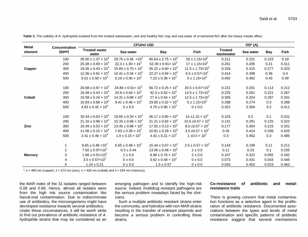
Atomic force Micrography of the bacteria morphology (Figures 2a-b) showed that A. hydrophila after the mercury
exposure (5 ppm), have changed form and become coccoid. These morphological modifications allow the adaptation to mercury stress.
Slime production under heavy metals
The objective to determine the effect of the tested metal on the slime production in isolated A. hydrophila, we found that after exposure to mercury, 3/10 of the treated wastewater isolates, 2/8 of sea water strains and only one strain from fish changed their phenotypical profile and became non slime producer and thereafter produced new morphotype (brown, pink and orange colonies). On the other hand, A. hydrophila of bay and those isolated from moribund fish did not modify their morphotype (Figure 3).
The Table 4 showed the resistance of all isolates to the effects of copper, zinc and cobalt and no morphotypic modification was registered in bay case. Similar results were found in the case of the treated wastewater isolates except for case of cobalt; indeed, only one strain changed profile and became non slime producer.
Whereas, A. hydrophila isolated from fish and sea water presented the most significant modifications, indeed, for copper, zinc and cobalt the percentages of morphotype

Saidi et al. 5701
Table 1. MAR Index and Model of resistance of the A. hydrophila.
Strain MAR index Model of antibiotics resistance
Treated wastewater
WT1 0.33 AN-TIC-FOX- SULF/TMP
WT2 0.41 IMP-AN-TIC-FOX -SULF/TMP
WT3 0.16 AN-FOX
WT4 0.16 TE -UB
WT5 0.33 TE-CL50-FOX- AO
WT6 0.33 AN-TIC-CL50-FOX
WT7 0.5 IMP-AN-TIC-CL50-FOX -OTC
WT8 0.5 AN-TIC-TE-CL50-FOX -OTC
WT9 0.58 N-AN-TIC-TE-CL50-FOX-UB
WT10 0.33 N-TIC-CL50-FOX
Bay
R1 0.33 IMP-TIC-TE-FOX
R2 0.25 AN-TIC-FOX
R3 0.41 AN-CL50-FOX -AO-SULF/TMP
R4 0.33 AN-CL50-FOX -UB
R5 0.25 N-AN-FOX
R6 0.41 TIC-CL50-FOX- OTC-SULF/TMP
Fish
E2 0.25 AN-FOX -UB
E3 0.5 AN-FOX -UB-AO-OTC-SULF/TMP
E4 0.33 N-TIC-FOX-OTC
E5 0.5 IMP-AN-TIC-TE-CL50-FOX
E6 0.33 IMP-N-AN-CL50
E7 0.66 IMP-N-AN-TIC-GEN-TE-CL50-FOX
Sea water
S1 0.08 CL50
S2 0.5 AN-FOX -UB-AO-OTC-SULF/TMP
S3 0.66 IMP-N-AN-TIC-GEN-TE-CL50-FOX-CF
S4 0.08 FOX
S5 - -
S6 0.08 AN
S7 0.16 AN-TIC
S8 0.16 AN-FOX
IMP: Imipenem. N: Neomycin. AN: Nalidixic acid. TIC: Ticarcilline. GEN: Gentamicin. TE: Tetracycline.
CL50: Colistine. FOX: Cefoxitine. UB: Flumequine. AO: Oxolinic acid. OTC: Oxytetracycline. SULF/TMP: Sulfamide/Trimethoprime.
modification of the sea water isolates were 1/8, 2/8 and 2/8, respectively. On the other hand, these values were 0, 1/6 and 2/6 for A. hydrophila isolated from fish.
DISCUSSION
The Aeromonas hydrophila resistance to antibiotics
The study of antibiotic resistance in water organisms is important, as it might indicate the extent of alteration of water ecosystems by human action. Actually, water bac-
teria could be indigenous to aquatic environments, or exogenous, transiently and occasionally present in the water as a result of shedding from animal, vegetal, or soil surfaces.
According to our results, bay and treated waste water A. hydrophila isolates were more tolerant to almost tested antibiotics than sea water and fish A. hydrophila. Martinez (2003) has found similar results and he has shown that more than 90% of bacterial strains originated from sea-water are tolerant to more than one antibiotic, and 20% are tolerant at least to five. The resistance of the strains to

5702 Afr. J. Microbiol. Res.
Table 2. Tolerance of A. hydrophila isolated from treated wastewater. fish. bay and sea water to heavy metals.
Metal/environment N Heavy metal concentrations (ppm)
1*/100 2*/200 3*/300 4*/400 5*/500
Copper
Treated wastewater 10 100 100 100 100 60
Sea water 08 100 100 100 100 25
Bay 06 100 100 100 100 100
Fish 06 100 100 100 100 25
Zinc
Treated wastewater 10 100 100 100 100 70
Sea water 08 100 100 100 100 37.5
Bay 06 100 100 100 100 83.33
Fish 06 100 100 100 100 10
Cobalt
Treated wastewater 10 100 100 100 100 50
Sea water 08 100 100 100 100 0
Bay 06 100 100 100 100 66.66
Fish 06 100 100 100 100 0
Mercury*
Treated wastewater 10 100 100 100 10 10
Sea water 08 100 100 100 0 0
Bay 06 100 100 100 10 10
Fish 06 100 100 100 0 0
Reference of test: Minimal Inhibiting Concentration of the standard strain Escherichia coli K12.
antibiotics could be explained by the possibility of the heavy use of these compounds in aquaculture (bay), se-veral of which are non biodegradable increases antibiotic selective pressure in water, facilitates the transfer of anti-biotic resistance determinants between aquatic bacteria, including fish and human pathogens, and allows the pre-sence of residual antibiotics in commercialized fish and shellfish products (Rhodes et al., 2000; Cabello, 2006). Antibiotic residues entering this aquatic environment from different sources may increase the distribution of poten-tial drug-resistant pathogen bacteria (Matyara et al., 2008). However, some studies indicate that increasing heavy metal concentrations lead to a decrease of antibio-tic resistance (Stepanauskas et al., 2005; McArthur and Tuckfield, 2008; Hölzel et al., 2012). The secontradicting results were investigated by Hölzel et al. (2012). In con-sequence of the addition of mercury chloride (HgCl2) to the antimicrobial test procedure, the MIC for a wide range of antibiotics decreased. This observation could be due to an interaction of Hg with enzymes or nucleic acids which cause antibiotic resistance. HgCl2 could also have aco-toxic effect with antibiotics that interfere with ribosomes because the generation of the Hg-degraded enzyme would be inhibited. Furthermore, Hölzel et al. (2012) mentioned also a possible metal induced shift within the bacterial community to ward Hg tolerant bacteria where
by the benefit of antibiotic resistance in the presence of antibiotics would be out competed. The increased anti-biotic susceptibility in consequence of Hg exposure could also play a role in the observations of other field studies (Seiler and Berendonk, 2012). Multiple antibiotic resistance (MAR) index of Aeromonas hydrophila Like Gram negative bacilli, the emergence of resistance among Aeromonads will be accelerated by the extensive clinical use of antibiotics (Ko and Chung, 1995; Chaudhury et al., 1996). Such high level of multiple drug resistance may arise from selective pressure due to the indiscriminate use of antibiotics. The variation in the drug resistance may be related to the source of A. hydrophila isolated and the frequency prescribed for treating Aeromonas infections in geographical areas (Radu et al., 1997).
These reports revealed that geographical, socio econo-mical parameters and local selective pressures could influence antibiotic resistance among Aeromonas spp. Growing incidence of MAR among A. hydrophila strains isolated from various sources has been reported from many parts of the world (Radu et al., 2003). In our study,
Saidi et al. 5703
Table 3. The viability of A. hydrophila isolated from the treated wastewater; sick and healthy fish; bay and sea water of ornamental fish after the heavy metals effect.
Metal
element
Concentration (ppm)
CFU/ml ±SD OD* (A)
Treated waste water
Sea water Bay Fish Treated
wastewater Sea water Bay Fish
Copper
100 35.00 ± 1.37 × 10⁴ 29.76 ± 0.36 ×10
2 65.64 ± 2.75 × 10
4 26 ± 1.15×10
2 0.211 0.231 0.153 0.18
200 25.38 ± 0.69 × 10⁴ 22.3 ± 1.00 × 10
2 52.39 ± 0.91× 10
4 17 ± 1.15×10
2 0.261 0.295 0.21 0.311
300 19.45 ± 0.43 × 10⁴ 15.65 ± 0.70 × 10
2 35.22 ± 0.60 × 10
4 11.5 ± 1.73×10
2 0.334 0.315 0.277 0.323
400 12.36 ± 0.91 × 10⁴ 10.41 ± 0.34 × 10
2 22.27 ± 0.94 × 10
4 6.5 ± 0.57×10
2 0.414 0.398 0.36 0.4
500 5.01 ± 0.50 × 10⁴ 5.16 ± 0.50 × 10
2 7.22 ± 0.36 × 10
4 5 ± 1.15×10
2 0.492 0.482 0.43 0.49
Cobalt
100 26.68 ± 0.97 × 10⁴ 24.86 ± 0.51× 10
2 56.72 ± 0.25 × 10
4 20.5 ± 0.57×10
2 0.221 0.201 0.113 0.212
200 16.48 ± 0.43 × 10⁴ 20.8 ± 0.64 × 10
2 42.3 ± 0.62 × 10
4 14.5 ± 1.73×10
2 0.225 0.261 0.221 0.267
300 15.58 ± 0.24 × 10⁴ 14.31 ± 0.88 × 10
2 27.4 ± 0.64 × 10
4 12.5 ± 1.73×10
2 0.305 0.324 0.287 0.331
400 10.83 ± 0.68 × 10⁴ 9.41 ± 0.46 × 10
2 19.65 ± 0.32 × 10
4 5 ± 1.15×10
2 0.298 0.274 0.3 0.288
500 4.63 ± 0.42 × 10⁴ 0 ± 0.0 4.76 ± 0.90 × 10
4 0 ± 0.0 0.322 0.394 0.2 0.411
Zinc
100 30.44 ± 0.63 × 10⁴ 19.65 ± 0.34 × 10
2 36.17 ± 0.80 × 10
4 15 ±1.15 × 10
2 0.103 0.2 0.1 0.231
200 21.16 ± 0.98 × 10⁴ 15.33 ± 0.68 × 10
2 21.21 ± 0.92 × 10
4 10.5 ±0.57 × 10
2 0.141 0.291 0.125 0.322
300 16.49 ± 0.53 × 10⁴ 13.45 ± 0.88 × 10
2 17.92 ± 0.13 × 10
4 8.5 ±0.57 × 10
2 0.177 0.304 0.215 0.331
400 11.06 ± 0.10 × 10⁴ 7.63 ± 0.39 × 10
2 10.81 ± 0.28 × 10
4 3.5 ±0.57 × 10
2 0.26 0.414 0.208 0.425
500 3.41 ± 0.46 × 10⁴ 1.9 ± 0.15 × 10
2 4.42 ± 0.21 × 10
4 1 ±0.0 × 10
2 0.3 0.462 0.3 0.485
Mercury
1 9.65 ± 0.48 ×10² 6.65 ± 0.48 × 10
2 15.44 ± 0.67 × 10
2 2.5 ± 0.57 × 10
2 0.143 0.199 0.11 0.211
2 7.92 ± 0.97×10² 6.5 ± 0.44 13.06 ± 0.48 × 10
2 2 ± 0.0 0.11 0.23 0.1 0.235
3 1.66 ± 0.51×10² 2 ± 0.0 9.96 ± 0.76 × 10
2 2 ± 0.0 0.107 0.322 0.055 0.342
4 3.5 ± 0.57×10² 0 ± 0.0 3.62 ± 0.48 × 10
2 0 ± 0.0 0.073 0.431 0.043 0.445
5 1.24 ± 0.21 0 ± 0.0 1.5 ± 0.57 0 ± 0.0 0.043 0.452 0.023 0.463
*: λ = 480 nm (copper). λ = 213 nm (zinc). λ = 425 nm (cobalt) and λ = 254 nm (mercury).
the MAR index of the 31 isolates ranged between 0.08 and 0.66. Hence, almost all isolates were from the high risk source contamination like faecal-oral contamination. Due to indiscriminate use of antibiotics, the microorganisms might have developed resistance towards several antibiotics. Under these circumstances, it will be worth while to find out prevalence of antibiotic resistance of A. hydrophila strains that may be considered as an
emerging pathogen and to identify the high-risk source. Indeed, multidrug resistant pathogens are the serious problem nowadays faced by the clini-cians.
Such a multiple antibiotic resistant strains enter the community, and hybridize with non-MAR strains resulting in the transfer of resistant plasmids and become a serious problem in controlling these strains.
Co-resistance of antibiotic and metal-resistance traits There is growing concern that metal contamina-tion functions as a selective agent in the prolife-ration of antibiotic resistance. Documented asso-ciations between the types and levels of metal contamination and specific patterns of antibiotic resistance suggest that several mechanisms
5704 Afr. J. Microbiol. Res.
Figure 2. Morphological modification of A. hydrophila examined by Atomic force Microscope (AFM): (a) bacillus form to (b) coccoid form
after mercury exposure.
underlie this co-selection process. These co-selection mechanisms include co-resistance (different resistance determinants present on the same genetic element) and cross-resistance (the same genetic determinant respon-sible for resistance to antibiotics and metals) (Clutterbuck et al., 2007).
Our results revealed that the wastewater and bay iso-lates were more tolerant to heavy metals (copper, zinc, cobalt and mercury) than sea water and fish strains; simi-
lar results of resistances have been shown for antibiotics. The association of antibiotic-resistance and resistance
to heavy metals is very frequent in the same organism (also in the same plasmid, transposon, or integron) so that industrial pollution probably selects for antibiotic-resistance and vice versa (Baker-Austin et al., 2006). The studies of Seiler and Berendonk (2012) have investigated the co-selection in the environment and they showed the presence of correlation between increased heavy metal

Saidi et al. 5705
(a) Before heavy metal exposure
(b) After heavy metal exposure
Figure 3. Morphotypes of A. hydrophila based
Figure 3. Morphotypes of A. hydrophila based on the colorimetric scale obtained on Congo red agar before heavy metals exposure (a): Very Black colonies (A. hydrophila ATCC7966); Black colonies; Red colonies and after heavy metals exposure (b): Brown colonies; Burgundy colonies; Orange colonies and Pinkish colonies. Staphylococcus
epidermidis producing and non-producing slime were used as negative and positive
controls.
concentrations with increased phenotypic or genotypic antibiotic resistance. In addition, others researches proved that metal contamination represent a long-standing, wide-spread, and recalcitrant selection pressure for multi-resis-tant organisms (Pathak and Gopal, 2005). For the non aquatic organisms, obviously the density of antibiotic - resistance organisms and antibiotic-resistance genes in fresh water varies with the proximity to areas with increased antibiotic consumption, metal pollution, and between sea-sons, being more frequently found in rainy seasons (Peak et al., 2007).
Evidence for co-selection of antibiotic and metal resis-tance in the environment originates from diverse habitats contaminated with a variety of metals, which indicates that co-selection is not limited to a subset of metals, envi-ronments or microbial taxonomic groups. The strength of evidence presented by these studies ranges considerably between anecdotal reports of co-resistances to experi-mental studies that unambiguously implicate metals in antibiotic resistance co-selection (Baker-Austin et al., 2006).
Actually, the results of this work has proven that
A. hydrophila isolated from fish were tolerant to high con-centration of heavy metals (copper, zinc and cobalt (100 to 400 ppm) and mercury (3 ppm)). Although these studies do not directly address the hypothesis that metal exposure co-selects for antibiotic resistance, they high-light the fact that metal and antibiotic resistances are commonly found within the same bacteria. Indeed, poten-tial public health concerns for the co-resistance of metal and antibiotic resistances were raised by Pathak and Gopal (2005), who observed that bacterial isolates obtained from fish tissue commonly consumed by humans exhi-bited resistance to multiple metals and antibiotics.
As evidenced in previous studies, subsequent expo-sure to elements of heavy metal leads to direct selection for metal-resistance while co-selection for antibiotic resis-tance. Maintenance of the co-selection antibiotic resistance was accomplished by co-resistance, cross resistance, and co-regulation of the resistant genes (Miranda and Castillo, 1998; Spain and Alm, 2003; Stepanauskas et al., 2005; Wright et al., 2006).
Nonetheless, bacterial resistance to heavy metal was

5706 Afr. J. Microbiol. Res.
Table 4. The effect of heavy metals on the slime production of A.
hydrophila isolated from treated wastewater, sea water, bay and fish.
Origin/metal N Slime production (%)
Before After
Copper
Treated wastewater 10 07/10 07/10
Bay 06 03/06 03/06
Sea water 08 04/06 03/06
Fish 06 06/06 06/06
Zinc
Treated wastewater 10 07/10 07/10
Bay 06 03/06 03/06
Sea water 08 04/08 02/08
Fish 06 06/06 05/06
Cobalt
Treated wastewater 10 07/10 06/10
Bay 06 03/06 03/06
Sea water 08 04/08 02/08
Fish 06 06/06 04/06
Mercury
Treated wastewater 10 07/10 04/10
Bay 06 03/06 03/06
Sea water 08 04/08 02/08
Fish 06 06/06 05/06
emphasized in the present study because substantial number of reports have been alerting on maintenance and
proliferation of antibiotic resistance. Resistance genes to both substances were presumably residing closely on the bacterial plasmid and transported together in the environ-ment (Sabry et al., 1997; Spain and Alm, 2003; Wright et al., 2006).
Interaction between antibiotics, heavy metals resistance and slime production in A. hydrophila
Many bacteria in the environment exist in surface-attached communities; in fact, the initial bacterial monolayer adhe-ring to polymeric surfaces is converted to a typical biofilm consisting of bacteria plus an extracellular substance (Heilmann et al., 1996). As compared with planktonic bacteria, biofilm bacteria are more tolerant to several antimicrobial agents or other environmental stresses. It has been postulated that large amounts of biofilm formed by these microorganisms play an important role in the degradation and transformation of pollutants in the increasingly polluted soil and water environment (Meng-Ying et al., 2009).
Moreover, biofilm bacteria are usually embedded in an extracellular polymeric substance (EPS) matrix composed of polysaccharides, proteins, and nucleic acids (Whitfield, 1988; Flemming and Wingender, 2001; Sutherland, 2001;
Whitchurch et al., 2002). Furthermore, the production of this nature substance termed “slime” appears to play a relevant role (Cristhensen et al., 1982; An and Friedmann, 1998).
The result of this work has shown that the antibiotic and heavy metals resistant strains were biofilm positive and producing slime on CRA. Besides, A. hydrophila isolated from treated wastewater and bay has presented the important viability under heavy metals effect than fish and sea water strains. Therefore, these findings were con-firmed by Liberto et al. (2007), these researchers have shown that adhesion, bacterial proliferation and slime production increase antibiotic resistance, since drugs may not be able to reach bacteria kept in rein in biofilm.
Further, this work proved the importance of the function played by slime to protect the water environment from selective events caused by the antibiotic and heavy metal release and reduced antibiotic susceptibility, which are acting more effectively on planktonic bacteria (Baquero et al., 2008) and proved that the degree of penetration is dependent on the biofilm and the antimicrobial agent. Clutterbuck et al. (2007) have demonstrated that EPS also can act as an ion exchange and is able to sequester hydrophobic and positively charged antibiotics such as aminoglycosides. On the other hand, Teitzel and Parsek (2003) have suggested that bacteria have developed a variety of resistance mechanisms to counteract heavy

metal stress. These mechanisms include the formation and sequestration of heavy metals in complexes, reduc-tion of a metal to a less toxic species, and direct efflux of a metal out of the cell (Outten et al., 2000). A proposed mechanism that contributes to this increa-sed resistance is binding and sequestration of antimicro-bial agents by EPS components, such as negatively charged phosphate, sulphate, and carboxylic acid groups (Hunt, 1986). Another factor that may contribute to the resistance of biofilms is that many antimicrobial agents target metabolically active cells (Teitzel and Parsek, 2003). However, slime production and association in biofilm are two parameters of great complexity; they are highly corre-lated with the environment.
The present study focuses on a part of the northern Mediterranean region, wastewater, bay and seawater samples from a polluted aquatic environment. It was established that there is a possible association between heavy antimicrobial consumption within a population and the frequent recovery of antibiotic resistant bacteria. However, it is apparent that a range of other agents might represent important mechanisms that drive the selection of antibiotic-resistance determinants.
Current advances in microbial genomics, physiology and biochemistry could provide the basis for the precise determination of important processes involved in metal–antibiotic resistance interactions. Areas of particular inte-rest include the multifunctional properties of co-resistance determinants and the relative contributions of these resis-tance systems to the fitness of bacteria in different envi-ronmental and clinical settings. It is necessary to evaluate potential mechanisms at several levels of biological orga-nization to comprehensively assess the role of metal con-taminants as a selective force in maintaining and propa-gating the pool of antibiotic-resistance determinants in the environment.
The geographic scope of this study should include other parts of the Tunisian coasts on the Mediterranean Sea. Furthermore, more studies should be developed - cheap and reliable: first, bacterial clones and resistance genes source tracking; second, detection of antibiotics in water environments; third, identification of the mecha-nisms involved in the association between antibiotic, metal resistances and slime producing, fourth, disinfec-tion of water from antibiotic-metal-resistant populations and the resistance gene pool, and removal of antibiotics from wastewater. REFERENCES
Akinbowale OL, Peng H, Grant P, Barton MD (2007). Antibiotic and
heavy metal resistance in motile aeromonads and pseudomonads from rainbow trout (Oncorhynchus mykiss) farms in Australia. Int. J.
Antimicrob. Ag. 30:177-82.
Alonso A, Sánchez P, Martínez JL (2001). Environmental selection of antibiotic resistance genes. Environ. Microbiol. 3:1-9.
An YH, Friedmann RJ (1998). Concise review of mechanisms of
bacterial adhesion to biomaterial surfaces. J. Biomed. Mater. Res. 43:338-48.
Saidi et al. 5707 Baker-Austin C, Wright MS, Stepanauskas R, McArthur JV (2006). Co-
selection of antibiotic and metal resistance. Trends Microbiol. 14:176-182.
Balows A, Hausler WJ, Herrmann KL (1991). (ed.) Manual of clinical microbiology, 5th ed. Americ. Society of Microbiol. Washington, D.C.
Baquero F, Martinez JL, Canton R (2008). Antibiotics and antibiotic
resistance in water environments. Curr. Opin. Biotechnol. 19:260-265.
Bauer AW, Kirby WM, Sherris JC, Turck M (1966). Antibiotic suscepti-
bility testing by a standardized single disk method. Am. J. Clin. Pathol. 45(4):493-496.
Braga PC, Ricci D (1998). Atomic force microscopy, application to investigation of Escherichia coli morphology before and after
exposure to cefodizime. Antimicrob. Agent. Chemother. 42:18-22. Burridge L, Weis JS, Cabello F, Pizarro J, Bostick K (2010). Chemical
use in salmon aquaculture: are view of current practices and possible environmental effects. Aquaculture 306: 7-23.
Cabello FC (2006). Heavy use of prophylactic antibiotics in aquaculture,
a growing problem for human and animal health and for the environment. Environ. Microbiol. 8:1137-1144.
Chaieb K, Chehab O, Zmantar T, Rouabhia M, Mahdouani K, Bakhrouf A (2007). In vitro effect of pH and ethanol on biofilm formation by clinical ica-positive Staphylococcus epidermidis strains. Ann.
Microbiol. 57(3):431-437.
Chapman JS (2003). Disinfectant resistance mechanisms, cross resistance, and co-resistance. Int. Biodeterior. Biodegr. 51:271-276.
Chaudhury A, Nath G, Shukla BN, Sanyal SC (1996). Biochemical
characterization, enteropathologenicity and antimicrobial resistance plasmids of clinical and environmental Aeromonas isolates. J. Med.
Microbiol. 44:434-437.
Clinical and Laboratory Standards Institute (CLSI, formerly NCCLS) antimicrobial susceptibility testing standards M2-A9 and M7-A7 (2007). Performance Standards for Antimicrobial Susceptibility
Testing; Seventeenth Informational Supplement, 26(3). Clutterbuck AL, Woods EJ, Knottenbelt DC, Clegg PD, Cochrane CASL
(2007). Percival Biofilms and their relevance to veterinary medicine.
Vet. Microbiol. 121:1-17. Cristhensen GD, Simpson WA, Bisno AL, Beachey EH (1982).
Adherence of slime-producing Staphyloccus epidermidis to smooth
surfaces. Infect. Immun. SS 2870-7.
Flemming HC, Wingender J (2001). Relevance of microbial extracellular polymeric substances (EPSs). Part I. Structural and ecological
aspects. Water Sci. Technol. 43:1-8. Freeman DJ, Falkiner FR, Keane CT (1989). New method for detecting
slime production by coagulase negative staphylococci. J. Clin. Pathol.
42:872-874. Han FX, Banin A, Su Y, Monts DL, Plodinec JM, Kingery WL, Glover ET
(2002). Industrial age anthropogenicin puts of heavy metals in to the pedosphere. Naturwissenschaften, 89:497-504.
Harrison JJ, Turner RJ, Ceri H (2005). Persister cells, the biofilm matrix and tolerance to metal cations in biofilm and planktonic Pseudomonas aeruginosa. Environ. Microbiol. 7:981-989.
Heilmann C, Gerke C, Perdreau-Remington F, Gotz F (1996). Charac-terization of Tn917 insertion mutants of Staphylococcus epidermidis
affected in biofilm formation. Infect. Immun. 64:277-82.
Holt JG, Krieg NR, Sneath PHA, Staley JT, Williams ST (1994). Bergey‟s manual of determinative bacteriology, 9th ed. Williams and
Wilkins, Co., Baltimore, Md.
Hölzel CS, Müller C, Harms KS, Mikolajewski S, Schäfer S, Schwaiger K, Bauer J (2012). Heavy metals in liquid pig manure in light of bacterial antimicrobial resistance. Environ. Res. 113:21-27.
Hunt S (1986). Diversity of biopolymer structure and its potential for ion binding applications, p. 15-46. In H. Eccles and S. Hunt (ed.),
Immobilisation of ions by bio-sorption. Ellis Horwood Ltd., West
Sussex, United Kingdom. Ko WC, Chung VC (1995). Aeromonas bacteraemia review of 59
episodes. Clin. Infect. Dis. 20:1298-1304.
Krulwich TA, Lewinson O, Padan E, Bibi E (2005). Do physiological roles foster persistence of drug/multidrug-efflux transporters? A case study. Nat. Rev. Microbiol. 3:566-572.
Liberto MC, Matera G, Quirino A, Lamberti AG, Capicotto R, Puccio R, Barreca GS (2007). Phenotypic and genotypic evaluation of slime

5708 Afr. J. Microbiol. Res.
production by conventional and molecular microbiological techniques Microbiol. Res. doi,10.1016/j.micres.2007.04.004
Martinez JL (2003). Recent advances on antibiotic resistance genes. In
Recent Advances in Marine Biotechnology. Molecular Genetics of Marine Organisms, vol 10. Edited by Fingerman, Nagabhushanam
pp. 13-32.
Matyara F, Kayab A, Dinçerb S (2008). Antibacterial agents and heavy metal resistance in Gram-negative bacteria isolated from seawater, shrimp and sediment in Iskenderun Bay, Turkey. Sci. Tot. Environ.
407:279-285. McArthur JV, Tuckfield RC (2000). Spatial patterns in antibiotic
resistance among stream bacteria: effects of industrial pollution. Appl.
Environ. Microbiol. 66:3722-3726. Meng-Ying LI, Zhang JI, Peng LU, Jing-Liang XU, Shun-Peng LI (2009).
Evaluation of Biological Characteristics of Bacteria Contributing to
Biofilm Formation. Pedosphere, 19(5):554-561. Miranda CD, Castillo G (1998). Resistance to antibiotic and heavy
metals of motile aeromonads from Chilean freshwater. Sci. Total
Environ. 224:167-176. Nicholson FA, Smith SR, Alloway B J, Carlton-Smith C, Chambers BJ
(2003). An inventory of heavy metals in puts to agricultural soils in
England and Wales. Sci. Total Environ. 311:205-219. Outten FW, Outten CE, O‟Halloran T (2000). Metalloregulatory systems
at the interface between bacterial metal homeostasis and resistance,
pp. 145-157. In G. Storz and R. Hengge-Aronis (ed.), Bacterial stress
responses. ASM Press, Washington, D.C. Pathak SP, Gopal K (2005). Occurrence of antibiotic and metal
resistance in bacteria from organs of river fish. Environ. Res. 98:100-103.
Pankaj KJ, Ramachandran S, Vibhava S, Deepti B, Sanjay KV (2009).
Characterization of metal and antibiotic resistance in a bacterial population isolated from a Copper mining industry. Int. J. Integr. Biol. 6(2):57.
Peak N, Knapp CW, Yang RK, Hanfelt MM, Smith MS, Aga DS, Graham DW (2007). Abundance of six tetracycline resistance genes in wastewater lagoons at cattle feedlots with different antibiotic use
strategies. Environ. Microbiol. 9:143-151. Radu S, Ahamed N, Ling FH, Reezal A (2003). Prevalence and resis-
tance to antibiotics for Aeromonas species from retail fish in
Malaysia. Int. J. Food. Micrbiol. 81:261-266. Radu S, Resul G, Sahilah AM, Zainuri A, Raha AR, Salmah I (1997).
Antibiotic resistance and plasmid profile of Aeromonas hydrophila
isolated from cultured fish, Tilapia (Tilapia mossambica). Lett. Appl.
Microbiol. 26:479-482. Rhodes G, Huys G, Swings J, Mcgann P, Hiney M, Smith P, Pickup RW
(2000). Distribution of oxytetracycline resistance plasmids between aeromonads in hospital and aquaculture environments, implication of Tn 1721 in dissemination of the tetracycline resistance determinant TetA. Appl. Environ. Microbiol. 66:3883-3890.
Sabry SA, Ghozlan HA, Abou-Zeid DM (1997). Metal tolerance and
antibiotic resistance patterns of a bacterial population isolated from sea water. J. Appl. Microbiol. 82:245-252.
Saidi N, Snoussi M, Usai D, Zanetti S, Bakhrouf A (2011). Adhesive properties of Aeromonas hydrophila strains isolated from Tunisian
aquatic biotopes. Afr. J. Microbiol. Res. 5(31):5644-5655.
Seiler C, Berendonk UT (2012). Heavy metal driven co selection of antibiotic resistance in soil and water bodies impacted by agriculture and aquaculture. Front. Microbiol. 3(2).
Sidhu MS, Heir E, Sørum H, Holck A (2001). Genetic linkage between resistance to quaternary ammonium compounds and β-lactam antibiotics in food related Staphylococcus spp. Microb. Drug. Resist.
7:363-371. Snoussi M, Chaieb K, Rouabhia M, Bakhrouf A (2006). Quantitative
study, identification and antibiotics sensitivity of some Vibrionaceae
associated to a marine fish hatchery. An. Microbiol. 56(4):289-293. Spain A, Alm E (2003). Implications of microbial heavy metal tolerance
in the environment. Reviews Under. Res. 2:1-6.
Stepanauskas R, Glenn TC, Jagoe CH, Tuckfield RC, Lindell AH, McArthur JV (2005). Elevated microbial tolerance to metals and antibiotics in metal-contaminated industrial environments. Environ.
Sci. Technol. 39:3671-3678. Subashkumar R, Thayumanavan T, Vivekanandhan G, Perumalsamy L
(2006). Occurrence of Aeromonas hydrophila in acute gastero-
enteritis among children. Indian. J. Med. Res. 123(1):61-66. Summers AO (2002). Generally overlooked fundamentals of bacterial
genetics and ecology. Clin. Infect. Dis. 34:S85-S92.
Sutherland IW (2001). Exopolysaccharides in biofilms, flocs and related structures. Water Sci. Technol. 43:77-86.
Teitzel GM, Parsek MR (2003). Heavy Metal Resistance of Biofilm and Planktonic Pseudomonas aeruginosa. Appl. Environ. Microbiol.
69(4):2313-2320. Thangavel M (2004). Studies on mesophilic Aeromonas hydrophila in
drinking water supply of coimabotre, south India. Thesis. Bharthiar University. India.
Whitchurch CB, Tolker-Nielsen T, Ragas PC, Mattivk JS (2002).
Extracellular DNA required for bacterial biofilm formation. Science 295-487.
Whitfield C (1988). Bacterial extracellular polysaccharides. Can. J.
Microbiol. 34:415-420. World Human Organization (WHO) (2006). Guidelines for the Safe Use
of Waste water, Excreta and Greywater.Volume3: Waste water and
Excreta Use in Aquaculture. Geneva: World Health Organization. Wright MS, Peltier GL, Stepanauskas R, McArthur JV (2006). Bacterial
tolerance to metals and antibiotics in metal contaminated and
reference streams. FEMS Microbiol. Ecol. 58:293-302. Zanetti S, Deriu A, Volterra L, Falchi MP, Molicotti P, Fadda G, Sechi
LA (2000). Virulence factors in Vibrio alginolyticus strains isolated
from aquatic environments. Ann. Ig.12(6):487-49.