Sleep-disordered breathing in unilateral diaphragm...
34
Sleep-disordered breathing in unilateral diaphragm paralysis or severe weakness Authors: Joerg Steier 1 , Caroline J Jolley 1 , John Seymour 1 , Sunny Kaul 1 , Yuan M Luo 2 , Gerrard F Rafferty 1 , Nicholas Hart 3 , Michael I Polkey 4 , John Moxham 1 Institution: 1 King´s College London School of Medicine, King´s College Hospital, London, UK 2 Guangzhou Medical College, The State Key Laboratory of Respiratory Disease, Guangzhou, China 3 St Thomas´ Hospital, London, UK 4 Royal Brompton Hospital, London, UK Correspondence: Joerg Steier King´s College London School of Medicine King´s College Hospital Chest Unit – 2 nd floor Cheyne Wing Denmark Hill London SE5 9PJ, United Kingdom Tel.: + 44 – (0) 203 2999 000 ext 2080 Fax: + 44 – (0) 203 299 3589 e-mail: [email protected] Word count: manuscript 3624 / abstract 200 This article has an online data supplement. Short title: Hemidiaphragm paralysis and sleep Key words: REM-sleep, respiratory muscles, electromyogram . Published on August 6, 2008 as doi: 10.1183/09031936.00018808 ERJ Express Copyright 2008 by the European Respiratory Society.
-
Upload
phungtuong -
Category
Documents
-
view
219 -
download
0
Transcript of Sleep-disordered breathing in unilateral diaphragm...

Sleep-disordered breathing
in unilateral diaphragm paralysis or severe weakness
Authors: Joerg Steier1, Caroline J Jolley1, John Seymour1, Sunny Kaul1, Yuan M
Luo2, Gerrard F Rafferty1, Nicholas Hart3, Michael I Polkey4, John
Moxham1
Institution: 1King´s College London School of Medicine,
King´s College Hospital, London, UK
2Guangzhou Medical College, The State Key Laboratory of Respiratory
Disease, Guangzhou, China
3St Thomas´ Hospital, London, UK
4Royal Brompton Hospital, London, UK
Correspondence: Joerg Steier
King´s College London School of Medicine
King´s College Hospital
Chest Unit – 2nd floor Cheyne Wing
Denmark Hill
London SE5 9PJ, United Kingdom
Tel.: + 44 – (0) 203 2999 000 ext 2080
Fax: + 44 – (0) 203 299 3589
e-mail: [email protected]
Word count: manuscript 3624 / abstract 200
This article has an online data supplement.
Short title: Hemidiaphragm paralysis and sleep
Key words: REM-sleep, respiratory muscles, electromyogram
. Published on August 6, 2008 as doi: 10.1183/09031936.00018808ERJ Express
Copyright 2008 by the European Respiratory Society.

ABSTRACT
Rationale Few data exist concerning sleep in patients with hemidiaphragm paralysis or
weakness. Traditionally such patients are considered to sustain normal ventilation in sleep.
Patients&Methods We measured diaphragm strength to identify patients with unilateral
paralysis or severe weakness. Patients underwent polysomnography with additional
recordings of the transesophageal electromyogram of the diaphragm and surface
electromyogram of extra-diaphragmatic respiratory muscles. We compared these data to 11
normal, healthy subjects matched for sex, age and body-mass-index.
Results We studied 11 patients (6men, mean(SD) age 56.5(10.0)years, body-mass-index
28.7(2.8)kg/m2) with hemidiaphragm paralysis or severe weakness (unilateral twitch
transdiaphragmatic pressure 3.3(1.7)cmH2O). They had a respiratory disturbance index of
8.1(10.1)/h during non-rapid-eye-movement sleep, and 26.0(17.8)/h (p<0.0001) during rapid-
eye-movement sleep (control group 0.4(0.4) and 0.7(0.9)/h; patient vs control group: p=ns,
and p<0.0001). The diaphragm electromyogram, as a percentage of maximum, was double
that of the control group in non-rapid-eye-movement sleep (15.3(5.3) vs 8.9(4.9)%max;
p<0.05) and increased in rapid-eye-movement sleep (20.0(6.9)%max; p<0.05), whilst normal
subjects sustained the same level of activation (6.2(3.1)%max;p=ns).
Conclusion Patients with unilateral diaphragm dysfunction are at risk of developing sleep-
disordered breathing during rapid-eye-movement sleep. The diaphragm electromyogram,
reflecting neural respiratory drive, is doubled in patients compared to normal subjects, and
increases further in rapid-eye-movement-sleep.
INTRODUCTION
Patients with diaphragm paralysis may develop breathlessness as a consequence of the
reduced capacity of the respiratory system [1, 2]. During sleep in normal subjects ventilation
depends particularly on diaphragm function [3]. In patients with diaphragm dysfunction,
during both rapid-eye-movement-sleep (REM-sleep) and wakefulness, electromyographic
activity of the extradiaphragmatic respiratory muscles [4-7] is higher than normal as a
compensation for diaphragm weakness.
The results of studies looking at breathing patterns in unilateral (UDP) or bilateral (BDP)
diaphragm paralysis have been inconsistent [8-13]. Some investigators have reported
disturbed sleep, inadequate ventilation during sleep, and daytime sleepiness caused by
diaphragm dysfunction [8, 11] while others have found little impact on the normal sleep
pattern unless there is additional load on the ventilatory system [9, 10, 14]. Previous studies
either did not focus on UDP (had mixed populations of BDP and UDP [8, 9] or exclusively
BDP [5, 11]), did not distinguish between REM and NREM-sleep associated SDB [10], did
not characterize the subjects other than with non-invasive measurements [15] or studied
animal models. The studies that did not perform invasive respiratory muscle tests therefore
have not tested a homogenous population of UDP patients and it is likely that the different
results reflect the heterogeneity of the patients studied, in terms of weakness and ventilatory
load.
Clinically it is often assumed that patients with UDP have no problems during sleep unless
other comorbidities are present [9, 10]. We have previously noted that some patients with
clinically diagnosed UDP present with daytime symptoms, including breathlessness and
sleepiness [14]. In these patients sleep-disordered breathing (SDB) and, in particular during
phases of REM-sleep, hypoventilation could occur [8].

Hypothesis
We hypothesized that patients with unilateral diaphragm paralysis or severe weakness and no
other respiratory abnormalities can develop SDB during REM-sleep, we therefore
investigated a group of UDP patients during sleep. To our knowledge, this is the first study
looking at REM-sleep related SDB in an accurately diagnosed population with UDP.
METHODS, MATERIALS AND PATIENTS
Study Subjects
39 patients referred with a clinical diagnosis of hemidiaphragm paralysis had comprehensive
respiratory muscle function assessment. These tests demonstrated that 14 of them had BDP,
17 had UDP or severe weakness, 8 had normal diaphragm function. Eleven of the patients
with UDP or severe weakness consented to polysomnography. All of them had a unilateral
twitch transdiaphragmatic pressure (Pdi) of <6cmH2O with normal twitch pressures on the
contralateral side.
Eleven normal subjects served as a control group. All participants gave written consent, the
study was approved by King´s College Hospital Research Ethics Committee.
Methods
The following assessments were made (additional detail on the methods for making these
measurements is provided in an online supplement):
1.) Questionnaires:
a) Medical Research Council (MRC) dyspnoea scale.
b) The Chronic Respiratory Diseases Questionnaire (CRDQ) and St George’s
Respiratory Questionnaire (SGRQ).
c) The Epworth Sleepiness Scale (ESS).

d) History of changes in sleep, problems with posture and breathlessness were noted.
2.) Lung function tests and arterialised earlobe blood gases:
a) The patients underwent spirometry according to the British Thoracic Society
(BTS) guidelines [16]. Vital capacity was measured in the sitting and supine
position.
b) Arterialised earlobe blood was collected into a capillary tube and analysed (Bayer
Rapidlab 248, Diamond Diagnostics, MA/USA).
3.) Respiratory muscle tests:
We measured inspiratory, diaphragm specific, and expiratory muscle strength
according to the ATS/ERS statement on respiratory muscle testing [17].
Electromyogram of the Diaphragm (EMGdi)
A multipair electrode catheter (Yinghui Medical Tech Ltd®, Guangzhou, China)
was inserted via one nostril to record the transoesophageal EMGdi, as previously
described [18, 19], connected to RA-8® amplifiers (Yinghui Medical Tech Ltd®,
Guangzhou, China) that further transmitted the signal to an analog-to-digital
converter, Powerlab® 16/30 (ADInstruments®, Colorado Springs/CO, USA),
running Chart® 5.4 (ADInstruments®, Colorado Springs/CO, USA).
Compound muscle action potential (CMAP) amplitude and latency after magnetic
stimulation of the phrenic nerves were measured. Reference values for the right
phrenic nerve values are CMAP 1.45 (0.35) mV, latency 6.9 (0.9) ms and for the
left phrenic nerve CMAP 1.68 (0.47) mV and latency 7.6 (0.7) ms [20].

Electromyogram of extra-diaphragmatic respiratory muscles
The EMG of the neck muscles, parasternal intercostals, and abdominal muscles
was recorded using surface electrodes (Kendall Arbo, Tyco Healthcare,
Neustadt, Germany) from standard positions [21-24].
The EMG was recorded during the following manoeuvres that have been described
to achieve maximum activation of the diaphragm [25, 26]; at least 5 attempts were
performed for each, until consistent results were achieved.
− whilst breathing in as much as possible (total lung capacity manoeuvre)
− whilst breathing in as hard as possible against a closed airway (PImax
manoeuvre)
− maximal sniffs
− maximum voluntary ventilation over 15s (“sprint MVV”)
− Additionally, during maximal coughs and expiration against an occluded valve
(PEmax manoeuvre).
4.) Overnight surveillance
Full polysomnography was performed using Alice 4® equipment (Respironics®,
Murrysville/PA, USA). Sleep and respiratory events were scored with standard
terminology [27].
Analysis
The overnight EMG data were normalised to maximum EMG activity during a maximal effort
and expressed as a percentage of maximum (%max) EMG activity. The researcher analysing
the EMG data was blinded to the results of the polysomnography analysis. We analysed
10min of a deep sleep cycle and compared this to 10min of a REM sleep cycle. To select

these periods we chose the first occurrence of such periods over the night. If a single sleep
stage did not last 10 minutes, the remaining time was analysed from the next sleep cycle.
There was a significant impact of posture and arousal on the EMGdi signal. Therefore, we
only compared REM to NREM data that were obtained in the supine position and excluded
arousal events. We only included sleep stage 2 and 3 in the analysis, because of the unstable
nature of sleep stage 1. Results were further analysed using SPSS®13 (SPSS®, Chicago/IL,
USA) and following testing for normalities expressed as mean (SD). A p-value <0.05 was
considered significant. The independent variables t-test was used for group comparisons. In
addition, we used a repeated measure one-way ANOVA for the analysis of REM and NREM-
sleep parameters within and between the groups, with post-hoc analysis using Bonferroni´s
correction for multiple comparisons. A multiple linear regression analysis to find independent
predictors of REM-related SDB was not possible due to the low sample size (n=22), therefore,
we report the r-values for the best fit between potential predictors (sex, age, BMI and
diaphragm weakness) and REM-related SDB [28].
RESULTS
The characteristics of the patients are given in Table 1. Nine of eleven patients preferred to
sleep with the normal hemidiaphragm downwards, two had no preference, and none preferred
sleeping with the abnormal hemidiaphragm downwards (normal subjects: n=11 without
preferred sleeping side; p=0.035). Thoracoabdominal paradox was observed in eight patients
(no 1-5,9-11) and was associated with a weaker hemidiaphragm (unilateral Twitch Pdi 2.6
(1.3) vs 4.9 (1.5) cmH2O; p=0.029). Although there was a trend for patients to be older and
have a slightly higher BMI, the patient group was not significantly different from the control
group in terms of age (56.5(10.0) vs 44.5(21.4) years; p=0.106), sex (both groups 6m : 5f;
p=1.0), and BMI (28.7(2.8) vs 26.3(2.6) kg/m2; p=0.051) (Table 1).

All but one patient (Patient 8) performed the volitional respiratory muscle tests satisfactorily.
Twitch Pdi and Sniff Pdi were reduced in the patients. PImax, Sniff Poes and Sniff Pnasal
were also reduced, whereas PEmax, cough Pgas and Twitch T10 were normal (Table 2,
additional detail on the individual results is provided in an online data supplement).
Phrenic nerve latency was prolonged in nine out of eleven patients on the weak side, and
prolonged in five out of eleven patients on the strong side. The compound muscle action
potentials (CMAP) were markedly reduced on the weaker side, with ten out of eleven patients
having a CMAP less than the lower limit of normal [20]. On the strong side the CMAP was
markedly reduced in four patients (Table 3).
The FEV1 was low in the patient group and compared to the normal group the FEV1/VC ratio
indicated a mild obstructive defect. Vital capacity was reduced in the patient group and
showed a further fall when supine; patient no 8 was excluded from the analysis of these data
because of inability to perform the VC manoeuvre lying supine. Although none of the patients
was hypercapnic, PaO2 was mildly reduced (Table 4).
During polysomnography we observed frequent hypopnoeas and apnoeas in REM-sleep
(Figure 1). Only two patients (no 7 and 8) had no SDB, and they had the highest Twitch Pdi
on their weak side (5-6cmH2O). Baseline oxygenation in the group was normal during
NREM-sleep, but dropped during hypopnoeas in REM-sleep. The RDI in NREM-sleep was
more variable in the UDP patients, but not significantly higher than in the normal subjects
(Table 5). The RDI was, in NREM-sleep, elevated in a subgroup of patients who had a
thoracoabdominal paradox (p=0.002).

Quality of life was significantly reduced in patients compared to the control group (additional
results on quality of life are provided in an online data supplement). All domains of the
SGRQ and the CRDQ were statistically different in the patient group compared to control
subjects, reaching more than the minimal clinical important difference. The largest differences
were observed in the CRDQ “dyspnoea” domain and SGRQ “activity” and “symptoms”
scores. The MRC dyspnoea scale (2.4 (1.0) vs 1.2 (0.4) points, p=0.002) and the Epworth
sleepiness scale (11.9 (5.7) vs 3.7 (2.8) points, p<0.001) were significantly increased in
patients (Table 1).
The electromyogram of the diaphragm (%max EMGdi) was elevated in patients with UDP in
sleep (15.3 (5.3) vs 8.9 (4.9) %maxEMGdi in NREM; p<0.05; Figure 2a and 3). While levels
of EMGdi were sustained during NREM and REM-sleep phases in the control group (8.9
(4.9) and 6.2 (3.1) %maxEMGdi; p=ns) they were increased in the patients during REM-sleep
(from 15.3 (5.3) to 20.0 (6.8) %maxEMGdi; p<0.05; Figure 2a and 3). There was no
difference in the activation of the neck muscles between patients and control subjects in
NREM sleep (p=ns; Figure 2b). In REM-sleep activation of the neck muscles decreased only
in the normal subjects (p=0.032, Figure 2b).
The parasternal intercostals were equally activated in NREM and REM sleep in patients and
control subjects (p=ns). There was no significant difference between patients and normal
subjects in the activation of the abdominal muscles (p=ns), although additional activation was
observed in some patients during REM-sleep.
The variability of EMGdi during sleep, as measured by the standard deviation of the mean of
the EMGdi (as percent of maximum) of every breath for a 10-minute period, in patients with
UDP was higher than in normal subjects in both REM- and NREM-sleep (p<0.01). It was
6.9%maxEMGdi in REM and 5.4%maxEMGdi in NREM sleep in UDP patients, whilst it was

2.1%maxEMGdi in REM and 2.4%maxEMGdi in NREM sleep in healthy subjects. Only one
patient with UDP, who had no SDB, had a standard deviation < 4%maxEMGdi (patient no
7=2.9%), whilst the only normal subject who reached a SD >3%maxEMGdi was a snorer.
The variability of the EMG signal of the neck muscles over a 10-minute period, measured as
described above, was similar in both groups in NREM-sleep (3.2 vs 4.0%maxEMGneck;
p=ns) and lower in the normal subjects in REM-sleep (2.6 vs 4.4%maxEMGneck; p<0.05).
The only variable significantly correlated to REM-RDI (1/h) was diaphragm weakness
(r=0.784, p<0.001). Age (r=0.332, p=0.132), sex (r=-0.118, p=0.60), and BMI (r=0.381,
p=0.08) were not significantly correlated to REM-sleep related SDB.
DISCUSSION
In patients with hemidiaphragm paralysis or severe weakness who present with related
respiratory symptoms neural respiratory drive to the diaphragm in NREM-sleep is
significantly increased compared to that of healthy individuals. While there is a decrease in
the activation of peripheral skeletal muscle in the healthy population during REM-sleep [29],
and levels of neural respiratory drive remain the same, the response of UDP patients is
different. They activate the diaphragm more during REM-sleep. In addition, like BDP patients
[5], they also activate their accessory respiratory muscles (neck muscles) during REM-sleep
to attenuate hypoventilation, although the use of these muscles for posture, as well as
movement artefacts, probably contributed to the wide spread of the data. During inspiration,
in REM-sleep, the abdominal muscles are active in some patients. Whilst this is likely to be a
compensatory response, its function is not completely understood and requires further
exploration.
We found a significant increase in respiratory-related events during REM-sleep, causing SDB
– this is consistent with the finding in patients with BDP having only REM-sleep related SDB

[11, 30]. Most of the events were hypopnoeas, although there was some obstruction in two
patients that became worse during REM-sleep. All patients with a Twitch Pdi of less than
5cmH2O on the weaker side had REM-related SDB. Two patients with a Twitch Pdi over
5cmH2O had no SDB, suggesting a threshold level of diaphragm dysfunction below which
SDB is more likely.
The predominant mechanism of respiratory events during REM-sleep in this patient
population was of central origin, according to the criteria of the American Academy of Sleep
Medicine Task Force (point 4.2.2.1 – definition central apnea/hypopnea events) [31].
In contrast neural respiratory drive, whilst relatively low during the onset of an obstructive
apnoea or hypopnoea, rises parallel to inspiratory pressures during the time of upper airway
occlusion, reflecting the respiratory effort against a high resistance [32]. In the UDP patients
there is a different pattern: mean drive is relatively high during REM-sleep falling only
slightly during phasic REM, and flow indicates that the upper airway is open, and there is no
snoring. In three UDP patients in whom we measured transdiaphragmatic pressure overnight
we could show that the respiratory events during REM-sleep were not associated with
increased inspiratory pressures.
We conclude that in the population studied the main mechanism leading to REM-related SDB
was central hypopnoea that occurred despite an elevated overall mean neural drive, because
the respiratory muscle pump is not able to sustain sufficient alveolar ventilation (Figure 4).
The observed changes in sleep pattern and habits indicate compensatory mechanisms are
active even in patients without overt SDB. In some of the patients with UDP
thoracoabdominal paradox was observed and these patients had weaker diaphragms and more
severe SDB. In addition, the self reported changes of sleeping side preference make it likely
that subjects have a more improved respiratory function when lying with the stronger

hemidiaphragm dependent. Changes in sleep habits or postures should be inquired for when
UDP is suspected.
The level of neural drive, in terms of EMGdi%max, is a measure of the load:capacity balance
of the ventilatory system. Measuring neural respiratory drive in terms of transoesophageal
EMGdi may offer a way of detecting SDB, assessing the response to therapy, and may be a
sensitive tool to record changes occurring in response to increased resistance in the pharynx
as in obstructive sleep apnoea [32]. The measurement of the EMG of the neck muscles, in
REM-sleep, may also be useful. We have shown that healthy subjects, provided they do not
snore, have little variation in EMGdi%max. Variability of the signal, normalised to maximum
manoeuvres, may thus be a marker for how disturbed sleep is. The same is true for the
EMGneck%max during REM-sleep.
The quality of life measures show that patients with UDP or severe weakness are more
symptomatic than normal subjects. UDP can cause breathlessness and sleepiness, as indicated
by changes in the CRDQ sypnoea score, the MRC dyspnoea scale and the Epworth sleepiness
scale. It is of note that thoracoabdominal paradox only occurred in patients with a unilateral
Twitch Pdi of less than 5cmH2O and all such patients had SDB. This physical sign may serve
to identify patients at risk of SDB.
Whereas bilateral diaphragm paralysis is considered a potential health risk [13], unilateral
paralysis of the diaphragm is usually considered to be benign. Our data show that UDP can
lead to sleep-disordered breathing and reduced quality of life. The weaker the hemidiaphragm
the more likely is sleep-disordered breathing to develop.

It is important to consider factors that could increase the risk of SDB in UDP. Obesity would
be expected to be important. It imposes an additional load on the reduced capacity of the
respiratory system, thereby predisposing to hypoventilation [6]. However, over the range of
BMI of the patient population studied diaphragm weakness, as measured by unilateral Twitch
Pdi, was the only factor correlated with REM-related sleep-disordered breathing.
Hart et al [7] showed an unexpectedly large reduction in exercise capacity in patients with
UDP. Laroche et al [33] found UDP patients to be more breathless than expected. An early
study by Douglas and Clagett [34] found 10-24% of patients with hemidiaphragm paralysis to
be breathless. Patakas et al [15] described twelve patients with nocturnal hypoxia and
presumed unilateral diaphragmatic paralysis, but characterised the patients insufficiently to
diagnose unilateral phrenic nerve dysfunction accurately. As we have shown in this study,
only 17 out of 39 patients who were clinically suspected to have UDP actually had unilateral
involvement when assessed with electrophysiological tests of the phrenic nerves.
Nevertheless, the findings from previous studies lend support to the hypothesis that patients
with UDP may be importantly symptomatic.
Limitations to this study
The clinical diagnosis of hemidiaphragm paralysis or weakness is not accurate and definitive
assessment requires measurement of transdiaphragmatic pressures [35]. Clinically, patients
suspected as having hemidiaphragm paralysis can have either bilateral involvement or no
weakness at all, despite the chest X-ray showing an elevated hemidiaphragm [35]. This causes
problems when recruiting patients to clinical studies. Patients with severe symptoms and
weakness are more likely to be referred, and subsequently participate in clinical studies. Thus,
the results of the present study may not be generalizable to all patients with hemidiaphragm

paralysis. However, our data clearly show that UDP can be symptomatic and reduce quality of
life.
This study did not measure PaCO2 overnight and therefore cannot define the degree of REM-
sleep related hypoventilation. However, the observed pathophysiology seems to be similar to
that of patients with BDP in whom CO2-retention has been measured and REM-sleep
hypoventilation confirmed [12,13]. It is therefore likely that the REM-related frequent
hypopnoeas and apnoeas in UDP patients cause hypoventilation.
We acknowledge that the RDI for the total sleep time is relatively low in the UDP patients.
This is because the sleep-disturbance is largely confined to the REM-sleep period with only
mild or no sleep-disordered breathing in NREM-sleep. However, the relatively severe
disturbance during REM-sleep causes clinically relevant symptoms, as shown by the reported
symptom scores and the quality of life data. These patients show compensatory patterns of
respiratory muscle recruitment, indicating a response of the nervous system to compensate for
weakness, particularly during REM-sleep. Similar to the total sleep time RDI, the periods of
oxygen desaturation are also relatively short in these patients.
A potential limitation in the study is that the patient group tended to be more obese than
controls. However, in the patients studied, REM-related SDB was independent of BMI.
In this study arousals occurred more often than in a normal healthy population in both patients
and control subjects. This is likely to be due to the complexity and invasiveness of the study
(polysomnography, transoesophageal catheter, surface EMGs). However, the arousals in the
normal subjects were not caused by respiratory effort (RERAs). These minor disturbancies of

sleep patterns were the same in patients and the control group. The REM-related hypopnoeas
in the patient group cannot therefore be attributed to the study setup.
The surface EMG provides a relatively nonspecific way to measure the activity of a particular
muscle. We are aware that the positioning of the electrodes on the surface of the
sternocleidomastoid and the rectus abdominis may have recorded EMG signals of other neck
or abdominal muscles (hence the labelling of traces as EMGneck or EMGabdomen). In addition,
we acknowledge that the maximisation manoeuvres for expiratory abdominal muscle
activation have not been standardized in previous studies. However, two standard manoeuvres
(PEmax and Cough Pgas) showed good reproducibility of EMG data and expiratory muscle
strength was similar between the two groups.
It might be of concern that the inspiratory activation of the abdominal muscles observed in
some patients could have been diaphragm activity detected by the surface EMG electrodes.
No inspiratory surface EMG activity was detected in REM-sleep in normal subjects. Our own
in-house data from patients with BDP, and therefore no spontaneous diaphragm EMG
activity, show the same inspiratory activity of the abdominal muscles during sleep. We are
therefore confident that we measured activation of the abdominal muscles, without
contamination from the diaphragm. The abdominal EMG activity referred to was observed
during inspiration. It is not completely understood how inspiratory acitivity of otherwise
expiratory muscles can help to compensate for diaphragmatic muscle weakness. However, we
wanted to clarify that this inspiratory activity was also observed in BDP patients in whom no
spontaneous diaphragmatic EMG could be recorded. The observed surface EMG signals in
such patients have to originate in the abdominal muscles. Abdominal muscle activation in
patients with a thoracoabdominal paradox might result in a stabilization of the anterior
abdominal wall, avoiding inspiratory inward movement of the abdomen. This would also help

to stabilize the weaker hemidiaphragm which otherwise would be pulled upwards by the
intrathoracic negative pressure.
Conclusion
Patients with hemidiaphragm paralysis or severe weakness can develop REM-related SDB,
independent of body-mass, sex and age. In such patients, frequent hypopnoeas during REM-
sleep with compensatory activation of the accessory respiratory muscles is observed. There
seems to be a threshold of unilateral Twitch Pdi (5cmH2O) above which patients with
unilateral diaphragm weakness do not develop SDB. It is sensible to screen for and address
long-term risks of SDB in symptomatic patients with UDP. Once the diagnosis is confirmed
treatment, including non-invasive ventilation, should then be considered to improve health
status.

Acknowledgments: Joerg Steier is a recipient of a Long-Term-Research-Fellowship of the
European Respiratory Society (No 18).
Conflict of interests: The multipair electrode used to record the diaphragm EMG was
developed by Dr YM Luo, no patent is pending. All other authors state that they have no
conflict of interest.

REFERENCES
(1) Schoenhofer B, Koehler D, Polkey MI. Influence of Immersion in Water on Muscle
Function and Breathing Pattern in Patients With Severe Diaphragm Weakness. Chest
2004;125(6):2069-2074.
(2) Moxham J. Respiratory Muscles. In: Hughes JMB, Pride NB, editors. Lung Function
Tests, physiological principles and clinical applications. W.B.Saunders ed. London:
2001.58-68.
(3) Laghi F, Tobin MJ. Disorders of the respiratory muscles. Am J Respir Crit Care Med
2003;168(1):10-48.
(4) Teitelbaum J, Borel CO, Magder S, Trystman RJ, Hussain SN. Effect of selective
diaphragmatic paralysis on the inspiratory motor drive. J Appl Physiol 1993;74:2261-
2268.
(5) Bennett JR, Dunroy HMA, Corfield DR, Hart N, Simonds AK, Polkey MI, Morrell
MJ. Respiratory muscle activity during REM sleep in patients with diaphragm
paralysis. Neurology 2004;62:134-137.
(6) Arnulf I, Similowski T, Salachas F, Garma L, Mehiri S, Attali V, Behin-Bellhesen V,
Meininger V, Derenne JP. Sleep Disorders and Diaphragmatic Function in Patients
with Amyotrophic Lateral Sclerosis. Am J Respir Crit Care Med 2000;161(3):849-
856.
(7) Hart N, Nickol AH, Cramer D, Ward SP, Lofaso F, Pride NB, Moxham J, Polkey MI.
Effect of severe isolated unilateral and bilateral diaphragm weakness on exercise
performance. Am J Respir Crit Care Med 2002;165(9):1265-1270.

(8) White JES, Drinnan MJ, Smithson AJ, Griffiths CJ, Gibson GJ. Respiratory muscle
activity and oxygenation during sleep in patients with muscle weakness. Eur Respir J
1995;8:807-814.
(9) Mulvey DA, Aquilina RJ, Elliott MW, Moxham J, Green M. Diaphragmatic
dysfunction in neuralgic amyotrophy: an electrophysiologic evaluation of 16 patients
presenting with dyspnea. Am Rev Respir Dis 1993;147(1):66-71.
(10) Laroche CM, Carroll N, Moxham J, Green M. Clinical significance of severe isolated
diaphragm weakness. Am Rev Respir Dis 1988;138(4):862-866.
(11) Skatrud J, Iber C, McHugh W, Rasmussen H, Nichols D. Determinants of
hypoventilation during wakefulness and sleep in diaphragmatic paralysis. Am Rev
Respir Dis 1980;121:587-593.
(12) Stradling J, Warley A. Bilateral diaphragm paralysis and sleep apnoea without
diurnal respiratory failure. Thorax 1988;43:75-77.
(13) Newsom-Davies J, Goldman M, Loh L, Casson M. Diaphragm function and alveolar
hypoventilation. Q J Med 1976;177:87-100.
(14) Hughes PD, Polkey MI, Moxham J, Green M. Long-term recovery of diaphragm
strength in neuralgic amyotrophy. Eur Respir J 1999;13(2):379-384.
(15) Patakas D, Tsara V, Zoglopitis F, Daskalopoulou E, Argyropoulou P, Maniki E.
Nocturnal hypoxia in unilateral diaphragmatic paralysis. Respiration 1991;58(2):95-9.
(16) British Thoracic Society. Guidelines for the measurement of respiratory function.
Respir Med 1994;88:165-194.
(17) ATS/ERS joint statement on respiratory muscle testing. Am J Respir Crit Care
Med 2002;166:518-624.
(18) Polkey MI, Duguet A, Luo YM, Hughes PD, Hart N, Hamnegard CH, Green M,
Similowski T, Moxham J. Anterior magnetic phrenic nerve stimulation: laboratory and
clinical evaluation. Intensive Care Med 2000;26(8):1065-1075.
(19) Luo YM, Harris ML, Lyall RA, Watson A, Polkey MI, Moxham J. Assessment of
diaphragm paralysis with oesophageal electromyography and unilateral magnetic
phrenic nerve stimulation. Eur Respir J 2000;15(3):596-599.
(20) Luo YM, Lyall RA, Harris ML, Rafferty GF, Polkey MI, Moxham J. Quantification
of the esophageal diaphragm electromyogram with magnetic phrenic nerve
stimulation. Am J Respir Crit Care Med 160;5Pt1:1629-1634.
(21) Maarsingh EJW, van Eykern LA, Sprikkelman AB, Hoekstra MO, van Aalderen
WMC. Respiratory muscle activity measured with a noninvasive EMG technique:
technical aspects and reproducibility. J Appl Physiol 2000;88(6):1955-1961.
(22) Duivermann ML, van Eykern LA, Vennik PW, Koeter GH, Maarsingh EJW,
Wijkstra PJ. Reproducibility and responsiveness of a noninvasive EMG technique of
the respiratory muscles in COPD patients and in healthy subjects. J Appl Physiol
2004;96:1723-1729.
(23) White JE, Drinnan MJ, Smithson AJ, Griffiths CJ, Gibson GJ. Respiratory muscle
activity during rapid eye movement (REM) sleep in patients with chronic obstructive
pulmonary disease. Thorax 1995;50(4):376-382.
(24) Konrad P, Schmitz K, Denner A. Neuromuscular Evaluation of Trunk-Training
Exercises. J Athl Tr 2001;36(2):109-118.

(25) Sinderby C, Beck J, Spahija J, Weinberg J, Grassino A. Voluntary activation of the
human diaphragm in health and disease. J Appl Physiol 1998;85(6):2146-2158.
(26) Luo YM, Moxham J. Measurement of neural respiratory drive in patients with COPD.
Respir Physiol Neurobiol 2005;146(2-3):165-174.
(27) Rechtschaffen A, Kales A. A manual of standardized terminology, techniques and
scoring system for sleep stages of human subjects. Washington D.C.: U.S.
Government Printing Office; 1968. NIH Publication No 204.
(28) Petrie A, Sabin C. Medical Statistics at a Glance. 2nd edition; Blackwell Publishing
Ltd, Oxford;2005:16,18,49-55,72-79
(29) Rechtschaffen A, Siegel J. Sleep and Dreaming. In: Rechtschaffen A, Siegel J,
editors. Principles of Neuroscience. 4th Edition. New York: McGraw-Hill; 2000:936-
947.
(30) Stradling JR, Warley AR. Bilateral diaphragm paralysis and sleep apnoea without
diurnal respiratory failure. Thorax 1988;43(1):75-77.
(31) The report of an American Academy of Sleep Medicine Task Force. Sleep-related
breathing disorders in adults: recommendations for syndrome definition and
measurement techniques in clinical research. SLEEP 1999;22:667-689.
(32) Luo YM, Wu HD, Tang J, Jolley C, Steier J, Moxham J, Zhong NS, Polkey MI.
Neural respiratory drive during apnoeic events in obstructive sleep apnoea. Eur Respir
J 2008;31(3):650-657.
(33) Laroche CM, Mier AK, Moxham J, Green M. Diaphragm strength in patients with
recent hemidiaphragm paralysis. Thorax 1988;43(3):170-174.

(34) Douglas B, Clagett O. The prognosis in idiopathic diaphragmatic paralysis. Chest
1960;37:294-297.
(35) Chetta A, Rehman AK, Moxham J, Carr DH, Polkey MI. Chest radiography cannot
predict diaphragm function. Respir Med 2005;99(1):39-44.

Table Legends
Table 1: Clinical features of patients. Age in years, BMI=Body-Mass-Index in kg/m2, MRC =
Medical Research Council dyspnoea scale, ESS = Epworth Sleepiness Scale, UDP =
unilateral diaphragm paralysis or severe weakness, Paradox = thoraco-abdominal paradox by
inspection and analysis of traces of oesophageal and gastric pressure swings, Sleeping Side =
preferred sleeping position.
Table 2: Results of respiratory muscle function tests for control group and patients. All
pressures recorded in cm H2O. Tw Pdi = twitch transdiaphragmatic pressure, Sniff Pdi = sniff
transdiaphragmatic pressure, Sniff Poes = sniff oesophageal pressure, Sniff Pnasal = sniff
nasal pressure, PImax = maximal inspiratory mouth pressure, PEmax = maximal expiratory
mouth pressure, cough Pgas = cough gastric pressure, Tw T10 = gastric pressure following
magnetic stimulation at level of T10. Tw T10 was not obtained in four patients (N/A). All
twitches were elicited with magnetic stimulation. The significance of the differences between
patient and control group are given by the p-values.
a unilateral Twitch Pdi in normal subjects with normal hemidiaphragm strength measured on
the less strong side.
b unilateral Twitch Pdi in normal subjects with normal hemidiaphragm strength measured on
the stronger side.
Table 3: Phrenic nerve conduction studies. Latencies in ms, CMAP=compound muscle action
potential in mV. The significance of the differences between patient and control group are
given by the p-values. Weaker sides of the hemidiaphragm are indicated, with a ratio (R:L).
a Phrenic nerve latencies and CMAPs in normal subjects with normal hemidiaphragm strength
measured on the less strong side.

b Phrenic nerve latencies and CMAPs in normal subjects with normal hemidiaphragm strength
measured on the stronger side.
Table 4: Pulmonary function test results. FEV1 as percent predicted, vital capacity (VC) as
percent predicted, FEV1/VC ratio as a percentage, VCdrop=fall in VC when changing from
the sitting to the supine position as a percentage (patient no 8 was excluded due inability to
perform the manoeuvre), PaO2 and PaCO2=partial pressures of oxygen and carbon dioxide in
kPa. Differences between patients and control group are indicated by the p-values.
Table 5: Results of polysomnography. TST=total sleep time in minutes, REM=rapid-eye-
movement sleep in minutes, RDI=respiratory disturbance index in events/h, SaO2=oxygen
saturation (%). Differences between patients and control group are indicated by the p-value.

Table 1 Pt Sex Age BMI UDP MRC ESS Paradox Sleeping
side
Comorbidities
1 M 53 26.8 Right 1 18 Yes Left None
2 F 64 28.8 Right 2 10 Yes None Ulcerative colitis
3 M 58 28.7 Right 2 16 Yes Left None
4 M 76 28.4 Right 4 13 Yes Left Gastrooesophageal reflux,
chronic anaemia
5 M 59 29.6 Right 2 6 Yes Left Coronary artery disease
6 F 62 29.6 Right 2 3 No Left None
7 F 47 32.5 Right 3 14 No Left Depression
8 F 44 22.9 Right 4 17 No None Depression
9 F 57 30.9 Left 4 21 Yes Right Diabetes mellitus
10 M 41 31.8 Right 2 9 Yes Left None
11 M 61 25.8 Right 3 7 Yes Left None

Table 2 UDP Group Control Group p-value
Tw Pdi, bilateral 17.3 (5.0) 26.2 (5.5) 0.001
Tw Pdi, weak side 3.3 (1.7) 10.4 (1.9)a <0.001
Tw Pdi, strong side 11.2 (2.2) 13.4 (3.3)b 0.083
Sniff Pdi 71.0 (40.2) 136.7 (39.9) 0.001
Sniff Poes 63.6 (34.5) 112.7 (39.5) 0.006
Sniff Pnasal 56.2 (31.3) 99.6 (43.6) 0.014
PImax 49.4 (29.8) 110.0 (43.7) 0.001
PEmax 96.8 (33.7) 118.4 (50.9) 0.253
Cough Pgas 186.1 (50.8) 202.7 (63.2) 0.504
Tw T10 43.7 (33.5) 35.3 (11.0) 0.444

Table 3 UDP Group Control Group p-value
Latency on weak side (ms) 11.3 (3.0) 6.7 (0.8)a <0.001
CMAP on weak side (mV) 0.370 (0.254) 1.405 (0.430)a <0.001
Latency on strong side (ms) 9.3 (2.2) 6.9 (1.0)b 0.004
CMAP on strong side (mV) 0.891 (0.299) 1.359 (0.370)b 0.004
Ratio weak:strong side 10:1 (r:l)

Table 4 UDP Group Control Group p-value
FEV1 62.5 (16.6) 99.0 (13.1) <0.001
VC 74.4 (18.4) 102.3 (22.8) 0.005
FEV1/VC ratio 71.3 (10.5) 80.8 (5.2) 0.014
VC drop 20.7 (5.7) 6.4 (4.7) <0.001
PaO2 10.08 (1.19) 12.10 (1.42) 0.002
PaCO2 4.92 (0.33) 5.07 (0.45) 0.389

Table 5 UDP Group Control Group p-value
TST 260.4 (108.2) 298.5 (77.7) 0.353
REM-time 36.5 (21.0) 53.7 (22.3) 0.078
RDI, REM 26.0 (17.8) 0.7 (0.9) <0.001
RDI, NREM 8.1 (10.1) 0.4 (0.4) 0.080
SaO2, REM average 92.9 (2.3) 95.9 (1.6) 0.002
SaO2, NREM average 93.6 (1.9) 95.5 (1.6) 0.024
SaO2, Nadir 85.9 (4.6) 92.0 (2.1) 0.001

Figure legends
Figure 1: Mean (SEM) of the respiratory disturbance index (RDI) for patients (circles) and
control group (squares). There is a significant increase of the RDI in REM sleep in patients
with UDP (p<0.0001). The group-difference is significant for REM-sleep (p<0.0001). ns=not
significant.
Figure 2a: Electromyogram of the diaphragm. Box-Whisker Plot showing the median (thick
black bar), quartiles, and extreme values. The box represents the interquartile range which
contains 50% of the values, the whiskers extend from the box to the highest and lowest value.
Neural respiratory drive of patients (left) as measured by the EMGdi (%max) during NREM-
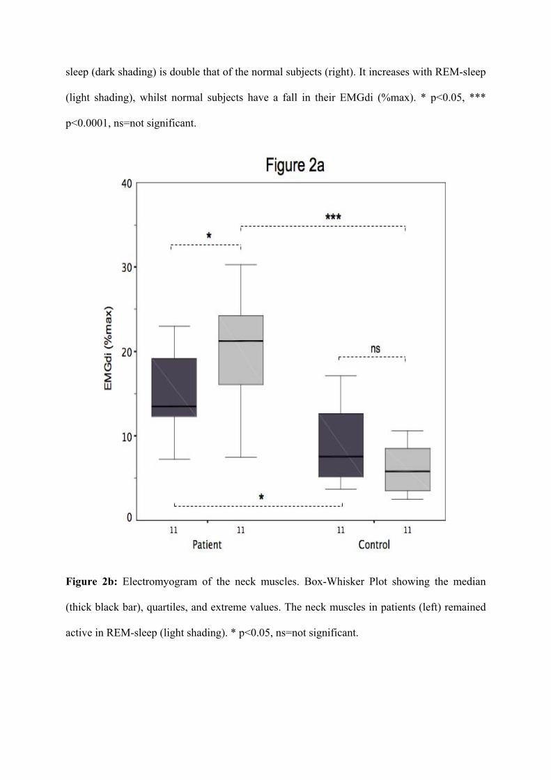
sleep (dark shading) is double that of the normal subjects (right). It increases with REM-sleep
(light shading), whilst normal subjects have a fall in their EMGdi (%max). * p<0.05, ***
p<0.0001, ns=not significant.
Figure 2b: Electromyogram of the neck muscles. Box-Whisker Plot showing the median
(thick black bar), quartiles, and extreme values. The neck muscles in patients (left) remained
active in REM-sleep (light shading). * p<0.05, ns=not significant.

Figure 3: 15s sample of an EMG signal of the diaphragm (EMGdi, µV) and the root-mean-
square (RMS, 50ms time constant, moving average, µV) during NREM (left) and REM (right)
sleep in a patient with UDP (top) and in a normal subject (bottom). Inspiratory activity of the
diaphragm is marked with arrows. Note the increase of neural respiratory drive in the patient
during REM-sleep, whilst the normal subject exhibits a lower level of activation.

Figure 4: Neural respiratory drive, as measured with the diaphragm EMG (EMGdi),
transdiaphragmatic pressure (Pdi), flow (pneumotachograph), genioglossus EMG (EMGgg)
and oxygenation (SaO2) during REM-sleep in a patient with UDP. The vertical dotted line
indicates the first occurrence of phasic REM. Following a loss of initially high central drive,
inspiratory effort reduces and the patient develops a hypopnoea that is eventually overcome
by an arousal-reaction.