Sleep and Arousal
33
Sleep and Arousal Lecture 9 NRS201S John Yeomans
description
Sleep and Arousal. Lecture 9 NRS201S John Yeomans. EEG Changes in Sleep. Waking: Alpha (10 Hz) and beta/gamma waves (40 Hz). Slow-Wave sleep: From alpha to spindles (14 Hz) and delta (1-4 Hz). REM sleep: Cortical arousal and muscular atonia. Also called paradoxical or dream sleep. - PowerPoint PPT Presentation
Transcript of Sleep and Arousal
Sleep and Arousal
Lecture 9
NRS201S
John Yeomans
EEG Changes in Sleep
• Waking: Alpha (10 Hz) and beta/gamma waves (40 Hz).
• Slow-Wave sleep: From alpha to spindles (14 Hz) and delta (1-4 Hz).
• REM sleep: Cortical arousal and muscular atonia. Also called paradoxical or dream sleep.
• Triggered in pontine reticular formation.
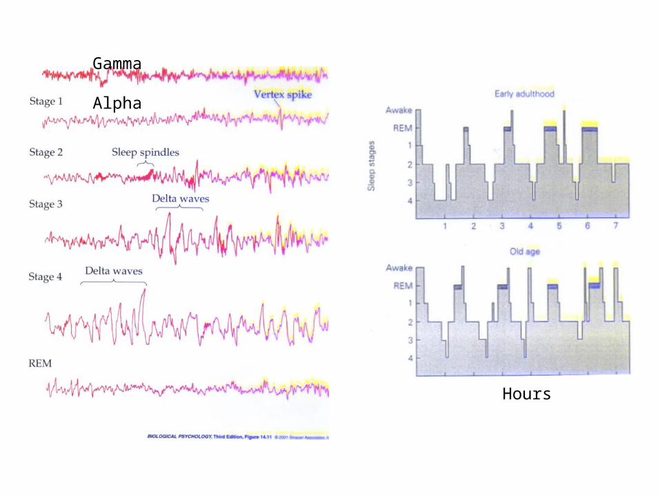
Gamma
Alpha
Hours

REM Sleep
• Brain is active, and eyes are active.
• Muscles of body are profoundly inhibited (atonia).
• Subjects report dreams, when awoken.
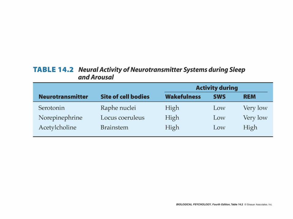
• NE and 5HT neurons silent. Ch neurons active.
• In slow-wave sleep, brain and eyes are quiet, but muscles are more active.

Transection studies
Brain Areas--Early Studies
• Coma (prolonged unconsciousness) due to injury in dorsal reticular formation.
• Stimulation of RF leads to arousal.
• Ascending path for cortical arousal.
• Descending path for atonia.
• Critical area in dorsal pontine reticular formation.
Diffuse Arousal Systems
• Locus coeruleus Norepinephrine neurons (A6).
• Mesopontine Cholinergic neurons (Ch5,6).• Raphe Serotonin neurons (B5,6).• Tuberomammilary Histamine neurons.• Lateral hypothalamus Orexin/Hypocretin
neurons.• Basal forebrain Cholinergic neurons (Ch1-
4).
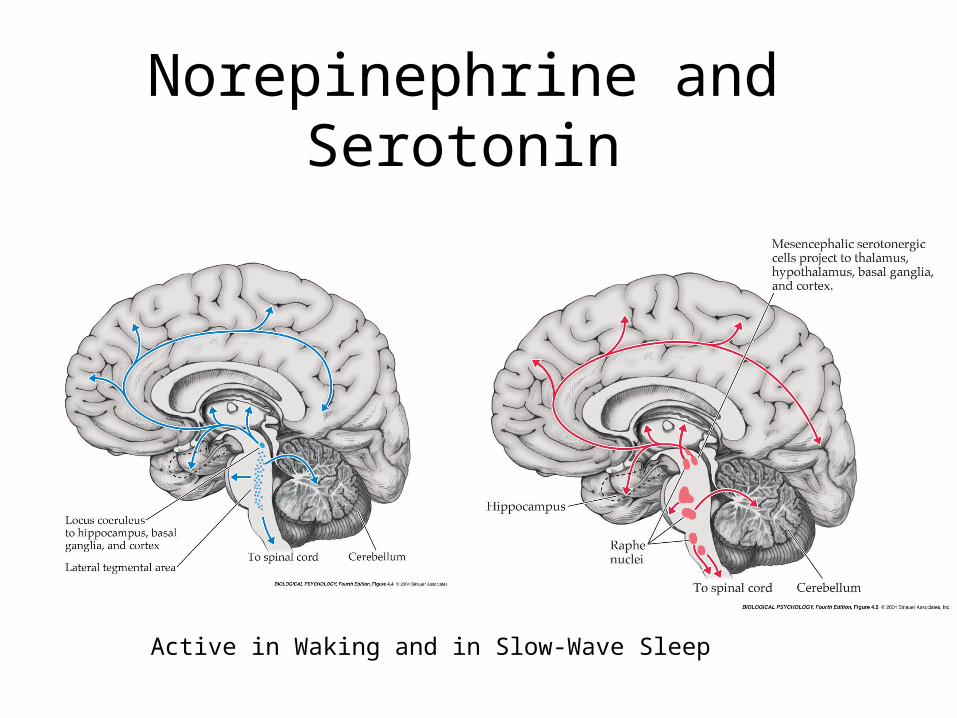
Norepinephrine and Serotonin
Active in Waking and in Slow-Wave Sleep
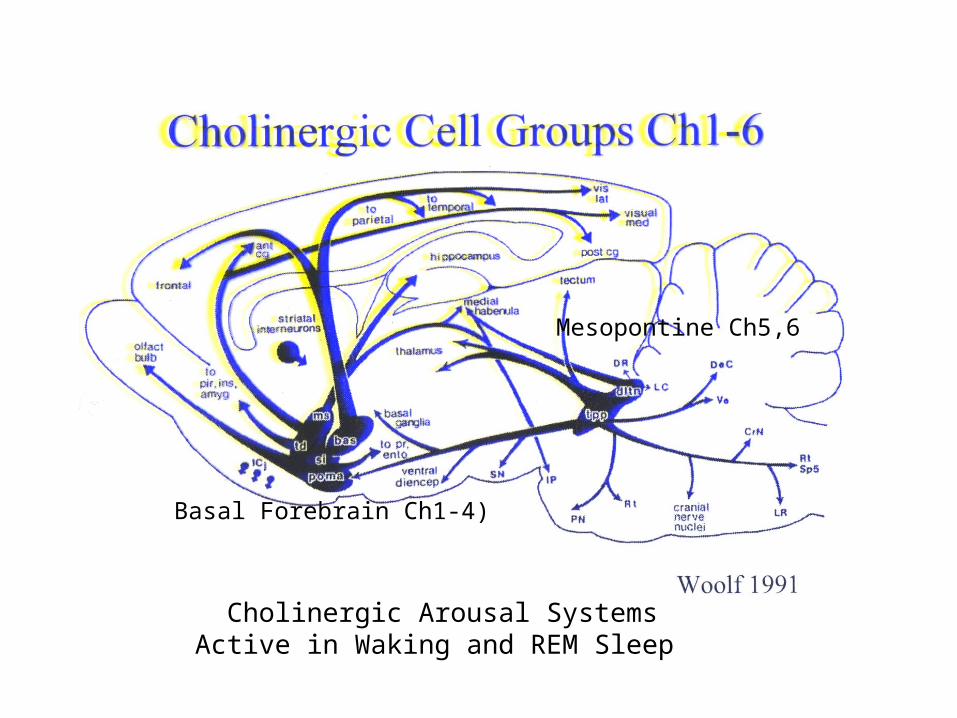
Cholinergic Arousal SystemsActive in Waking and REM Sleep
Basal Forebrain Ch1-4)
Mesopontine Ch5,6
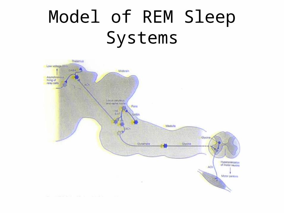
Model of REM Sleep Systems
Sleep Disorders
• Insomnia (too little sleep).• Sleep apnea (loss of breathing in REM,
too much atonia?).• Narcolepsy/cataplexy (daytime sleepiness
and REM/atonia attacks).• Triggered by arousal (e.g. laughing,
running).• Due to loss of orexin/hypocretin neurons in
humans, or receptors in dogs and mice.

Narcolepsy
• Orexin 2 receptors lost in dogs (Mignon).
• O/H neurons lost in humans.
• O/H gene or receptors in mice.
• O/H neurons active in waking arousal, and needed to inhibit atonia.
• In narcolepsy, arousal can activate REM/atonia neurons, if O/H signal is lost.
• Which neurons and how? Ch5,6?
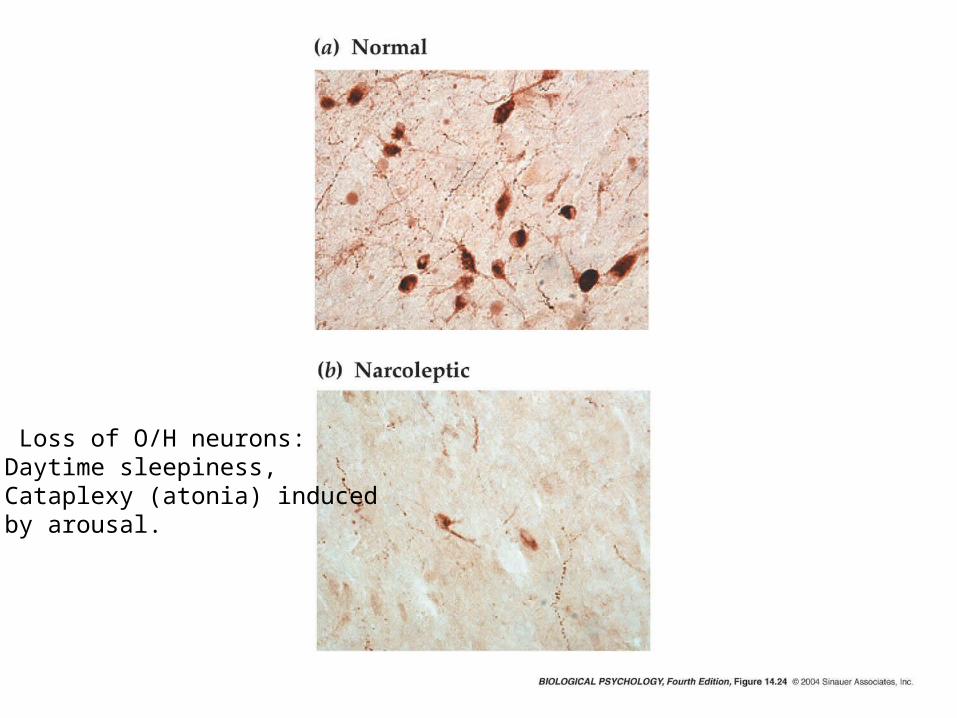
Loss of O/H neurons:Daytime sleepiness,Cataplexy (atonia) inducedby arousal.
Circadian Rhythms
March 17, 2006
PSY391S
John Yeomans

Timing of Motivated Behaviors• When is best season to feed and mate?
Seasonal periods of activity and breeding based on availability of food. Based on axis of earth around sun.
• When is best time of day to feed? Diurnal/nocturnal to find food and avoid predators. Based on earth’s rotation relative to sun.
• Circadian clock built into all plants and animals to help survival.
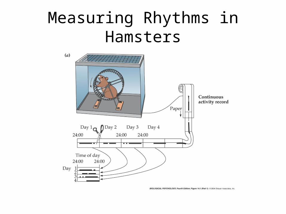
Measuring Rhythms in Hamsters
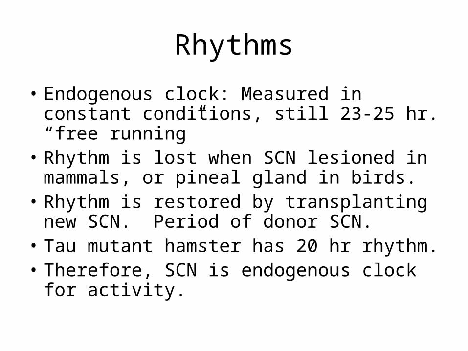
Rhythms
• Endogenous clock: Measured in constant conditions, still 23-25 hr. “free running”
• Rhythm is lost when SCN lesioned in mammals, or pineal gland in birds.
• Rhythm is restored by transplanting new SCN. Period of donor SCN.
• Tau mutant hamster has 20 hr rhythm.• Therefore, SCN is endogenous clock for
activity.
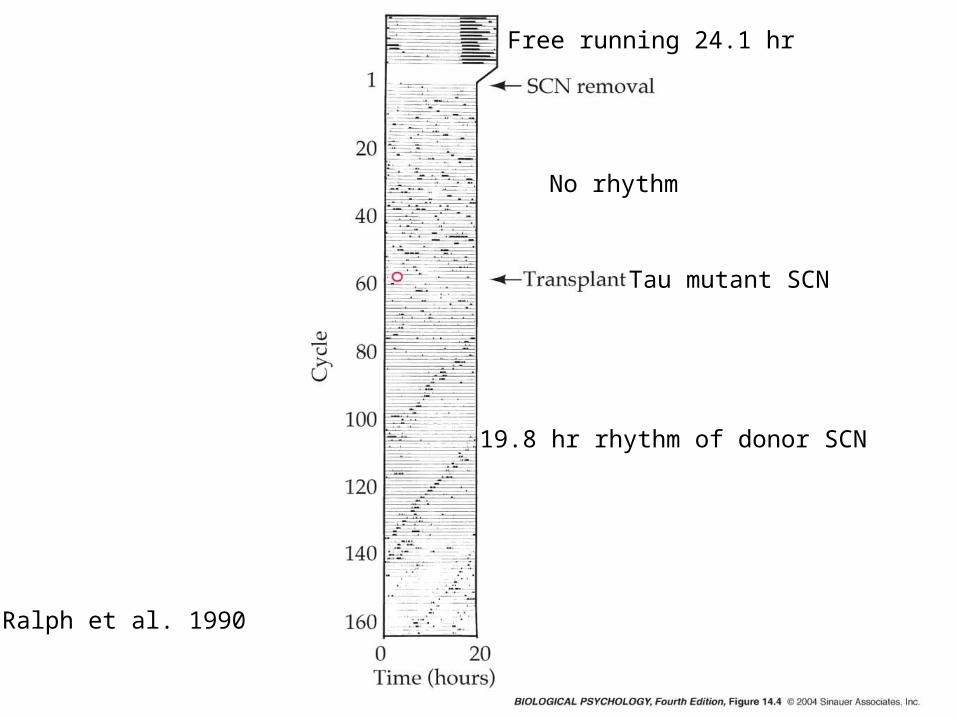
Free running 24.1 hr
No rhythm
19.8 hr rhythm of donor SCN
Tau mutant SCN
Ralph et al. 1990

Retinal Paths to SCN and IGL
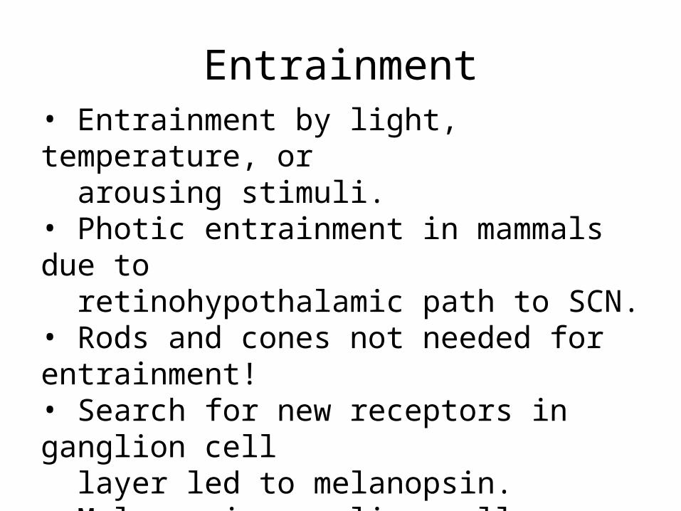
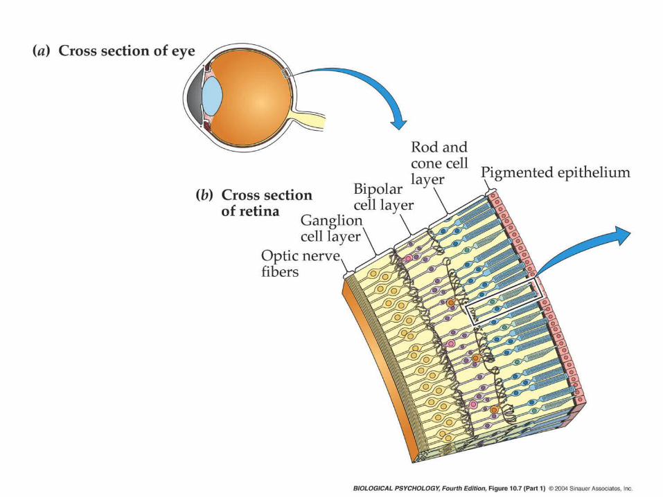
Entrainment• Entrainment by light, temperature, or arousing stimuli.• Photic entrainment in mammals due to retinohypothalamic path to SCN.• Rods and cones not needed for entrainment!• Search for new receptors in ganglion cell layer led to melanopsin.• Melanopsin ganglion cells directly activated by light, indirectly by rods and cones.• Huge dendrites and receptive fields, insensitive to light, but stable (no adaptation)
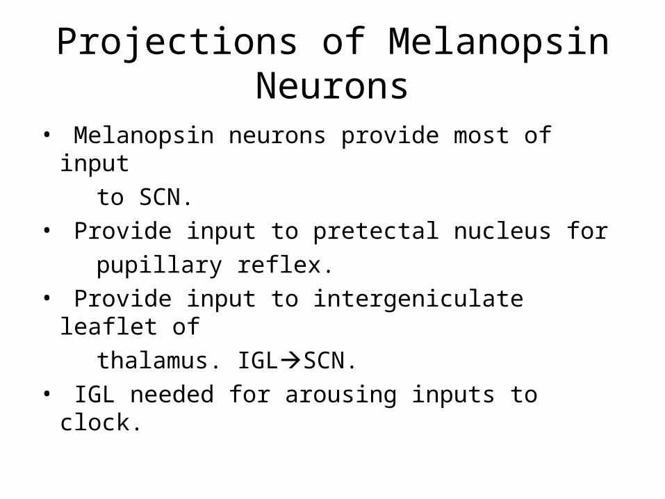
Projections of Melanopsin Neurons
• Melanopsin neurons provide most of input
to SCN.
• Provide input to pretectal nucleus for
pupillary reflex.
• Provide input to intergeniculate leaflet of
thalamus. IGLSCN.
• IGL needed for arousing inputs to clock.
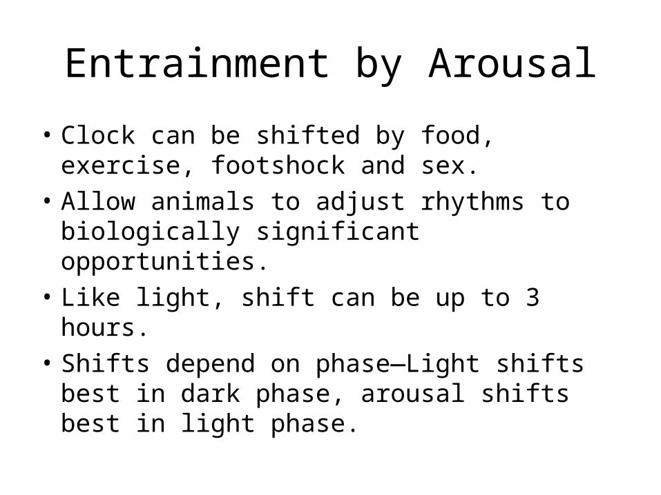
Entrainment by Arousal
• Clock can be shifted by food, exercise, footshock and sex.
• Allow animals to adjust rhythms to biologically significant opportunities.
• Like light, shift can be up to 3 hours.
• Shifts depend on phase—Light shifts best in dark phase, arousal shifts best in light phase.
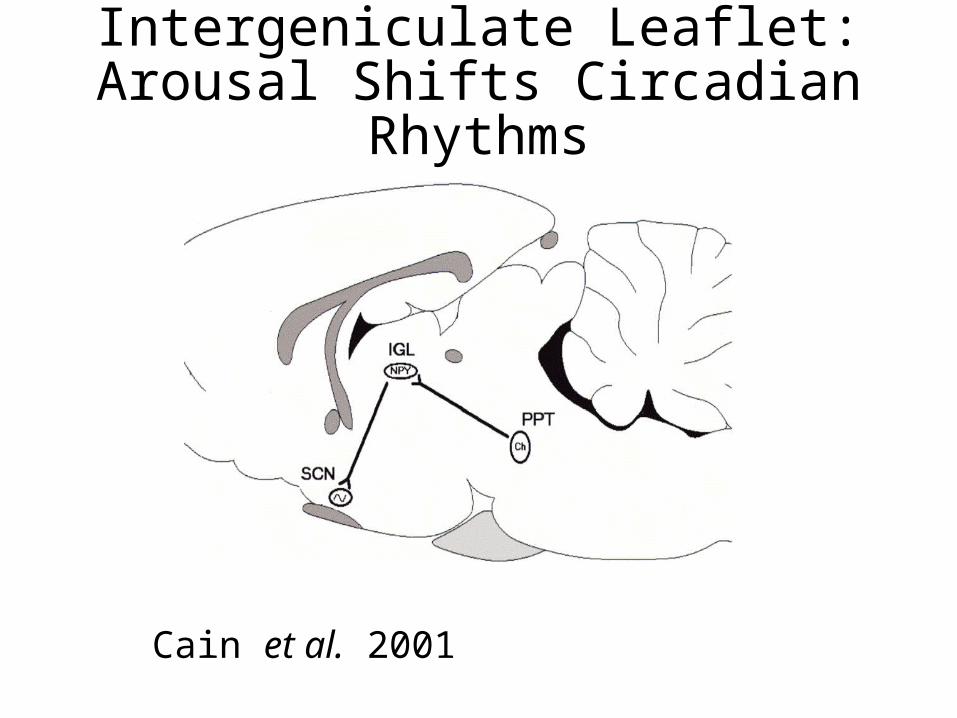
Intergeniculate Leaflet: Arousal Shifts Circadian Rhythms
Cain et al. 2001
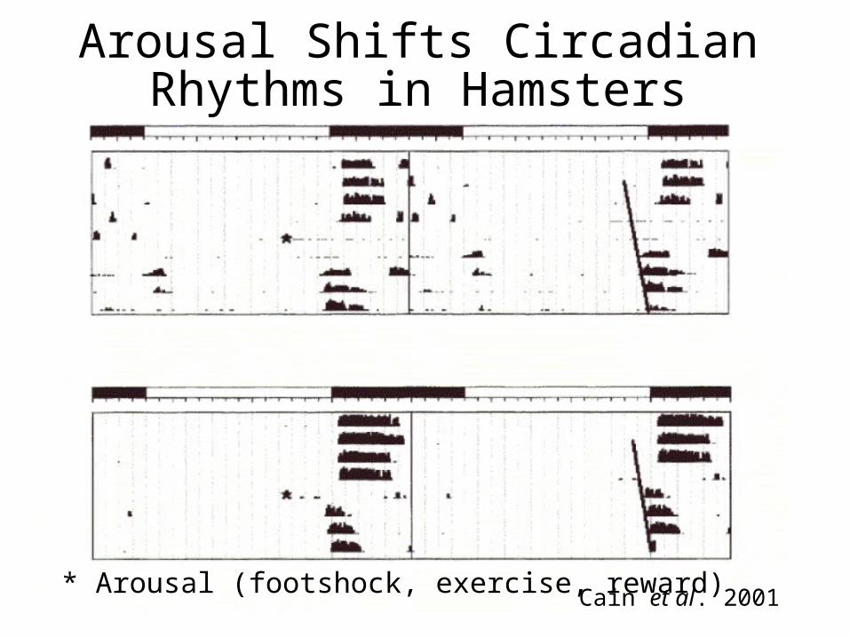
Arousal Shifts Circadian Rhythms in Hamsters
Cain et al. 2001* Arousal (footshock, exercise, reward)
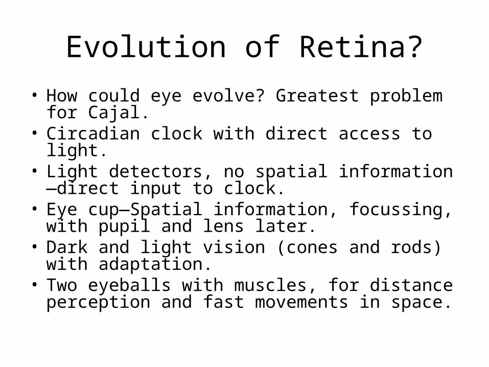
Evolution of Retina?
• How could eye evolve? Greatest problem for Cajal.
• Circadian clock with direct access to light.• Light detectors, no spatial information—direct
input to clock.• Eye cup—Spatial information, focussing, with
pupil and lens later.• Dark and light vision (cones and rods) with
adaptation.• Two eyeballs with muscles, for distance
perception and fast movements in space.
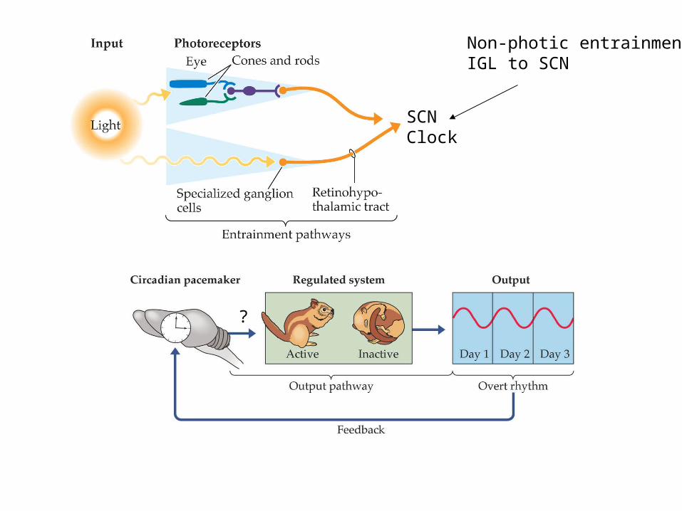
Non-photic entrainment:IGL to SCN
SCNClock
?
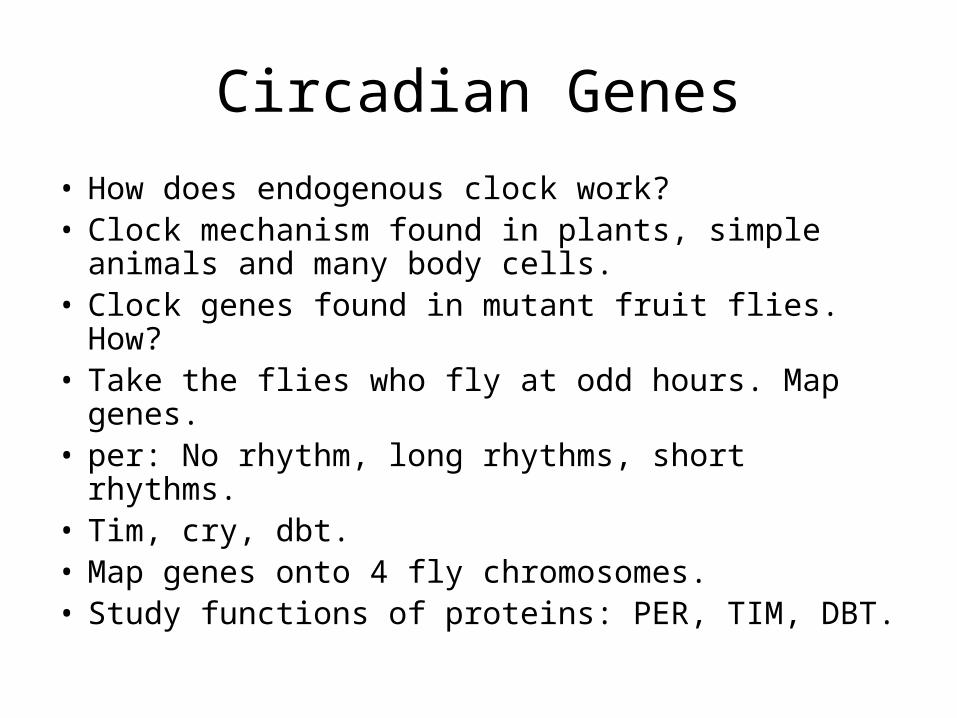
Circadian Genes
• How does endogenous clock work?• Clock mechanism found in plants, simple
animals and many body cells.• Clock genes found in mutant fruit flies. How?• Take the flies who fly at odd hours. Map genes.• per: No rhythm, long rhythms, short rhythms.• Tim, cry, dbt.• Map genes onto 4 fly chromosomes.• Study functions of proteins: PER, TIM, DBT.
Mutations Alter Rhythms in Flys and Mice
• per, tim are needed for 24 hr rhythms.
• Mutations lead to short, long or no rhythm.
• dbt mutations alter enzyme, casein kinase, leading to short rhythm in Drosophila.
• Homologous genes (per1-3, cry, tau) found in mice and humans.
• Transcription factors Clock and Cycle start each cycle. These are also regulated.
Clock Genes and Negative Feedback
• per, cry genes transcribed in nucleus.• Per, Cry proteins are translated in
cytoplasm.• Per/Cry dimers inhibit Clock/Cycle
transcription factors in nucleus.• Less Per, Cry less inhibition.• New per, cry transcribed 24 hrs later.• Tau gene makes a casein kinase that
degrades Per.
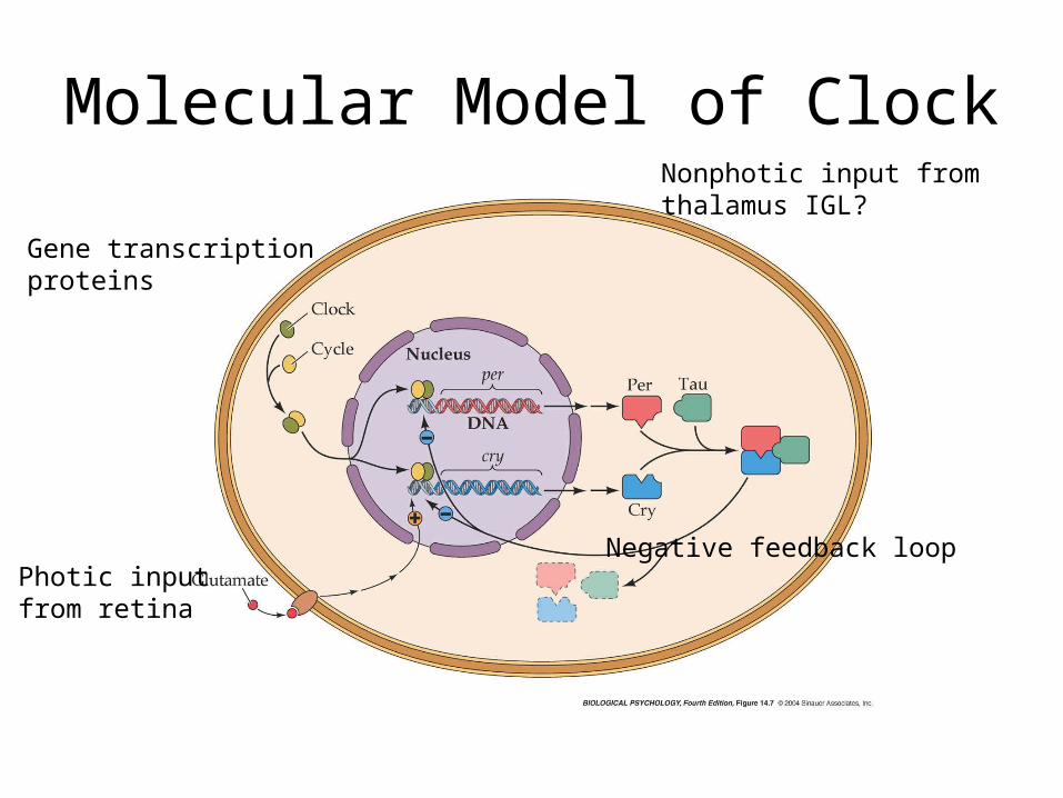
Molecular Model of Clock
Photic inputfrom retina
Nonphotic input from thalamus IGL?
Gene transcriptionproteins
Negative feedback loop