Skin
42
-
Upload
jessica-morgan -
Category
Education
-
view
87 -
download
0
Transcript of Skin
General Facts:
A.First line of defense, skin is an organ
B.Vital to homeostasis
C.Largest body organ
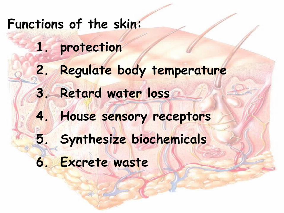
Functions of the skin:
1. protection
2. Regulate body temperature
3. Retard water loss
4. House sensory receptors
5. Synthesize biochemicals
6. Excrete waste

Regions
Epidermis – outermost superficial region
Dermis – middle region
Subcutaneous (superficial fascia) – deepest region
Epidermis
• Composed of epithelial cells, consisting of four distinct cell types and four or five layers and lacks blood vessels
• Cell types include keratinocytes, melanocytes, Merkel cells, and Langerhans’ cells
• Outer portion of the skin is exposed to the external environment and functions in protection

Cells of the Epidermis
• Keratinocytes – produce the fibrous protein keratin
• Melanocytes – produce the brown pigment melanin
• Langerhans’ cells – epidermal macrophages that help activate the immune system
• Merkel cells – function as touch receptors in association with sensory nerve endings

Layers of the Epidermis

Layers of the Epidermis: Stratum Basale
(Basal Layer)• Deepest epidermal
layer firmly attached to the dermis
• Consists of a single row of the youngest keratinocytes
• Cells undergo rapid division
• Well nourished by dermal blood vessels

Cells are pushed upward as new cells are formed and become keratinized as they die.
Protects against water loss, injury, harmful chemicals & bacteria.
Melanocytes lie deep in the epidermis & the underlying dermis, produce a pigment called melanin that protects deeper cells from the sun’s UV rays.
Melanocytes pass melanin to nearby cells through cytocrine secretion.

Skin Color• Three pigments contribute to
skin color• Melanin – yellow to reddish-
brown to black pigment, responsible for dark skin colors
• Freckles and pigmented moles – result from local accumulations of melanin
• Carotene – yellow to orange pigment, most obvious in the palms and soles of the feet
• Hemoglobin – reddish pigment responsible for the pinkish hue of the skinGenetic differences result from different amounts of
melanin & in the size of pigment granules.
Sunlight causes the melanin production to increase.
Circulation with dermal blood vessels affects skin color.
Dermis
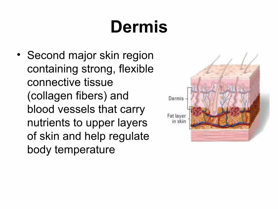
• Second major skin region containing strong, flexible connective tissue (collagen fibers) and blood vessels that carry nutrients to upper layers of skin and help regulate body temperature
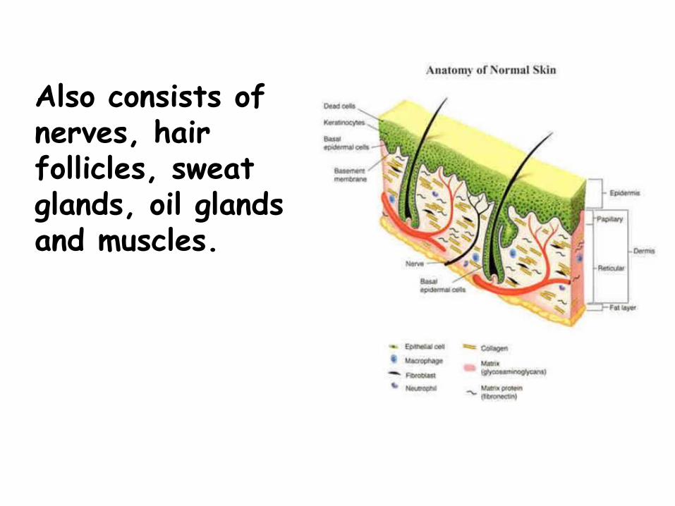
Also consists of nerves, hair follicles, sweat glands, oil glands and muscles.
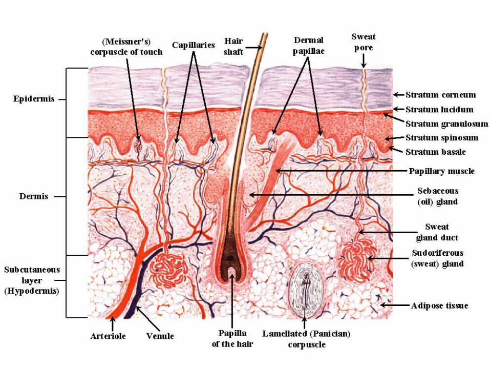
Subcutaneous (Hypodermis)
• Subcutaneous layer deep to the skin
• Composed of adipose (fat layer) and areolar (loose) connective tissue
• It binds the skin to underlying organs and contains the blood vessels that supply the skin.
• There is no sharp boundary between the dermis and the hypodermis
Accessory Organs of the Skin
Hair Function and Distribution
• Hair is distributed over the entire skin surface except:
• Palms, soles, and lips
• Nipples and portions of the external genitalia
Hair Follicle• Individual hairs develop from cells at the base of the
hair follicle, an invagination of the lower epidermis that dips down into the dermis
• As new cells are formed, old cells are pushed outward and become keratinized, forming the hair shaft
• A bundle of smooth muscle cells, called the arrector pili muscle, is attached to each hair follicle
• Hair color is determined by genetics; melanin from melanocytes is responsible for most hair colors, but red hair also contains the pigment trichosiderin
Hair Function and Distribution
• Functions of hair include:
• Helping to maintain warmth
• Alerting the body to presence of insects on the skin
• Guarding the scalp against physical trauma, heat loss, and sunlight
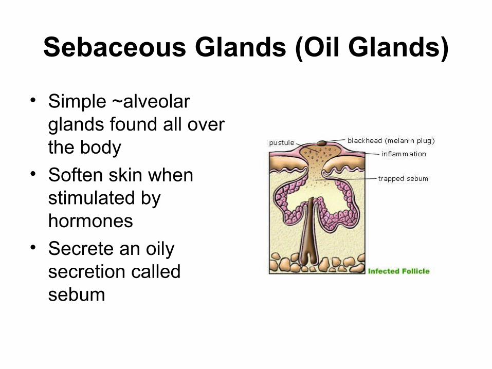
Sebaceous Glands (Oil Glands)
• Simple ~alveolar glands found all over the body
• Soften skin when stimulated by hormones
• Secrete an oily secretion called sebum
Sweat Glands• Different types prevent overheating of the body;
secrete cerumen~ and milk
• Eccrine sweat glands – found in palms, soles of the feet, and forehead. Respond to body temperature
• Apocrine sweat glands – found in axillary and anogenital areas. Respond to body temperature, stress, & sexual arrousal
• ~Eruminous glands – modified apocrine glands in external ear canal that secrete cerumen (wax)
• ~Mammary glands – specialized sweat glands that secrete milk

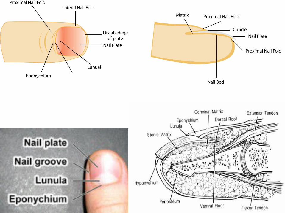
Structure of a Nail
• Scalelike modification of the epidermis on the distal, dorsal surface of fingers and toes
• Consist of epithelial cells overlying the nail bed, with the lunula as the most actively growing region of the nail root
• As new cells are produced, older ones are pushed outward and become keratinized.
Functions of the Integumentary System: Regulation of Body Temperature
• Body temperature regulation is accomplished by:
• Dilation (cooling) and constriction (warming) of dermal vessels
• Increasing sweat gland secretions to cool the body
• Excessive cooling: inactivates sweat glands, shivering

Healing of Wounds &
Burns
Wounds•Inflammation, in which blood vessels dilate and become more permeable, causing tissues to become red and swollen, is the body’s normal response to injury.•Superficial cuts are filled in by reproducing epithelial cells.•Deeper cuts are closed off by clots, covered by scabs, and eventually filled in by fibroblasts, making connective tissue. Blood vessels extend into the area, injured tissues are replaced, and the scab falls off.•Large wounds leave scars and healing may be accompanied by the formation of granulations.

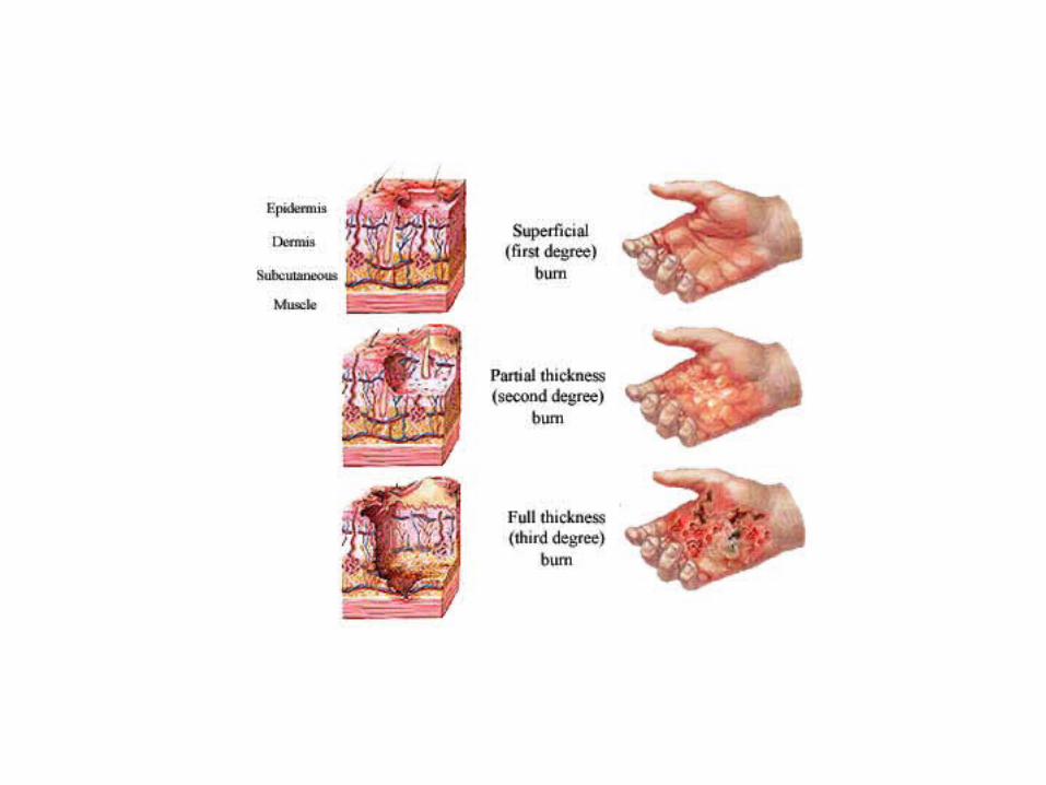
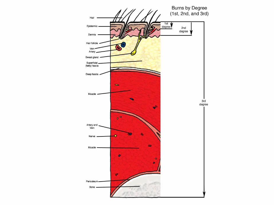
Burns
• First-degree – only the epidermis is damaged– Symptoms include localized redness, swelling, and
pain• Second-degree – epidermis and upper regions
of dermis are damaged– Symptoms mimic first degree burns, but blisters also
appear• Third-degree – entire thickness of the skin is
damaged– Burned area appears gray-white, cherry red, or black;
there is no initial edema or pain (since nerve endings are destroyed)

Skin Cancer
• Most skin tumors are benign (not harmful) and do not metastasize (spread to other parts of the body)
• A crucial risk factor for non-melanoma skin cancers is the disabling of the p53 gene
• Newly developed skin lotions can fix damaged DNA
Skin Cancer
• The three major types of skin cancer are:
– Squamous cell carcinoma
– Basal cell carcinoma
– Malignant Melanoma
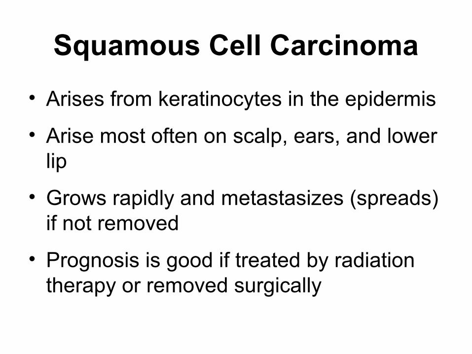
Squamous Cell Carcinoma
• Arises from keratinocytes in the epidermis
• Arise most often on scalp, ears, and lower lip
• Grows rapidly and metastasizes (spreads) if not removed
• Prognosis is good if treated by radiation therapy or removed surgically


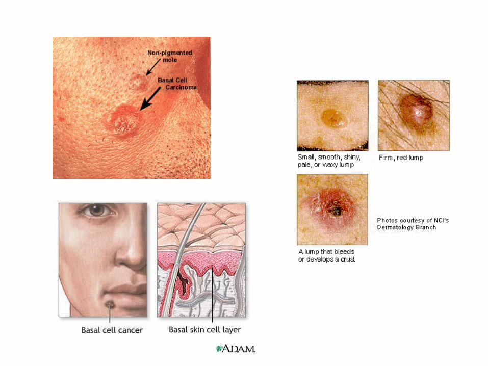
Basal Cell Carcinoma
• Least malignant and most common skin cancer
• Stratum basale cells proliferate and invade the dermis and hypodermis
• Slow growing and do not often metastasize
• Can be cured by surgical excision in 99% of the cases
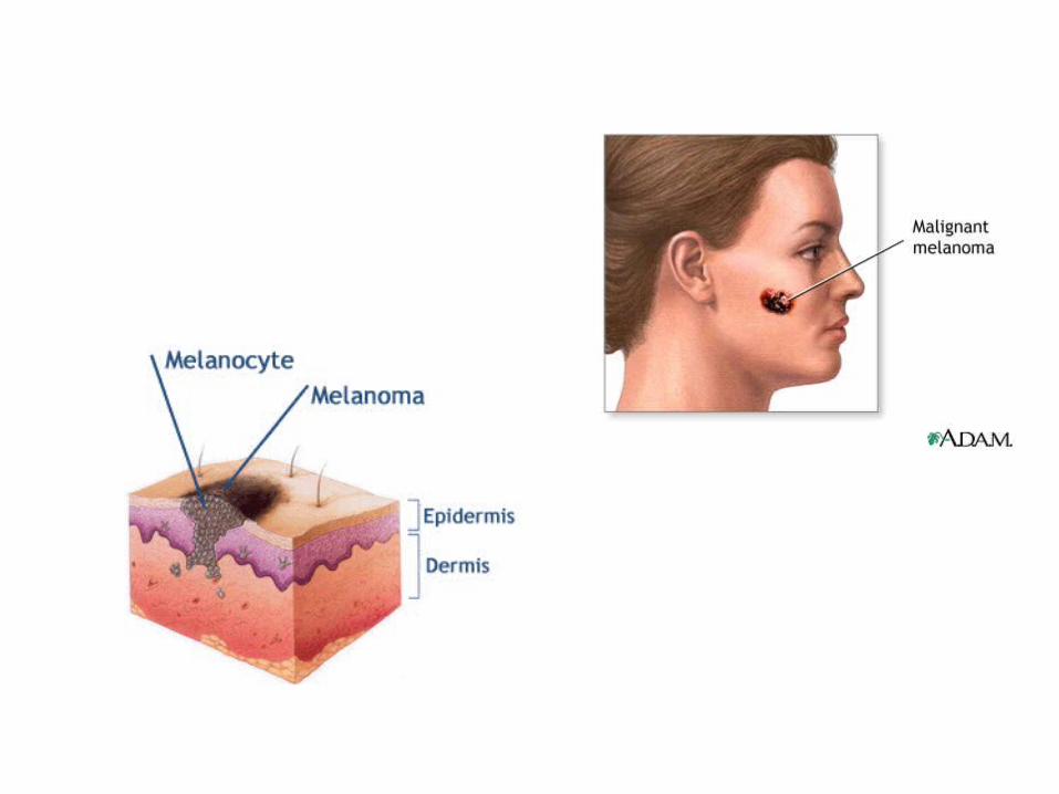
Malignant Melanoma
• Cancer of melanocytes is the most dangerous type of skin cancer because it is:
– Highly metastatic
– Resistant to chemotherapy
Melanoma (cont.)
• Melanomas have the following characteristics (ABCD rule)– A: Asymmetry; the two sides of the
pigmented area do not match – B: Border is irregular and exhibits
indentations– C: Color (pigmented area) is black, brown,
tan, and sometimes red or blue– D: Diameter is larger than 6 mm (size of a
pencil eraser)

Prognosis and Treatment
• Treated by wide surgical excision accompanied by immunotherapy
• Chance of survival is poor if the lesion is over 4 mm thick