Skin of the thigh Cutaneous Nerves 1- The femoral branch of the genitofemoral nerve. It enters the...
34
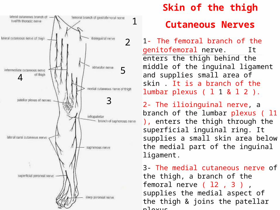
Skin of the thigh Cutaneous Nerves 1- The femoral branch of the genitofemoral nerve. It enters the thigh behind the middle of the inguinal ligament and supplies small area of skin . It is a branch of the lumbar plexus ( l 1 & l 2 ). 2- The ilioinguinal nerve, a branch of the lumbar plexus ( l1 ), enters the thigh through the superficial inguinal ring. It supplies a small skin area below the medial part of the inguinal ligament. 3- The medial cutaneous nerve of the thigh, a branch of the femoral nerve ( l2 , 3 ) , supplies the medial aspect of the thigh & joins the patellar plexus. 2 1 4 5 3
-
Upload
marian-tamsin-bruce -
Category
Documents
-
view
214 -
download
0
Transcript of Skin of the thigh Cutaneous Nerves 1- The femoral branch of the genitofemoral nerve. It enters the...
Skin of the thigh
Cutaneous Nerves
1- The femoral branch of the genitofemoral nerve. It enters the thigh behind the middle of the inguinal ligament and supplies small area of skin . It is a branch of the lumbar plexus ( l 1 & l 2 ).
2- The ilioinguinal nerve, a branch of the lumbar plexus ( l1 ), enters the thigh through the superficial inguinal ring. It supplies a small skin area below the medial part of the inguinal ligament.
3- The medial cutaneous nerve of the thigh, a branch of the femoral nerve ( l2 , 3 ) , supplies the medial aspect of the thigh & joins the patellar plexus.
4- The intermediate cutaneous nerve of the thigh, a branch of the femoral nerve ( l1, 2 ). It divides into 2 branches that supply the anterior aspect of the thigh & joins the patellar plexus.
2
1
45
3

5- Branches from the anterior division of the obturaror nerve ( l2, 3 ; 4 ) supply area of the skin on the medial aspect of the high.
6- The lateral cutaneous nerve of the thigh, a branch of the lumbar plexus ( L2; 3 ). It enters thigh behind the lateral end of the inguinal ligament. It divides into A & P branches to supply the skin of the lateral aspect of the thigh & knee . Also, the skin of the lower lateral quadrant of the buttock
5
6
56

Superficial fascia of the thigh The membranous layer of the superficial fascia of the anterior abdominal wall extends into the thigh and is attached to the deep fascia ( fascia lata ) about a fingerbreadth ( .8 cm) below the inguinal ligament . The fatty layer of the superficial fascia on the anterior abdominal wall extends into the thigh and continues down over the lower limb without interruption.
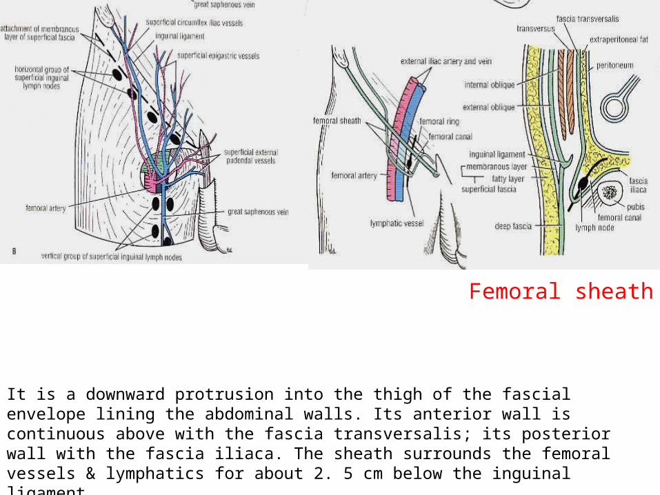
Femoral sheath It is a downward protrusion into the thigh of the fascial envelope lining the abdominal walls. Its anterior wall is continuous above with the fascia transversalis; its posterior wall with the fascia iliaca. The sheath surrounds the femoral vessels & lymphatics for about 2. 5 cm below the inguinal ligament .
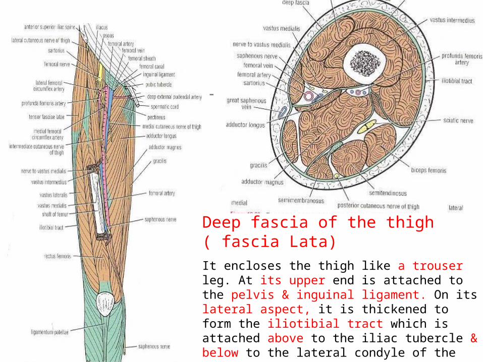
Deep fascia of the thigh ( fascia Lata)It encloses the thigh like a trouser leg. At its upper end is attached to the pelvis & inguinal ligament. On its lateral aspect, it is thickened to form the iliotibial tract which is attached above to the iliac tubercle & below to the lateral condyle of the tibia . The iliotibial tract receives the insertion of the tensor fasciae latae & the greater part of the gluteus maximus muscles.

Deep fascia
In it, there is a gap in the front of the thigh just below the inguinal ligament. It is the Saphenous opening which transmits the great saphenous vein, small branches of the femoral artery; lymph vessels. It is situated about 4 cm below & lateral to the pubic tubercle.
The lower lateral border of the opening is called the falciform margin . It lies anterior to the femoral vessels.
The border of the opening then curves upward & medially then laterally behind the femoral vessels, to be attached to the pectineal line of the superior ramus of the pubis.
The saphenous opening is filled with loose connective tissue called the Cribriform fascia .
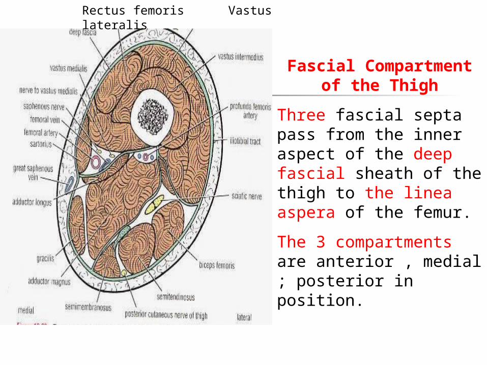
Fascial Compartment of the Thigh
Three fascial septa pass from the inner aspect of the deep fascial sheath of the thigh to the linea aspera of the femur.
The 3 compartments are anterior , medial ; posterior in position.
Rectus femoris Vastus lateralis
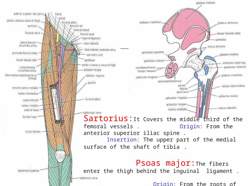
Sartorius:It Covers the middle third of the femoral vessels . Origin: From the anterior superior iliac spine . Insertion: The upper part of the medial surface of the shaft of tibia .
Psoas major:The fibers enter the thigh behind the inguinal ligament . Origin: From the roots of the T. processes; sides of the vertebral bodies; the intervertebral discs, from the 12th thoracic to the 5th lumbar vertebrae. Insertion: Into the lesser trochanter of the femur.

Pectineus Origin : From the superior ramus of the pubis .
Insertion : It is attached to the upper end of the linea aspera just below the lesser trochanter .
Iliacus : Origin : Arises from the iliac fossa within the abdomen . Insertion : Its fibers join the tendon of the psoas to form the iliopsoas muscle which are inserted into the lesser trochanter of the femur & the adjoining area .

Muscles of the anterior compartment of the thigh
1- Sartorius : N . Supply : femoral nerve. Action : flexes both hip & knee joints. Abduct ; laterally rotates the thigh and medially rotates the leg at the knee .
2- Iliacus : N. supply : A branch of the femoral nerve within the abdomen. Action : The iliopsoas flexes the thigh on the trunk at the hip joint or if the thigh is fixed , it flexes the trunk on the thigh ; it medially rotates the thigh .
3- Psoas : N. supply : Branches from the lumbar plexus. Action : like the iliacus .
Pectineus : N. supply : femoral & a branch from the obturator nerves . Action : Flexes & adducts the thigh at the hip joint .

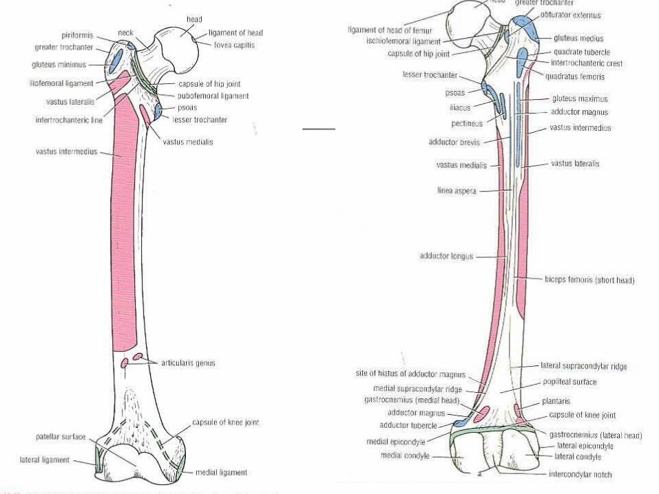
Origin of quadriceps femoris 1-Rectus femoris : A straight head from the A. inferior iliac spine & A reflected head from the ilium above the acetabulum . 2- Vastus lateralis : From the upper part of intertrochanteric line , the base of the greater trochanter , lateral margin of gluteal tuberosity & linea aspera of the femur . 3- Vastus medialis : from lower part of intertrochanteric line ; spiral line ; linea aspera of the femur & upper part of medial supracondylar ridge . 4- Vastus intermedius: From the upper 2l3 of the A.& Lat surfaces of the shaft of the femur .

Insertion of The Quadriceps Femoris
The four muscles have a common tendon of insertion into the upper, lateral; medial borders of the patella & then via the ligamentum patellae into the tubercle of the tibia. So, They are inserted into the quadriceps tendon & so into the patella .
N. B. The Vastus intermedius fibers join the deep aspect of the quadriceps tendon. The articularis genus is a small part of the vastus intermedius that is inserted into the upper part of the synovial membrane of the knee joint. It serves to retract the synovial membrane superiorly during extension of the knee joint .
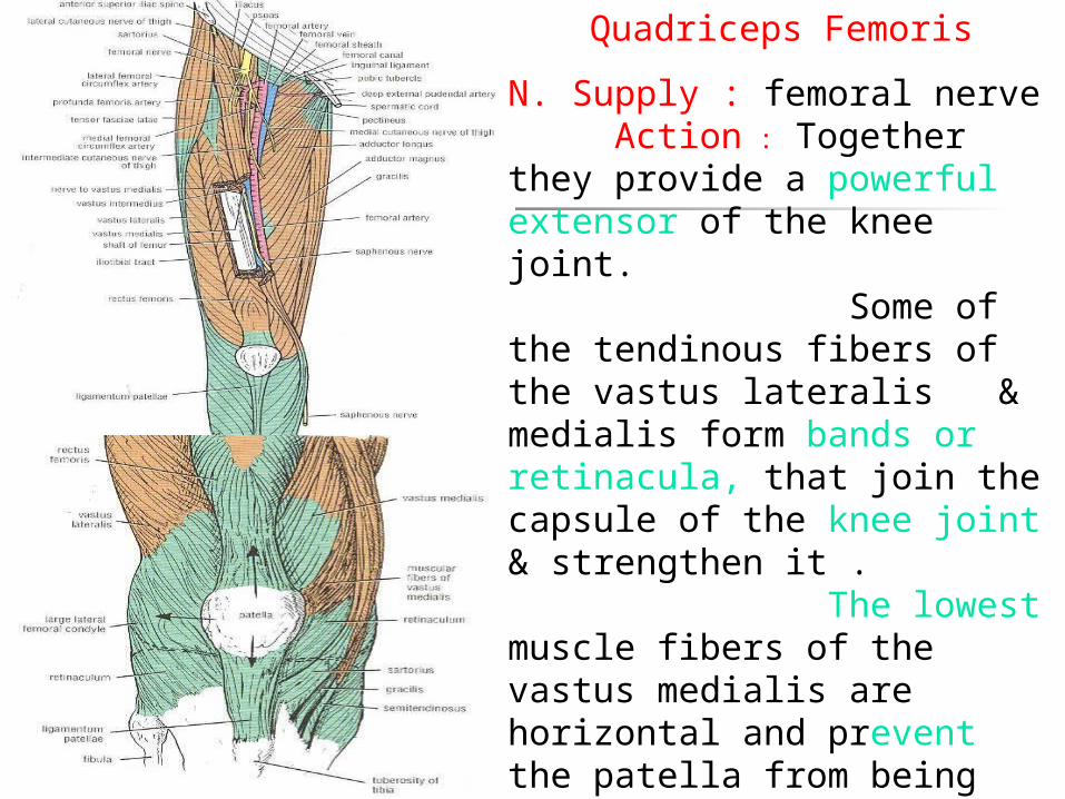
Quadriceps Femoris
N. Supply : femoral nerve Action : Together they provide a powerful extensor of the knee joint. Some of the tendinous fibers of the vastus lateralis & medialis form bands or retinacula, that join the capsule of the knee joint & strengthen it . The lowest muscle fibers of the vastus medialis are horizontal and prevent the patella from being pulled laterally during contraction of the quadriceps muscle. Their tone strengthens the knee joint . The rectus femoris flexes hip joint
Femoral sheath
It is a downward protrusion into the thigh of the fascial envelope lining the abdominal walls. Its anterior wall is continuous above with the fascia transversalis; its posterior wall with the fascia iliaca. The sheath surrounds the femoral vessels & lymphatics for about 2. 5 cm below the inguinal ligament .
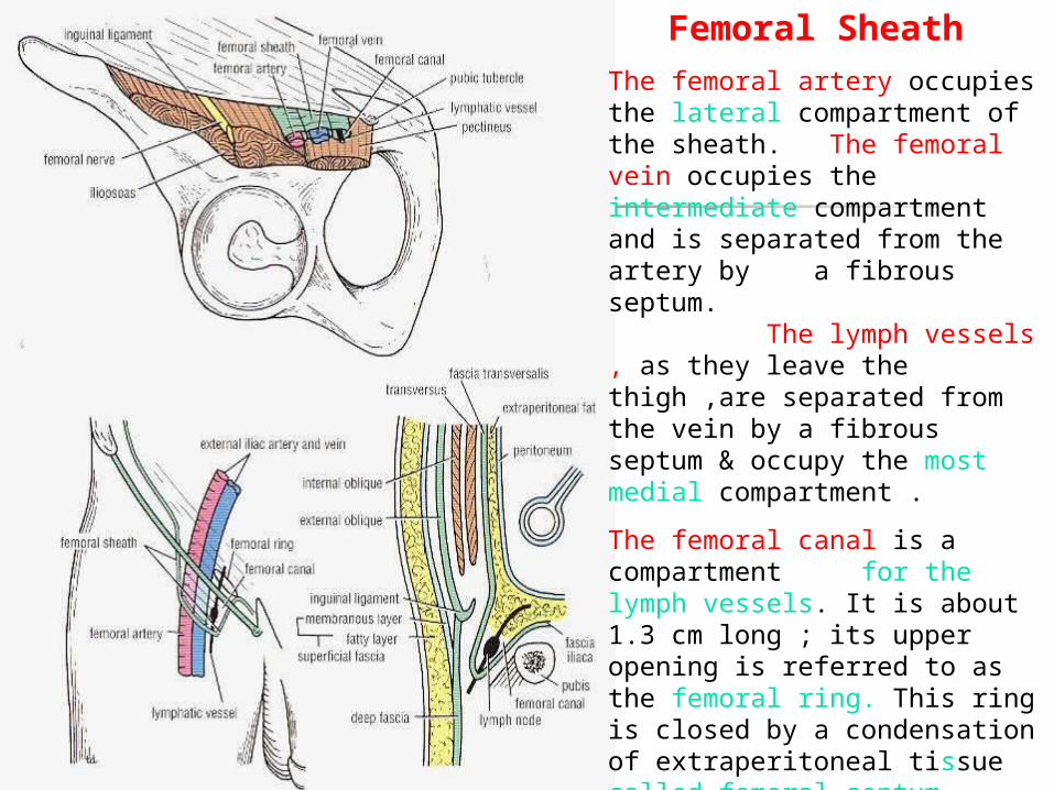
Femoral Sheath The femoral artery occupies the lateral compartment of the sheath. The femoral vein occupies the intermediate compartment and is separated from the artery by a fibrous septum. The lymph vessels , as they leave the thigh ,are separated from the vein by a fibrous septum & occupy the most medial compartment .
The femoral canal is a compartment for the lymph vessels. It is about 1.3 cm long ; its upper opening is referred to as the femoral ring. This ring is closed by a condensation of extraperitoneal tissue called femoral septum. This canal contains fatty connective tissue ; all the efferent lymph vessels ( come out ) from the deep inguinal lymph nodes and one of the deep inguinal lymph nodes.

The femoral sheath
It is adherent to the walls of the blood vessels and inferiorly blends with the tunica adventitia of these vessels. It is not adherent to the walls of the lymph vessels & this site is a potentially weak area in the abdomen. A protrusion of the abdominal parietal peritoneum into the canal
push the femoral septum, forming a femoral hernia .The upper end of the canal ( ring ) is related Anteriorly: to the inguinal ligament Posteriorly: to the superior ramus of the pubis .Medially : into the lacunar ligament .Laterally : The femoral vein . The lower end of the canal is closed by the adherence of its medial wall to the tunica adventitia of the femoral vein. It lies close to the saphenous opening in the deep fascia of the thigh.

Femoral Hernia It is more in female than male ( because of their wide pelvis & femoral canal ).
The hernial sac ( abdominal parietal peritoneum ) passes through the femoral canal pushing the femoral septum before it . At the lower end of the femoral canal it expands to form a swelling in the upper part of the thigh deep to the deep fascia .
The hernial sac may turn upward to cross the anterior surface of the inguinal ligament .
The neck of the sac is narrow & lies at the femoral ring . It can not expand due to the surrounding anatomic structures .
It is irreducible hernia .
After coughing a piece of bowel may be forced through the neck , and its blood vessels may be compressed by the femoral ring forming strangulated hernia. It is dangerous & should treated surgically

Femoral Hernia
The Neck of the sac is narrow & lies at the femoral ring .
The neck of the sac lies below & lateral to the pubic tubercle. This serves to differentiate it from the inguinal hernia which lies above & medial to the pubic tubercle .

Femoral triangle
It is situated at the upper part of the medial aspect of the thigh just below the inguinal ligament .It is bounded superiorly by the inguinal ligament , Laterally by sartorius & medially by medial border of the adductor longus muscle .
Its floor is formed from lateral to medial by the iliopsaos , pectineus; adductor longus .
Its roof is formed by skin & fasciae of the thigh .
It contains the terminal branches of the femoral nerve ,F. A. and its branches ; F. v. And its tributaries ; F. sheath and deep inguinal lymph nodes .

Adductor canal ( Subsartorial )It is an intermuscular cleft situated on the medial aspect of the middle third of the thigh beneath the sartorius muscle . It begins above at the apex of the femoral triangle & ends below at the opening in the adductor magnus .
In cross section it is triangular having anteromedial ; posterior and lateral walls .
The anteromedial wall is formed by sartorius muscle .
The posterior wall is formed by the adductor longus & magnus ,
The lateral wall is formed by the vastus medialis .
It contains the terminal part of the femoral artery ; femoral vein ; saphenous nerve ;nerve to vastus medialis ; the terminal part of the obturator nerve and the deep lymph vessels .
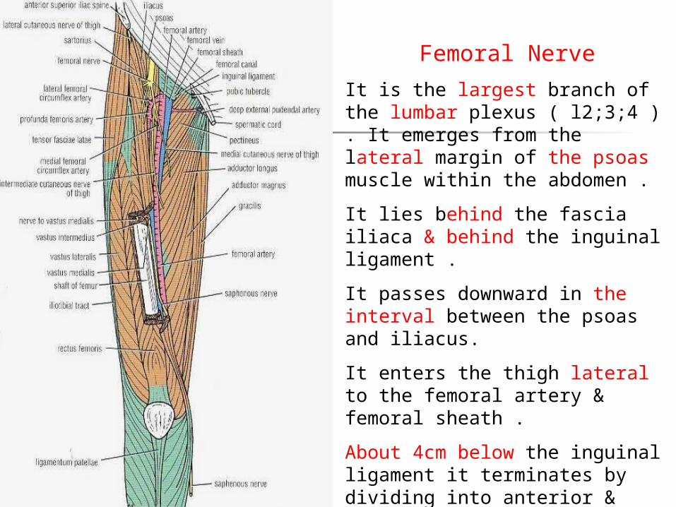
Femoral Nerve
It is the largest branch of the lumbar plexus ( l2;3;4 ) . It emerges from the lateral margin of the psoas muscle within the abdomen .
It lies behind the fascia iliaca & behind the inguinal ligament .
It passes downward in the interval between the psoas and iliacus.
It enters the thigh lateral to the femoral artery & femoral sheath .
About 4cm below the inguinal ligament it terminates by dividing into anterior & posterior divisions .

Femoral Nerve Branches Anterior Division 1;2;3
and 4 .
Posterior Division : 1- muscular to quadriceps . The branch to rectus femoris also supplies the hip joint ; the branches to the 3 vasti also supply the knee joint .
2- saphenous N:It is cutaneous It runs downward & medially. It crosses the femoral artery from the its lateral to its medial side .
It emerges on the medial side of the knee between the tendon of sartorius & gracilis .
It runs down the medial side of the leg in company with the great saphenous vein . It passes in front of medial malleolus
1 2
3
4
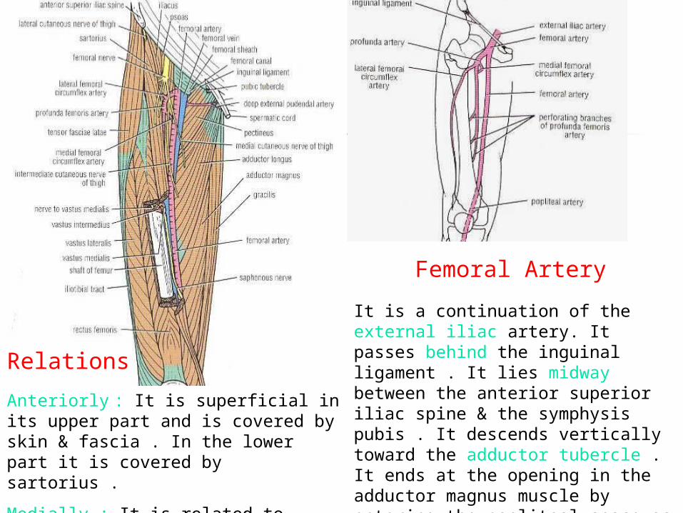
Femoral Artery
It is a continuation of the external iliac artery. It passes behind the inguinal ligament . It lies midway between the anterior superior iliac spine & the symphysis pubis . It descends vertically toward the adductor tubercle . It ends at the opening in the adductor magnus muscle by entering the popliteal space as the politeal artery.
:
Anteriorly : It is superficial in its upper part and is covered by skin & fascia . In the lower part it is covered by sartorius .
Medially : It is related to femoral vein in the upper part .
Relations
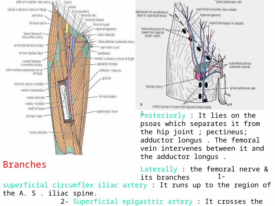
: Posteriorly : It lies on the psoas which separates it from the hip joint ; pectineus; adductor longus . The femoral vein intervenes between it and the adductor longus .
Laterally : the femoral nerve & its branchesBranches
1- superficial circumflex iliac artery : It runs up to the region of the A. S . iliac spine. 2- Superficial epigastric artery : It crosses the inguinal ligament & runs to the umbilicus. 3- Superficial external pudendal artery : It runs medially to supply the skin of scrotum. 4- Deep external pudendal artery: It runs medially to supply the scrotum or labia majora

5- Descending genicular artery: It arises from the femoral artery near its termination . It supply the knee joint .
6-Profunda femoris artery :
It is a large branch that arises from the lateral side of the femoral artery about 4 cm below the inguinal ligament . It passes medially behind the femoral vessels & enter the medial fascial compartment . It descends in the interval between the adductor longus & adductor brevis , then lies on the adductor magnus , where It ends by becoming the 4th perforating artery .

BranchesMedial femoral circumflex artery : It passes backward between the muscles of the floor of the femoral triangle and gives off muscular branches to the muscles of the medial fascial compartment . It shares in the formation of the cruciate anastomosis.
Lateral femoral circumflex artery : It passes laterally between the terminal branches of the femoral nerve . It gives off muscular branches to this area and shares in the formation of the cruciate anastomosis .
Four perforating arteries : They run backward and supply the surrounding muscles . They are anastomosing with one another & with the inferior gluteal & circumflex femoral arteries above & the muscular branches of the popliteal artery below .
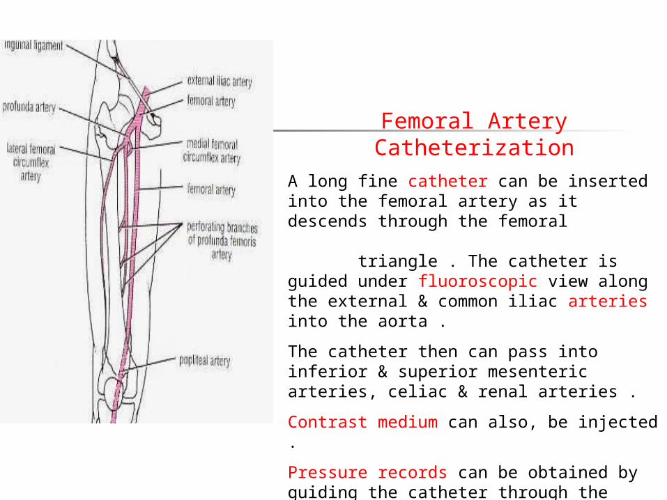
Femoral Artery CatheterizationA long fine catheter can be inserted into the femoral artery as it descends through the femoral triangle . The catheter is guided under fluoroscopic view along the external & common iliac arteries into the aorta .
The catheter then can pass into inferior & superior mesenteric arteries, celiac & renal arteries .
Contrast medium can also, be injected .
Pressure records can be obtained by guiding the catheter through the aortic valve into the left ventricle .
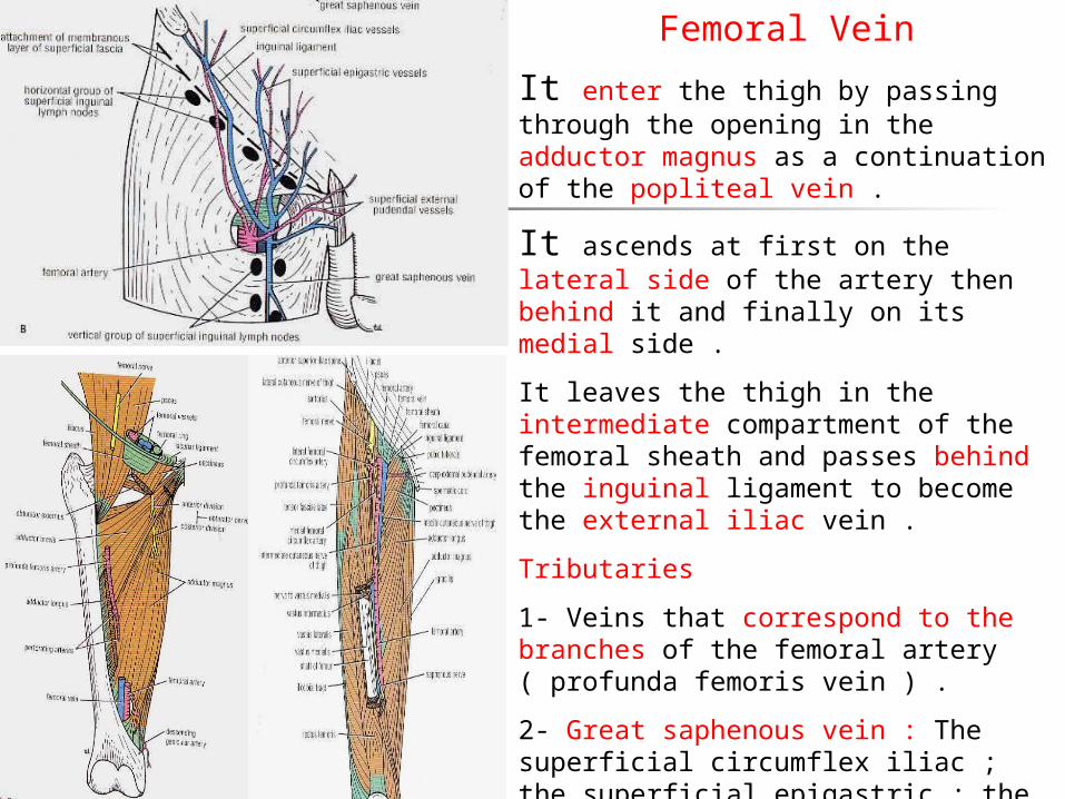
Femoral Vein
It enter the thigh by passing through the opening in the adductor magnus as a continuation of the popliteal vein .
It ascends at first on the lateral side of the artery then behind it and finally on its medial side .
It leaves the thigh in the intermediate compartment of the femoral sheath and passes behind the inguinal ligament to become the external iliac vein .
Tributaries
1- Veins that correspond to the branches of the femoral artery ( profunda femoris vein ) .
2- Great saphenous vein : The superficial circumflex iliac ; the superficial epigastric ; the external pudendal veins drain into it .
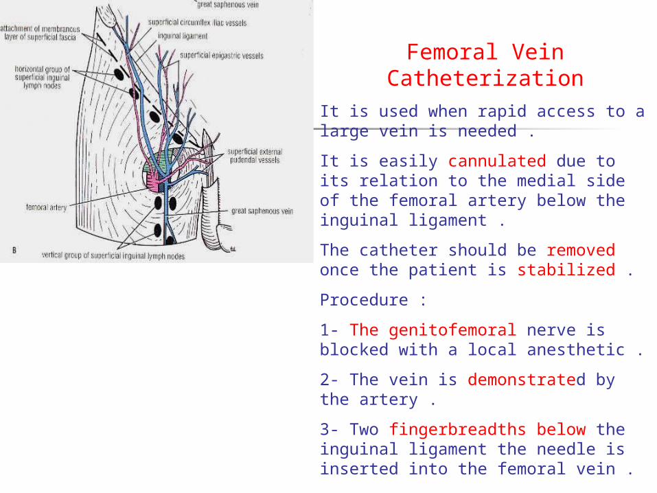
Femoral Vein CatheterizationIt is used when rapid access to a large vein is needed .
It is easily cannulated due to its relation to the medial side of the femoral artery below the inguinal ligament .
The catheter should be removed once the patient is stabilized .
Procedure :
1- The genitofemoral nerve is blocked with a local anesthetic .
2- The vein is demonstrated by the artery .
3- Two fingerbreadths below the inguinal ligament the needle is inserted into the femoral vein .
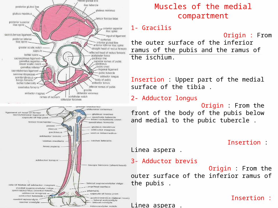
Muscles of the medial compartment
1- Gracilis Origin : From the outer surface of the inferior ramus of the pubis and the ramus of the ischium. Insertion : Upper part of the medial surface of the tibia .
2- Adductor longus Origin : From the front of the body of the pubis below and medial to the pubic tubercle . Insertion : Linea aspera .
3- Adductor brevis Origin : From the outer surface of the inferior ramus of the pubis . Insertion : Linea aspera .
4- Adductor magnus Origin : From the outer surface of the inferior ramus of the pubis ; the ramus of the ischium and the ischial tuberosity Insertion : The adductor portion is attached to the posterior surface of the shaft of the femur . The hamstring portion : On the adductor tubercle.
5- Obturator externus : Origin: From the outer surface of the obturator membrane and pubic & ischial rami . Insertion : Onto the medial surface of the greater trochanter of the femur .
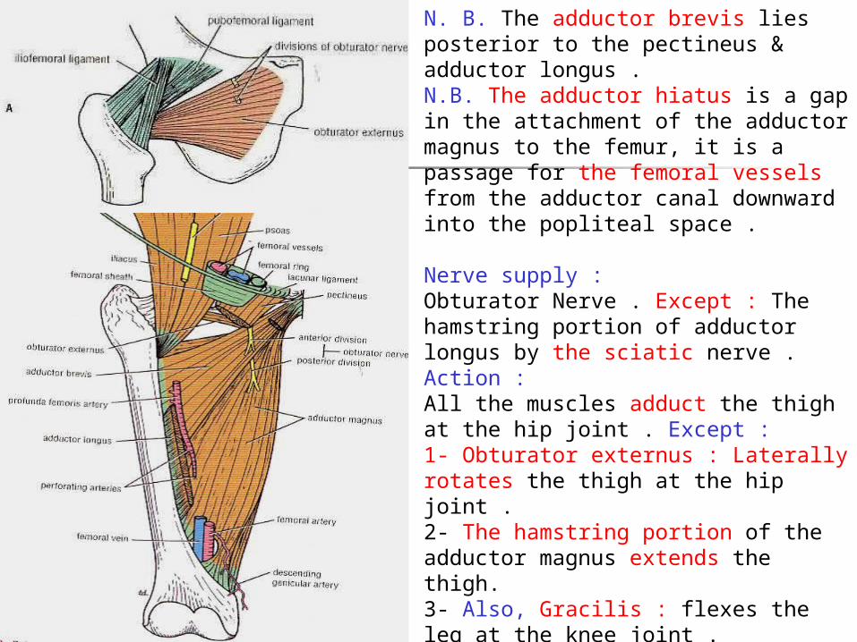
N. B. The adductor brevis lies posterior to the pectineus & adductor longus .N.B. The adductor hiatus is a gap in the attachment of the adductor magnus to the femur, it is a passage for the femoral vessels from the adductor canal downward into the popliteal space .
Nerve supply : Obturator Nerve . Except : The hamstring portion of adductor longus by the sciatic nerve .Action :All the muscles adduct the thigh at the hip joint . Except :1- Obturator externus : Laterally rotates the thigh at the hip joint .2- The hamstring portion of the adductor magnus extends the thigh. 3- Also, Gracilis : flexes the leg at the knee joint .4- Also, The adductor longus & brevis and the adductor part of the magnus . Assist in lateral rotation of the thigh .
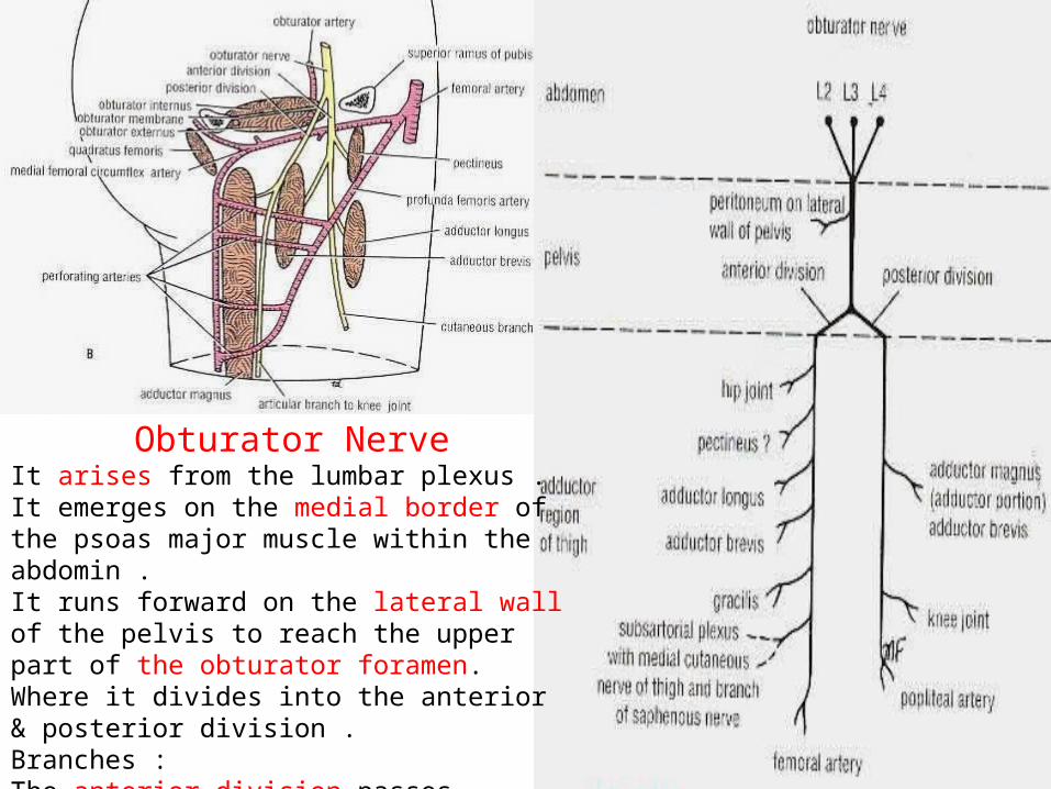
Obturator NerveIt arises from the lumbar plexus . It emerges on the medial border of the psoas major muscle within the abdomin .It runs forward on the lateral wall of the pelvis to reach the upper part of the obturator foramen. Where it divides into the anterior & posterior division .Branches :The anterior division passes downward in front of the obturator externus. The adductor longus.
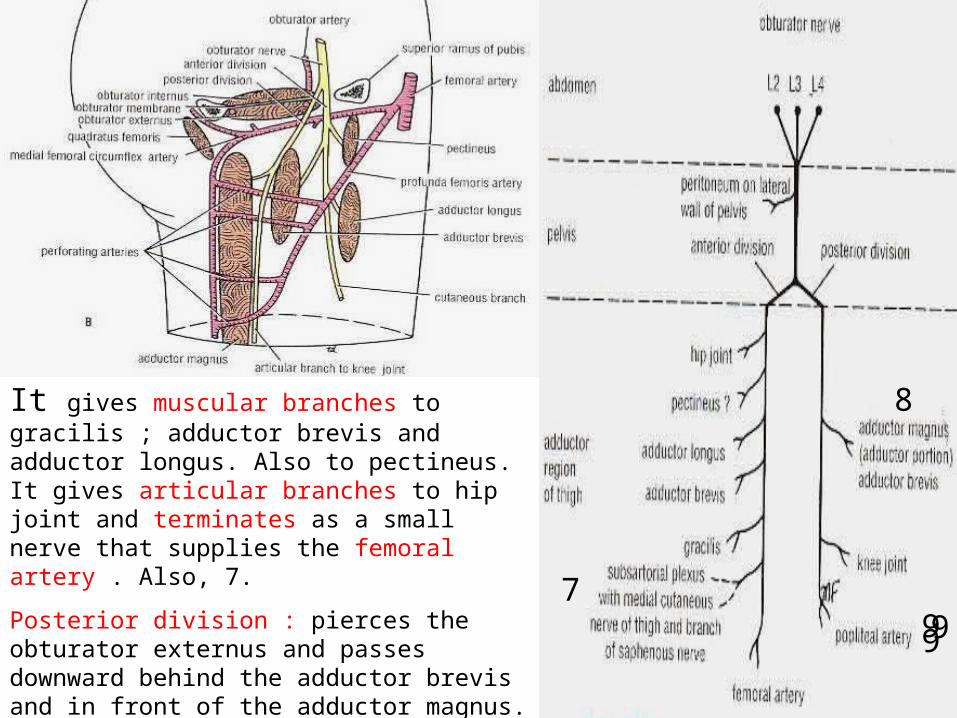
It gives muscular branches to gracilis ; adductor brevis and adductor longus. Also to pectineus. It gives articular branches to hip joint and terminates as a small nerve that supplies the femoral artery . Also, 7.
Posterior division : pierces the obturator externus and passes downward behind the adductor brevis and in front of the adductor magnus. It terminates by descending through the opening in the adductor magnus to supply the knee joint. It supplies obturator externus; 8 ; 9 .
7
8
999
99
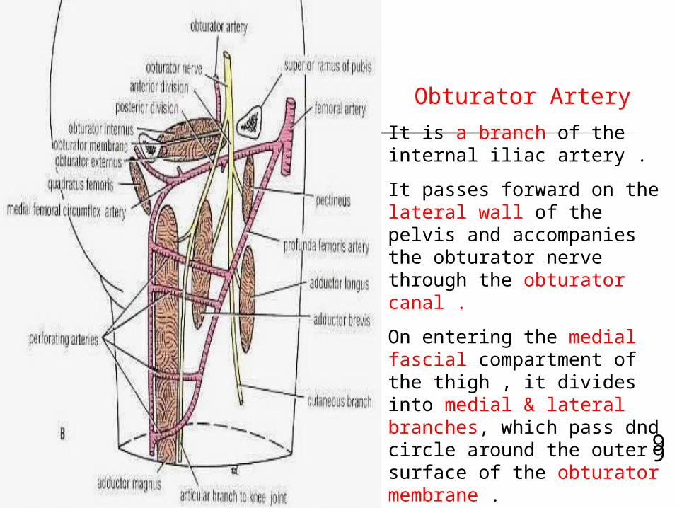
Obturator Artery
It is a branch of the internal iliac artery .
It passes forward on the lateral wall of the pelvis and accompanies the obturator nerve through the obturator canal .
On entering the medial fascial compartment of the thigh , it divides into medial & lateral branches, which pass dnd circle around the outer surface of the obturator membrane .
It gives muscular branches and articular branche to hip joint .