Skin Cancer Management Options - GP CME CME/Friday/C1 0830 McDonald.pdf · Skin Cancer Cell...
70
Skin Cancer Management Options Dr Ken Macdonald Dermatologist/Dermatologic Surgeon
Transcript of Skin Cancer Management Options - GP CME CME/Friday/C1 0830 McDonald.pdf · Skin Cancer Cell...
Skin Cancer Management Options
Dr Ken Macdonald
Dermatologist/Dermatologic Surgeon
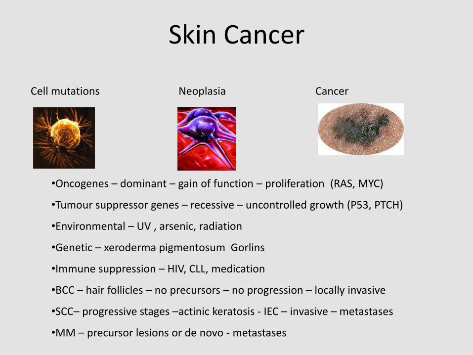
Skin Cancer
Cell mutations Neoplasia Cancer
•Oncogenes – dominant – gain of function – proliferation (RAS, MYC)
•Tumour suppressor genes – recessive – uncontrolled growth (P53, PTCH)
•Environmental – UV , arsenic, radiation
•Genetic – xeroderma pigmentosum Gorlins
•Immune suppression – HIV, CLL, medication
•BCC – hair follicles – no precursors – no progression – locally invasive
•SCC– progressive stages –actinic keratosis - IEC – invasive – metastases
•MM – precursor lesions or de novo - metastases
Skin Cancer – Accurate Diagnosis
•Identify skin cancer – don’t miss skin cancer – think skin cancer •Biopsies do not spread cancer – better to biopsy than not to biopsy •Pigmented lesions can be biopsied -not all pigmented lesions are melanocytic •Biopsies must contain dermal tissue (punch, pyramid, incisional vs shave) •Sampling error - take more than one biopsy of a lesion or excision biopsy •Describe the lesion in detail on the pathology form and note biopsy type •Identify the exact location of the lesion – map it on a diagram •Transport medium – formalin – saline – frozen (cryostat on site) •Most special stains on ‘permanents’. Any doubt get deeper levels cut

Skin Cancer – Histology The Definitive Diagnosis
BCC SCC MM

Skin Cancer – Management options
Physical treatment modalities •Cryosurgery For: -superficial cancer •Curettage -low recurrence risk •Electrosurgery -no metastatic risk Laser ablation (CO² laser) -accurate tissue destruction •Photodynamic therapy -precise targeting -good aesthetic results •Radiotherapy -Definitive, aduvant or palliative treatment
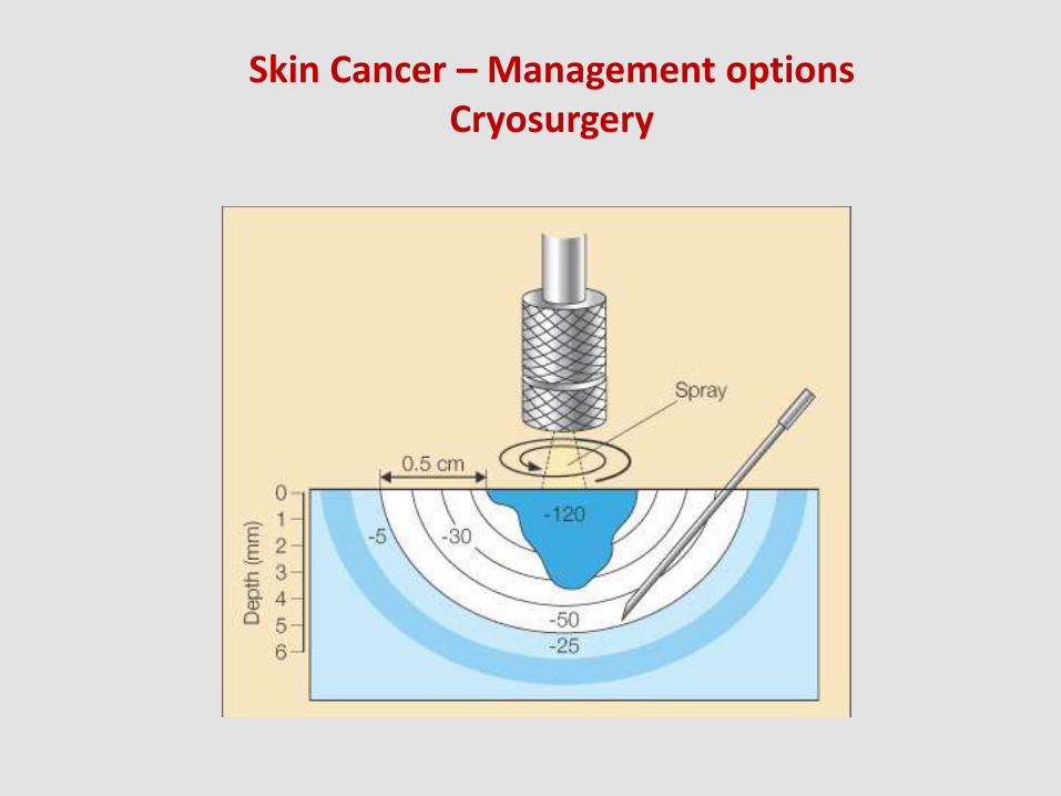
Skin Cancer – Management options Cryosurgery

Skin Cancer – Management options
Cryosurgery (liquid nitrogen @ -196°C) •Spray tip(diameter)– preferred type or probe not cotton bud •Single vs double cycle – fast freeze – slow thaw •Ice field volume – temperature gradient - 60°C required for cancer •Ice crystals – cell wall rupture – osmotic apoptosis – vascular stasis •Morbidity -significant discomfort/pain
-delayed healing after treatment of cancer -hypo-pigmentation (melanocytes sensitive) -scars with more aggressive treatments -nerve/tendon damage/ skin retraction Negatives: Incomplete cancer treatment and lack of histology Positives: Quick and simple, cost effective and predictable.
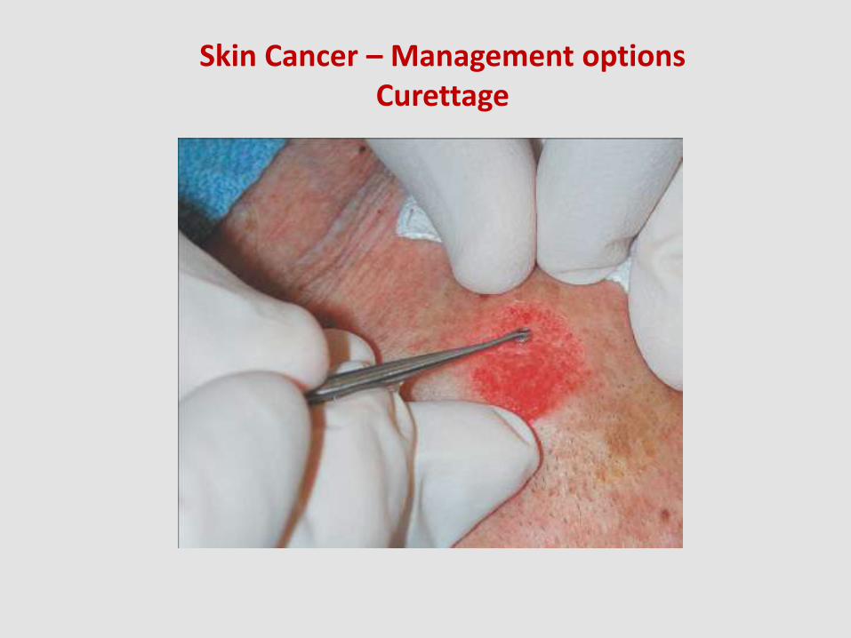
Skin Cancer – Management options Curettage

Skin Cancer – Management options
Curettage -excellent for debulking ‘soft’ tumours -excellent for removing cutaneous debris and preparing for PDT -option for treating superficial basal cell carcinomas and Bowen’s disease (squamous cell carcinoma in situ). Additional electrosurgery required Biopsy material can be saved but fragments may be difficult to interpret
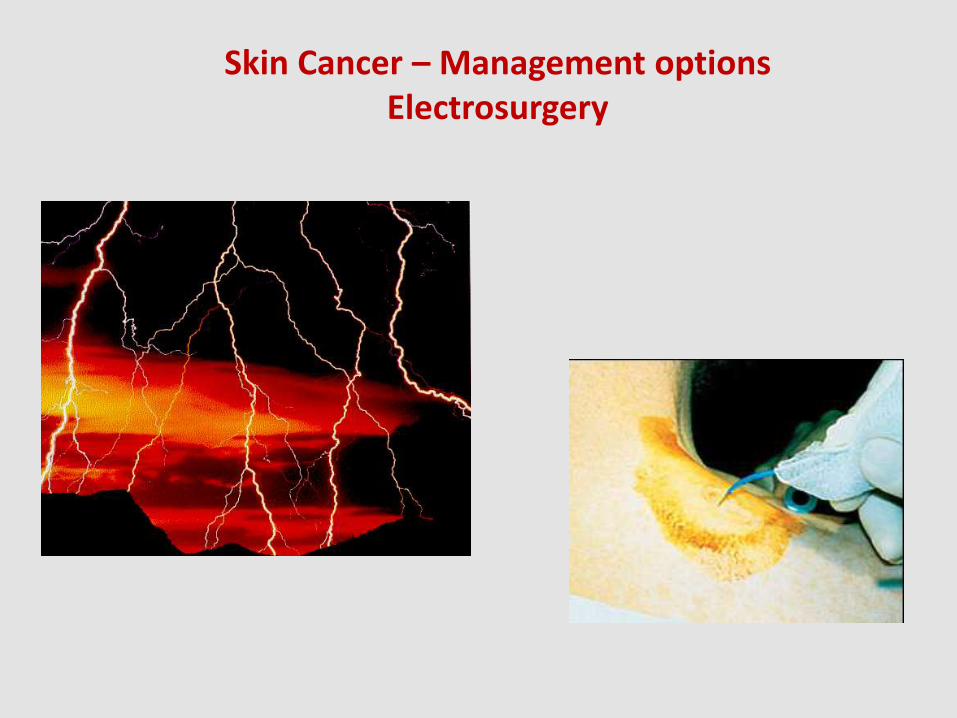
Skin Cancer – Management options Electrosurgery
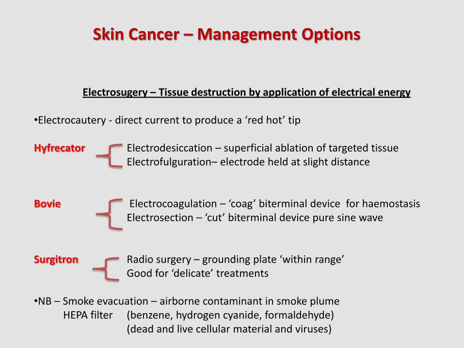
Skin Cancer – Management Options
Electrosugery – Tissue destruction by application of electrical energy •Electrocautery - direct current to produce a ‘red hot’ tip Hyfrecator Electrodesiccation – superficial ablation of targeted tissue Electrofulguration– electrode held at slight distance Bovie Electrocoagulation – ‘coag’ biterminal device for haemostasis Electrosection – ‘cut’ biterminal device pure sine wave Surgitron Radio surgery – grounding plate ‘within range’ Good for ‘delicate’ treatments •NB – Smoke evacuation – airborne contaminant in smoke plume HEPA filter (benzene, hydrogen cyanide, formaldehyde) (dead and live cellular material and viruses)

Skin Cancer – Management Options Laser Ablation
Laser resurfacing for ‘field change’ superficial cancer and actinic dermatitis •CO² laser collimated hand piece, Ultrapulse mode -good visual ablation -precise tissue removal -haemostasis immediate •Disadvantages -possible scarring and hypopigmentation -no histology to confirm tumour removal -recurrance from follicular epithelium -smoke plume requires evacuation
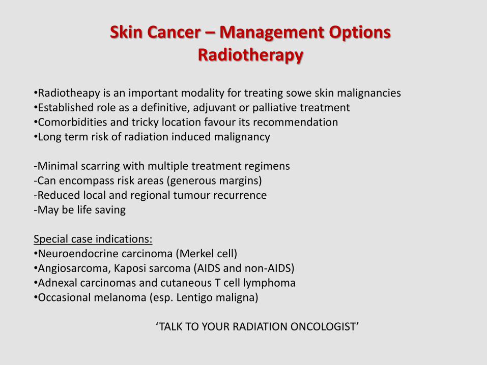
Skin Cancer – Management Options Radiotherapy
•Radiotheapy is an important modality for treating sowe skin malignancies •Established role as a definitive, adjuvant or palliative treatment •Comorbidities and tricky location favour its recommendation •Long term risk of radiation induced malignancy
-Minimal scarring with multiple treatment regimens -Can encompass risk areas (generous margins) -Reduced local and regional tumour recurrence -May be life saving Special case indications: •Neuroendocrine carcinoma (Merkel cell) •Angiosarcoma, Kaposi sarcoma (AIDS and non-AIDS) •Adnexal carcinomas and cutaneous T cell lymphoma •Occasional melanoma (esp. Lentigo maligna)
‘TALK TO YOUR RADIATION ONCOLOGIST’

Photodynamic Therapy (PDT)
ADVANTAGES -No surgical excision -Excellent cosmetic result -Large areas can be treated
DISADVANTAGES -Uncomfortable -Recurrence of cancer not uncommon -Repeat treatments may be required -Inadequate drugs/light delivery at depth -Not for invasive BCC or SCC or MM
•Lesions are prepared and ALA or methyl ALA is applied. •After 3 hours placed under red LED light for approx 7 minutes •Specific responses at cellular level – iatrogenic porphyria •Response depends on pattern of tissue localisation of photosensitizer
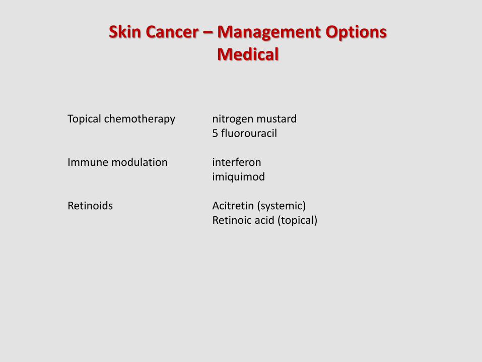
Skin Cancer – Management Options Medical
Topical chemotherapy nitrogen mustard 5 fluorouracil Immune modulation interferon imiquimod Retinoids Acitretin (systemic) Retinoic acid (topical)
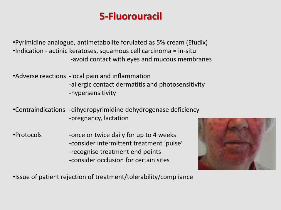
5-Fluorouracil
•Pyrimidine analogue, antimetabolite forulated as 5% cream (Efudix) •Indication - actinic keratoses, squamous cell carcinoma = in-situ -avoid contact with eyes and mucous membranes •Adverse reactions -local pain and inflammation -allergic contact dermatitis and photosensitivity -hypersensitivity •Contraindications -dihydropyrimidine dehydrogenase deficiency -pregnancy, lactation •Protocols -once or twice daily for up to 4 weeks -consider intermittent treatment ‘pulse’ -recognise treatment end points -consider occlusion for certain sites •Issue of patient rejection of treatment/tolerability/compliance
Immunomodulators Imiquimod
•Commercially available as a 5% cream (Aldara) •Approved for superficial basal cell carcinoma, but also effective for flat actinic keratoses •Has shown efficacy for lentigo maligna, SCC insitu and extramammary Paget’s disease •Not evaluated for sBCC within 1cm of eyes, nose, mouth or ears •Immune response modifier stimulates innate immunity via INF α and TNF α •Induces targeted cell mediated immunity •Percutaneous absorption is minimal •No reports of systemic immune alteration •Not contraindicated in organ transplant recipients •Has efficacy in patients on immunesuppressive therapies and with HIV •SBCC clearance rates (70-75%) are less than for surgery •Subset of non-responders (TLR 7 deficiency or impairment) •Pregnancy category B. Not known to be excreted in breast milk

Immunomodulators Imiquimod
•Superficial BCC diagnosis – digital pressure with skin spreading to demonstrate thread – like margin. Biopsy confirmation but avoid areas of regression •Superficial BCCs can resemble SCC in situ but can also occasionally minic amelanotic radial growth phase melanoma – biopsy important •Biopsy sampling error may occur when there is non uniform histology. (Important reason for treatment failure in the large ‘sBCC’ Aldara trails.) •Some patients react vigorously to Aldara. Crusting and skin erosion may occur after only the first 5 days. Treatment must be withheld until skin heals ‘rest days’ essential •Excessive inflammation and super infection can cause ulceration and scarring with permanent loss of pigmentation (esp. In sun damaged skin sites) – stop treatment •A small subset of patients will suffer severe ‘flu-like symptoms and may become depressed and suicidal - beware •Good phone support, patient information and knowledge able nursing staff essential •All patients must have long term follow up
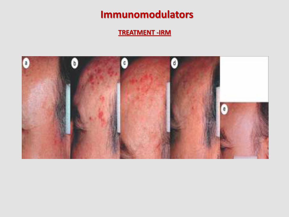
TREATMENT -IRM
Immunomodulators
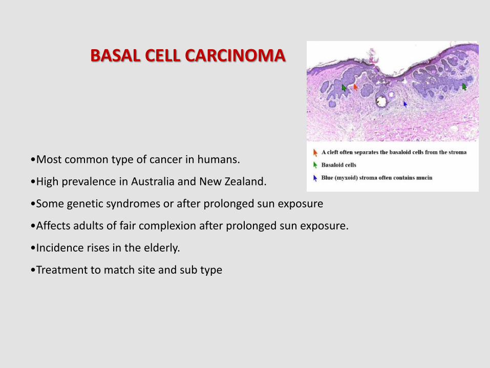
BASAL CELL CARCINOMA
•Most common type of cancer in humans.
•High prevalence in Australia and New Zealand.
•Some genetic syndromes or after prolonged sun exposure
•Affects adults of fair complexion after prolonged sun exposure.
•Incidence rises in the elderly.
•Treatment to match site and sub type
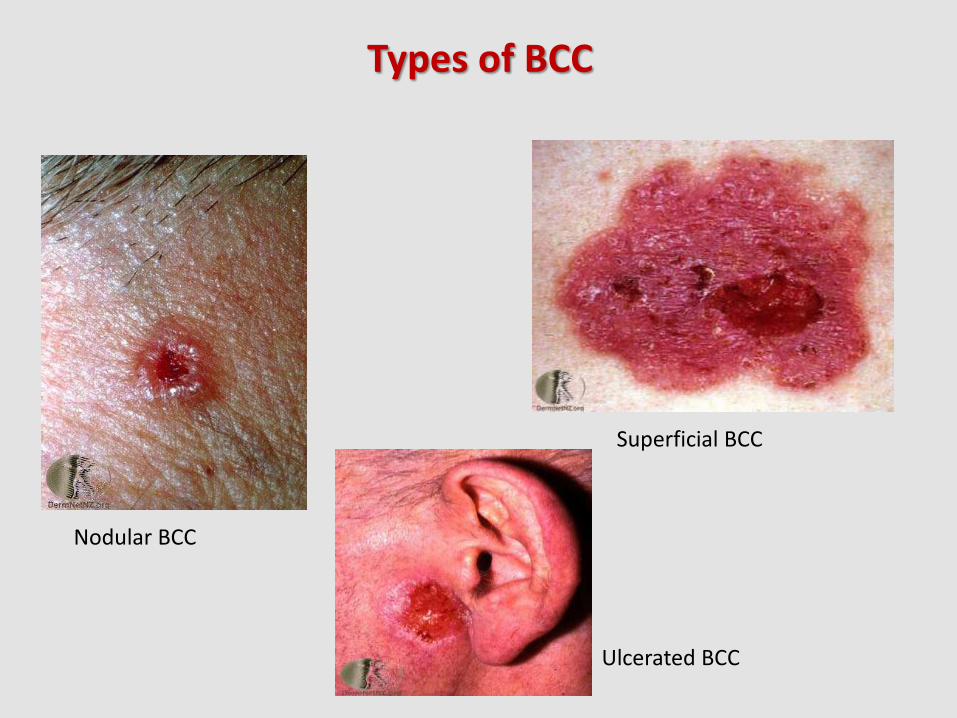
Types of BCC
Nodular BCC
Superficial BCC
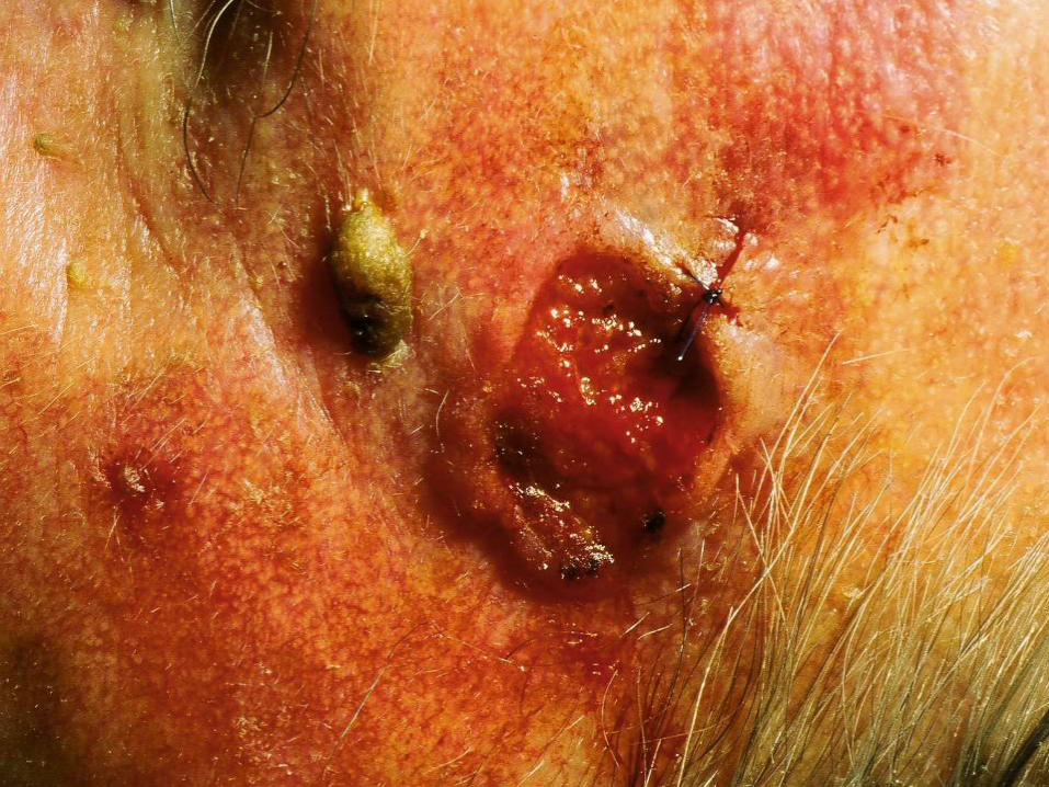
Ulcerated BCC
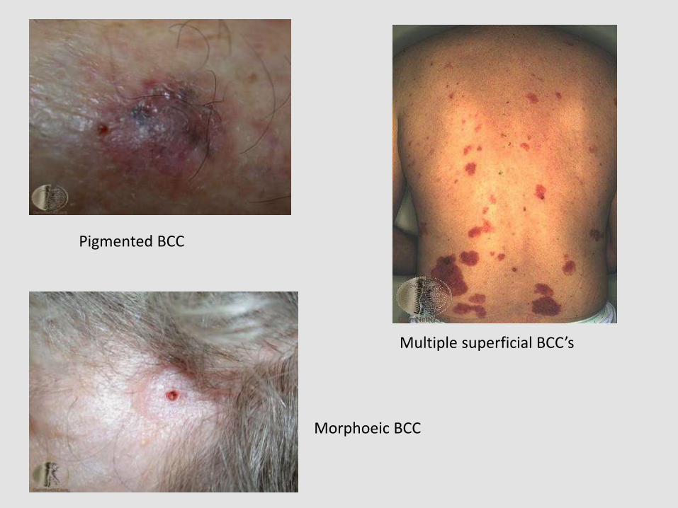
Pigmented BCC
Multiple superficial BCC’s
Morphoeic BCC
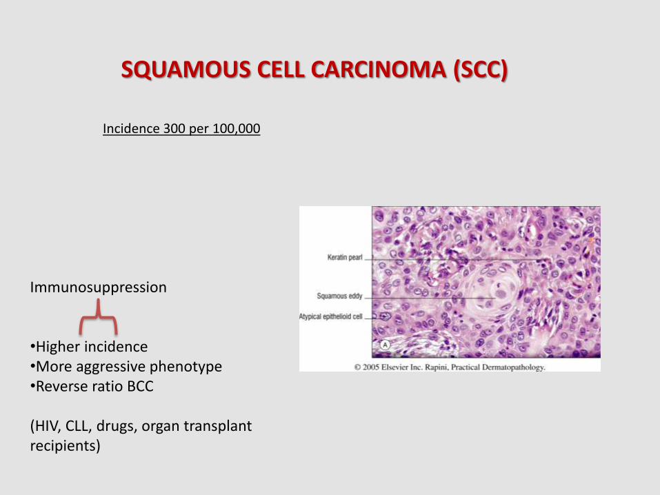
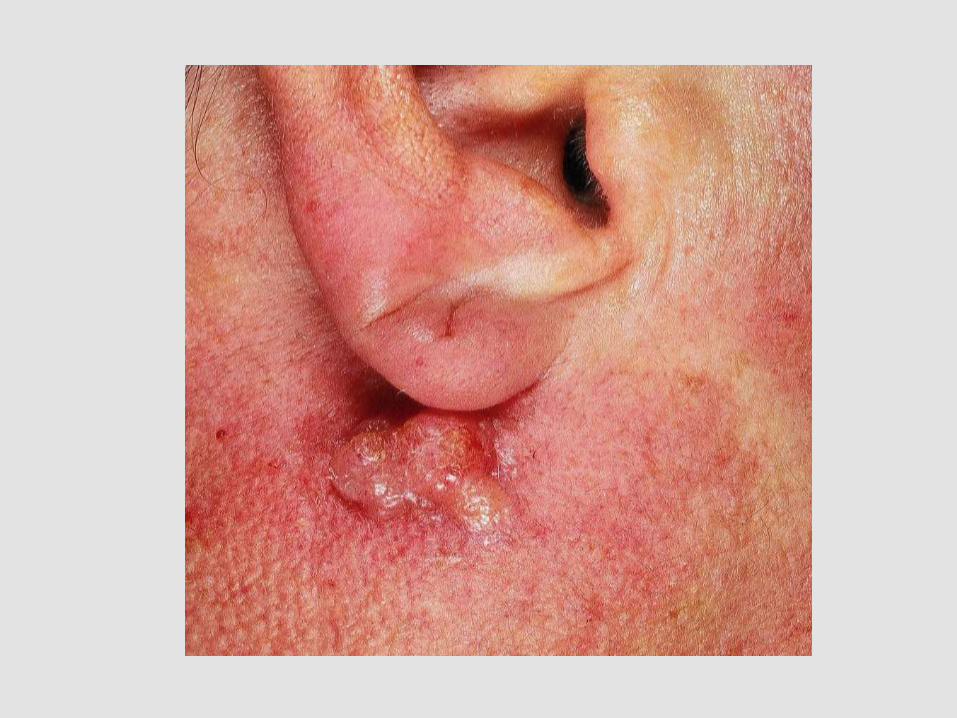
SQUAMOUS CELL CARCINOMA (SCC)
Incidence 300 per 100,000
Immunosuppression •Higher incidence •More aggressive phenotype •Reverse ratio BCC (HIV, CLL, drugs, organ transplant recipients)
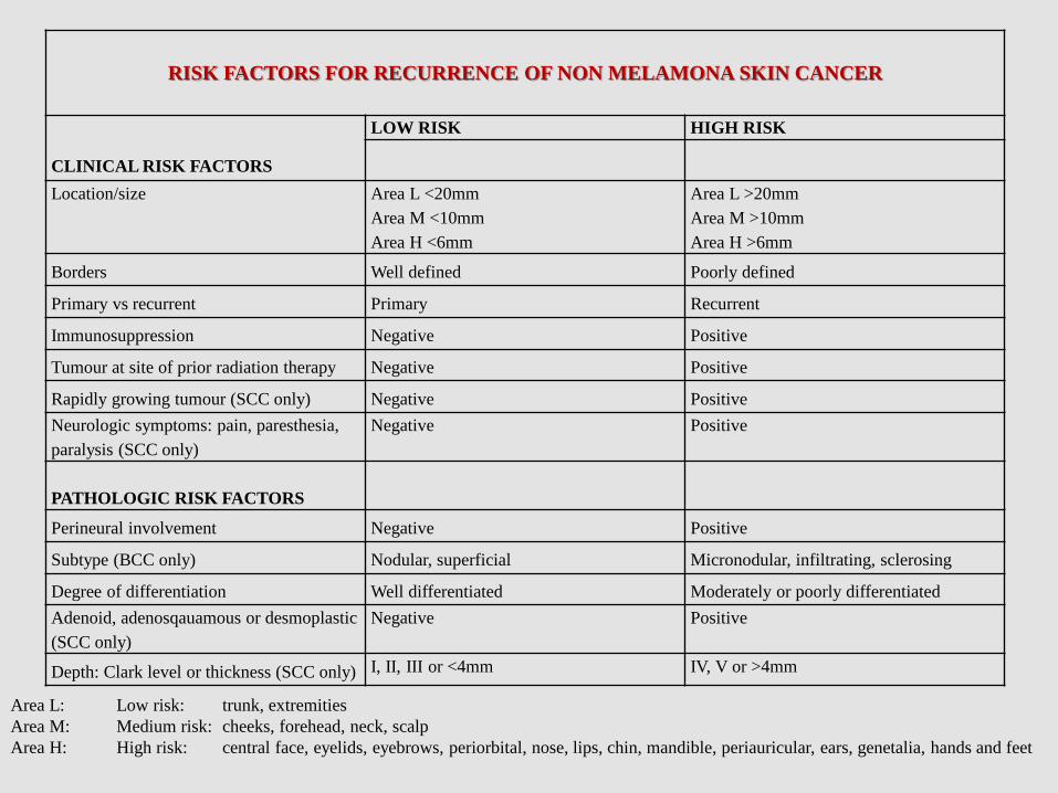
RISK FACTORS FOR RECURRENCE OF NON MELAMONA SKIN CANCER
LOW RISK HIGH RISK
CLINICAL RISK FACTORS
Location/size Area L <20mm Area L >20mm
Area M <10mm Area M >10mm
Area H <6mm Area H >6mm
Borders Well defined Poorly defined
Primary vs recurrent Primary Recurrent
Immunosuppression Negative Positive
Tumour at site of prior radiation therapy Negative Positive
Rapidly growing tumour (SCC only) Negative Positive
Neurologic symptoms: pain, paresthesia,
paralysis (SCC only)
Negative Positive
PATHOLOGIC RISK FACTORS
Perineural involvement Negative Positive
Subtype (BCC only) Nodular, superficial Micronodular, infiltrating, sclerosing
Degree of differentiation Well differentiated Moderately or poorly differentiated
Adenoid, adenosqauamous or desmoplastic
(SCC only)
Negative Positive
Depth: Clark level or thickness (SCC only) I, II, III or <4mm IV, V or >4mm
Area L: Low risk: trunk, extremities
Area M: Medium risk: cheeks, forehead, neck, scalp
Area H: High risk: central face, eyelids, eyebrows, periorbital, nose, lips, chin, mandible, periauricular, ears, genetalia, hands and feet

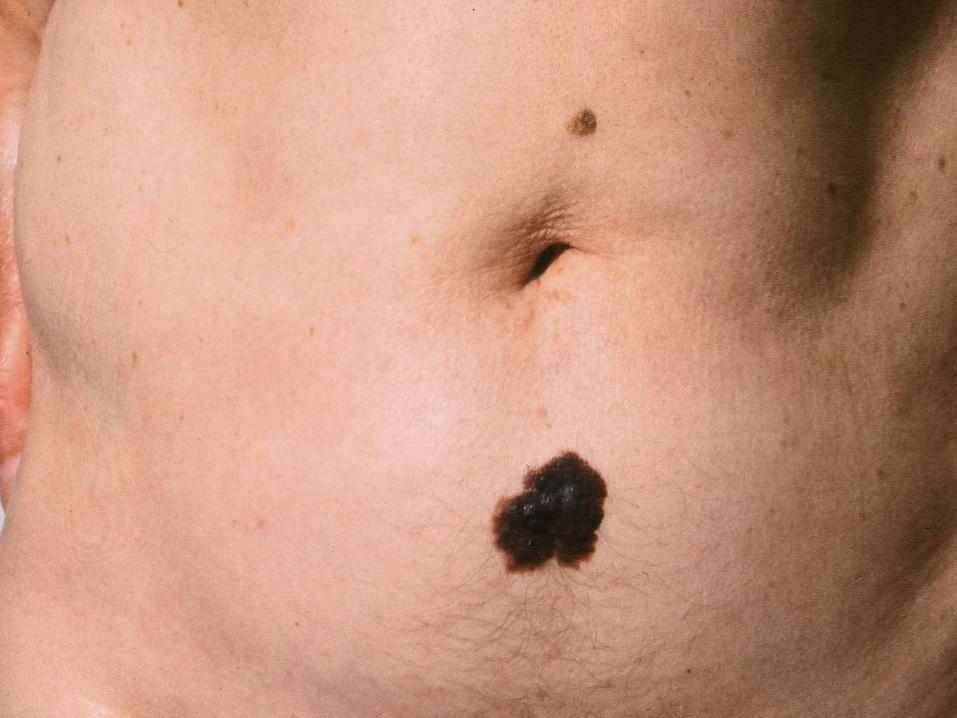
•Each year 2000 New Zealanders are diagnosed. •New Zealand has the highest melanoma rate in the world •90% are cured by early surgical management •Melanoma is associated with fair skin types, high mole count and UV expsoure
Melanoma in New Zealand
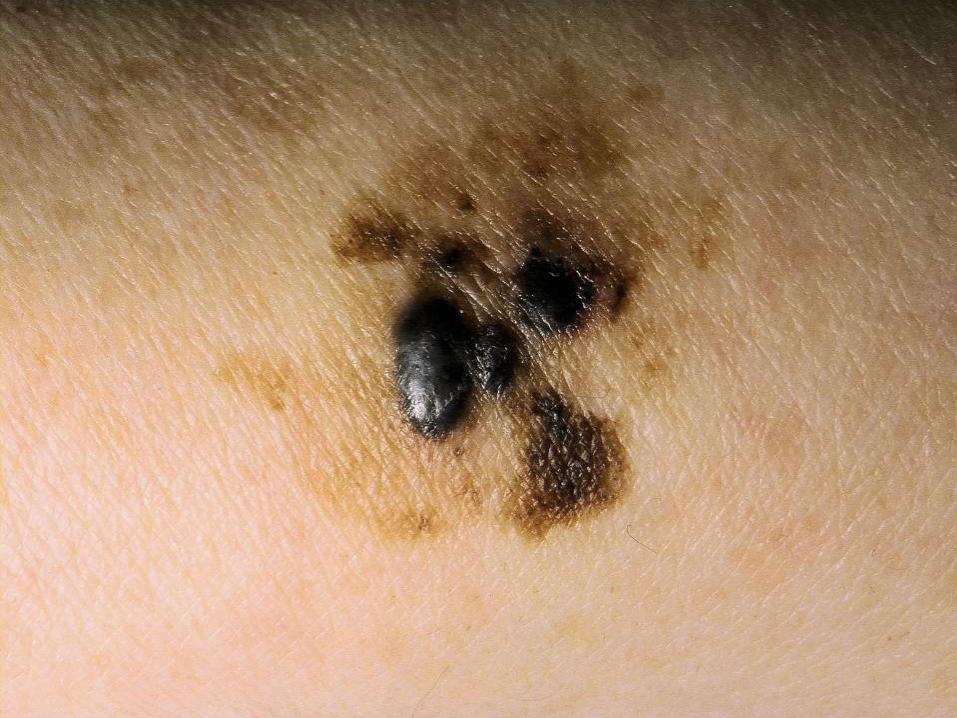
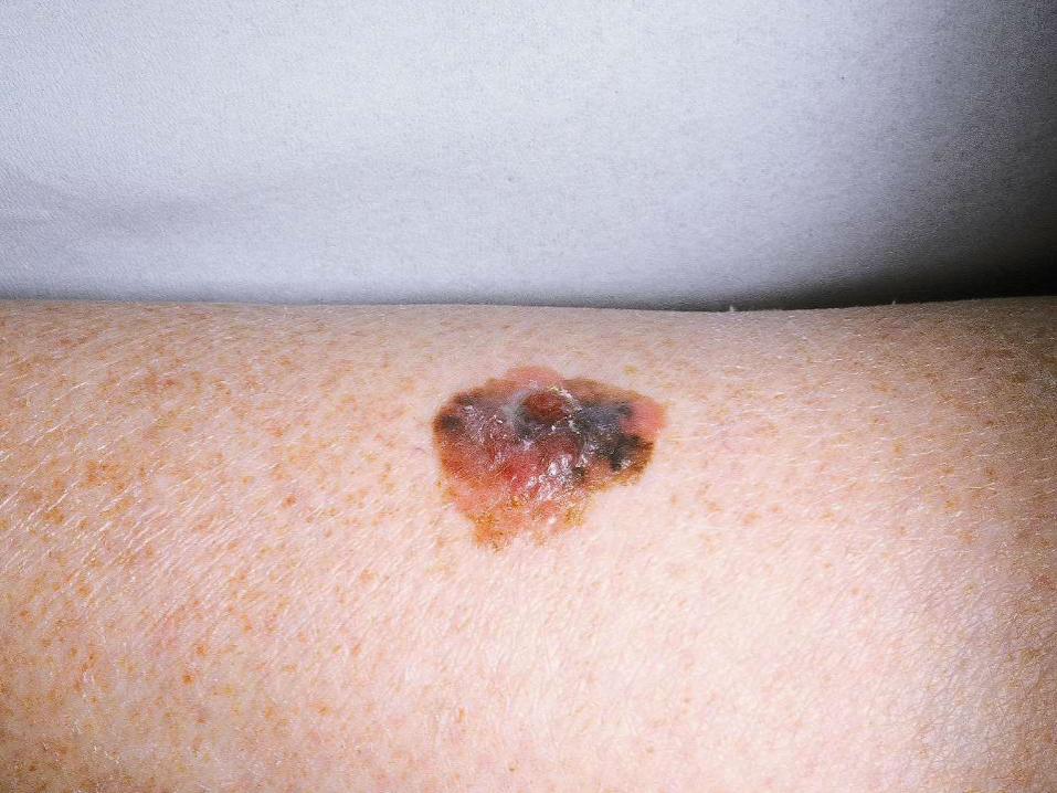
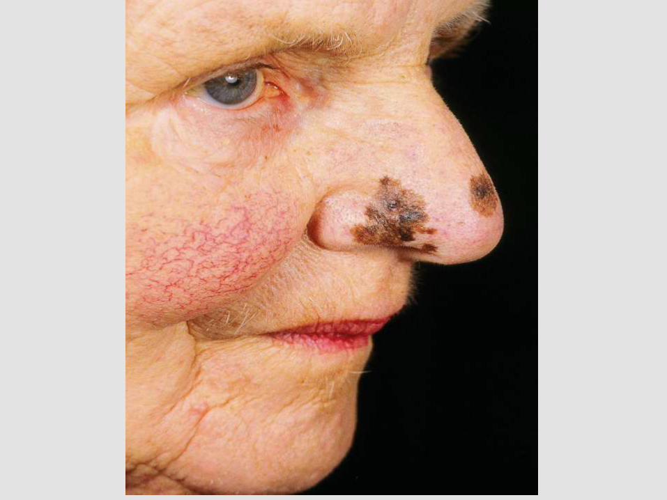
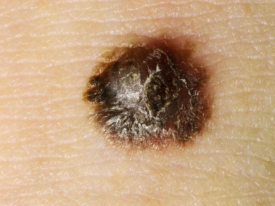
•Melanoma precursor lesions – dysplastic nevi, cellular blue nevi, atypical melanocytic hyperplasia, atypical lentiginous naevus • Melanomas are described according to their appearance and behaviour •Initial horizontal growth phase lesions include: •Superficial spreading melanoma •Lentigo maligna melanoma (sun damaged skin of face, scalp and neck) •Acral lentiginous melanoma (on soles of feet, palms of hands or subungual) •Amelanotic macular melanoma •ABCD acronym •Melanomas sub types that rapidly invade deeper tissues with early vertical growth phase include: •Nodular melanoma (presenting as a rapidly enlarging lump) may be a melanotic •Mucosal melanoma (arising on lips, eyelids, vulva, penis, anus) •Desmoplastic melanoma (fibrous tumour with a tendency to grow down nerves) •Nevoid melanoma (indeterminate papule) •EFG acronym
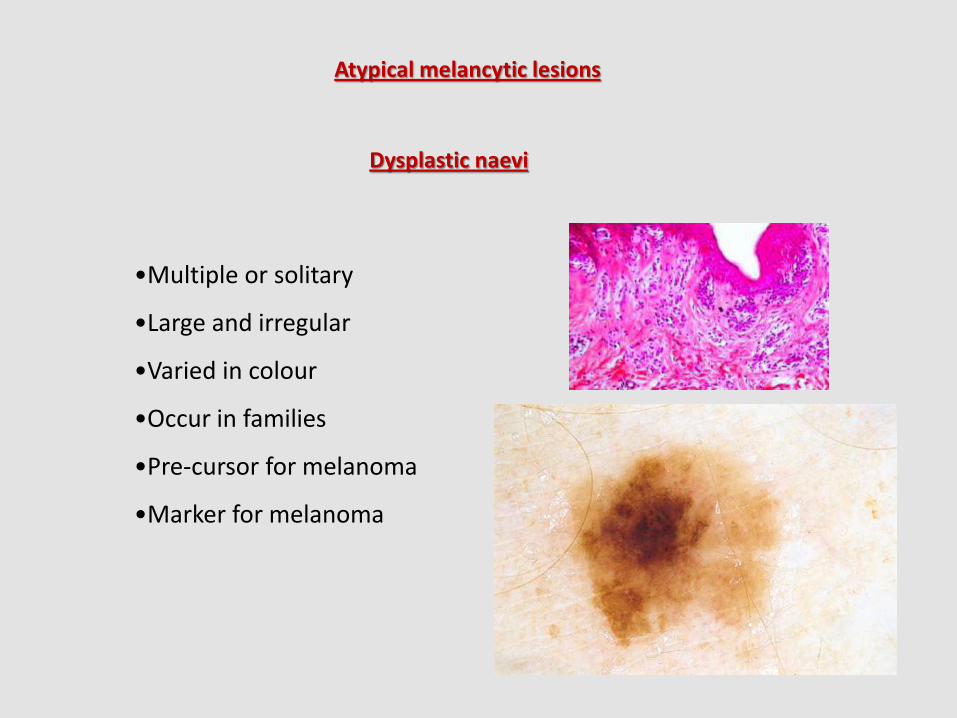
Atypical melancytic lesions
Dysplastic naevi
•Multiple or solitary
•Large and irregular
•Varied in colour
•Occur in families
•Pre-cursor for melanoma
•Marker for melanoma
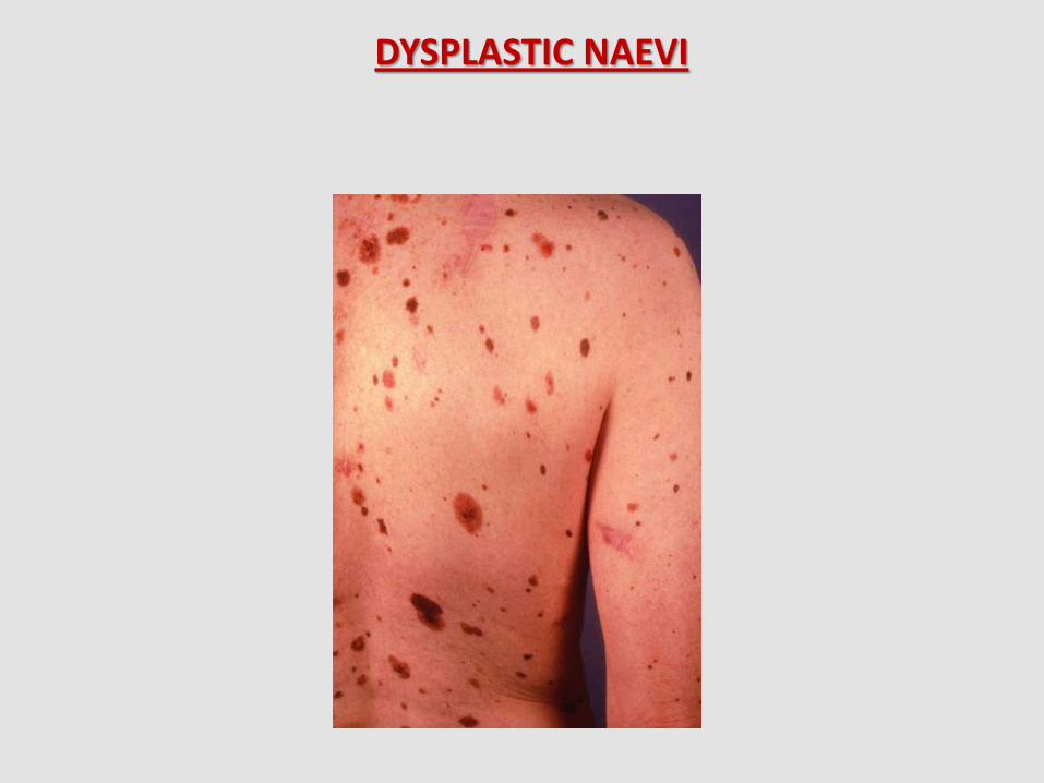
DYSPLASTIC NAEVI
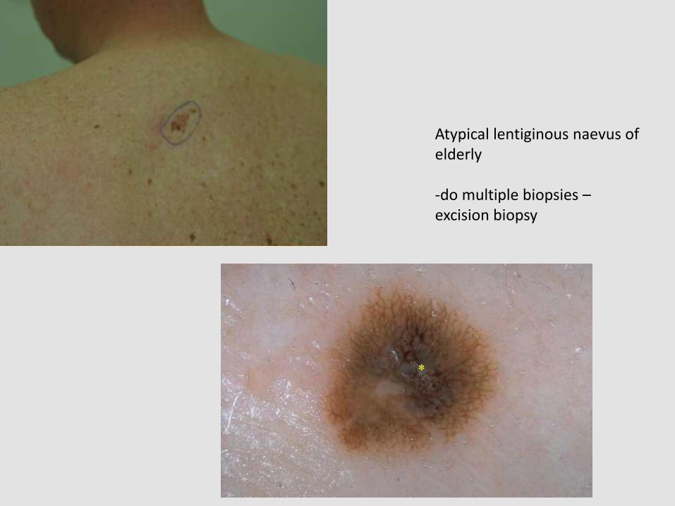
Atypical lentiginous naevus of elderly -do multiple biopsies – excision biopsy
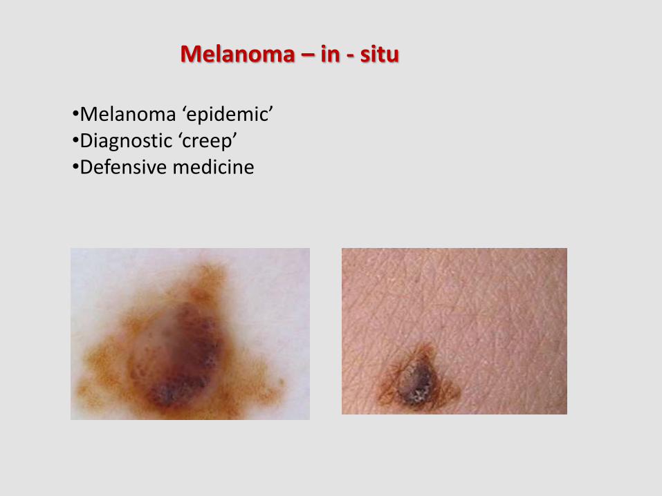
Melanoma – in - situ
•Melanoma ‘epidemic’ •Diagnostic ‘creep’ •Defensive medicine
Skin Cancer – Treatment Options - Surgical
Cancer which may be suitable for surgery in the general practice context •Small < 1cm diameter, well defined NMSC on the trunk and limbs •To melanomas on trunk and limbs -surgical treatment protocols strictly followed •Larger NMSCon trunk and limbs and facial lesions depending on skill level General practitioners with an interest in skin cancer surgery should: -be practiced in simple skin surgery and understand excision margins -be able to perform basic random pattern skin flaps and grafts -meet patient’s expectations with regard to skin cancer clearance and aesthetics -have good collegial relationships in place. -have low threshold for referring onto experienced colleagues Be well prepared for audit – poor outcomes will happen
‘Keep it simple and sensible’
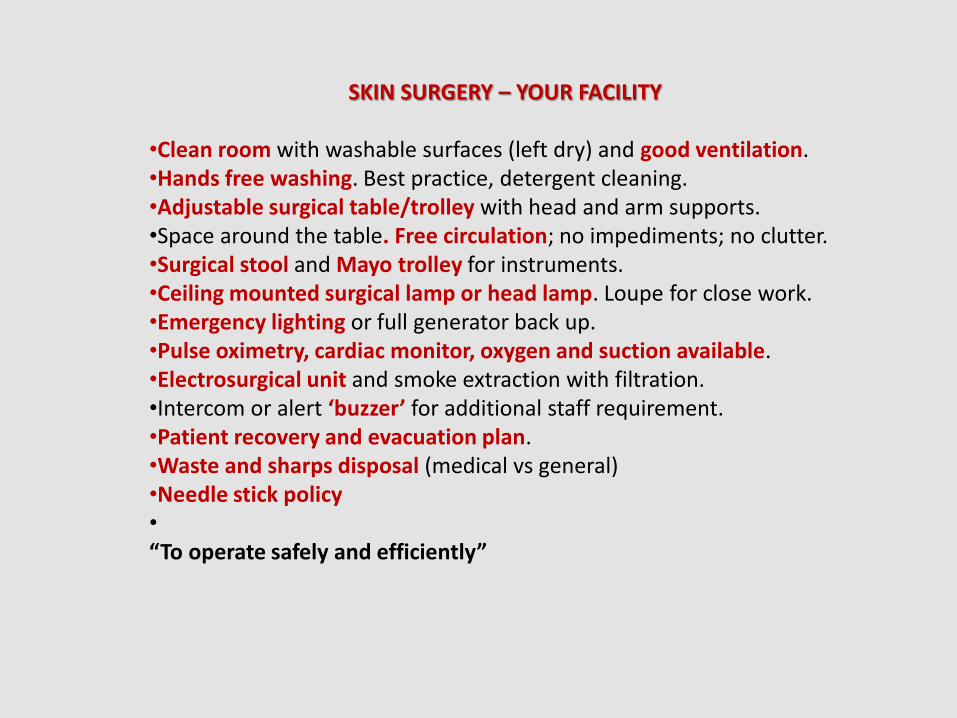
SKIN SURGERY – YOUR FACILITY •Clean room with washable surfaces (left dry) and good ventilation. •Hands free washing. Best practice, detergent cleaning. •Adjustable surgical table/trolley with head and arm supports. •Space around the table. Free circulation; no impediments; no clutter. •Surgical stool and Mayo trolley for instruments. •Ceiling mounted surgical lamp or head lamp. Loupe for close work. •Emergency lighting or full generator back up. •Pulse oximetry, cardiac monitor, oxygen and suction available. •Electrosurgical unit and smoke extraction with filtration. •Intercom or alert ‘buzzer’ for additional staff requirement. •Patient recovery and evacuation plan. •Waste and sharps disposal (medical vs general) •Needle stick policy • “To operate safely and efficiently”
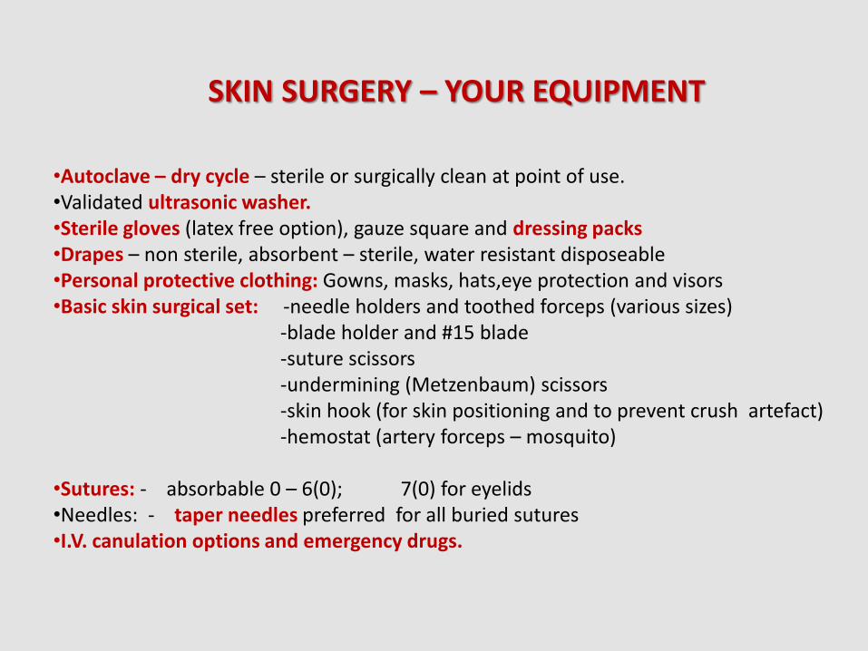
SKIN SURGERY – YOUR EQUIPMENT •Autoclave – dry cycle – sterile or surgically clean at point of use. •Validated ultrasonic washer. •Sterile gloves (latex free option), gauze square and dressing packs •Drapes – non sterile, absorbent – sterile, water resistant disposeable •Personal protective clothing: Gowns, masks, hats,eye protection and visors •Basic skin surgical set: -needle holders and toothed forceps (various sizes)
-blade holder and #15 blade -suture scissors -undermining (Metzenbaum) scissors -skin hook (for skin positioning and to prevent crush artefact) -hemostat (artery forceps – mosquito)
•Sutures: - absorbable 0 – 6(0); 7(0) for eyelids •Needles: - taper needles preferred for all buried sutures •I.V. canulation options and emergency drugs.
SKIN SURGERY – YOUR PATIENT
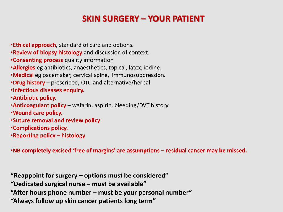
•Ethical approach, standard of care and options. •Review of biopsy histology and discussion of context. •Consenting process quality information •Allergies eg antibiotics, anaesthetics, topical, latex, iodine. •Medical eg pacemaker, cervical spine, immunosuppression. •Drug history – prescribed, OTC and alternative/herbal •Infectious diseases enquiry. •Antibiotic policy. •Anticoagulant policy – wafarin, aspirin, bleeding/DVT history •Wound care policy. •Suture removal and review policy •Complications policy. •Reporting policy – histology •NB completely excised ‘free of margins’ are assumptions – residual cancer may be missed.
“Reappoint for surgery – options must be considered” “Dedicated surgical nurse – must be available” “After hours phone number – must be your personal number” “Always follow up skin cancer patients long term”
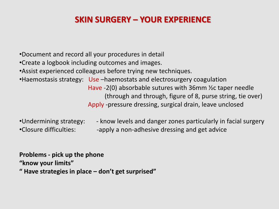
SKIN SURGERY – YOUR EXPERIENCE •Document and record all your procedures in detail •Create a logbook including outcomes and images. •Assist experienced colleagues before trying new techniques. •Haemostasis strategy: Use –haemostats and electrosurgery coagulation
Have -2(0) absorbable sutures with 36mm ½c taper needle (through and through, figure of 8, purse string, tie over) Apply -pressure dressing, surgical drain, leave unclosed
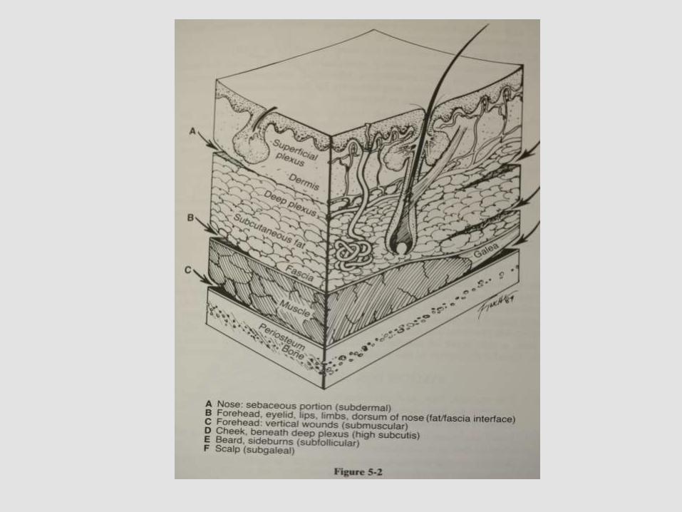
•Undermining strategy: - know levels and danger zones particularly in facial surgery •Closure difficulties: -apply a non-adhesive dressing and get advice Problems - pick up the phone “know your limits” “ Have strategies in place – don’t get surprised”
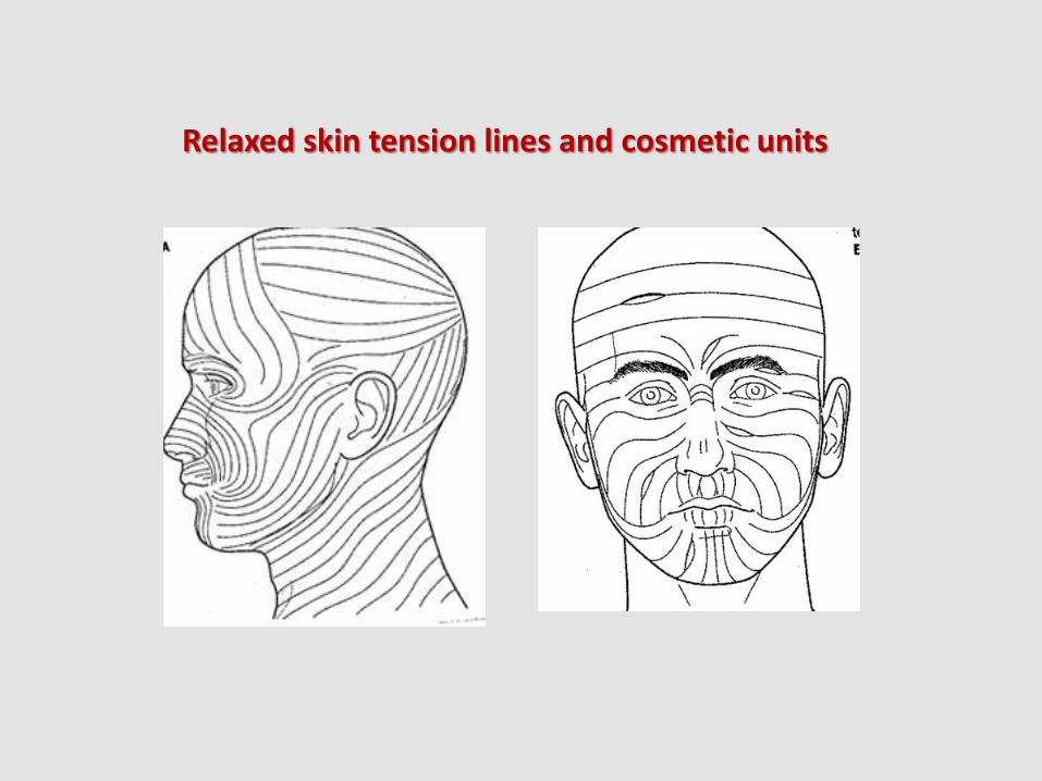
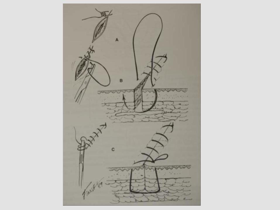
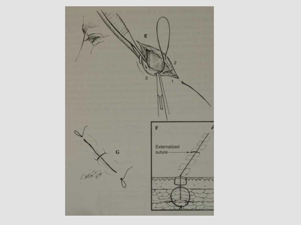
Relaxed skin tension lines and cosmetic units
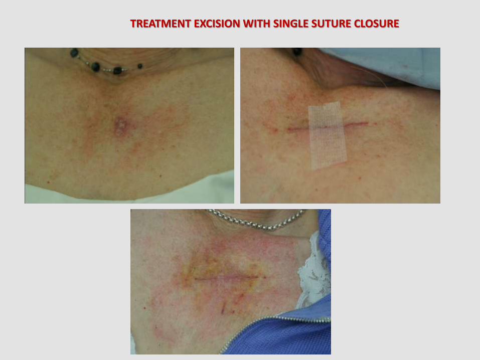
TREATMENT EXCISION WITH SINGLE SUTURE CLOSURE
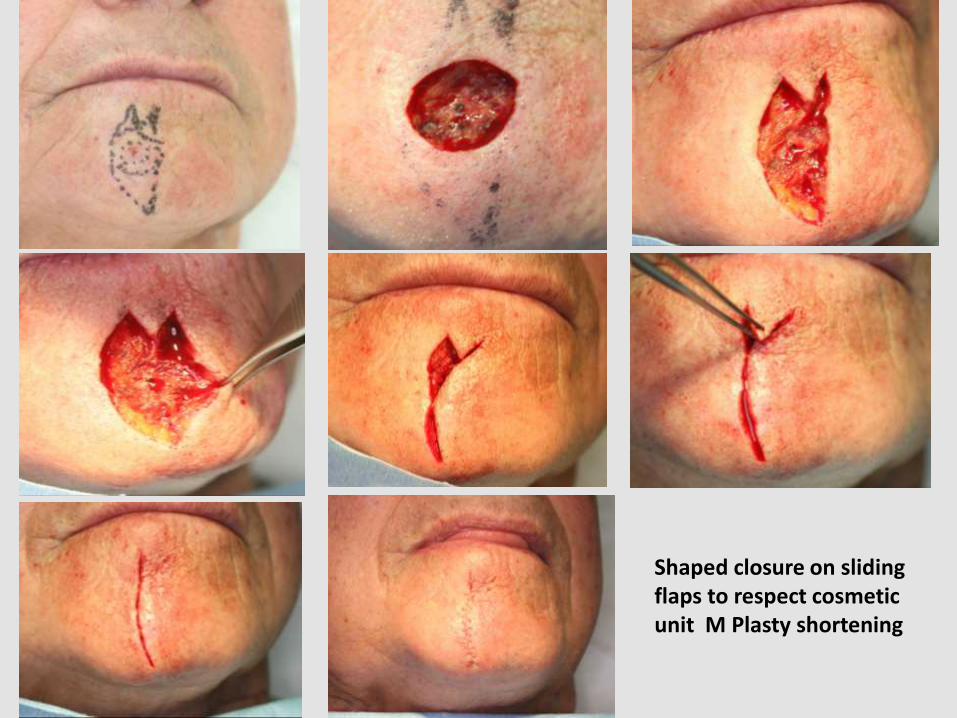
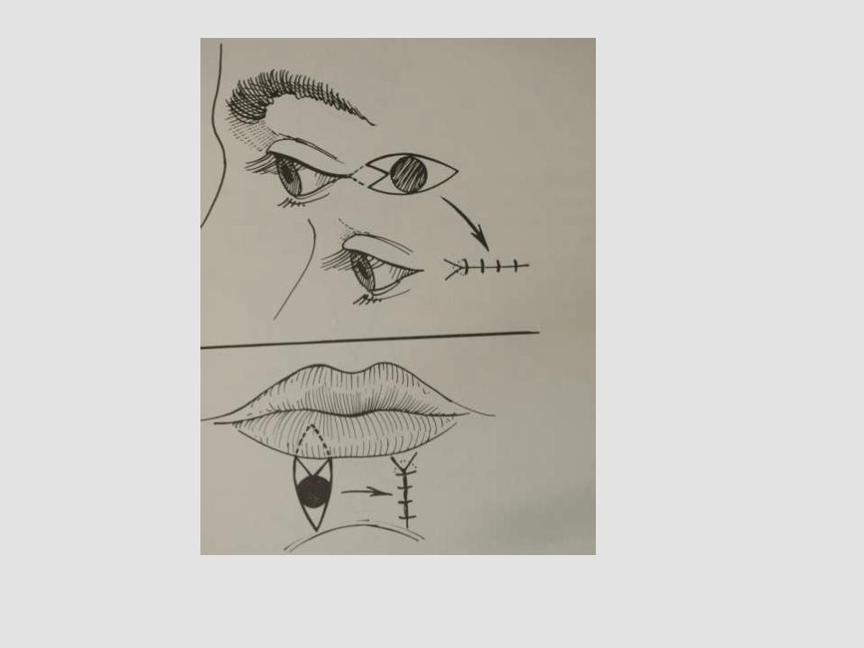
Shaped closure on sliding flaps to respect cosmetic unit M Plasty shortening
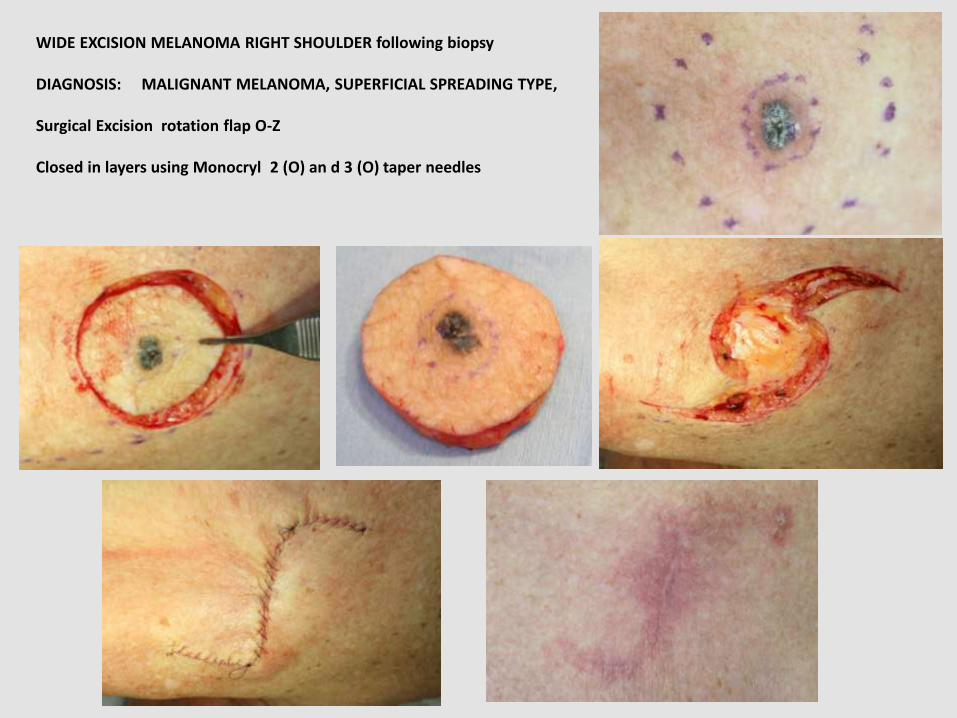
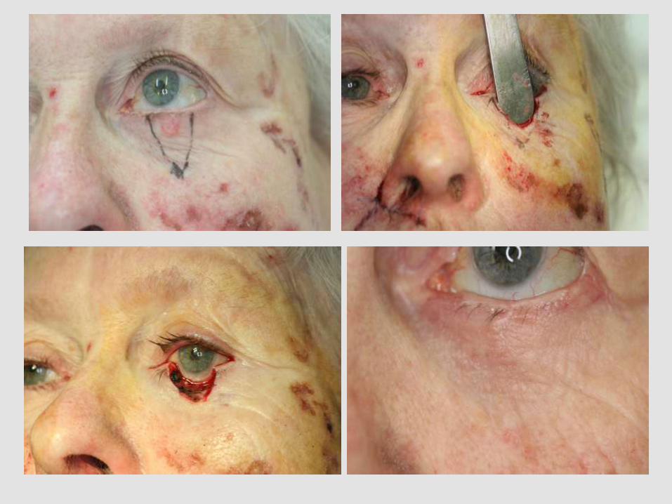
WIDE EXCISION MELANOMA RIGHT SHOULDER following biopsy DIAGNOSIS: MALIGNANT MELANOMA, SUPERFICIAL SPREADING TYPE, Surgical Excision rotation flap O-Z Closed in layers using Monocryl 2 (O) an d 3 (O) taper needles
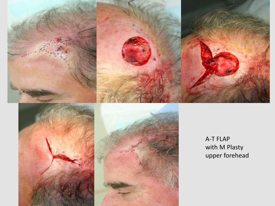
A-T FLAP with M Plasty upper forehead
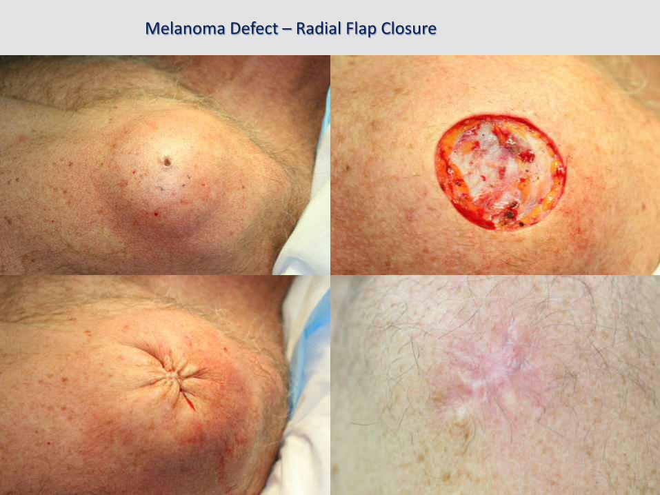
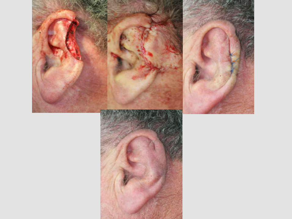
Melanoma Defect – Radial Flap Closure
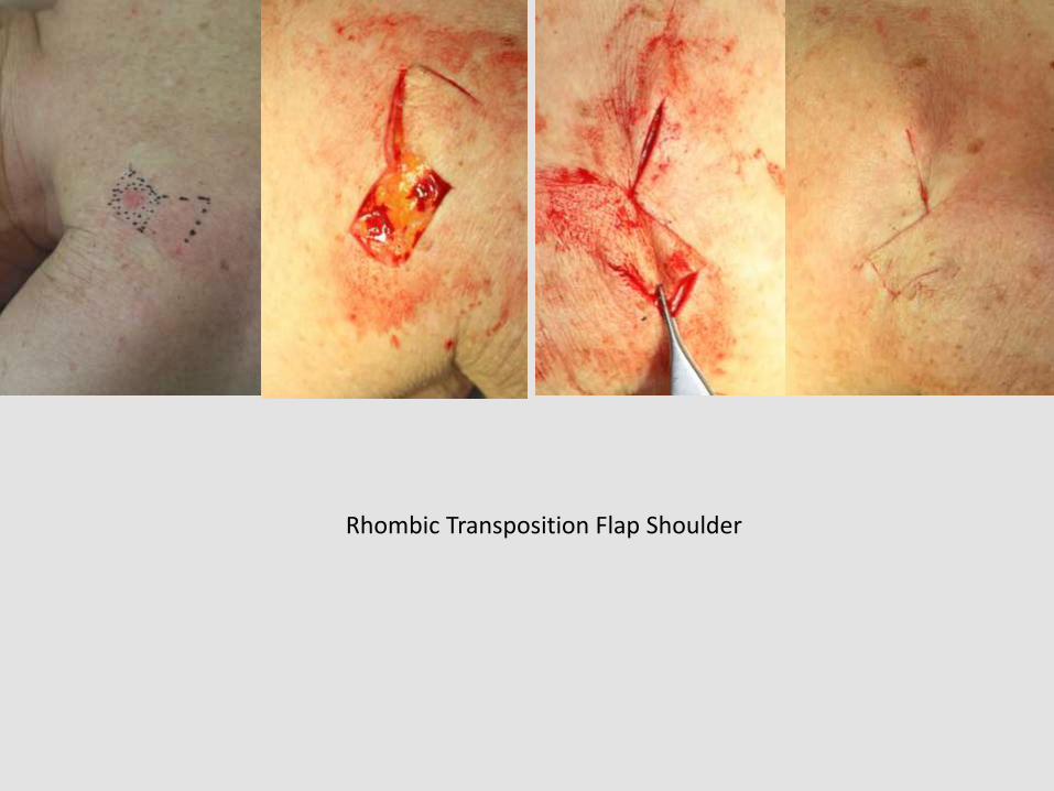
Rhombic Transposition Flap Shoulder

Tension Suture FT SG
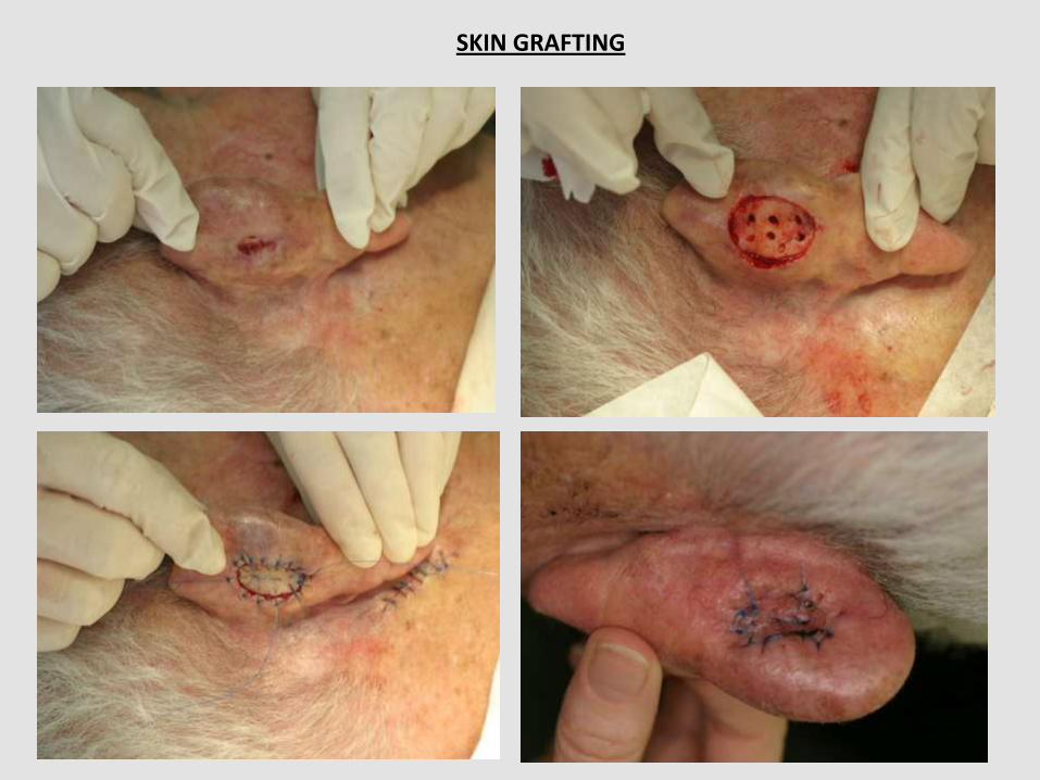
SKIN GRAFTING
MOHS SURGERY
Mohs micrographic surgery is a specialised procedure for the microscopically controlled excision of skin cancer.
Available at KM Surgical Ltd & Dermatology Associates Ltd
Dr Paul Weber, MD
Fellow of the American College of Mohs surgery
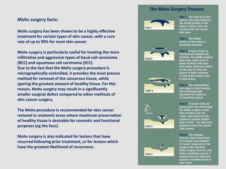
Mohs surgery facts: Mohs surgery has been shown to be a highly effective treatment for certain types of skin cancer, with a cure rate of up to 99% for most skin cancer. Mohs surgery is particularly useful for treating the more infiltrative and aggressive types of basal cell carcinoma (BCC) and squamous cell carcinoma (SCC). Due to the fact that the Mohs surgery procedure is micrographically controlled, it provides the most precise method for removal of the cancerous tissue, while sparing the greatest amount of healthy tissue. For this reason, Mohs surgery may result in a significantly smaller surgical defect compared to other methods of skin cancer surgery. The Mohs procedure is recommended for skin cancer removal in anatomic areas where maximum preservation of healthy tissue is desirable for cosmetic and functional purposes (eg the face). Mohs surgery is also indicated for lesions that have recurred following prior treatment, or for lesions which have the greatest likelihood of recurrence.
Patients with basal cell, squamous cell and certain rare neoplasm should ideally be treated by this technique if they fall within one of the following criteria:
-Recurrent skin cancer. -Skin cancer with aggressive/infiltrative pathology. -Skin cancer with indistinct margins. -Skin cancer in younger persons where tissue sparing might be important. -Skin cancers occurring in sites were recurrence rates with traditional surgery are high: eg around the eyes, nose, lips, ears, fingers, toes, genitalia. -Incomplete removal of skin cancer following traditional surgery. -Larger skin cancers (greater than 1cm in diameter)
Which patients are suitable for Mohs Surgery
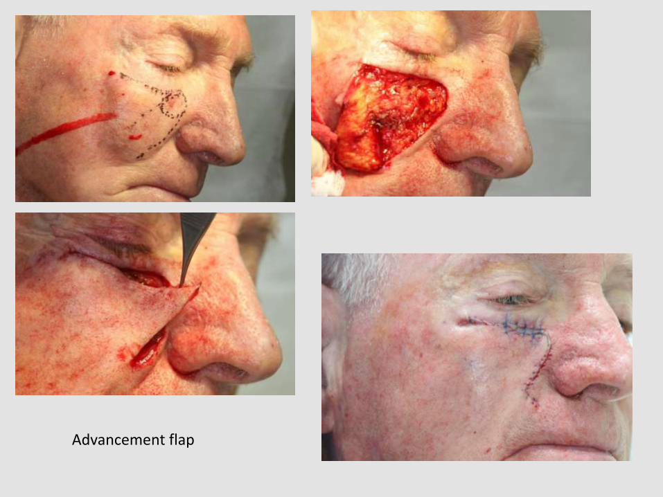
Advancement flap
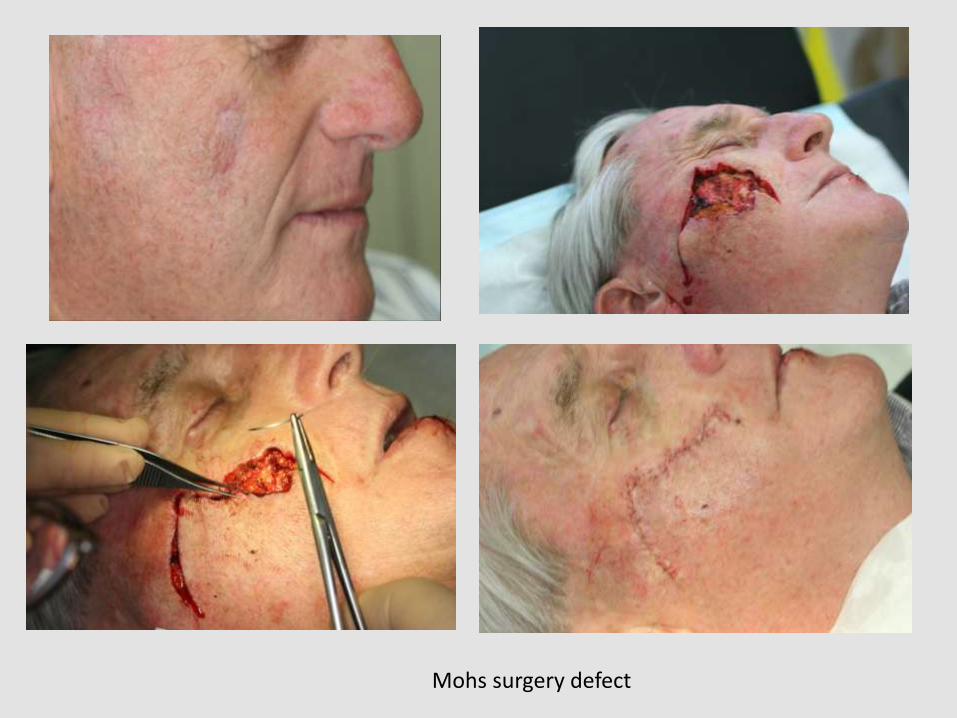
Mohs surgery defect
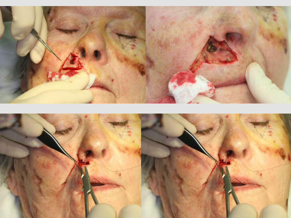
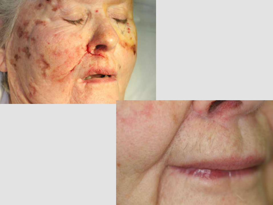

Mohs Surgery defect
Maximum tension scalp closure with full thickness undersized skin graft from base of neck
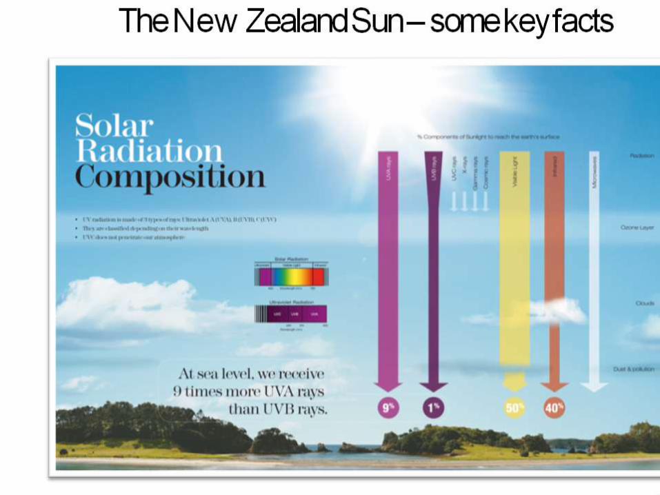

UV Irradiation and Skin Cancer
Vitamin D

Thank you ! Remember
your sunscreen
Skin Cancer More Information Websites: www.dermnetnz.org www.kmsurgical.co.nz Email: [email protected] Invitation: Come and watch some procedures! Dr Ken Macdonald Dr Paul Weber Dr Paul Maurice