Skeletal System
24
The Skeletal The Skeletal System: System: Structure, Function, Structure, Function, and Diseases and Diseases of the bones and of the bones and joints joints
Transcript of Skeletal System

The Skeletal The Skeletal System:System:
Structure, Function, Structure, Function, and Diseasesand Diseases
of the bones and of the bones and jointsjoints

The Skeletal SystemThe Skeletal System
Copyright © 2003 Pearson Education, Inc. publishing as Benjamin Cummings
Parts of the skeletal system
Bones (skeleton)
Joints
Cartilages
Ligaments (bone to bone)(tendon=bone to muscle)
Divided into two divisions
Axial skeleton- skull, spinal column
Appendicular skeleton – limbs and girdle
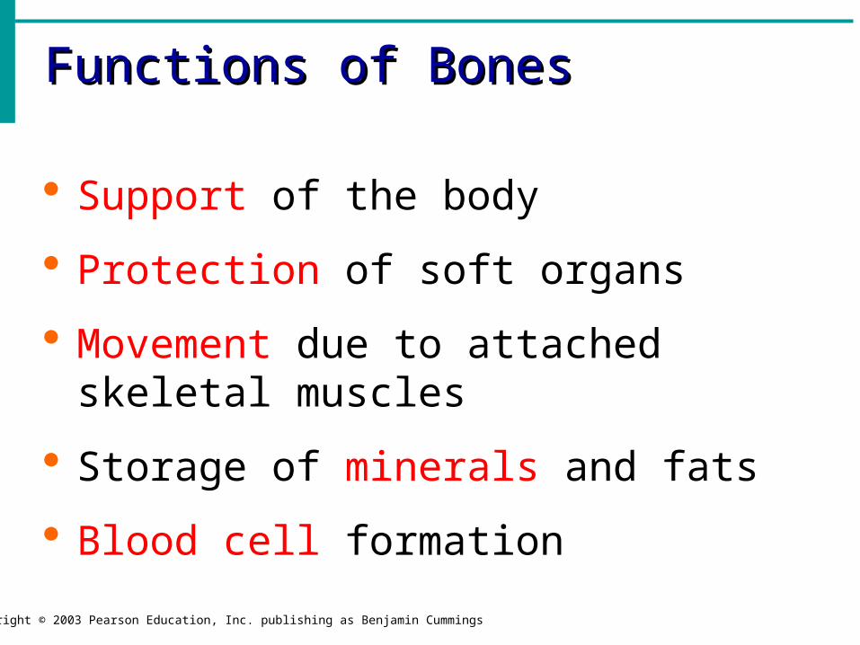
Functions of BonesFunctions of Bones
Copyright © 2003 Pearson Education, Inc. publishing as Benjamin Cummings
Support of the body
Protection of soft organs
Movement due to attached skeletal muscles
Storage of minerals and fats
Blood cell formation
Bones of the Human BodyBones of the Human Body
Copyright © 2003 Pearson Education, Inc. publishing as Benjamin Cummings
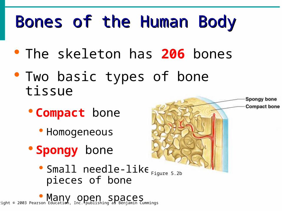
The skeleton has 206 bones
Two basic types of bone tissue
Compact bone
Homogeneous
Spongy bone
Small needle-like pieces of bone
Many open spacesFigure 5.2b

Bones are classified by their shape:
1.Long- bones are longer than they are wide (arms, legs)
2.Short- usually square in shape, cube like (wrist, ankle)
3.Flat- flat , curved (skull, Sternum)
4.Irregular- odd shapes (vertebrae, pelvis)
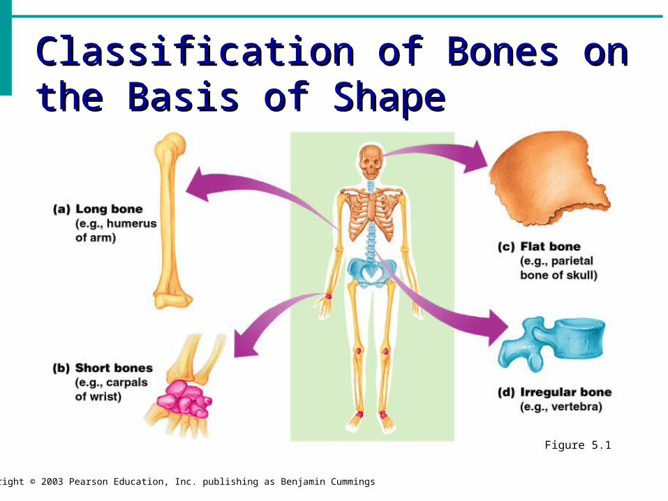
Classification of Bones on the Classification of Bones on the Basis of ShapeBasis of Shape
Copyright © 2003 Pearson Education, Inc. publishing as Benjamin Cummings
Figure 5.1

Types of Bone CellsTypes of Bone Cells
Copyright © 2003 Pearson Education, Inc. publishing as Benjamin Cummings
Osteocytes Mature bone cells
Osteoblasts Bone-forming cells
Osteoclasts Bone-destroying cells
Break down bone matrix for remodeling and release of calcium
Bone remodeling is a process by both osteoblasts and osteoclasts
Changes in the Human SkeletonChanges in the Human Skeleton
Copyright © 2003 Pearson Education, Inc. publishing as Benjamin Cummings
In embryos, the skeleton is primarily hyaline cartilage
During development, much of this cartilage is replaced by bone
Cartilage remains in isolated areas
Bridge of the nose
Parts of ribs
Joints
Bone FracturesBone Fractures
Copyright © 2003 Pearson Education, Inc. publishing as Benjamin Cummings
A break in a bone
Types of bone fractures
Closed (simple) fracture – break that does not penetrate the skin
Open (compound) fracture – broken bone penetrates through the skin
Greenstick- frays, hard to repair, breaks like a green twig
Bone fractures are treated by reduction and immobilization
Realignment of the bone

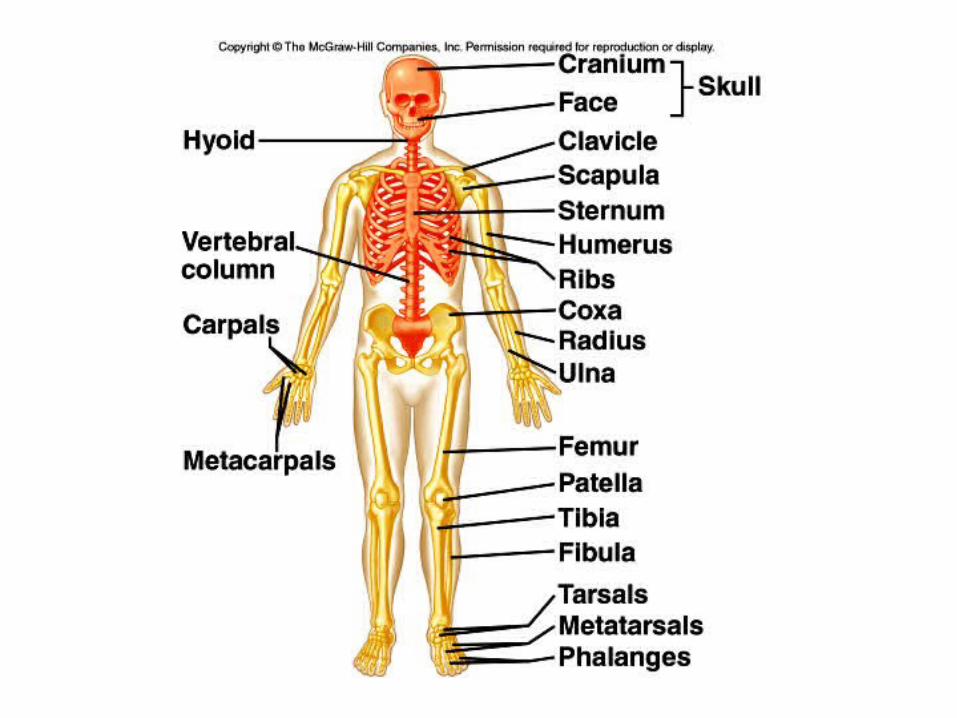
Axial skeleton supports and protects organs of head, neck and trunk Axial skeleton:
skull (cranium and facial bones) hyoid bone (anchors tongue and
musclesassociated with swallowing)
vertebral column (vertebrae and disks)
bony thorax (ribs and sternum)

Appendicular skeleton includes bones of limbs and
bones that anchor them to the axial skeletonAppendicular skeleton:
pectoral girdle (clavicle, scapula)
upper limbs (arms)pelvic girdle (sacrum, coccyx)lower limbs (legs)
Articulation- where joints meet, connect, and are formed.


The Axial SkeletonThe Axial Skeleton
Slide 5.20a
Copyright © 2003 Pearson Education, Inc. publishing as Benjamin Cummings
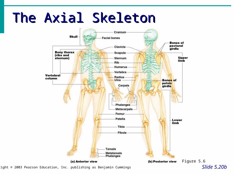
Forms the longitudinal part of the body
Divided into three parts
Skull
Vertebral Column
Rib Cage
The Axial SkeletonThe Axial Skeleton
Slide 5.20b
Copyright © 2003 Pearson Education, Inc. publishing as Benjamin Cummings
Figure 5.6
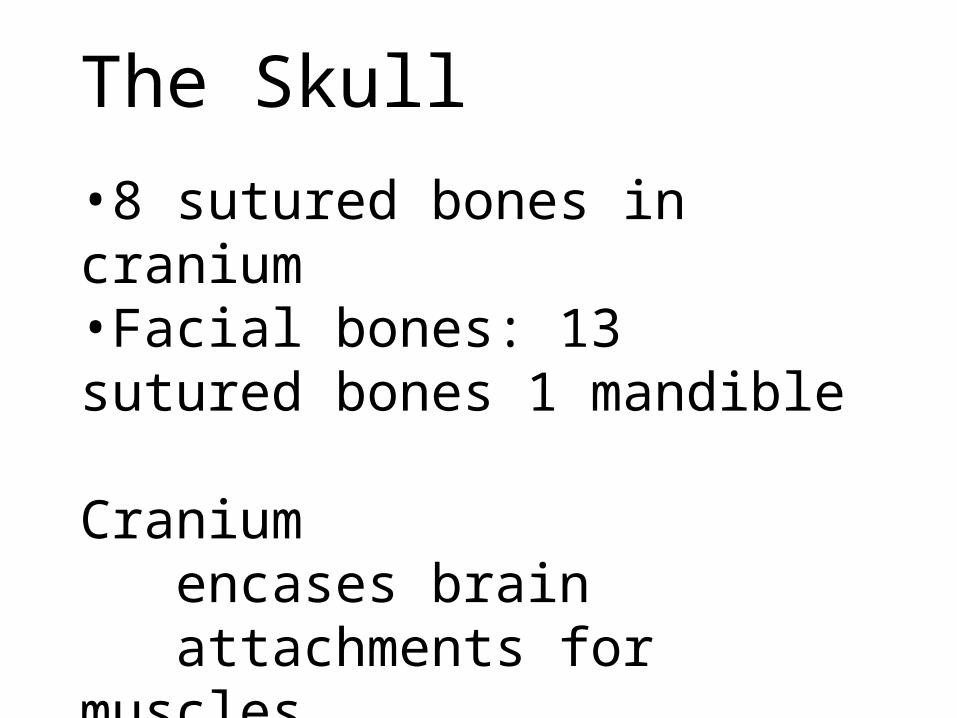
The Skull
•8 sutured bones in cranium•Facial bones: 13 sutured bones 1 mandible
Craniumencases brainattachments for musclessinuses


Bones of the SkullBones of the Skull
Copyright © 2003 Pearson Education, Inc. publishing as Benjamin Cummings
Figure 5.11

Allows forgrowth

Paranasal SinusesParanasal Sinuses
Slide 5.25a
Copyright © 2003 Pearson Education, Inc. publishing as Benjamin Cummings
Hollow portions of bones surrounding the nasal cavity
Figure 5.10

The Hyoid BoneThe Hyoid Bone
Slide 5.26Copyright © 2003 Pearson Education, Inc. publishing as Benjamin Cummings
The only bone that does not articulate with another bone
Serves as a moveable base for the tongue, and other muscle attachments
Figure 5.12
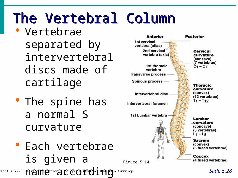
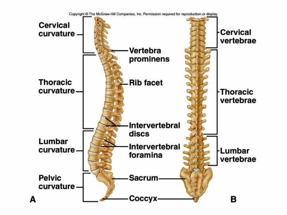
The Vertebral ColumnThe Vertebral Column
Slide 5.28Copyright © 2003 Pearson Education, Inc. publishing as Benjamin Cummings
Vertebrae separated by intervertebral discs made of cartilage
The spine has a normal S curvature
Each vertebrae is given a name according to its location Figure 5.14

Thoracic cageribsthoracic
Vertebraesternumcostal cartilages
•True ribs are directly attached to the sternum(first seven pairs)•Three false ribs are joined to the 7th rib•Two pairs of floating ribs