Skeletal Muscle Gross muscle Plasma membrane Neuromuscular junction Action potential.
23
Skeletal Muscle Gross muscle Plasma membrane Neuromuscular junction Action potential
-
Upload
doreen-banks -
Category
Documents
-
view
220 -
download
0
Transcript of Skeletal Muscle Gross muscle Plasma membrane Neuromuscular junction Action potential.

Skeletal Muscle
Gross muscle
Plasma membrane
Neuromuscular junction
Action potential

Muscle Connective Tissue
• provides structure & form to muscle • allows force to be transmitted to
tendons/bones• three layers of connective tissue--
composed primarily of collagen fibers– epimysium (outer layer)– perimysium (groups fibers into bundles
(fascicles))– endomysium (surrounds each fiber)

Muscle Connective
Tissue

Skeletal Muscle

Endomysial connective tissue
within skeletal muscle

Connective Tissue Functions
• provides “scaffolding” upon which fibers can form
• holds fibers together• perimysium provides conduit for
arterioles/venules and intramuscular nerves• distributes strain/force over entire muscle• endomysium conveys part of contractile force
to tendon• fibers taper near tendon attachment; folding
of plasma membrane

Myon
Myonuclei of skeletal fiber

Sarcolemma
• surrounds each fiber and composed of:– basement membrane (outer side)– plasma membrane
• basement membrane contains:– acetylcholinesterase – collagen
• functions of basement membrane– termination of synaptic transmission– attachment of fiber to endomysium– scaffolding for muscle fiber regeneration
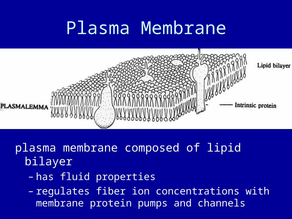
Plasma Membrane
plasma membrane composed of lipid bilayer– has fluid properties– regulates fiber ion concentrations with
membrane protein pumps and channels

Plasma Membrane Proteins• myonuclei and satellite cells
– bound to inter surface of plasma membrane
• peripheral proteins (plasma membrane receptors)– associated with surface of bilayer– e.g., adenylate cyclase, kinases, hormone receptors– integrins
• class of connective proteins• link basement membrane to plasma membrane and
cytoskeletal structures
• integral proteins function as “gatekeepers”– embedded in phospholipid bilayer– selectively let ions pass

Methods of transport
• osmosis (i.e., water)
• simple diffusion (e.g., O2, CO2)
• facilitated diffusion (e.g., glucose, lactate)
• active transport (e.g., Na+, K+)

`• several thousand
amino acids arranged in 1 or more subunits
• hundreds of sugar residues linked
• controlled by voltage- or receptor-regulated gate

Transport Times
movement of side chain on protein 10-10 s
movement of Na+ through a pore 10-8 s
fastest enzyme turnover 10-6 s
activation of a channel (rate-limiting step) 10-4 s
actin-myosin turnover 10-2 s

Na+
channel
K+
Na+
Na+ Na+
Na+
Na+ Na+
Na+
Na+ Na+
Na+
Na+ Na+Na+ Na+Na+
Na+Na+Na+
Na+K+ K+
K+K+
K+ K+K+ K+
K+ K+K+ K+
K+ K+K+
K+K+K+K+
Mem
bra
ne
po
ten
tial
(m
V)
+20
0
-20
-40
-60
-80
Time (ms)
ATP
PiADP
ATPase
intracellular
K+
channelNa+-K+
exchange pump

Distribution of Na+-K+ pumps in skeletal muscle and muscle-nerve bundles (N). Pumps are lit from exposure to a labeled antibody

Action Potential
results from disturbance (e.g. electrical) to membrane
affects membrane permeability to Na+ and K+
follows “all-or-nothing” principle

depolarization – influx of Na+
repolarization – efflux of K+
hyperpolarization – overshoot of K+ efflux
Phases of Action Potential

Action Potential

Motor Unit

Motoneuron
• inputs to motoneuron are both excitatory and inhibitory
• continuous nerve from spinal cord to neuromuscular junction
• are all myelinated– wrapped with myelin (Schwann cells)– nodes of Ranvier– AP conducted by saltatory conduction– greatly conduction velocity
• extrafusal motor units innervated by motoneurons

Motor End Plate

Neuromuscular Junction
AP at motor end plate (active zones) causes Ca2+ influx
stimulates vesicles to migrate/fuse to membrane and release acetylcholine (ACh)
ACh diffuses across synapse and binds with postsynaptic ACh receptors
most ACh metabolized by cholinesterase postsynaptic ACh binding causes Na+ influx and
K+ efflux depolarization causes development of APs

Curare blocks ACh receptors Anticholinesterase drugs (e.g., mustard
gas, sarin) prevent hydrolysis of ACh Botulism (bacterium) blocks release of
ACh


![NP 16 Excitation of Skeletal Muscle [호환 모드]web.khu.ac.kr/~tskim/NP_16_Excitation of Skeletal Muscle.pdf · Ch. 7 Excitation of Skeletal Muscle •Neuromuscular Junction –Motor](https://static.fdocuments.net/doc/165x107/5f8d1c7c5988837821360b96/np-16-excitation-of-skeletal-muscle-eeoewebkhuackrtskimnp16excitation.jpg)