Size Influences the Effect of Hydrophobic Nanoparticles · PDF fileSize Influences the Effect...
17
Size Influences the Effect of Hydrophobic Nanoparticles on Lung Surfactant Model Systems Mridula V. Dwivedi, †‡ Rakesh Kumar Harishchandra, † Olga Koshkina, §k Michael Maskos, {k and Hans-Joachim Galla † * † Institute of Biochemistry, Westfa ¨ lische Wilhelms Universita ¨t Mu ¨ nster, Germany; ‡ NRW International Graduate School of Chemistry, University of Muenster, Muenster, Germany; § BAM Federal Institute for Materials Research and Testing, Berlin, Germany; { Institute of Physical Chemistry, Johannes Gutenberg University, Mainz, Germany; and k Institut fu ¨r Mikrotechnik Mainz (IMM), Mainz, Germany ABSTRACT The alveolar lung surfactant (LS) is a complex lipid protein mixture that forms an interfacial monolayer reducing the surface tension to near zero values and thus preventing the lungs from collapse. Due to the expanding field of nanotech- nology and the corresponding unavoidable exposure of human beings from the air, it is crucial to study the potential effects of nanoparticles (NPs) on the structural organization of the lung surfactant system. In the present study, we investigated both, the domain structure in pure DPPC monolayers as well as in lung surfactant model systems. In the pure lipid system we found that two different sized hydrophobic polymeric nanoparticles with diameter of ~12 nm and ~136 nm have contrasting effect on the functional and structural behavior. The small nanoparticles inserted into fluid domains at the LE-LC phase transition are not visibly disturbing the phase transition but disrupting the domain morphology of the LE phase. The large nanoparticles led to an expanded isotherm and to a significant decrease in the line tension and thus to a drastic disruption of the domain structures at a much lower number of nanoparticles with respect to the lipid. The surface activity of the model LS films again showed drastic variations due to presence of different sized NPs illustrated by the film balance isotherms and the atomic force microscopy. AFM revealed laterally profuse multilayer protrusion formation on compression but only in the presence of 136 nm sized nanopar- ticles. Moreover we investigated the vesicle insertion process into a preformed monolayer. A severe inhibition was observed only in the presence of ~136 nm NPs compared to minor effects in the presence of ~12 nm NPs. Our study clearly shows that the size of the nanoparticles made of the same material determines the interaction with biological membranes. INTRODUCTION Lung surfactant (LS) is a complex lipid-protein film span- ning the air-water interface of the alveoli in the lungs. Its primary function is to reduce the surface tension at the air-water interface, which thus is most essential for the normal breathing process. During the process of inhalation, the reduction in surface tension renders the process effort- less and during exhalation, this property prevents the lungs from collapse. In view of the essential functional behavior of the lung surfactant, it is known to reduce the surface tension to near zero values during compression and respread instan- taneously without breaking during subsequent expansion (1–3). The mammalian lung surfactant consists mainly of ~85–90% phospholipids and 8–10% of fatty acids, choles- terols, and surfactant-specific proteins (4–6). The domi- nantly present phospholipids are zwitterionic saturated phosphatidylcholines (PC) (40–50%) primarily responsible for decreasing the surface tension to near zero values (7). However, it has poor respreading properties and rigidifies the surfactant film. To enhance the respreading properties, the second most abundant lipids present are the negatively charged phosphatidylglycerols (PG) and unsaturated phos- pholipids that fluidize the lung surfactant film. Along with these, surfactant-specific proteins also play a crucial role in the normal functioning of the surfactant film. The surfac- tant-specific proteins SP-B and SP-C are mainly involved in enhancing the surface activity of the surfactant film during compression and expansion (5,8). Whereas SP-A and SP- D are related to the immunological and host defense mech- anism (9). Over the years, a good amount of literature has cleared our knowledge about action of the lung surfactant film. On compression, the fluid components of the lipid- protein monolayer squeeze out to form three-dimensional (3D) multilayer stacks or protrusion structures, which acts as a surfactant reservoir. On expansion, these multilayer stacks are reincorporated into the monolayer and assist the respreading of the film to form a stable lung surfactant film. The presence of these structures in vivo has been evi- denced by atomic force microscopy (AFM) and electron microscopy (EM) studies. These multilayer stacks are formed and held together by the aid of SP-B and SP-C and hence the lipid and protein components of the surfactant film interact and harmonize with each other during the breathing cycle (10–13). The surfactant-specific proteins SP-B and SP-C also play an important role in the recycling and replenishment of the lipid material by the vesicle inser- tion process (14–16). Although advances in nanotechnology has brought to us novel methods in medical diagnostics and targeted drug Submitted May 17, 2013, and accepted for publication October 10, 2013. *Correspondence: [email protected] Rakesh Kumar Harishchandra’s present address is Department of Chemis- try and Biochemistry, Worcester Polytechnic Institute (WPI), 60 Prescott Street, Worcester, MA 01605. Editor: Klaus Gawrisch. Ó 2014 by the Biophysical Society 0006-3495/14/01/0289/10 $2.00 http://dx.doi.org/10.1016/j.bpj.2013.10.036 Biophysical Journal Volume 106 January 2014 289–298 289
Transcript of Size Influences the Effect of Hydrophobic Nanoparticles · PDF fileSize Influences the Effect...

Biophysical Journal Volume 106 January 2014 289–298 289
Size Influences the Effect of Hydrophobic Nanoparticles on LungSurfactant Model Systems
Mridula V. Dwivedi,†‡ Rakesh Kumar Harishchandra,† Olga Koshkina,§k Michael Maskos,{k
and Hans-Joachim Galla†*†Institute of Biochemistry, Westfalische Wilhelms Universitat Munster, Germany; ‡NRW International Graduate School of Chemistry,University of Muenster, Muenster, Germany; §BAM Federal Institute for Materials Research and Testing, Berlin, Germany; {Institute ofPhysical Chemistry, Johannes Gutenberg University, Mainz, Germany; and kInstitut fur Mikrotechnik Mainz (IMM), Mainz, Germany
ABSTRACT The alveolar lung surfactant (LS) is a complex lipid protein mixture that forms an interfacial monolayer reducingthe surface tension to near zero values and thus preventing the lungs from collapse. Due to the expanding field of nanotech-nology and the corresponding unavoidable exposure of human beings from the air, it is crucial to study the potential effectsof nanoparticles (NPs) on the structural organization of the lung surfactant system. In the present study, we investigatedboth, the domain structure in pure DPPC monolayers as well as in lung surfactant model systems. In the pure lipid systemwe found that two different sized hydrophobic polymeric nanoparticles with diameter of ~12 nm and ~136 nm have contrastingeffect on the functional and structural behavior. The small nanoparticles inserted into fluid domains at the LE-LC phase transitionare not visibly disturbing the phase transition but disrupting the domain morphology of the LE phase. The large nanoparticles ledto an expanded isotherm and to a significant decrease in the line tension and thus to a drastic disruption of the domain structuresat a much lower number of nanoparticles with respect to the lipid. The surface activity of the model LS films again showed drasticvariations due to presence of different sized NPs illustrated by the film balance isotherms and the atomic force microscopy. AFMrevealed laterally profuse multilayer protrusion formation on compression but only in the presence of 136 nm sized nanopar-ticles. Moreover we investigated the vesicle insertion process into a preformed monolayer. A severe inhibition was observedonly in the presence of ~136 nm NPs compared to minor effects in the presence of ~12 nm NPs. Our study clearly showsthat the size of the nanoparticles made of the same material determines the interaction with biological membranes.
INTRODUCTION
Lung surfactant (LS) is a complex lipid-protein film span-ning the air-water interface of the alveoli in the lungs. Itsprimary function is to reduce the surface tension at theair-water interface, which thus is most essential for thenormal breathing process. During the process of inhalation,the reduction in surface tension renders the process effort-less and during exhalation, this property prevents the lungsfrom collapse. In view of the essential functional behavior ofthe lung surfactant, it is known to reduce the surface tensionto near zero values during compression and respread instan-taneously without breaking during subsequent expansion(1–3). The mammalian lung surfactant consists mainly of~85–90% phospholipids and 8–10% of fatty acids, choles-terols, and surfactant-specific proteins (4–6). The domi-nantly present phospholipids are zwitterionic saturatedphosphatidylcholines (PC) (40–50%) primarily responsiblefor decreasing the surface tension to near zero values (7).However, it has poor respreading properties and rigidifiesthe surfactant film. To enhance the respreading properties,the second most abundant lipids present are the negativelycharged phosphatidylglycerols (PG) and unsaturated phos-
Submitted May 17, 2013, and accepted for publication October 10, 2013.
*Correspondence: [email protected]
Rakesh Kumar Harishchandra’s present address is Department of Chemis-
try and Biochemistry, Worcester Polytechnic Institute (WPI), 60 Prescott
Street, Worcester, MA 01605.
Editor: Klaus Gawrisch.
� 2014 by the Biophysical Society
0006-3495/14/01/0289/10 $2.00
pholipids that fluidize the lung surfactant film. Along withthese, surfactant-specific proteins also play a crucial rolein the normal functioning of the surfactant film. The surfac-tant-specific proteins SP-B and SP-C are mainly involved inenhancing the surface activity of the surfactant film duringcompression and expansion (5,8). Whereas SP-A and SP-D are related to the immunological and host defense mech-anism (9). Over the years, a good amount of literature hascleared our knowledge about action of the lung surfactantfilm. On compression, the fluid components of the lipid-protein monolayer squeeze out to form three-dimensional(3D) multilayer stacks or protrusion structures, which actsas a surfactant reservoir. On expansion, these multilayerstacks are reincorporated into the monolayer and assist therespreading of the film to form a stable lung surfactantfilm. The presence of these structures in vivo has been evi-denced by atomic force microscopy (AFM) and electronmicroscopy (EM) studies. These multilayer stacks areformed and held together by the aid of SP-B and SP-Cand hence the lipid and protein components of the surfactantfilm interact and harmonize with each other during thebreathing cycle (10–13). The surfactant-specific proteinsSP-B and SP-C also play an important role in the recyclingand replenishment of the lipid material by the vesicle inser-tion process (14–16).
Although advances in nanotechnology has brought to usnovel methods in medical diagnostics and targeted drug
http://dx.doi.org/10.1016/j.bpj.2013.10.036

290 Dwivedi et al.
delivery, it has also made us prone to exposure of nanopar-ticles (NPs) through inhalation from the atmosphere. It ishence of utmost importance to study the effect of theintentional or unintentional exposure of nanosized particlesto the lung surfactant. The important parameters to be inves-tigated in these nanoparticle interaction studies are—distur-bance of the structural and functional behavior of the LSfilm, interaction with the individual components of the LSfilm, retention/penetration of the NPs in the film, and inter-action with epithelial cells and alveolar macrophages.Recently, numerous amounts of research have been dedi-cated tomonitor the toxicological influence of different kindsof NPs on the lungs and the further fate of the NPs (17–19). Afew reports have focused on the lung surfactant dysfunctioncaused by the inhalation and penetration of NPs throughthe lung surfactant film. In most cases, the lung surfactantdysfunction has been caused in a dose- and time-dependentmanner by the frequently used metallic NPs, which indeedform a major part of the particulate matter in the atmosphereand are also employed in medical diagnostics (20–23). How-ever, seldom have the dosages been physiologically relevant.It has also now become clear that the toxicological effect ofNPs are more or less dependent upon the surface properties,shape, size, solubility, and surface charge of the NPs (24). Ithas been previously shown that 20 nm AmorSil20 polymerichydrophobic NPs tend to inhibit the lung surfactant functionat high concentrations and a detailed study employingvarious applications of AFM and EM showed that thesehydrophobic NPs are retained into the surfactant film evenafter repetitive compression expansion cycles (22,25). Thisbehavior of a few NPs to retain into the surfactant film couldbe exploited by the drug delivery applications for long-termrelease of the drugs into the systembefore theNPs are clearedout. Hence, these NPs are required to cause minimal distur-bance to the lung surfactant function. In this report, wehave studied the effect of size of NPs on the biophysicalbehavior of model LS films and proved that the size of theNPs is an important factor responsible for causing severedamage to the lung surfactant structure.
In view of the highly complex and dynamic nature ofthe natural lung surfactant film, it is technically challengingto extract any valuable information from such a systemand hence a well-characterized and highly reproduciblemodel system has been developed that closely mimics thenatural lung surfactant. It consists mainly of the zwitterionic1,2-dipalmitoyl-sn-glycero-3-phosphocholine (DPPC), ne-gatively charged 1,2-dipalmitoyl-sn-glycero-3-phosphogly-cerol (DPPG) and surfactant-specific protein SP-C(26–28). In this study, we have investigated the influenceof size of hydrophobic polymeric NPs on the lung surfactantmodel system. We have studied the variation in the surfaceactivity of the lung surfactant model system in the presenceof small NPs (diameter ¼ 12.4 5 2.6 nm, determined bytransmission electron microscopy (TEM), particles depos-ited from chloroform) and large NPs (diameter ¼ 136 5
Biophysical Journal 106(1) 289–298
27.0 nm, TEM, particles deposited from chloroform) withthe same surface characteristics as the previously studiedNPs (AmorSil20) with a diameter ~20 nm consisting of inertSi-CH3 groups making it essentially hydrophobic in nature.The evidently contrasting surface activity behavior of thelung surfactant model system in the presence of differentsized NPs has been further explored using the AFM tech-nique. The factors that are focused upon in this studyare—variation in the surface activity of the LS modelsystem and hence inhibition of surfactant function, theeffect of size variance on the line tension and domainmorphology, the effect of size on the 3D protrusion struc-tures, localization of the different sized NPs around theprotrusion structures, and analysis of the multilayer stepsof the protrusion structures. Furthermore, the size-depen-dent effect of hydrophobic NPs on the vesicle insertion pro-cess is studied by vesicle insertion kinetic studies.
MATERIALS AND METHODS
Materials
DPPC and DPPGwere purchased fromAvanti Polar Lipids (Alabaster, AL).
SP-C was isolated from porcine bronchoalveolar lavage fluid by the butanol
extraction method (29,30). 2-(4, 4-Difluoro-5-methyl-4-bora-3a, 4a-diazas-
indacene-3-dodecanoyl)-1-hexadecanoyl-sn-glycero-3-phosphocholine (b-
BODIPY 500/510 C12-HPC, BODIPY-PC) was purchased from Molecular
Probes (Eugene, OR). Chloroform and methanol were high pressure liquid
chromatography grade and obtained from Sigma-Aldrich (Steinheim, Ger-
many) and Merck (Darmstadt, Germany), respectively. N-(2-Hydroxyethyl)
piperazine-N0-(2-ethanesulfonic acid) sodium salt (Na-HEPES), and cal-
cium chloride (CaCl2) were purchased from Sigma-Aldrich. Water was
purified and deionized by a multicartridge system (Sartorius, Goettingen,
Germany) and had a resistivity >18 MUm at 25�C. Lipids were dissolvedin chloroform/methanol solution (1:1, v/v). All chemicals were used
as received: trimethoxymethylsilane, diethoxydimethylsilane, (p-chloro-
methyl)phenyltrimethoxysilane, (ABCR, Germany), dodecylbenzenesul-
fonic acid, chloroform (Sigma-Aldrich).
Sample preparation
Poly(organosiloxane) NPs were synthesized by condensation of di- and tri-
alkoxysilanes as described previously (31–33). Briefly, the synthesis of
12 nm core-shell NPs (R ¼ 6.2 5 1.3 nm (TEM)) was carried out under
alkaline conditions using benzethonium chloride as surfactant at a fleet ratio
of 0.3. The core was fluorescently labeled by addition of rhodamine
B-(p-trimethoxysilyl)benzylester to the monomer mixture. 136 nm NPs
(R ¼ 68 5 13.5 nm (TEM)) were prepared with dodecylbenzenesulfonic
acid as surfactant (fleet ratio 0.001) by sequential condensation of polydi-
methylsiloxane-core and two shells; the inner shell was labeled by rhoda-
mine B during polycondensation.
The characterization of particle size in aqueous solution was carried out
by multiangle dynamic light scattering (DLS) using an ALV/CGS-3
Compact Goniometer System, with HeNe Laser (l0¼ 633 nm). All samples
were filtered (Millex-LCR filter, 0.45 mm pore size, Millipore) before
measurement. For determination of hydrodynamic radii, the autocorrelation
data were evaluated by applying a biexponential fit, m2 values were derived
from a cumulant fit at 90� (34). DLS measurements showed a hydrody-
namic radius Rh of 7.9 nm (m2 ¼ 0.11) for 12 nm particles and Rh of
70.9 nm (m2 ¼ 0.05) for 136 nm particles in aqueous dispersion.
Size Influences Effect of Hydrophobic NPs on LS Model System 291
After polycondensation and characterization by DLS the free silanol
groups on the nanoparticle’s surface were endcapped with trimethylethox-
ysilane. The particles were washed or dialyzed in methanol to remove the
surfactant. By this procedure the surface properties of the NPs were
changed to hydrophobic and the particles were redispersed in chloroform
and visualized by TEM (33). The hydrodynamic radii of the hydrophobic
particles were determined by DLS in tetrahydrofuran. The resulting values
are Rh¼ 110 nm (m2 (90�)¼ 0.06) for the big particles and Rh¼ 9.1 nm (m2
(90�) ¼ 0.07) for the small particles.
The NPs were dissolved in chloroform to obtain 3.784 � 1012 NPs/mL
(5.98 mg/mL) of 136 nm NPs stock solution and 1.430 � 1016 NPs/mL
(17.13 mg/mL) of 12 nm NPs stock solution. The NPs were mixed in lipid
solutions (1 mg/mL) in concentrations of 1, 10, 20, 50, 100, and
1000 mg/mL for both 136 nm and 12 nm NPs to acquire physiological
relevance and hence the number of NPs in the case of 136 nm NPs is 103
times smaller than that of 12 nm NPs. The lipid-nanoparticle mixture, dis-
solved in chloroform/methanol (1:1, v/v) solution, was sonicated and in-
jected onto the subphase.
Surface pressure-Area isotherms
All the film balance experiments were performed on an analytical Wilhelmy
film balance (Riegler and Kirstein, Mainz, Germany) with an operational
area of 144 cm2. All surface pressure measurements were done on
25 mM HEPES with 3 mM CaCl2 at pH 7.0 as buffered subphase at
20�C. The sample solutions consisting of lipid mixtures and NPs was
injected onto the subphase and was left for an equilibration time of
10–15 min for the solvent to evaporate after which the lipid monolayer
formed was compressed at the rate of 2.9 cm2/min using a Teflon barrier.
The monolayer isothermal compressibility of the DPPC film was calcu-
lated using
Cs ¼ ð�1=AÞðdA=dpÞ;
where Cs is the compressibility coefficient, A is the molecular area at a
given surface pressure, and p is the corresponding surface pressure. The
compressibility coefficient is known to indicate the lateral stiffness of the
monolayer at a given surface pressure.
Video-enhanced epifluorescence microscopy
Domain structures of DPPC in the presence of NPs were observed by
doping the lipid solution with 0.5 mol% BODIPY-PC and visualizing by
means of an epifluorescence microscope (Olympus STM5-MJS,Olympus,
Hamburg, Germany) equipped with xy-stage and connected to a charge-
coupled device camera (Hamamatsu, Herrsching, Germany). The images
were captured at desired surface pressures by stopping the barrier. All the
measurements were done in pure water as the subphase at 20�C.
Langmuir-Blodgett transfer
The lipid mixtures with and without NPs were spread onto the 25 mM
HEPES with 3 mM CaCl2 at pH 7.0 subphase in a film balance (Riegler
and Kirstein) with an operational area of 39 cm2 and the monolayer
obtained was compressed with a velocity of 1.5 cm2/min to achieve the
desired surface pressure and area per molecule. The lipid film was allowed
to stabilize at the attained surface pressure and area per molecule for around
10 min and then an already positioned vertically dipped freshly cleaved
mica sheet is drawn upward with the speed of 0.7 mm/min. For the lipid
film with 12 nm NPs, this transfer is carried out at around 52.2 mN/m,
i.e., at the plateau region where multilayer protrusion structures are
observed. For the lipid mixture with 136 nm NPs, the film is transferred
at the beginning of the plateau and at the end of the plateau where the sur-
face pressure is around 50.5 mN/m in either case. The area per molecule
value is attained by slowly stopping the barrier at the desired position
and stabilizing the lipid film for around 10 min before beginning the trans-
fer. All the transfers were done at 20�C.
AFM
The Lipid films transferred onto the mica sheet were scanned using
NanoWizard III (JPK Instruments, Berlin, Germany). Silicon nitride tips
(Budget Sensors, Sofia, Bulgaria) with a spring constant of 40 N/m and a
resonance frequency of 300 5 100 kHz were used. All the scanning was
performed in the intermittent contact mode in air at 20�C. The images
were obtained at 512 � 512 pixel resolution and processed using JPK
data processing software.
Vesicle insertion kinetic studies
Vesicle preparation
A phospholipid mixture containing DPPC/DPPG in the molar ratio 4:1 were
dissolved in a chloroform/methanol (1:1, v/v) solution and dried under a
stream of nitrogen at 50�C. Traces of solvent were removed for at least
3 h at 50�C in a vacuum oven. The dried lipid films were then hydrated
by adding a buffer containing 25 mM Hepes and 0.1 mM EDTA. The
suspension was kept for 30 min at 50�C water bath and vortexed every
10 min. The resulting multilamellar vesicles solution was extruded from
a 100 nm pore size polycarbonate membrane to obtain a 100 nm large uni-
lamellar vesicle solution.
Vesicle insertion studies
The insertion experiments were performed using model LS monolayer on
25 mM Hepes and 3 mM CaCl2 at pH 7.0 as buffer subphase at 20�C.The lipid film with and without NPs was spread at the air-water interface
on a film balance (Riegler and Kirstein) with an operational area of
39 cm2 and the monolayer obtained was compressed with a velocity of
1.5 cm2/min to achieve the desired surface pressure (25 mN/m in this
case). The lipid film was stabilized for at least 10 min before injecting
the large unilamellar vesicle (LUV) solution into the subphase. The in-
crease in surface pressure is measured with respect to time and the isotherm
obtained is used for analyzing the insertion kinetics of the model LS film in
the presence of different sized NPs.
RESULTS
Size-dependent effect of NPs on surface activityof DPPC monolayer
The size-dependent effect of hydrophobic NPs on thesurface activity of DPPC monolayers was studied usingthe Langmuir film balance. DPPC forms the major com-ponent of the model LS film and hence alteration in its phasebehavior consequently causes alteration in the phasebehavior of model LS films. The isotherm of a pure DPPCmonolayer consists of an initial gaseous phase succeededby a liquid-expanded phase (LE), a liquid-condensed (LC)phase, and a solid phase. The phase transition from the LEphase to the LC phase is intermediated by the LE-LC phasecoexistence region, which is signified by a plateau at asurface pressure of around 5 mN/m. The isotherms illus-trating the size-dependent effect of NPs on DPPC mono-layer are presented in Fig. 1. In the presence of 12 nm
Biophysical Journal 106(1) 289–298

FIGURE 1 Surface pressure-Area per lipid mole-
cule isotherm for (a) DPPC þ 12 nm NPs and (b)
DPPC þ 136 nm NPs with 25 mM Hepes þ3 mM CaCl2 as the subphase. To see this figure in
color, go online.
292 Dwivedi et al.
NPs no change in the isotherm occurred up to the measuredconcentrations (Fig. 1 a). However, in the presence of136 nm NPs, the isotherm is expanded toward a higherarea per molecule values diminishing the LE-LC coexis-tence region (Fig. 1 b).
The compressibility values (Cs) (Table 1) are calculatedfor the DPPC films in the LC phase at the area per moleculevalue of 45 A2. With increasing concentration of the136 nm NPs the compressibility values increased for theDPPC monolayer.
Size-dependent effect of NPs on line tension anddomain morphology of DPPC monolayer
A lipid monolayer forms phase separated domains oncompression depending on the surface pressure. The domainmorphology can be studied using epifluorescence micro-scopy by doping the lipid monolayer with BODIPY-PCdye, which is known to preferentially partition into the LEphase. Hence, the LC phase appears as dark domains.
The size-dependent effect of NPs on the domains formedby DPPC films is presented in Fig. 2. Increasing concentra-tions of 12 nm NPs cause disruption of the domainmorphology beginning with formation of outgrowths atthe periphery of the LC domains at low concentrations ofthe NPs (Fig. 2 a). The fluorescent NPs are visible at highsurface pressure values as bright stretches (shown in red)against the compressed lipid monolayer backgroundbecause of the fluorescent Rhodamine B core of thenanoparticles.
With increasing concentrations of 136 nm NPs the LCdomains form branched structures, which at high surfacepressure forms a mesh-like network (Fig. 2 b). The signifi-cant decrease in the line tension between phases responsiblefor domain morphology is noticeable along with profusedisruption of the domain structures. The effect is consider-ably strong even at a very low concentration of 1 mg/mL,
TABLE 1 Compressibility values for DPPC film at the area per mol
CaCl2 buffer subphase in the presence of varying concentrations of
w/o NPs 10 mg/mL NPs
Cs(m/mN) 3.92 � 10�3 4.0 � 10�3
Biophysical Journal 106(1) 289–298
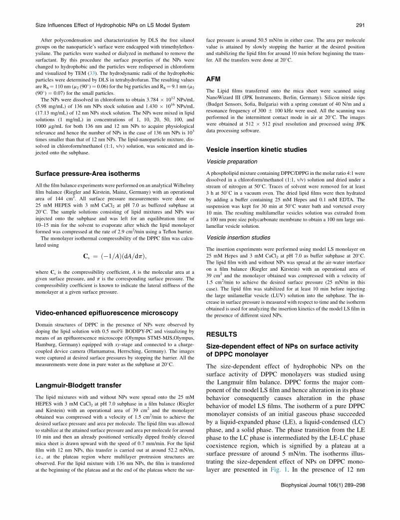
which amounts to a much lower number of NPs (106 lipidmolecules per nanoparticle) as compared to that of 12 nmNPs (102 lipid molecules per nanoparticle) (Fig. S2 in theSupporting Material).
Hence, the presence of 136 nm NPs significantly altersthe phase behavior of DPPC monolayers and decreasesphase separation. To further understand the size-dependenteffect of NPs on model LS films, a DPPC/DPPG/SP-C(80:20:0.4 mol%) mixture was used.
Size-dependent effect of NPs on surface activityof LS model system
The characteristic surface pressure-area isotherm for a pureDPPC/DPPG/SP-C (80:20:0.4 mol %) lipid mixture shows acontinuous increase in the surface pressure upon compres-sion until it reaches the surface pressure value of around52 mN/m. There it forms a plateau signifying a squeezeout of some of the lipid component of the monolayer, thuspreventing an increase in the surface pressure values foraround 5–10 A2 area per molecule region. This is explainedby the selective squeeze out of the fluid component of thelipid monolayer forming multilayer stacks held togetherby surfactant-specific proteins (SP-C in this case).
The size-dependent effect of NPs on the surface proper-ties of the LS model system is illustrated in Fig. 3. The pres-ence of 12 nm NPs in the LS model system monolayer doesnot cause any significant change in the surface pressure-areaisotherm except at a very high concentration where theplateau formation is inhibited (Fig. 3 a).
With increasing concentration of 136 nm NPs, however,the plateau region of the LS film isotherms is enhancedunlike for 12 nm NPs (Fig. 3 b). At the highest concentrationof 136 nm NPs that we measured (i.e., 100 mg/mL), thecompressibility of the lipid film at the plateau region isincreased immensely to achieve very low area per moleculevalues on continuous compression. In addition, we observed
ecule value of 45.1 A2 in LC region on 25 mM HEPES D 3 mM
136 nm NPs at 20�C.
25 mg/mL NPs 50 mg/mL NPs 100 mg/mL NPs
4.45 � 10�3 6.0 � 10�3 7.08 � 10�3

FIGURE 2 Epifluorescence microscopy images for
DPPC in the presence of increasing concentration of
(a) 12 nmNPs and (b) 136 nm NPs on water as the sub-
phase at 20�C. The DPPC monolayer is doped with
0.5 mol% BODIPY-PC, which preferentially partitions
into the LE phase. The images are taken at 10 mN/m
surface pressure. Scale bar is 50 mm. To see this figure
in color, go online.
Size Influences Effect of Hydrophobic NPs on LS Model System 293
a slight expansion of the isotherm toward higher area permolecule values with increasing nanoparticle concentration.From the repetitive cyclic compression expansion cycles forthe model LS system in the presence of 12 nm as well as136 nm NPs, we observed no variation in the hysteresiseffect suggesting that there is no loss of material from thelipid monolayer into the subphase in either case (Fig. S3).This also implies the retention of NPs in the lipid monolayerafter three compression and expansion cycles for both 12 nmas well as 136 nm NPs.
Surface activity studies of bare NPs
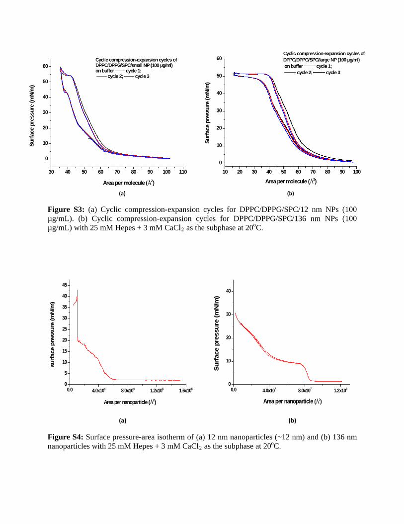
To study the surface activity of bare NPs, the nanoparticlesolutions were spread onto the buffered subphase and com-pressed to achieve very low area per nanoparticle values astechnically feasible. Using film balance, often, moleculararrangements can be contemplated based on the surfaceactivity of the molecules (or particles) of interest. Highersurface activity for a wide range of area per nanoparticle im-plies a higher level of interaction between the molecules andhence lesser affinity between the NPs and the aqueous sub-phase. This consequently gives an idea of the hydrophobic-ity of the nanoparticle and hence the high free energy barrierencountered by the NPs to enter into the subphase.
As shown in Fig. S4 the wide range of area per moleculevalues for 136 nm NPs reveal the high surface activity ofthese particles, which can be compressed to very low areaper nanoparticle values without collapsing into the sub-phase. The 12 nm NPs, on the other hand, show compara-tively less surface activity properties and display a suddenincrease in surface pressure and a sudden collapse oncompression. The wider range of surface activity can beexplained by the very high free energy barrier encounteredby the 136 nm hydrophobic NPs at the aqueous interfaceto enter into the aqueous subphase. The free energy barrieris also encountered by the 12 nm NPs; however, the free en-ergy barrier experienced by the 136 nm NPs is much largerinhibiting the collapse of the NPs into the subphase.
Size-dependent effect of NPs on multilayerprotrusion structures
To study the surface topology of the LS model system at theplateau region in the presence of both 12 and 136 nm NPsthe AFM technique was used. The lipid film was transferredat the plateau region of the isotherm from the aqueoussurface onto a solid substrate and scanned under AFM. Tostudy the surface topology of the model LS film in thepresence of 136 nm NPs at the enhanced plateau region,
FIGURE 3 (a) Pressure-Area isotherm for
DPPC/DPPG/SP-C (80:20:0.4 mol%) þ 12 nm
NPs. (b) Pressure-Area isotherm for DPPC/
DPPG/SP-C (80:20:0.4 mol%) þ 136 nm NPs.
All measurements were done on 25 mM Hepes þ3 mM CaCl2 as the subphase at 20�C. To see this
figure in color, go online.
Biophysical Journal 106(1) 289–298

294 Dwivedi et al.
the lipid film was transferred at both the initial plateau re-gion and at the end plateau region.
The AFM scan for pure DPPC/DPPG/SP-C(80:20:0.4 mol%) monolayer lipid film at the plateau regionshows network structures that are multilayer protrusions aspreviously observed (22).
Surface topology analysis of LS model system in the pres-ence of 12 nm NPs
The protrusion structures are not affected noticeably by thepresence of 12 nm NPs, in the applied concentration(Fig. 4 b). They are visibly localized around the protrusionstructures (marked by arrows). High-resolution scanningconfirms the presence of NPs associated with the protrusionstructures (see Fig. 5 a).
Surface topology analysis of LS model system in the pres-ence of 136 nm NPs
AFM scans for the LS model system in the presence of136 nm NPs reveal a significant lateral disruption of the
FIGURE 4 AFM topography images of (a) pure DPPC/DPPG/SP-C (80:20:0
region; (c) with 100 mg/mL 136 nm NPs transferred at initial plateau region; (d) w
on 25 mM Hepes þ 3 mM CaCl2 as the subphase at 20�C. The clusters of NPs a
see this figure in color, go online.
Biophysical Journal 106(1) 289–298
network structures (Fig. 4, c and d). At the initial plateauregion, the network structures consisting of multilayer pro-trusions are intact (Fig. 4 c). However, at the end plateauregion, the network structures are considerably disruptedforming laterally disordered profuse multilayer protrusions(Fig. 4 d). With increasing concentration of NPs the lateraldisorder increases significantly as shown in Fig. S6. Thehighest clusters with a height of around 100 nm (markedby arrows) were imaged under high resolution. Theseimages clearly show the presence of 136 nm NPs associatedwith the protrusion structures (Fig. 5 b). The topographyimages are supplemented by phase images that have higherclarity revealing a cluster of NPs (Fig. 5 c). It should benoted that the size measured for these NPs are in aqueousdispersions. However, the 136 nm NPs present on themica substrate within the LS model system film are driedrendering the size to be around 100 nm because of the elasticproperties manifested by the 136 nm NPs.
The 136 nm NPs visible in the phase images are similar tothose obtained in TEM micrograph images (Fig. S7). The
.4 mol%) monolayer lipid film; (b) with 100 mg/mL 12 nm NPs at plateau
ith 100 mg/mL 136 nm NPs at end plateau region. The film was compressed
round the protrusion structures are marked by arrows. Scale bar is 5 mm. To

FIGURE 5 High-resolution imaging for DPPC/DPPG/SP-C (80:20:0.4
mol%) monolayer lipid films. (a) 3D topography image with 12 nm NPs;
(b) 3D topography image with 136 nm NPs; (c) Phase images showing
136 nm NPs with higher clarity; (d) Height profile analysis for 136 nm
NPs clusters. To see this figure in color, go online.
Size Influences Effect of Hydrophobic NPs on LS Model System 295
136 nm NPs appear to be nonrigid as evidenced by surfacedeformation during packing (Fig. 5 c, Fig. S7 b). Hence, thenegative phase change at the top of the 136 nm NPs could beexplained by the ability of the cantilever to press into thenanoparticle during measurement. The height profile ofthe nanoparticle cluster shows height up to 100 nm withcurved edges corresponding to the shape of the nanoparticle.
As shown by the hysteresis studies, the continuouscompression of the LS model system film in the presenceof 136 nm NPs does not show any irreversible damage tothe model surfactant film due to a loss of any componentinto the subphase (Fig. S3). This is consistent with theAFM studies, which show the formation of multilayer stepswith step heights of 5 nm, i.e., for a bilayer (Fig. 6, markedby arrows) around the NPs. Hence, in the presence of
FIGURE 6 AFM images of for DPPC/DPPG/SP-C (80:20:0.4 mol%) mono
(marked by arrows). The AFM image is accompanied by its height profiles to t
136 nm NPs, the structures formed are indeed multilayerprotrusion structures and not collapse structures due to adisruption of the film.
Effect of nanoparticle size on vesicle insertionkinetics of model LS films
The natural LS monolayer is constantly recycled and replen-ished with new lipid material from the subphase by thevesicle insertion process. This process is essential for main-taining the integrity and functional activity of the lung sur-factant monolayer (32). We have tried to mimic this processby inserting DPPC/DPPG (4:1) LUVs into the subphasecontaining model LS film at the air-water interface. Themodel LS monolayer is compressed up to 25 mN/m, stabi-lized for 10 min, and the LUVs are inserted into the sub-phase. The increase in surface pressure is monitored withtime to obtain an isotherm, which gives information aboutthe insertion kinetics of the vesicles into the monolayer.
Fig. 7 illustrates the effect of different sized NPs on thevesicle insertion kinetics of model LS monolayer. The pres-ence of higher numbers of 12 nm NPs does not cause a sig-nificant effect on the vesicle insertion process of this modelLS film. However, in the presence of a comparatively lowernumber of 136 nm NPs, the vesicle insertion process isconsiderably inhibited.
DISCUSSION
Previously, we studied the effect of hydrophobic NPs(AmorSil20) with a diameter of 20 nm and showed thatthese NPs are localized around the multilayer protrusionstructures. The NPs with 20 nm diameter did not causeany significant damage to the lung surfactant surface activ-ity except at a very high concentration of 1000 mg/mLwherein the plateau formation was inhibited. The multilayerprotrusion structures consequently were also unaffected by
layer lipid films with 136 nm NPs showing presence of multilayer steps
he right. To see this figure in color, go online.
Biophysical Journal 106(1) 289–298

FIGURE 7 Vesicle insertion kinetic studies for DPPC/DPPG/SP-C
(0.4 mol%) monolayer with different numbers of 12 nm and 136 nm
NPs. The DPPC/DPPG (4:1) vesicles were inserted at 25 mN/m surface
pressure. All measurements were done on 25 mM Hepes and 3 mM
CaCl2 as subphase at 20�C. To see this figure in color, go online.
296 Dwivedi et al.
the presence of these hydrophobic NPs. Furthermore, theNPs were confirmed to retain in the model LS film aftercontinuous compression expansion cycles. In this study,we report that this effect of hydrophobic NPs is size depen-dent and that the effect of 136 nm NPs is much more struc-ture damaging.
The surface activity of model LS films remained unaf-fected in the presence of 12 nm NPs, except at very highconcentrations similar to the 20 nm NPs. However, withthe increasing concentration of 136 nm NPs, the plateauregion was considerably enhanced, rendering the surfactantfilm incapable of increasing the surface pressure to thedesired value, thus causing potential lung surfactantdysfunction.
The AFM scans of the model LS films at the plateauregion in the presence of 12 nm NPs displayed resultssimilar to those obtained with 20 nm NPs. The 12 nm hydro-phobic NPs are localized around the protrusion structures,also evidenced by the high-resolution images. The networkstructures consisting of multilayer protrusions are unalteredin the presence of both 12 and 20 nm hydrophobic NPs. For136 nm NPs the lipid film was transferred at two differentregions of the plateau to examine the surface topographyof multilayer protrusion structures at different compressedstates. The AFM scans for initial plateau regions show nosignificant change in the network structures. However, atthe end plateau region, the network structures are laterallydisrupted forming profuse multilayer protrusion structures.The 136 nm NPs can be spotted as clusters associatedwith the protrusion structures, also confirmed by the high-resolution images. Furthermore, we show that the profusestructures present are indeed multilayers formed due tocontinuous compression and not random collapse structures.
These enhanced multilayered protrusion structures can bewell correlated with the enhanced plateau region in the
Biophysical Journal 106(1) 289–298
isotherm of the model LS film in the presence of 136 nmNPs. The multilayer protrusion formation is explained byselective squeeze out of the fluid component of the film.Enhanced multilayer protrusion formation implies anincreased amount of lipid molecules in the fluid componentof the film. In addition, lateral disruption of the networkstructures implies increased mixing of rigid and fluiddomains, i.e., decreased phase separation. Furthermore,the LS model film does not exhibit any variation in thehysteresis of the isotherm in the presence of any of theNPs with respect to multiple compression reexpansioncycles. Hence, the NPs are retained in the surfactant filmpossibly due to their highly hydrophobic surface and conse-quently high free energy barrier to enter into the subphase.
The isotherms for bare NPs illustrate high surface activityof the 136 nm NPs suggesting a high-energy barrier encoun-tered by the 136 nm hydrophobic NPs for penetrating intothe aqueous subphase. This phenomenon also partially ex-plains the noncollapse behavior of the LS model system inthe presence of the 136 nm NPs even after continuouscompression. The hydrophobic NPs interact with the hydro-phobic acyl chains of the lipid molecules and retain the lipidmolecules along with it preventing collapse. We expect asimilar interaction in the case of 12 nm NPs; however, itis not prominently visible in these data sets.
Because DPPC forms the major component of the modelLS film, any alteration in its phase behavior due to the pres-ence of differently sized NPs can be extrapolated to themodel LS film. Hence, the size-dependent effect of NPson phase behavior and domain morphology of DPPC mono-layers was studied. The isotherm of a DPPC monolayer didnot display any appreciable change in the presence of 12 nmNPs. However, in the presence of 136 nm NPs, the LE-LCcoexistence region was diminished. To emphasize the effectof 136 nm NPs on the isotherms of the DPPC monolayer, wehave calculated the compressibility values at the LC phaseregion. With increasing concentration of 136 nm NPs thecompressibility values increase, which imply decreasedcooperativity between lipid molecules. Hence, 136 nmNPs interfere with the lipid packing order and decreaseintermolecular interactions. This can also be true for12 nm NPs but the isotherms data alone for 12 nm NPsare not conclusive.
The epifluorescence microscopy images reveal slightlydisrupted domain morphology for DPPC monolayers inthe presence of 12 nm NPs. Furthermore, due to the pres-ence of the high numbers of these fluorescent 12 nm NPs,they are clearly visible at high surface pressure. In the pres-ence of 136 nm NPs, however, the domain morphology istremendously disrupted causing branching of the domainsat low surface pressure and formation of mesh-like networkstructures at high surface pressure. The disruption in thedomain morphology implies probably localization of theNPs at the phase boundaries and decreased line tensionsuggests decreased phase separation. This is in accordance

Size Influences Effect of Hydrophobic NPs on LS Model System 297
with the AFM scans of DPPC/DPPG/SP-C mixtures whereDPPC is the major component. The lateral disruption ofthe network structures in the AFM scans of DPPC/DPPG/SP-C mixture are rationalized because of increased mixingof rigid and fluid domains, i.e., decreased phase separation.Hence, in the presence of 136 nm NPs, line tension betweendifferent phases is reduced largely causing mixing of do-mains in the lipid film, which is responsible for lateraldisruption of the network structures. This increase in thefluid component causes enhanced multilayer protrusionformation consequently causing potential lung surfactantdysfunction.
A 136 nm nanoparticle should occupy around 17,000 nm2
area on the aqueous subphase, whereas the area occupied bythe lipid molecule is 0.6 nm2, which is almost 106 timessmaller than that of the nanoparticle. Considering that thenumber of lipid molecules is 105 per nanoparticle, the totalarea occupied by the 136 nm NPs should be almost of thesame order as the total area occupied by the lipid molecules.Given this fact, the whole isotherm of a DPPC monolayershould be shifted toward higher area per molecule values(as opposed to expansion of LE phase region alone) toaccount for the area occupied by the 136 nm nanoparticle.However, this is not the case even at the highest concentra-tion of the 136 nm NPs. This suggests that the lipid mole-cules are displaced from the air-water interface to thesurface of the nanoparticle attributable to hydrophobic inter-action between the acyl chains and the hydrophobic nano-particle surface. Thus, it renders fewer lipid molecules onthe aqueous subphase leaving the total area occupied essen-tially the same. In addition, it should be noticed that thetransition pressure is unaltered in the presence of NPs indi-cating that there might be a possible phase separation be-tween the lipid bound to the nanoparticles and the freelipid molecules.
The presence of 136 nm NPs also has a significant effecton the vesicle insertion process as seen from the vesicleinsertion kinetics studies. The vesicle insertion process isthought to be a two-step mechanism—initial Caþ2-mediatedadsorption of the vesicles onto the model LS monolayer,rupture of the vesicles, and reincorporation of the lipidsfrom the vesicles into the monolayer facilitated by surfac-tant proteins. As seen in Fig. 7, the presence of highernumbers of 12 nm NPs has a negligible effect on the inser-tion kinetics of the model LS film. At very high concentra-tions, the inhibition effect of these NPs is much moreprominent (data not shown) where the half-time increaseswith increasing concentration. These results are similar tothose observed with the 20 nm diameter NPs. In thepresence of 136 nm NPs the inhibitory effect is visiblyprominent even at comparatively lower numbers of NPs.Another important observation is that the shape of the curveis hyperbolic in the presence of 12 nm NPs and sigmoidal inthe presence of 136 nm NPs. The inhibitory effect of the12 nm NPs can be explained by a direct or indirect inter-
action of the NPs with the surfactant protein-C, thus inhib-iting its function in vesicle insertion. This is alsoemphasized by the fact that the isotherm of model LS mono-layers show inhibited plateau regions in the presence of veryhigh concentrations of 12 nm NPs. As previously discussed,these NPs are known to be found associated with the protru-sion structures and hence with the fluid component of thefilm. The surfactant-specific proteins are also known to bepresent in the DPPG-enriched fluid component of the film.This increases the possibility of an interaction between thesurfactant protein-C and the 12 nm nanoparticle, which isobservable only at very high concentrations of the NPs.The effect of 136 nm nanoparticles on the vesicle insertioncurve however is significantly different and cannot be sim-ply explained with the previous theory of nanoparticle-protein interaction. From the previous discussion, wededuced a possible interaction of the fluid component ofthe model LS film with the nanoparticles. The fluid com-ponent is mainly enriched in DPPG. DPPG being negativelycharged is also involved in the adsorption of the vesicles,mediated by Caþ2 ions, and plays an important role in theelectrostatic interaction between the lipid monolayer andthe LUVs in the subphase (33,34). In the presence of136 nm NPs, DPPG molecules may possibly form a lipidlayer on the nanoparticle and hence the number of DPPGmolecules available for adsorption of LUVs would decreaseleading to an initial lag phase in the insertion curve. With theeventual adsorption and reincorporation of the new lipidmaterial from the subphase to the monolayer, the numberof DPPG molecules in the monolayer increases subse-quently leading to an increased adsorption of the vesicles.This process is similar to a cooperative process and hencewould result in a sigmoidal curve as obtained in Fig. 7.Due to its size, 136 nm NPs have much more area to occupylarger amounts of lipid molecules than the 12 nm NPs.Hence, even a small number of 136 nm NPs has a consider-ably higher inhibitory effect on the insertion kinetics thanthe 12 nm NPs.
CONCLUSION
This study shows the effect of different sized NPs on modelLS films and proves that the size of the NPs is an importantfactor responsible for causing severe structural and func-tional damage to the model LS films. The surface activityof the model LS film is almost unaffected in the presenceof 12 nm NPs, which is similar to the results obtained for20 nm NPs. However, in the presence of 136 nm NPs theplateau region is enhanced significantly rendering the modelLS film highly compressible. The AFM scans reveal forma-tion of profuse multilayer protrusion structures responsiblefor the enhanced plateau region. The fluorescence micro-scopy images for DPPC monolayers in the presence of136 nm NPs show a decreased tendency for a phase separa-tion due to reduction in the line tension. This is in
Biophysical Journal 106(1) 289–298

298 Dwivedi et al.
accordance with the AFM studies where the network struc-ture of model LS film is disrupted in the presence of 136 nmNPs implying diffusive mixing of rigid and fluid domains.The vesicle insertion kinetics is inhibited in the presenceof fewer numbers of 136 nm NPs as compared with the12 nm NPs where the effect is negligible. The mechanismfor the inhibitory effect is also discussed in the study basedon the size of the NPs. It is worth noticing that fewernumbers of 136 nm NPs have much more structural andfunctional damaging effect than the comparatively highernumber of 12 nm NPs.
SUPPORTING MATERIAL
Seven figures are available at http://www.biophysj.org/biophysj/
supplemental/S0006-3495(13)01246-0.
We acknowledge Amit Kumar Sachan for fruitful discussions especially
regarding the high pressure measurements on the film balance.
This work was supported by the Deutsche Forschungsgemeinschaft,
Schwerpunktprogramm 1313 and by the NRW International Graduate
School of Chemistry Munster.
REFERENCES
1. Goerke, J. 1974. Lung surfactant. Biochim. Biophys. Acta.344:241–261.
2. Hills, B. A. 1990. The role of lung surfactant. Br. J. Anaesth. 65:13–29.
3. Schurch, S., J. Goerke, and J. A. Clements. 1976. Direct determinationof surface tension in the lung. Proc. Natl. Acad. Sci. USA. 73:4698–4702.
4. Yu, S., P. G. R. Harding, ., F. Possmayer. 1983. Bovine pulmonarysurfactant: chemical composition and physical properties. Lipids.18:522–529.
5. Goerke, J. 1998. Pulmonary surfactant: functions and molecularcomposition. Biochim. Biophys. Acta. 1408:79–89.
6. Veldhuizen, R., K. Nag, ., F. Possmayer. 1998. The role of lipids inpulmonary surfactant. Biochim. Biophys. Acta. 1408:90–108.
7. Veldhuizen, E. J. A., and H. P. Haagsman. 2000. Role of pulmonarysurfactant components in surface film formation and dynamics.Biochim. Biophys. Acta. 1467:255–270.
8. Weaver, T. E., and J. J. Conkright. 2001. Function of surfactant proteinsB and C. Annu. Rev. Physiol. 63:555–578.
9. Reid, K. B. M. 1998. Functional roles of the lung surfactant proteinsSP-A and SP-D in innate immunity. Immunobiology. 199:200–207.
10. Schurch, S., R. Qanbar,., F. Possmayer. 1995. The surface-associatedsurfactant reservoir in the alveolar lining. Biol. Neonate. 67(Suppl 1):61–76.
11. Amrein, M., A. von Nahmen, and M. Sieber. 1997. A scanning force-and fluorescence light microscopy study of the structure and functionof a model pulmonary surfactant. Eur. Biophys. J. 26:349–357.
12. Krol, S., M. Ross, ., A. Janshoff. 2000. Formation of three-dimen-sional protein-lipid aggregates in monolayer films induced by surfac-tant protein B. Biophys. J. 79:904–918.
13. Perez-Gil, J. 2008. Structure of pulmonary surfactant membranes andfilms: the role of proteins and lipid-protein interactions. Biochim.Biophys. Acta. 1778:1676–1695.
Biophysical Journal 106(1) 289–298
14. Rugonyi, S., S. C. Biswas, and S. B. Hall. 2008. The biophysical func-tion of pulmonary surfactant. Respir. Physiol. Neurobiol. 163:244–255.
15. Oosterlaken-Dijksterhuis, M. A., H. P. Haagsman, ., R. A. Demel.1991. Characterization of lipid insertion into monomolecular layersmediated by lung surfactant proteins SP-B and SP-C. Biochemistry.30:10965–10971.
16. Ross, M., S. Krol,., H. J. Galla. 2002. Kinetics of phospholipid inser-tion into monolayers containing the lung surfactant proteins SP-B orSP-C. Eur. Biophys. J. 31:52–61.
17. Muhlfeld, C., B. Rothen-Rutishauser,., P. Gehr. 2008. Interactions ofnanoparticles with pulmonary structures and cellular responses. Am. J.Physiol. Lung Cell. Mol. Physiol. 294:L817–L829.
18. Konduru, N. V., Y. Y. Tyurina,., V. E. Kagan. 2009. Phosphatidylser-ine targets single-walled carbon nanotubes to professional phagocytesin vitro and in vivo. PLoS ONE. 4:e4398.
19. Kapralov, A. A., W. H. Feng, ., V. E. Kagan. 2012. Adsorption ofsurfactant lipids by single-walled carbon nanotubes in mouse lungupon pharyngeal aspiration. ACS Nano. 6:4147–4156.
20. Schleh, C., C. Muhlfeld, ., J. M. Hohlfeld. 2009. The effect of tita-nium dioxide nanoparticles on pulmonary surfactant function and ultra-structure. Resp. Res. 10:90.
21. Fan, Q. H., Y. E. Wang, ., Y. Y. Zuo. 2011. Adverse biophysicaleffects of hydroxyapatite nanoparticles on natural pulmonary surfac-tant. ACS Nano. 5:6410–6416.
22. Harishchandra, R. K., M. Saleem, and H. J. Galla. 2010. Nanoparticleinteraction with model lung surfactant monolayers. J. R. Soc. Interface.7 (Suppl 1):S15–S26.
23. Bakshi, M. S., L. Zhao, ., N. O. Petersen. 2008. Metal nanoparticlepollutants interfere with pulmonary surfactant function in vitro.Biophys. J. 94:855–868.
24. Nel, A., T. Xia, ., N. Li. 2006. Toxic potential of materials at thenanolevel. Science. 311:622–627.
25. Sachan, A. K., R. K. Harishchandra, ., H. J. Galla. 2012. High-reso-lution investigation of nanoparticle interaction with a model pulmonarysurfactant monolayer. ACS Nano. 6:1677–1687.
26. Krol, S., A. Janshoff, ., H. J. Galla. 2000. Structure and function ofsurfactant protein B and C in lipid monolayers: a scanning force micro-scopy study. Phys. Chem. Chem. Phys. 2:4586–4593.
27. von Nahmen, A., A. Post, ., M. Sieber. 1997. The phase behavior oflipid monolayers containing pulmonary surfactant protein C studied byfluorescence light microscopy. Eur. Biophys. J. 26:359–369.
28. von Nahmen, A., M. Schenk, ., M. Amrein. 1997. The structure of amodel pulmonary surfactant as revealed by scanning force microscopy.Biophys. J. 72:463–469.
29. Haagsman, H. P., S. Hawgood,., B. J. Benson. 1987. The major lungsurfactant protein, SP 28-36, is a calcium-dependent, carbohydrate-binding protein. J. Biol. Chem. 262:13877–13880.
30. Taneva, S. G., J. Stewart, ., K. M. W. Keough. 1998. Method ofpurification affects some interfacial properties of pulmonary surfactantproteins B and C and their mixtures with dipalmitoylphosphatidylcho-line. Biochim. Biophys. Acta. 1370:138–150.
31. Scherer, C., S. Utech,., M. Maskos. 2010. Synthesis, characterizationand fine-tuning of bimodal poly(organosiloxane) nanoparticles. Poly-mer (Guildf.). 51:5432–5439.
32. Schartl, W. 2007. Light Scattering from Polymer Solutions and Nano-particle Dispersions. Springer, Berlin Heidelberg.
33. Jungmann, N., M. Schmidt,., J. Ebenhoch. 2002. Synthesis of amphi-philic poly(organosiloxane) nanospheres with different core-shellarchitectures. Macromolecules. 35:6851–6857.
34. Koshkina, O., C. Bantz, ., M. Maskos. 2011. Fluorophore-labeledsiloxane-based nanoparticles for biomedical applications. Macromol.Symp. 309–310:141–146.

Size Influences the Effect of Hydrophobic
Nanoparticles on Lung Surfactant Model Systems
Mridula.V. Dwivedi†‡, Rakesh Kumar Harishchandra†║, Olga Koshkina┴¶, Michael Maskos§¶,
Hans-Joachim Galla†*
† Institute of Biochemistry, Westfälische Wilhelms Universität, Wilhelm-Klemm-Str.2, 48149 Münster, Germany.
‡ NRW International graduate school of chemistry, University of Muenster, Wilhelm-Klemm-Str. 10, 48149 Muenster, Germany
┴ BAM Federal Institute for Materials Research and Testing, Unter den Eichen 87, 12205 Berlin, Germany
§ Institute of Physical Chemistry, Johannes Gutenberg University, Jakob-Welder Weg 11, 55128 Mainz, Germany.
¶ Institut für Mikrotechnik Mainz (IMM), Carl-Zeiss-Str. 18-20, 55129, Mainz, Germany.
║current address: Dept. of Chemistry & Biochemistry Worcester Polytechnic Institute (WPI) 60 Prescott Street, Worcester, MA 01605 USA.
*Corresponding author: [email protected]
Mailing address: Institute of Biochemistry, Westfälische Wilhelms Universität, Wilhelm-Klemm-Str. 2, 48149 Münster, Germany. Tel.: +49-251-8333200. Fax: +49-251-8333206.
Keywords: Lung Surfactant, nanoparticle size, Surface activity, atomic force microscopy, vesicle insertion kinetics, Multilayer protrusion structures.

Supporting Material
Figure S1: Epi-fluorescence microscopy images for DPPC in the presence of increasing concentration of small nanoparticles (~12 nm) on water as the subphase at 20oC. The DPPC monolayer is doped with 0.5 mol% BODIPY-PC which preferentially partitions into the liquid expanded phase. The images are taken at varying surface pressure values. Scale bar is 50 µm.
Figure S2: Epi-fluorescence microscopy images for DPPC in the presence of increasing concentration of large nanoparticles (~136 nm) on water as the subphase at 20oC. The DPPC monolayer is doped with 0.5 mol% BODIPY-PC which preferentially partitions into the liquid expanded phase. The images are taken at varying surface pressure values. Scale bar is 50 µm.
(a) (b)
Figure S3: (a) Cyclic compression-expansion cycles for DPPC/DPPG/SPC/12 nm NPs (100 µg/mL). (b) Cyclic compression-expansion cycles for DPPC/DPPG/SPC/136 nm NPs (100 µg/mL) with 25 mM Hepes + 3 mM CaCl2 as the subphase at 20oC.
(a) (b)
Figure S4: Surface pressure-area isotherm of (a) 12 nm nanoparticles (~12 nm) and (b) 136 nm nanoparticles with 25 mM Hepes + 3 mM CaCl2 as the subphase at 20oC.
0.0 4.0x10⁷ 8.0x10⁷ 1.2x1080
10
20
30
40
Area per nanoparticle (Å2)
Surf
ace
pres
sure
(mN
/m)
0.0 4.0x10⁴ 8.0x10⁴ 1.2x10⁵ 1.6x1050
5
10
15
20
25
30
35
40
45
Area per nanoparticle (Å2)
surf
ace
pres
sure
(mN
/m)
10 20 30 40 50 60 70 80 90 100
0
10
20
30
40
50
60
Surfa
ce p
ress
ure
(mN/
m)
Area per molecule (Å2)
Cyclic compression-expansion cycles ofDPPC/DPPG/SPC/large NP (100 µg/ml) on buffer cycle 1; cycle 2; cycle 3
30 40 50 60 70 80 90 100 110
0
10
20
30
40
50
60
Area per molecule (Å2)
Surfa
ce p
ress
ure
(mN/
m)
Cyclic compression-expansion cycles of DPPC/DPPG/SPC/small NP (100 µg/ml)on buffer cycle 1; cycle 2; cycle 3

Figure S5: AFM topography images of (a) pure DPPC/DPPG/SPC (80:20:0.4 mol %) monolayer lipid film (b) with 50 µg/mL 12 nm nanoparticles (c) with 100 µg/mL 12 nm nanoparticles transferred at plateau region. The film was compressed on 25 mM Hepes + 3 mM CaCl2 as the subphase at 20oC. The clusters of nanoparticles around the protrusion structures are marked by arrows.

Figure S6: AFM topography images of DPPC/DPPG/SPC (80:20:0.4 mol %) monolayer lipid film with (a) 50 µg/mL 136 nm nanoparticles at initial plateau region (b) 50 µg/mL 136 nm nanoparticles at end plateau region (c) 100 µg/mL 136 nm nanoparticles at initial plateau region (d) 100 µg/mL 136 nm nanoparticles at end plateau region. The film was compressed on 25 mM Hepes + 3 mM CaCl2 as the subphase at 20oC. The clusters of nanoparticles around the protrusion structures are marked by arrows.

Figure S7: TEM micrograph images of (a) 12 nm nanoparticles (b) 136 nm nanoparticles. Samples were deposited from chloroform. Scale bar is 100 nm.