Site-specific acetylation of ISWI by GCN5
11
BioMed Central Page 1 of 11 (page number not for citation purposes) BMC Molecular Biology Open Access Research article Site-specific acetylation of ISWI by GCN5 Roger Ferreira 1,2 , Anton Eberharter 1 , Tiziana Bonaldi 1,3 , Mariacristina Chioda 1 , Axel Imhof 1 and Peter B Becker* 1 Address: 1 Adolf-Butenandt-Institut, Molekularbiologie, 80336 München, Germany, 2 European Patent Office – Biotechnology, D-80339 München, Germany and 3 Max-Planck Institut für Biochemie, D-82152 Martinsried, Germany Email: Roger Ferreira - [email protected]; Anton Eberharter - [email protected]; Tiziana Bonaldi - [email protected]; Mariacristina Chioda - [email protected]; Axel Imhof - [email protected]; Peter B Becker* - [email protected] muenchen.de * Corresponding author Abstract Background: The tight organisation of eukaryotic genomes as chromatin hinders the interaction of many DNA-binding regulators. The local accessibility of DNA is regulated by many chromatin modifying enzymes, among them the nucleosome remodelling factors. These enzymes couple the hydrolysis of ATP to disruption of histone-DNA interactions, which may lead to partial or complete disassembly of nucleosomes or their sliding on DNA. The diversity of nucleosome remodelling factors is reflected by a multitude of ATPase complexes with distinct subunit composition. Results: We found further diversification of remodelling factors by posttranslational modification. The histone acetyltransferase GCN5 can acetylate the Drosophila remodelling ATPase ISWI at a single, conserved lysine, K753, in vivo and in vitro. The target sequence is strikingly similar to the N- terminus of histone H3, where the corresponding lysine, H3K14, can also be acetylated by GCN5. The acetylated form of ISWI represents a minor species presumably associated with the nucleosome remodelling factor NURF. Conclusion: Acetylation of histone H3 and ISWI by GCN5 is explained by the sequence similarity between the histone and ISWI around the acetylation site. The common motif RK T / S xGx(K ac )xP R / K differs from the previously suggested GCN5/PCAF recognition motif GKxxP. This raises the possibility of co-regulation of a nucleosome remodelling factor and its nucleosome substrate through acetylation of related epitopes and suggests a direct crosstalk between two distinct nucleosome modification principles. Background Disruption of DNA-histone interactions by nucleosome remodelling ATPases may lead to a variety of transitions of chromatin structure, such as the partial or complete dis- assembly of nucleosomes, the exchange of histones, or the sliding of intact histone octamers on DNA [1-4]. In many cases their activity is focused on local disruption of the nucleosomal fibre through recruitment of DNA-binding regulators to promote access of factors further down- stream in the cascade of events that leads to promoter opening [2]. However, genome-wide processes like repli- cation, DNA damage responses or homologous recombi- Published: 30 August 2007 BMC Molecular Biology 2007, 8:73 doi:10.1186/1471-2199-8-73 Received: 10 April 2007 Accepted: 30 August 2007 This article is available from: http://www.biomedcentral.com/1471-2199/8/73 © 2007 Ferreira et al; licensee BioMed Central Ltd. This is an Open Access article distributed under the terms of the Creative Commons Attribution License (http://creativecommons.org/licenses/by/2.0 ), which permits unrestricted use, distribution, and reproduction in any medium, provided the original work is properly cited.
-
Upload
roger-ferreira -
Category
Documents
-
view
212 -
download
0
Transcript of Site-specific acetylation of ISWI by GCN5
BioMed CentralBMC Molecular Biology
ss
Open AcceResearch articleSite-specific acetylation of ISWI by GCN5Roger Ferreira1,2, Anton Eberharter1, Tiziana Bonaldi1,3, Mariacristina Chioda1, Axel Imhof1 and Peter B Becker*1Address: 1Adolf-Butenandt-Institut, Molekularbiologie, 80336 München, Germany, 2European Patent Office – Biotechnology, D-80339 München, Germany and 3Max-Planck Institut für Biochemie, D-82152 Martinsried, Germany
Email: Roger Ferreira - [email protected]; Anton Eberharter - [email protected]; Tiziana Bonaldi - [email protected]; Mariacristina Chioda - [email protected]; Axel Imhof - [email protected]; Peter B Becker* - [email protected]
* Corresponding author
AbstractBackground: The tight organisation of eukaryotic genomes as chromatin hinders the interactionof many DNA-binding regulators. The local accessibility of DNA is regulated by many chromatinmodifying enzymes, among them the nucleosome remodelling factors. These enzymes couple thehydrolysis of ATP to disruption of histone-DNA interactions, which may lead to partial orcomplete disassembly of nucleosomes or their sliding on DNA. The diversity of nucleosomeremodelling factors is reflected by a multitude of ATPase complexes with distinct subunitcomposition.
Results: We found further diversification of remodelling factors by posttranslational modification.The histone acetyltransferase GCN5 can acetylate the Drosophila remodelling ATPase ISWI at asingle, conserved lysine, K753, in vivo and in vitro. The target sequence is strikingly similar to the N-terminus of histone H3, where the corresponding lysine, H3K14, can also be acetylated by GCN5.The acetylated form of ISWI represents a minor species presumably associated with thenucleosome remodelling factor NURF.
Conclusion: Acetylation of histone H3 and ISWI by GCN5 is explained by the sequence similaritybetween the histone and ISWI around the acetylation site. The common motif RKT/SxGx(Kac)xPR/
K differs from the previously suggested GCN5/PCAF recognition motif GKxxP. This raises thepossibility of co-regulation of a nucleosome remodelling factor and its nucleosome substratethrough acetylation of related epitopes and suggests a direct crosstalk between two distinctnucleosome modification principles.
BackgroundDisruption of DNA-histone interactions by nucleosomeremodelling ATPases may lead to a variety of transitionsof chromatin structure, such as the partial or complete dis-assembly of nucleosomes, the exchange of histones, or thesliding of intact histone octamers on DNA [1-4]. In many
cases their activity is focused on local disruption of thenucleosomal fibre through recruitment of DNA-bindingregulators to promote access of factors further down-stream in the cascade of events that leads to promoteropening [2]. However, genome-wide processes like repli-cation, DNA damage responses or homologous recombi-
Published: 30 August 2007
BMC Molecular Biology 2007, 8:73 doi:10.1186/1471-2199-8-73
Received: 10 April 2007Accepted: 30 August 2007
This article is available from: http://www.biomedcentral.com/1471-2199/8/73
© 2007 Ferreira et al; licensee BioMed Central Ltd. This is an Open Access article distributed under the terms of the Creative Commons Attribution License (http://creativecommons.org/licenses/by/2.0), which permits unrestricted use, distribution, and reproduction in any medium, provided the original work is properly cited.
Page 1 of 11(page number not for citation purposes)
BMC Molecular Biology 2007, 8:73 http://www.biomedcentral.com/1471-2199/8/73
nation require chromatin to be dynamic on a global scale.In addition to generating local access to nucleosomalDNA, nucleosome disruption may also have profoundconsequences for the folding of the nucleosomal fibreinto higher order structures [5,6].
One largely unresolved issue to date is how the activity ofchromatin remodelling enzymes is regulated. Establishedregulatory principles that govern, for example, metabolicenzymes also apply to nucleosome remodelling ATPases,but our knowledge is still anecdotal. The expression ofnucleosome remodelling ATPases may be selective. Forexample, the fact that the mammalian ISWI isoformSNF2H is abundant in proliferating cells, whereas therelated SNF2L is enriched in terminally differentiated neu-rons points to functional diversification of related remod-elling enzymes and specialized roles in proliferation/differentiation control [7]. Such a role has also been sug-gested for the SWI2/SNF2-type ATPase hBRM by the earlyfinding that its expression is down-regulated when cellsreceive a mitogenic stimulus or during ras-mediated onco-genic transformation, whereas its forced expression par-tially reverses transformation [8].
A further regulatory strategy involves post-translationalmodifications of enzymes, such as their phosphorylation.Phosphorylation of hBRM and BRG-1 during mitosis cor-relates with dissociation of these remodellers from thechromosomes during condensation [9]. Muchardt andcolleagues also showed that acetylation of hBRM corre-lates with a reduced inhibition of cell growth [10]. Thepossibility of regulating nucleosome remodelling ATPasesby lysine acetylation is intriguing given that properties oftheir substrates, the histones, are most prominently mod-ulated by acetylation at their exposed N-termini [11].
Here we describe another example of potential regulationof a remodelling ATPase by acetylation. We found thatDrosophila ISWI, the founding member of a family ofnucleosome remodelling ATPases, was preferentiallyacetylated by GCN5 at a single lysine within a amino acidsequence of high similarity to the N-terminus of histoneH3. This acetylated lysine corresponds to lysine 14 in his-tone H3 (H3K14), a known target for GCN5, suggestingthat this acetyltransferase may affect the chromatin struc-ture by two distinct strategies: by acetylation of the nucle-osomes and by modification of a nucleosomeremodelling enzyme.
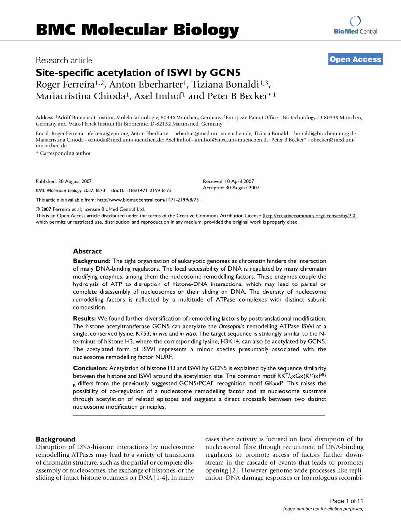
ResultsISWI is acetylated in vivoIn order to explore whether ISWI was acetylated in Dro-sophila cell lines we immunoprecipitated the ATPase fromextracts of SF4 cells (Figure 1A, lanes 1–3). Probing theprecipitate with a pan-acetyl-lysine antibody (αAcLysine)
we detected a labelled protein migrating at the position ofISWI (Figure 1A, lanes 4, 5). To facilitate detection ofacetylated ISWI we treated Kc cells with the histonedeacetylase inhibitor Trichostatin A (TSA), preparedwhole cell extracts and monitored ISWI levels (Figure 1B,lanes 1, 2). We then immunoprecipitated ISWI or proteinscontaining acetylated lysines from these extracts. In theabsence of TSA ISWI was barely detectable in the αAcLy-sine precipitate, which may be due to the inefficiency ofthe antibody and/or the small amounts of acetylated ISWIpresent in Kc cells (Figure 1B, lanes 3–5). However, uponovernight TSA treatment the levels of acetylated ISWIincreased significantly (Figure 1B, lanes 6–8). Takentogether these results suggested that a minor fraction ofISWI was acetylated in Kc and SF4 cells. In order to con-firm this notion by an independent experiment we meta-bolically labelled SF4 and Kc cells by addition of [3H]-acetic acid for 3 hrs to the growth medium, preparedextracts and determined the ISWI levels as before (Figure1C, lanes 9, 10). ISWI was immunoprecipitated fromthese extracts (Figure 1C, lanes 5–8) and acetylated pro-teins in the precipitate were detected by gel electrophore-sis and autoradiography. A band migrating at the positionof ISWI was only detectable in the αISWI precipitate, butwas absent in the control (Figure 1C, lanes 1–4). A secondlabelled band points to an acetylated ISWI-associated pro-tein of unknown identity (asterisk in Figure 1C). Collec-tively, these data demonstrate that a relatively minorfraction of ISWI is acetylated in Drosophila tissue culturecells.
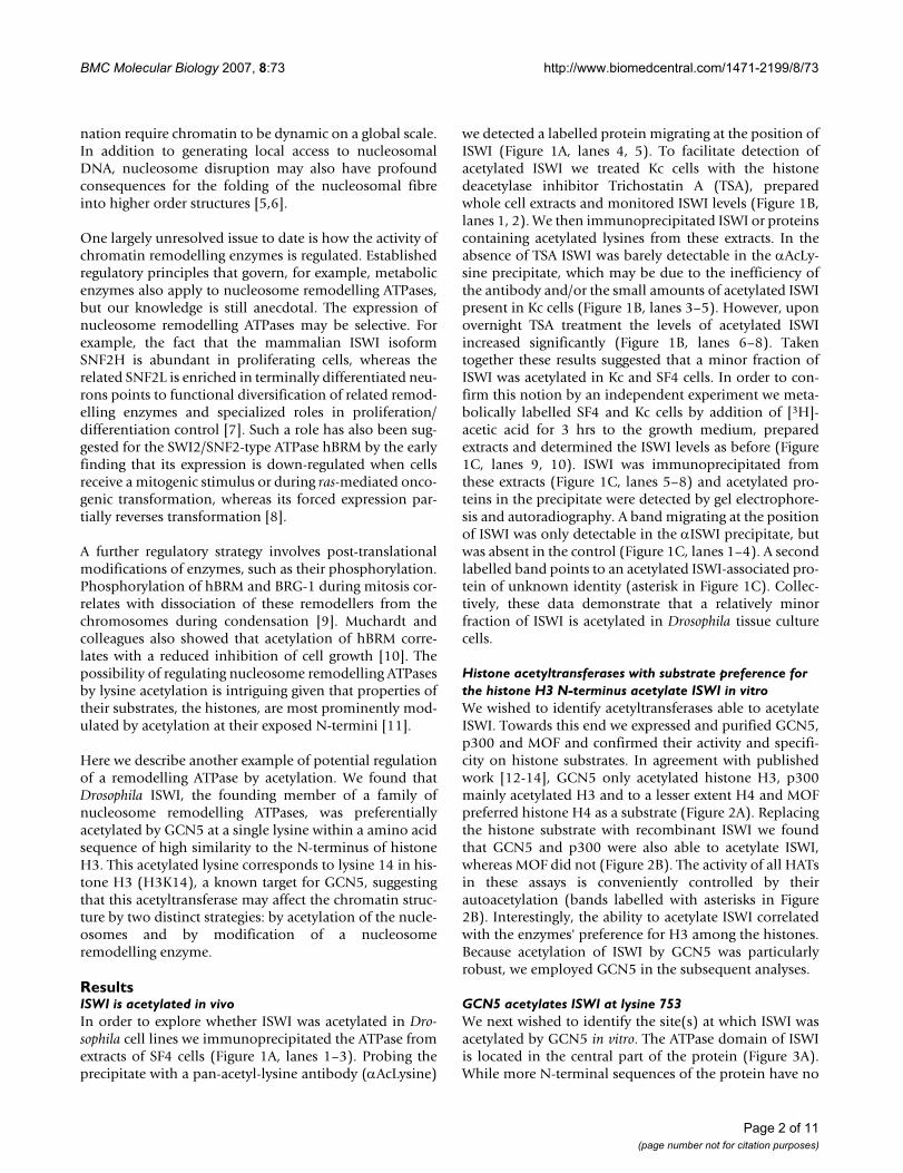
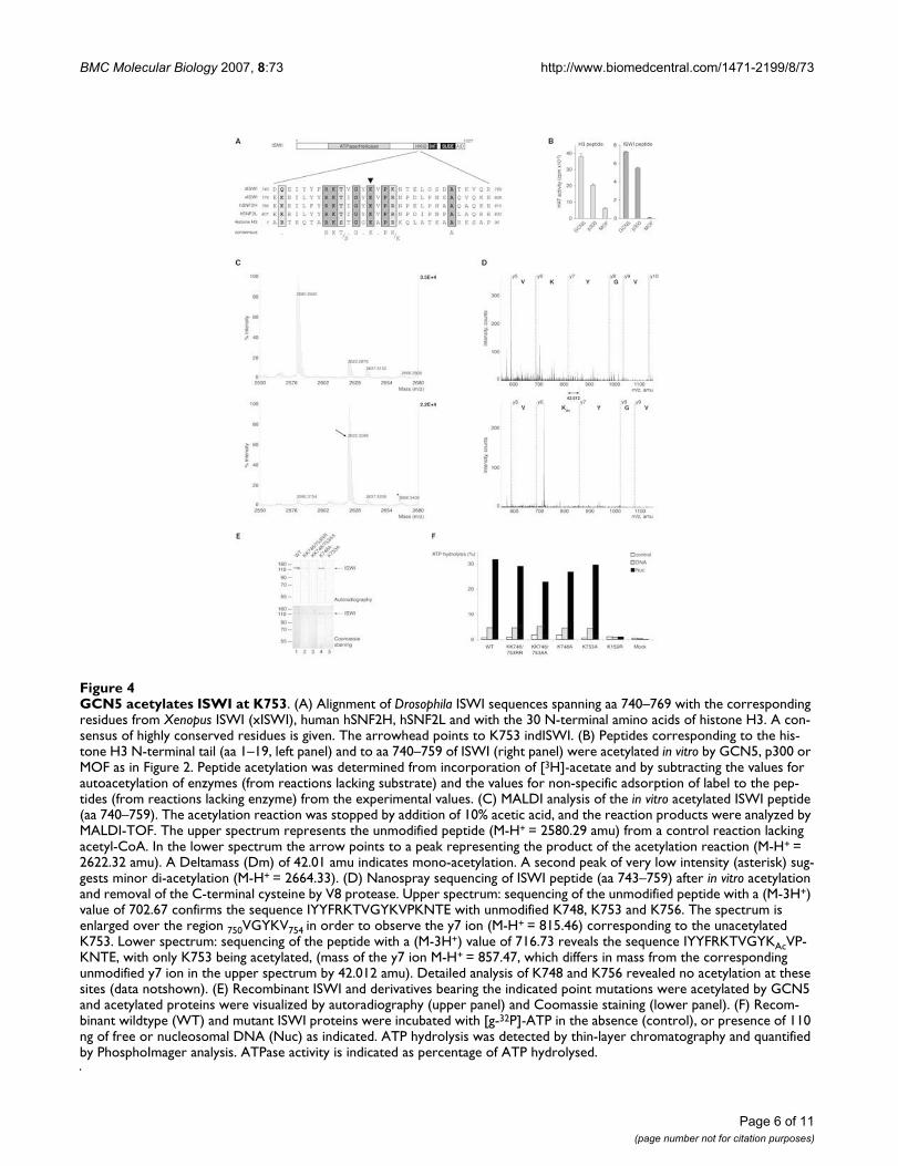
Histone acetyltransferases with substrate preference for the histone H3 N-terminus acetylate ISWI in vitroWe wished to identify acetyltransferases able to acetylateISWI. Towards this end we expressed and purified GCN5,p300 and MOF and confirmed their activity and specifi-city on histone substrates. In agreement with publishedwork [12-14], GCN5 only acetylated histone H3, p300mainly acetylated H3 and to a lesser extent H4 and MOFpreferred histone H4 as a substrate (Figure 2A). Replacingthe histone substrate with recombinant ISWI we foundthat GCN5 and p300 were also able to acetylate ISWI,whereas MOF did not (Figure 2B). The activity of all HATsin these assays is conveniently controlled by theirautoacetylation (bands labelled with asterisks in Figure2B). Interestingly, the ability to acetylate ISWI correlatedwith the enzymes' preference for H3 among the histones.Because acetylation of ISWI by GCN5 was particularlyrobust, we employed GCN5 in the subsequent analyses.
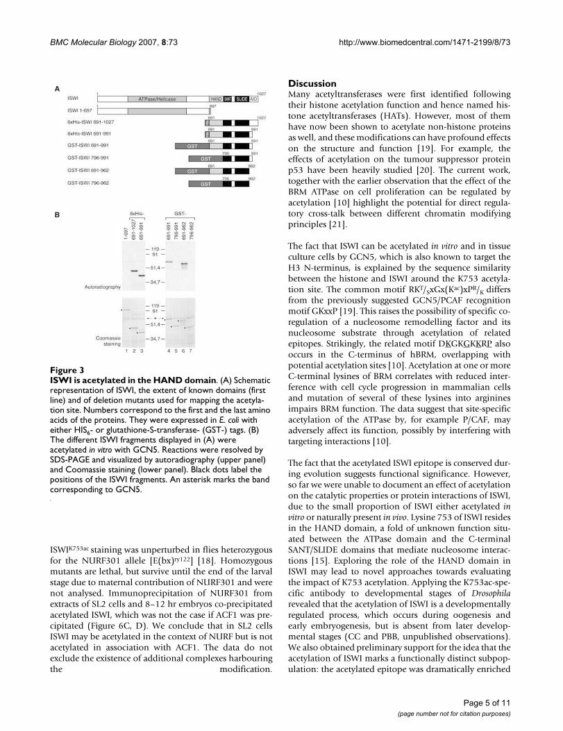
GCN5 acetylates ISWI at lysine 753We next wished to identify the site(s) at which ISWI wasacetylated by GCN5 in vitro. The ATPase domain of ISWIis located in the central part of the protein (Figure 3A).While more N-terminal sequences of the protein have no
Page 2 of 11(page number not for citation purposes)
BMC Molecular Biology 2007, 8:73 http://www.biomedcentral.com/1471-2199/8/73
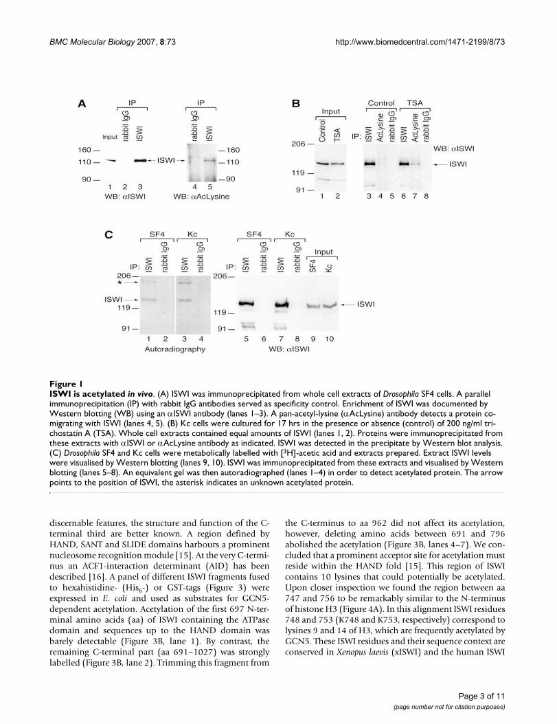
discernable features, the structure and function of the C-terminal third are better known. A region defined byHAND, SANT and SLIDE domains harbours a prominentnucleosome recognition module [15]. At the very C-termi-nus an ACF1-interaction determinant (AID) has beendescribed [16]. A panel of different ISWI fragments fusedto hexahistidine- (His6-) or GST-tags (Figure 3) wereexpressed in E. coli and used as substrates for GCN5-dependent acetylation. Acetylation of the first 697 N-ter-minal amino acids (aa) of ISWI containing the ATPasedomain and sequences up to the HAND domain wasbarely detectable (Figure 3B, lane 1). By contrast, theremaining C-terminal part (aa 691–1027) was stronglylabelled (Figure 3B, lane 2). Trimming this fragment from
the C-terminus to aa 962 did not affect its acetylation,however, deleting amino acids between 691 and 796abolished the acetylation (Figure 3B, lanes 4–7). We con-cluded that a prominent acceptor site for acetylation mustreside within the HAND fold [15]. This region of ISWIcontains 10 lysines that could potentially be acetylated.Upon closer inspection we found the region between aa747 and 756 to be remarkably similar to the N-terminusof histone H3 (Figure 4A). In this alignment ISWI residues748 and 753 (K748 and K753, respectively) correspond tolysines 9 and 14 of H3, which are frequently acetylated byGCN5. These ISWI residues and their sequence context areconserved in Xenopus laevis (xISWI) and the human ISWI
ISWI is acetylated in vivoFigure 1ISWI is acetylated in vivo. (A) ISWI was immunoprecipitated from whole cell extracts of Drosophila SF4 cells. A parallel immunoprecipitation (IP) with rabbit IgG antibodies served as specificity control. Enrichment of ISWI was documented by Western blotting (WB) using an αISWI antibody (lanes 1–3). A pan-acetyl-lysine (αAcLysine) antibody detects a protein co-migrating with ISWI (lanes 4, 5). (B) Kc cells were cultured for 17 hrs in the presence or absence (control) of 200 ng/ml tri-chostatin A (TSA). Whole cell extracts contained equal amounts of ISWI (lanes 1, 2). Proteins were immunoprecipitated from these extracts with αISWI or αAcLysine antibody as indicated. ISWI was detected in the precipitate by Western blot analysis. (C) Drosophila SF4 and Kc cells were metabolically labelled with [3H]-acetic acid and extracts prepared. Extract ISWI levels were visualised by Western blotting (lanes 9, 10). ISWI was immunoprecipitated from these extracts and visualised by Western blotting (lanes 5–8). An equivalent gel was then autoradiographed (lanes 1–4) in order to detect acetylated protein. The arrow points to the position of ISWI, the asterisk indicates an unknown acetylated protein.
Page 3 of 11(page number not for citation purposes)
BMC Molecular Biology 2007, 8:73 http://www.biomedcentral.com/1471-2199/8/73
isoforms hSNF2H and hSNF2L (Figure 4A). We thereforefocused our attention on this part of ISWI.
A peptide corresponding to aa 1–19 of histone H3 isacetylated rather well by GCN5 and p300 in a standardHAT reaction, but less by MOF (Figure 4B, left panel). Bydirect comparison, a peptide spanning the corresponding19 aa of ISWI (Figure 4A) is well acetylated by GCN5, butnot at all by MOF (Figure 4B, right panel). Maldi-TOFmass spectrometry and nano-electrospray sequencingdocumented that the peptide was mono-acetylated (Fig-ure 4C) exclusively at K753 (Figure 4D). In order to testfor the contribution of K753 to ISWI acetylation in thecontext of the entire protein we expressed recombinantISWI derivatives that had either K748 or K753 or bothlysines replaced by alanines (A) or arginines (R). Equiva-lent protein amounts (Figure 4E, lower panel) were usedas substrate for GCN5-dependent acetylation reactions.The ISWI derivative bearing the K748A mutation wasacetylated as the wildtype protein, whereas replacement oflysine 753 by alanine (K753A) led to a significantlyreduced acetylation (Figure 4E, compare lanes 4, 5). Col-lectively the data identify K753 of ISWI, which appearsrelated by sequence context to H3K14, as the major targetof acetylation by GCN5 in vitro.
We were not able to evaluate the effect of acetylation onISWI ATPase activity due to the small fraction of ISWImodified in these in vitro reactions. However, replacementof either lysine 753 or lysine 748 (which corresponds tolysine 9 in H3) by arginine or alanine did not affect theATPase activity in response to DNA or nucleosomes (Fig-ure 4F).
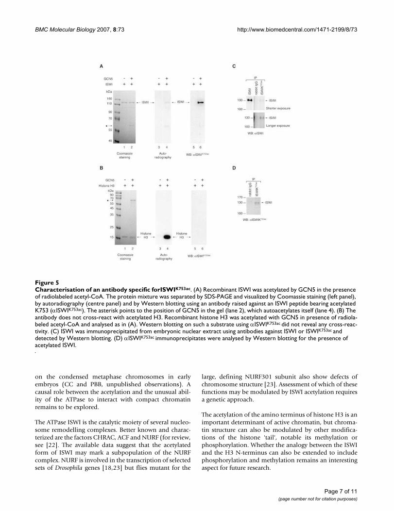
ISWI is acetylated at K753 by GCN5 in vivoIn order to monitor the distribution of ISWIK753ac in vivowe raised an antibody against an ISWI peptide acetylatedat K753. The antibody recognised ISWI only after acetyla-tion by GCN5 (Figure 5A, lanes 5, 6) and did not cross-react with H3 even when the histone was acetylated tomuch higher levels (Figure 5A, B; compare lanes 4 and 6).The antibody precipitated ISWI from Drosophila embryoextracts (Figure 5C) as well as a protein of the size of ISWIthat was stainable with the ISWIK753ac antibody (Figure5D).
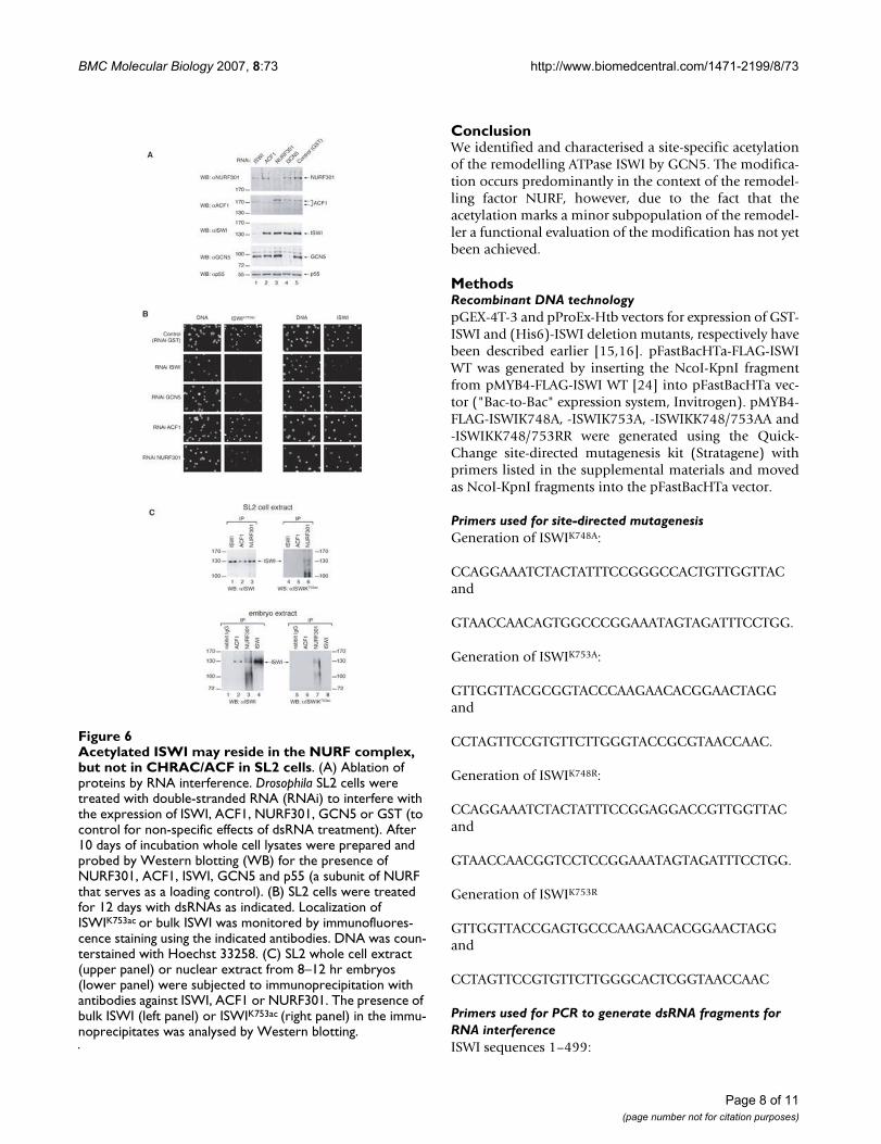
The antibody also recognised an epitope in nuclei of Dro-sophila SL2 cells (Figure 6B, upper left panel). To confirmthat the antibody reactivity was specific in vivo we per-formed RNA interference on SL2 cells with dsRNA corre-sponding to ISWI or GCN5 sequences and monitored thesuccess of the knockdown by Western blotting of wholecells extracts (Figure 6A) and immunofluorescence (Fig-ure 6B). Knocking down either ISWI or GCN5 slowed cellproliferation effectively increasing the doubling time twofold. Ablation of ISWI removed not only the signalobtained with the ISWI antibody, but also the ISWIK753ac-specific signal (Figure 6B). Remarkably, knockdown ofGCN5 also greatly reduced the signal obtained with theISWIK753ac antibody, but did not affect ISWI in general(Figure 6B). Taken together, these data document the spe-cificity of the antibody for ISWIK753ac in immunofluores-cence applications. They also show that ISWI is acetylatedat K753 by GCN5 in SL2 cells.
Acetylated ISWI may reside in the NURF complex, but not in CHRAC/ACFSo far ISWI is known to reside in two kinds of nucleosomeremodelling complexes in Drosophila: CHRAC/ACF,which are characterised by the ACF1 subunit, and NURF,which contains NURF301 as a defining feature [17]. Inorder to explore whether ISWIK753ac was preferentiallyassociated with one of these complexes we ablated the sig-nature subunits ACF1 and NURF301 by RNA interferencein SL2 cells (Figure 6A) and found that the ISWIK753ac
epitope was significantly reduced in the absence ofNURF301, but remained unperturbed if ACF1 wasknocked down (Figure 6B). In agreement with the notionthat ISWIK753ac is not a subunit of CHRAC/ACF we foundthat the expression pattern of the acetylated ISWI formwas unperturbed in acf1 null flies (data not shown).
In vitro, ISWI is specifically acetylated by HATs able to modify histone H3Figure 2In vitro, ISWI is specifically acetylated by HATs able to modify histone H3. (A) The histone acetyltransferase activities of recombinant GCN5, p300 and MOF were assayed in reactions containing all four histones. Incorpora-tion of [3H]-acetyl-coenzyme A leads to labelled proteins, which are separated by SDS-PAGE and detected by autoradi-ography of the gel. Bands corresponding to acetylated his-tones are marked by arrows. (B) Recombinant ISWI was incubated in acetylation reactions with GCN5, p300 or MOF. Acetylated ISWI (arrow) was detected as in (A). All other bands (asterisks) are due to auto-acetylation of the corre-sponding enzyme.
Page 4 of 11(page number not for citation purposes)
BMC Molecular Biology 2007, 8:73 http://www.biomedcentral.com/1471-2199/8/73
ISWIK753ac staining was unperturbed in flies heterozygousfor the NURF301 allele [E(bx)ry122] [18]. Homozygousmutants are lethal, but survive until the end of the larvalstage due to maternal contribution of NURF301 and werenot analysed. Immunoprecipitation of NURF301 fromextracts of SL2 cells and 8–12 hr embryos co-precipitatedacetylated ISWI, which was not the case if ACF1 was pre-cipitated (Figure 6C, D). We conclude that in SL2 cellsISWI may be acetylated in the context of NURF but is notacetylated in association with ACF1. The data do notexclude the existence of additional complexes harbouringthe modification.
DiscussionMany acetyltransferases were first identified followingtheir histone acetylation function and hence named his-tone acetyltransferases (HATs). However, most of themhave now been shown to acetylate non-histone proteinsas well, and these modifications can have profound effectson the structure and function [19]. For example, theeffects of acetylation on the tumour suppressor proteinp53 have been heavily studied [20]. The current work,together with the earlier observation that the effect of theBRM ATPase on cell proliferation can be regulated byacetylation [10] highlight the potential for direct regula-tory cross-talk between different chromatin modifyingprinciples [21].
The fact that ISWI can be acetylated in vitro and in tissueculture cells by GCN5, which is also known to target theH3 N-terminus, is explained by the sequence similaritybetween the histone and ISWI around the K753 acetyla-tion site. The common motif RKT/SxGx(Kac)xPR/K differsfrom the previously suggested GCN5/PCAF recognitionmotif GKxxP [19]. This raises the possibility of specific co-regulation of a nucleosome remodelling factor and itsnucleosome substrate through acetylation of relatedepitopes. Strikingly, the related motif DKGKGKKRP alsooccurs in the C-terminus of hBRM, overlapping withpotential acetylation sites [10]. Acetylation at one or moreC-terminal lysines of BRM correlates with reduced inter-ference with cell cycle progression in mammalian cellsand mutation of several of these lysines into argininesimpairs BRM function. The data suggest that site-specificacetylation of the ATPase by, for example P/CAF, mayadversely affect its function, possibly by interfering withtargeting interactions [10].
The fact that the acetylated ISWI epitope is conserved dur-ing evolution suggests functional significance. However,so far we were unable to document an effect of acetylationon the catalytic properties or protein interactions of ISWI,due to the small proportion of ISWI either acetylated invitro or naturally present in vivo. Lysine 753 of ISWI residesin the HAND domain, a fold of unknown function situ-ated between the ATPase domain and the C-terminalSANT/SLIDE domains that mediate nucleosome interac-tions [15]. Exploring the role of the HAND domain inISWI may lead to novel approaches towards evaluatingthe impact of K753 acetylation. Applying the K753ac-spe-cific antibody to developmental stages of Drosophilarevealed that the acetylation of ISWI is a developmentallyregulated process, which occurs during oogenesis andearly embryogenesis, but is absent from later develop-mental stages (CC and PBB, unpublished observations).We also obtained preliminary support for the idea that theacetylation of ISWI marks a functionally distinct subpop-ulation: the acetylated epitope was dramatically enriched
ISWI is acetylated in the HAND domainFigure 3ISWI is acetylated in the HAND domain. (A) Schematic representation of ISWI, the extent of known domains (first line) and of deletion mutants used for mapping the acetyla-tion site. Numbers correspond to the first and the last amino acids of the proteins. They were expressed in E. coli with either HIS6- or glutathione-S-transferase- (GST-) tags. (B) The different ISWI fragments displayed in (A) were acetylated in vitro with GCN5. Reactions were resolved by SDS-PAGE and visualized by autoradiography (upper panel) and Coomassie staining (lower panel). Black dots label the positions of the ISWI fragments. An asterisk marks the band corresponding to GCN5.
Page 5 of 11(page number not for citation purposes)
BMC Molecular Biology 2007, 8:73 http://www.biomedcentral.com/1471-2199/8/73
Page 6 of 11(page number not for citation purposes)
GCN5 acetylates ISWI at K753Figure 4GCN5 acetylates ISWI at K753. (A) Alignment of Drosophila ISWI sequences spanning aa 740–769 with the corresponding residues from Xenopus ISWI (xISWI), human hSNF2H, hSNF2L and with the 30 N-terminal amino acids of histone H3. A con-sensus of highly conserved residues is given. The arrowhead points to K753 indISWI. (B) Peptides corresponding to the his-tone H3 N-terminal tail (aa 1–19, left panel) and to aa 740–759 of ISWI (right panel) were acetylated in vitro by GCN5, p300 or MOF as in Figure 2. Peptide acetylation was determined from incorporation of [3H]-acetate and by subtracting the values for autoacetylation of enzymes (from reactions lacking substrate) and the values for non-specific adsorption of label to the pep-tides (from reactions lacking enzyme) from the experimental values. (C) MALDI analysis of the in vitro acetylated ISWI peptide (aa 740–759). The acetylation reaction was stopped by addition of 10% acetic acid, and the reaction products were analyzed by MALDI-TOF. The upper spectrum represents the unmodified peptide (M-H+ = 2580.29 amu) from a control reaction lacking acetyl-CoA. In the lower spectrum the arrow points to a peak representing the product of the acetylation reaction (M-H+ = 2622.32 amu). A Deltamass (Dm) of 42.01 amu indicates mono-acetylation. A second peak of very low intensity (asterisk) sug-gests minor di-acetylation (M-H+ = 2664.33). (D) Nanospray sequencing of ISWI peptide (aa 743–759) after in vitro acetylation and removal of the C-terminal cysteine by V8 protease. Upper spectrum: sequencing of the unmodified peptide with a (M-3H+) value of 702.67 confirms the sequence IYYFRKTVGYKVPKNTE with unmodified K748, K753 and K756. The spectrum is enlarged over the region 750VGYKV754 in order to observe the y7 ion (M-H+ = 815.46) corresponding to the unacetylated K753. Lower spectrum: sequencing of the peptide with a (M-3H+) value of 716.73 reveals the sequence IYYFRKTVGYKAcVP-KNTE, with only K753 being acetylated, (mass of the y7 ion M-H+ = 857.47, which differs in mass from the corresponding unmodified y7 ion in the upper spectrum by 42.012 amu). Detailed analysis of K748 and K756 revealed no acetylation at these sites (data notshown). (E) Recombinant ISWI and derivatives bearing the indicated point mutations were acetylated by GCN5 and acetylated proteins were visualized by autoradiography (upper panel) and Coomassie staining (lower panel). (F) Recom-binant wildtype (WT) and mutant ISWI proteins were incubated with [g-32P]-ATP in the absence (control), or presence of 110 ng of free or nucleosomal DNA (Nuc) as indicated. ATP hydrolysis was detected by thin-layer chromatography and quantified by PhosphoImager analysis. ATPase activity is indicated as percentage of ATP hydrolysed.
BMC Molecular Biology 2007, 8:73 http://www.biomedcentral.com/1471-2199/8/73
on the condensed metaphase chromosomes in earlyembryos (CC and PBB, unpublished observations). Acausal role between the acetylation and the unusual abil-ity of the ATPase to interact with compact chromatinremains to be explored.
The ATPase ISWI is the catalytic moiety of several nucleo-some remodelling complexes. Better known and charac-terized are the factors CHRAC, ACF and NURF (for review,see [22]. The available data suggest that the acetylatedform of ISWI may mark a subpopulation of the NURFcomplex. NURF is involved in the transcription of selectedsets of Drosophila genes [18,23] but flies mutant for the
large, defining NURF301 subunit also show defects ofchromosome structure [23]. Assessment of which of thesefunctions may be modulated by ISWI acetylation requiresa genetic approach.
The acetylation of the amino terminus of histone H3 is animportant determinant of active chromatin, but chroma-tin structure can also be modulated by other modifica-tions of the histone 'tail', notable its methylation orphosphorylation. Whether the analogy between the ISWIand the H3 N-terminus can also be extended to includephosphorylation and methylation remains an interestingaspect for future research.
Characterisation of an antibody specific forISWIK753acFigure 5Characterisation of an antibody specific forISWIK753ac. (A) Recombinant ISWI was acetylated by GCN5 in the presence of radiolabeled acetyl-CoA. The protein mixture was separated by SDS-PAGE and visualized by Coomassie staining (left panel), by autoradiography (centre panel) and by Western blotting using an antibody raised against an ISWI peptide bearing acetylated K753 (αISWIK753ac). The asterisk points to the position of GCN5 in the gel (lane 2), which autoacetylates itself (lane 4). (B) The antibody does not cross-react with acetylated H3. Recombinant histone H3 was acetylated with GCN5 in presence of radiola-beled acetyl-CoA and analysed as in (A). Western blotting on such a substrate using αISWIK753ac did not reveal any cross-reac-tivity. (C) ISWI was immunoprecipitated from embryonic nuclear extract using antibodies against ISWI or ISWIK753ac and detected by Western blotting. (D) αISWIK753ac immunoprecipitates were analysed by Western blotting for the presence of acetylated ISWI.
Page 7 of 11(page number not for citation purposes)
BMC Molecular Biology 2007, 8:73 http://www.biomedcentral.com/1471-2199/8/73
ConclusionWe identified and characterised a site-specific acetylationof the remodelling ATPase ISWI by GCN5. The modifica-tion occurs predominantly in the context of the remodel-ling factor NURF, however, due to the fact that theacetylation marks a minor subpopulation of the remodel-ler a functional evaluation of the modification has not yetbeen achieved.
MethodsRecombinant DNA technologypGEX-4T-3 and pProEx-Htb vectors for expression of GST-ISWI and (His6)-ISWI deletion mutants, respectively havebeen described earlier [15,16]. pFastBacHTa-FLAG-ISWIWT was generated by inserting the NcoI-KpnI fragmentfrom pMYB4-FLAG-ISWI WT [24] into pFastBacHTa vec-tor ("Bac-to-Bac" expression system, Invitrogen). pMYB4-FLAG-ISWIK748A, -ISWIK753A, -ISWIKK748/753AA and-ISWIKK748/753RR were generated using the Quick-Change site-directed mutagenesis kit (Stratagene) withprimers listed in the supplemental materials and movedas NcoI-KpnI fragments into the pFastBacHTa vector.
Primers used for site-directed mutagenesisGeneration of ISWIK748A:
CCAGGAAATCTACTATTTCCGGGCCACTGTTGGTTACand
GTAACCAACAGTGGCCCGGAAATAGTAGATTTCCTGG.
Generation of ISWIK753A:
GTTGGTTACGCGGTACCCAAGAACACGGAACTAGGand
CCTAGTTCCGTGTTCTTGGGTACCGCGTAACCAAC.
Generation of ISWIK748R:
CCAGGAAATCTACTATTTCCGGAGGACCGTTGGTTACand
GTAACCAACGGTCCTCCGGAAATAGTAGATTTCCTGG.
Generation of ISWIK753R
GTTGGTTACCGAGTGCCCAAGAACACGGAACTAGGand
CCTAGTTCCGTGTTCTTGGGCACTCGGTAACCAAC
Primers used for PCR to generate dsRNA fragments for RNA interferenceISWI sequences 1–499:
Acetylated ISWI may reside in the NURF complex, but not in CHRAC/ACF in SL2 cellsFigure 6Acetylated ISWI may reside in the NURF complex, but not in CHRAC/ACF in SL2 cells. (A) Ablation of proteins by RNA interference. Drosophila SL2 cells were treated with double-stranded RNA (RNAi) to interfere with the expression of ISWI, ACF1, NURF301, GCN5 or GST (to control for non-specific effects of dsRNA treatment). After 10 days of incubation whole cell lysates were prepared and probed by Western blotting (WB) for the presence of NURF301, ACF1, ISWI, GCN5 and p55 (a subunit of NURF that serves as a loading control). (B) SL2 cells were treated for 12 days with dsRNAs as indicated. Localization of ISWIK753ac or bulk ISWI was monitored by immunofluores-cence staining using the indicated antibodies. DNA was coun-terstained with Hoechst 33258. (C) SL2 whole cell extract (upper panel) or nuclear extract from 8–12 hr embryos (lower panel) were subjected to immunoprecipitation with antibodies against ISWI, ACF1 or NURF301. The presence of bulk ISWI (left panel) or ISWIK753ac (right panel) in the immu-noprecipitates was analysed by Western blotting.
Page 8 of 11(page number not for citation purposes)
BMC Molecular Biology 2007, 8:73 http://www.biomedcentral.com/1471-2199/8/73
TTAATACGACTCACTATAGGGAGAATGTCCAAAACAGA-TACAGCTG and
TTAATACGACTCACTATAGGGAGAGCAGAGATAT-GGTCTGCAGG;
GCN5 sequences 1–525:
TTAATACGACTCACTATAGGGAGAATGTCTGGTGGTC-CATCC and
TTAATACGACTCACTATAGGGAGAGCGCTGCATGGA-CATGAA;
ACF1 sequences 1–560
TTAATACGACTCACTATAGGGAGAATGCCCATTT-GCAAGCGG and
TTAATACGACTCACTATAGGGAGAAT-GCGCGAGCGACGTAAT;
NURF301 sequences 1–521
TTAATACGACTCACTATAGGGAGAATGAGCGGTCGCG-GCAG and
TTAATACGACTCACTATAGGGAGAGCTGCATACTGAC-GACCT;
GST sequences 1–527;
TTAATACGACTCACTATAGGGAGAATGTC-CCCTATACTAGGTTA and
TTAATACGACTCACTATAGGGAGAACGCATCCAGGCA-CATTG;
AntibodiesThe following antibodies were kind gifts: αISWI (J. Tam-kun, UC St. Cruz), αp55 (C. Wu, NIH, Bethesda), αGCN5(Jerry Workman, Stowers Institute for Medical Research)αNURF301 (Andreas Hochheimer). The monoclonalαACF1 antibody was raised in rats. Its specificity was con-firmed using the acf1 null fly strain [25] (C. Chioda et al,manuscript in preparation). We employed two differentpan-acetyl-lysine antibodies for immunoprecipitation[26] and Western blotting (Cell Signalling Technology #9441). The αISWIK753ac was raised against ISWI peptideTVGYKacVPKNT (aa 749–758) where K753 wasacetylated. The antibody is available from Abcam(ab10748). As control antibody we used rabbit IgG (sc-2027, Santa Cruz Biotechnology). The αH3S10p antibodywas from Upstate (RR002, catalog # 05–598).
Cell culture, RNA interference and immunostainingDrosophila SF4 cells (an SL2 clone sorted for diploidy andadherence) were provided by D. Arndt-Jovin (Göttingen).SF4, Kc and SL2 cells were maintained at 26°C in Schnei-der's Drosophila medium (Invitrogen) supplementedwith 10% fetal calf serum (FCS), antibiotics andglutamine. When needed, trichostatine A (TSA) wasadded at a final concentration of 200 ng/ml to themedium for 17 hrs before harvesting the cells. RNAiexperiments were performed as described [27]. The prim-ers used to generate the double-stranded RNA are listed inthe supplemental materials. Briefly, 1–2 × 10⁶ SL2 cellswere seeded into 6-wells plates in 1 ml of medium with-out FCS just before adding 10 µg of dsRNA. Plates werethen placed onto a shaking platform for 10 min and thenfor 50 min at 26°C. 2 ml of medium complemented withFCS were then added to the cells and incubated at 26°C.Cells were collected 10 or 12 days after dsRNA treatment.Protein extractions for Western blot analysis after RNAi,1–2 × 10⁶ cells were pelleted and lysed in 50 µl of ureabuffer (8 M urea, 5% SDS, 200 mM Tris-Cl pH 6.8, 0.1mM EDTA, 100 mM DTT) and incubated at 65°C for 15min. Immunostainings were performed as described [28].For immunostaining following RNAi, cells were collected12 days after dsRNA treatment. After fixation, permeabili-sation and blocking, cells were incubated for 1 hr withantibody against ISWI, diluted 1:600 in blocking solution(2% BSA and 5% goat serum in PBS), or aISWIK753ac,diluted 1:300. After washes, cells were incubated for 1 hrwith Cy3-conjugated secondary antibody (Jackson Immu-noresearch Laboratories) diluted in blocking buffer. Cellswere washed four times in PBS. DNA was counterstainedwith 1 µg/ml bisbenzimide (Hoechst 33258). Slides weremounted using 1.5% n-propyl gallate, 50% glycerol inPBS. Images were acquired using a Zeiss Axiophot micro-scope coupled to a Retiga Exi CCD Camera (Qimaging,Burnaby, Canada). Images were cropped and levelsadjusted in Photoshop.
ImmunoprecipitationTotal cell extracts were prepared as described [29] from 4× 10⁷ Drosophila cells. All steps were performed at 4°C.Cells were washed with PBS, resuspended in 1 volume oflysis buffer (50 mM Tris-Cl pH 8.0, 300 mM NaCl, 10 mMMgCl2, 0.4% NP40 and proteases inhibitors) and incu-bated for 15 min. The supernatant was cleared by centrif-ugation and mixed with 1 volume of dilution buffer (50mM Tris-Cl pH 8.0, 0.4% NP40). Diluted extracts werepre-cleared with protein A/protein G Sepharose beads(Amersham Biosciences) and incubated with αISWI,αAcLysine or irrelevant antibodies, and immunoprecipi-tated with protein A/protein G Sepharose beads. Follow-ing extensive washing with a 1:1 mix of lysis and dilutionbuffer, beads were resuspended in Laemmli buffer andproteins were analyzed by Western blot analysis.
Page 9 of 11(page number not for citation purposes)
BMC Molecular Biology 2007, 8:73 http://www.biomedcentral.com/1471-2199/8/73
Western Blot AnalysisImmunoprecipitated proteins were separated by SDS-PAGE, electro-transferred onto PVDF or nitrocellulosemembranes and detected using an ECL kit (AmershamBiosciences) according to the manufacturer's instructions.
In vivo labelingDrosophila cells were treated with 0.5 mCi/ml [3H]-aceticacid (TRK12, Amersham Biosciences) and 10 mM sodiumbutyrate (NaB) for 3 hrs at 26°C. Whole cell extracts andimmunoprecipitations were performed using standardprocedures, except that all buffers were complementedwith 5 mM sodium butyrate. Immunoprecipitated pro-teins were analyzed by Western blot or by autoradiogra-phy.
Recombinant proteinsFLAG-ISWI mutants were expressed and purified frombaculovirus vectors in Sf9 cells as described [30]. Expres-sion of proteins in E. coli and purification was accordingto the following published procedures: FLAG-ISWI andHIS6-ISWI deletion mutants [15]; GST-ISWI deletionmutants [16]; GST-hGCN5 and FLAG-p300 [31]; MOF[13].
In vitro acetylation assays200 ng of FLAG-ISWI, 200 ng of His- or GST-ISWI deletionmutants, 2 µg of histone octamers or 200 ng of bacteriallyexpressed histone H3 were incubated for 30 min at 26°C,in 20 µl final volume with 0.25 mCi of [3H]-acetyl-CoA(4.1 Ci/mmol, TRK688, Amersham Biosciences), 50 to100 ng of GST-hGCN5, HA-MOF or FLAG-p300 in HATbuffer (10 mM Tris-Cl pH 7.8, 0.1 mM EDTA, comple-mented with 1 mM PMSF, 1 mM DTT and 10 mM NaB).Reaction mixtures were analyzed by SDS-PAGE. The gelwas Coomassie stained, destained, treated with Amplify(Amersham Biosciences) for 30 min, dried and autoradi-ographed. 1 µg each of an H3 peptide (aa 1–19) or anISWI peptide (aa 740–759) were acetylated under thesame conditions. A 10 µl aliquot of each reaction wasspotted onto p81 filters (Whatman), which were washedthree times with 50 mM NaCarbonate pH 9.2 andcounted in a scintillation counter.
Sequence alignmentSequence alignment was performed using ClustalW soft-ware, using default parameters.
Mass spectrometryMALDI-TOF analysis of acetylated ISWI peptide. The in vitroacetylated peptides were purified on C18 reversed phaseZipTip mini-columns (Millipore) according to the manu-facturer's protocol. In short the peptide was washed 3times with 0.1% TFA, eluted with 1 µl of matrix solution[saturated a-cyanohydroxy-cinammic acid (Sigma) dis-
solved in 50% ACN (v/v)/0.3% TFA (v/v)] directly ontothe target plate. The peptide-matrix co-crystal was ana-lyzed in a Voyager DE STR spectrometer according to themanufacturer's instructions. Peptide mass fingerprintscovered the mass range between 700–3500 amu, with thelow mass gate set at 500 amu. The accelerating voltage wasset to 20 kV, the grid to 66% and the delay time to 100nsec. ESI-analysis. The ISWI peptide DQEIYYFRKTV-GYKVPKNTEC plus a C-terminal cysteine (aa 740–759,M-H+ = 2580,27 amu) was digested with V8 protease. Theresulting peptide IYYFRKTVGYKVPKNTE (M-H+ =2106.14 amu) was de-salted, concentrated using a C18reversed phase minicolumn (Eppendorf) and eluted in0.5 µl 50% Methanol, 0.1% FA into medium size nano-spray needles (Protana, Odense, Denmark). ESI massspectra were recorded on an Applied Biosystems QStar XLhybrid quadrupole time of flight mass spectrometer,equipped with a Protana nano-spray ion source in thestatic nano-spray mode according to the manufacturer'sinstructions. The needles were adjusted in front of the ori-fice and the spray voltage was set between 950 and 1100V. Product ion scans were acquired for 4–5 min contain-ing approximately 200–300 scans.
ATPase assaysThe ATPase assays were performed as described previously[30].
Authors' contributionsRF discovered ISWI acetylation and did most of the bio-chemical analysis, prepared the figures and contributed topreparing the manuscript. AE expressed recombinant ACFin vitro, contributed the ATPase assay and commented onthe manuscript. CC performed experiments not shownaimed at revealing a function of ISWI acetylation in tissueculture cells and helped prepare the manuscript. TB per-formed the mass spectrometrical analysis. AI contributedto interpretation of the mass spectometrical analysis. PBcoordinated the project, provided funds and wrote themanuscript.
AcknowledgementsWe thank I. Vetter for expert technical assistance and the following former and present lab members for reagents and advice: A. Akhtar, K. Bouazoune, V. Morales, C. Regnard, H. Würl. We also thank M. Heck, J. Tamkun, C. Wu, A. Hochheimer and J. Workman (Stowers Institute) for antibodies. This work was supported by Deutsche Forschungsgemeinschaft through SFB 594 (TP A6) and by the European Union via HPRN-CT-2000-00078 and the Network of Excellence FP6-503433.
References1. Smith CL, Peterson CL: ATP-dependent chromatin remode-
ling. Curr Top Dev Biol 2005, 65:115-148.2. Becker PB, Horz W: ATP-dependent nucleosome remodeling.
Annu Rev Biochem 2002, 71:247-273.3. Saha A, Wittmeyer J, Cairns BR: Chromatin remodelling: the
industrial revolution of DNA around histones. Nat Rev Mol CellBiol 2006, 7(6):437-447.
Page 10 of 11(page number not for citation purposes)
BMC Molecular Biology 2007, 8:73 http://www.biomedcentral.com/1471-2199/8/73
Publish with BioMed Central and every scientist can read your work free of charge
"BioMed Central will be the most significant development for disseminating the results of biomedical research in our lifetime."
Sir Paul Nurse, Cancer Research UK
Your research papers will be:
available free of charge to the entire biomedical community
peer reviewed and published immediately upon acceptance
cited in PubMed and archived on PubMed Central
yours — you keep the copyright
Submit your manuscript here:http://www.biomedcentral.com/info/publishing_adv.asp
BioMedcentral
4. Workman JL: Nucleosome displacement in transcription.Genes Dev 2006, 20(15):2009-2017.
5. Woodcock CL, Dimitrov S: Higher-order structure of chroma-tin and chromosomes. Curr Opin Genet Dev 2001, 11(2):130-135.
6. Varga-Weisz PD, Becker PB: Regulation of higher-order chro-matin structures by nucleosome-remodelling factors. CurrOpin Genet Dev 2006, 16(2):151-156.
7. de la Serna IL, Ohkawa Y, Imbalzano AN: Chromatin remodellingin mammalian differentiation: lessons from ATP-dependentremodellers. Nat Rev Genet 2006, 7(6):461-473.
8. Muchardt C, Bourachot B, Reyes JC, Yaniv M: ras transformationis associated with decreased expression of the brm/SNF2alpha ATPase from the mammalian SWI-SNF com-plex. Embo J 1998, 17(1):223-231.
9. Muchardt C, Reyes JC, Bourachot B, Leguoy E, Yaniv M: The hbrmand BRG-1 proteins, components of the human SNF/SWIcomplex, are phosphorylated and excluded from the con-densed chromosomes during mitosis. Embo J 1996,15(13):3394-3402.
10. Bourachot B, Yaniv M, Muchardt C: Growth inhibition by themammalian SWI-SNF subunit Brm is regulated by acetyla-tion. Embo J 2003, 22(24):6505-6515.
11. Kouzarides T: Acetylation: a regulatory modification to rivalphosphorylation? EMBO J 2000, 19(6):1176-1179.
12. Wang L, Mizzen C, Ying C, Candau R, Barlev N, Brownell J, Allis CD,Berger SL: Histone acetyltransferase activity is conservedbetween yeast and human GCN5 and is required for comple-mentation of growth and transcriptional activation. Mol CellBiol 1997, 17(1):519-527.
13. Akhtar A, Becker PB: Activation of transcription through his-tone H4 acetylation by MOF, an acetyltransferase essentialfor dosage compensation in Drosophila. Mol Cell 2000,5(2):367-375.
14. Ogryzko VV, Schiltz RL, Russanova V, Howard BH, Nakatani Y: Thetranscriptional coactivators p300 and CBP are histoneacetyltransferases. Cell 1996, 87(5):953-959.
15. Grüne T, Brzeski J, Eberharter A, Clapier CR, Corona DF, Becker PB,Müller CW: Crystal structure and functional analysis of anucleosome recognition module of the remodeling factorISWI. Mol Cell 2003, 12(2):449-460.
16. Eberharter A, Vetter I, Ferreira R, Becker PB: ACF1 improves theeffectiveness of nucleosome mobilization by ISWI throughPHD-histone contacts. Embo J 2004, 23(20):4029-4039.
17. Corona DF, Tamkun JW: Multiple roles for ISWI in transcrip-tion, chromosome organization and DNA replication. Bio-chim Biophys Acta 2004, 1677(1-3):113-119.
18. Badenhorst P, Xiao H, Cherbas L, Kwon SY, Voas M, Rebay I, CherbasP, Wu C: The Drosophila nucleosome remodeling factorNURF is required for Ecdysteroid signaling and metamor-phosis. Genes Dev 2005, 19(21):2540-2545.
19. Roth SY, Denu JM, Allis CD: Histone Acetyltransferases. AnnuRev Biochem 2001, 70:81-120.
20. Brooks CL, Gu W: Ubiquitination, phosphorylation andacetylation: the molecular basis for p53 regulation. Curr OpinCell Biol 2003, 15(2):164-171.
21. Eberharter A, Ferreira R, Becker P: Dynamic chromatin: con-certed nucleosome remodelling and acetylation. Biol Chem2005, 386(8):745-751.
22. Längst G, Becker PB: Nucleosome mobilization and positioningby ISWI-containing chromatin remodeling factors. J Cell Sci-ence 2001, 114:2561-2568.
23. Badenhorst P, Voas M, Rebay I, Wu C: Biological functions of theISWI chromatin remodeling complex NURF. Genes Dev 2002,16(24):3186-3198.
24. Corona DFV, Längst G, Clapier CR, Bonte EJ, Ferrari S, Tamkun JW,Becker PB: ISWI is an ATP-dependent nucleosome remode-ling factor. Molecular Cell 1999, 3:239-245.
25. Fyodorov DV, Blower MD, Karpen GH, Kadonaga JT: Acf1 confersunique activities to ACF/CHRAC and promotes the forma-tion rather than disruption of chromatin in vivo. Genes Dev2004, 18(2):170-183.
26. Hebbes TR, Turner CH, Thorne AW, Crane-Robinson C: A "mini-mal epitope" anti-protein antibody that recognises a singlemodified amino acid. Mol Immunol 1989, 26(9):865-873.
27. Maiato H, Sunkel CE, Earnshaw WC: Dissecting mitosis by RNAiin Drosophila tissue culture cells. Biol Proced Online 2003,5:153-161.
28. Morales V, Straub T, Neumann MF, Mengus G, Akhtar A, Becker PB:Functional integration of the histone acetyltransferase MOFinto the dosage compensation complex. Embo J 2004,23(11):2258-2268.
29. Ferreira R, Magnaghi-Jaulin L, Robin P, Harel-Bellan A, Trouche D:The three members of the pocket proteins family share theability to repress E2F activity through recruitment of a his-tone deacetylase. Proc Natl Acad Sci U S A 1998,95(18):10493-10498.
30. Eberharter A, Ferrari S, Langst G, Straub T, Imhof A, Varga-Weisz P,Wilm M, Becker PB: Acf1, the largest subunit of CHRAC, reg-ulates ISWI-induced nucleosome remodelling. EMBO J 2001,20(14):3781-3788.
31. Imhof A, Yang X, Ogryzko VV, Nakatani Y, Wolffe AP, Ge H:Acetylation of general transcription factors by histoneacetyltransferases. Current Biology 1997, 7:689-692.
Page 11 of 11(page number not for citation purposes)
http://www.ncbi.nlm.nih.gov/entrez/query.fcgi?cmd=Retrieve&db=PubMed&dopt=Abstract&list_uids=9427756
http://www.ncbi.nlm.nih.gov/entrez/query.fcgi?cmd=Retrieve&db=PubMed&dopt=Abstract&list_uids=9427756
http://www.ncbi.nlm.nih.gov/entrez/query.fcgi?cmd=Retrieve&db=PubMed&dopt=Abstract&list_uids=9427756
http://www.ncbi.nlm.nih.gov/entrez/query.fcgi?cmd=Retrieve&db=PubMed&dopt=Abstract&list_uids=8670841
http://www.ncbi.nlm.nih.gov/entrez/query.fcgi?cmd=Retrieve&db=PubMed&dopt=Abstract&list_uids=8670841
http://www.ncbi.nlm.nih.gov/entrez/query.fcgi?cmd=Retrieve&db=PubMed&dopt=Abstract&list_uids=8670841
http://www.ncbi.nlm.nih.gov/entrez/query.fcgi?cmd=Retrieve&db=PubMed&dopt=Abstract&list_uids=8972232
http://www.ncbi.nlm.nih.gov/entrez/query.fcgi?cmd=Retrieve&db=PubMed&dopt=Abstract&list_uids=8972232
http://www.ncbi.nlm.nih.gov/entrez/query.fcgi?cmd=Retrieve&db=PubMed&dopt=Abstract&list_uids=8972232
http://www.ncbi.nlm.nih.gov/entrez/query.fcgi?cmd=Retrieve&db=PubMed&dopt=Abstract&list_uids=8945521
http://www.ncbi.nlm.nih.gov/entrez/query.fcgi?cmd=Retrieve&db=PubMed&dopt=Abstract&list_uids=8945521
http://www.ncbi.nlm.nih.gov/entrez/query.fcgi?cmd=Retrieve&db=PubMed&dopt=Abstract&list_uids=8945521
http://www.ncbi.nlm.nih.gov/entrez/query.fcgi?cmd=Retrieve&db=PubMed&dopt=Abstract&list_uids=2481233
http://www.ncbi.nlm.nih.gov/entrez/query.fcgi?cmd=Retrieve&db=PubMed&dopt=Abstract&list_uids=2481233
http://www.ncbi.nlm.nih.gov/entrez/query.fcgi?cmd=Retrieve&db=PubMed&dopt=Abstract&list_uids=2481233
http://www.ncbi.nlm.nih.gov/entrez/query.fcgi?cmd=Retrieve&db=PubMed&dopt=Abstract&list_uids=9724731
http://www.ncbi.nlm.nih.gov/entrez/query.fcgi?cmd=Retrieve&db=PubMed&dopt=Abstract&list_uids=9724731
http://www.ncbi.nlm.nih.gov/entrez/query.fcgi?cmd=Retrieve&db=PubMed&dopt=Abstract&list_uids=9724731
http://www.ncbi.nlm.nih.gov/entrez/query.fcgi?cmd=Retrieve&db=PubMed&dopt=Abstract&list_uids=9285713
http://www.ncbi.nlm.nih.gov/entrez/query.fcgi?cmd=Retrieve&db=PubMed&dopt=Abstract&list_uids=9285713


![Myc regulation of histone acetylation TCA cycle [U- 13 C] Glucose [U- 13 C]-Pyruvate 13 C-FA, 13 C-acetate and 13 C-acetoacetate 13 C-Ac GCN5 [U- 13 C]-Acetyl-CoA.](https://static.fdocuments.net/doc/165x107/56649ec95503460f94bd72a6/myc-regulation-of-histone-acetylation-tca-cycle-u-13-c-glucose-u-13-c-pyruvate.jpg)