
Sirtuins and renal diseases: relationship with aging and ... · Sirtuins and renal diseases:...
12
Clinical Science (2013) 124, 153–164 (Printed in Great Britain) doi: 10.1042/CS20120190 Sirtuins and renal diseases: relationship with aging and diabetic nephropathy Munehiro KITADA ∗ , Shinji KUME†, Ai TAKEDA-WATANABE ∗ , Keizo KANASAKI ∗ and Daisuke KOYA ∗ ∗ Diabetology and Endocrinology, Kanazawa Medical University, 1-1 Daigaku, Uchinada, Ishikawa, Japan †Diabetes, Nephrology and Neurology, Shiga University of Medical Science, Setatsukinowa, Otsu, Shiga, Japan Abstract Sirtuins are members of the Sir2 (silent information regulator 2) family, a group of class III deacetylases. Mammals have seven different sirtuins, SIRT1–SIRT7. Among them, SIRT1, SIRT3 and SIRT6 are induced by calorie restriction conditions and are considered anti-aging molecules. SIRT1 has been the most extensively studied. SIRT1 deacetylates target proteins using the coenzyme NAD + and is therefore linked to cellular energy metabolism and the redox state through multiple signalling and survival pathways. SIRT1 deficiency under various stress conditions, such as metabolic or oxidative stress or hypoxia, is implicated in the pathophysiologies of age-related diseases including diabetes, cardiovascular diseases, neurodegenerative disorders and renal diseases. In the kidneys, SIRT1 may inhibit renal cell apoptosis, inflammation and fibrosis, and may regulate lipid metabolism, autophagy, blood pressure and sodium balance. Therefore the activation of SIRT1 in the kidney may be a new therapeutic target to increase resistance to many causal factors in the development of renal diseases, including diabetic nephropathy. In addition, SIRT3 and SIRT6 are implicated in age-related disorders or longevity. In the present review, we discuss the protective functions of sirtuins and the association of sirtuins with the pathophysiology of renal diseases, including diabetic nephropathy. Key words: aging, diabetic nephropathy, sirtuin, SIRT1 (sirtuin 1). INTRODUCTION The prevalence of diabetes mellitus has been increasing world- wide over recent years. Long-term diabetes results in vascular changes and dysfunction; diabetic complications are the major causes of morbidity and mortality in diabetic patients. Among diabetic vascular complications, nephropathy is recognized as not only a leading cause of end-stage renal disease, but also an independent risk factor for cardiovascular diseases. Large clinical studies indicate that hyperglycaemia is a major contributing factor to the pathogenesis of diabetic vascular com- plications, including nephropathy [1]. Hyperglycaemia-mediated alterations of extra- and intra-cellular metabolism, such as AGEs (advanced glycation end-products), enhancement of diacylgly- cerol/PKC (protein kinase C) activity and increased flux through polyol and hexosamine pathways, constitute the classical patho- Abbreviations: AGE, advanced glycation end-product; AMPK, AMP-activated protein kinase; AngII, angiotensin II; AT 1 R, angiotensin type 1 receptor; Atg, autophagy-related gene; BMAL1, brain and muscle ARNT (aryl hydrocarbon receptor nuclear translocator)-like 1; Bnip3, BCL2/adenovirus E1V 19-kDa interacting protein 3; CKD, chronic kidney disease; CR, calorie restriction; CRP , C-reactive protein; COX2, cyclo-oxygenase 2; α-ENaC, epithelial Na + channel α-subunit; eNOS, endothelial NO synthase; ER, endoplasmic reticulum; FOXO, forkhead box O; FXR, farnesoid X receptor; H3K9, histone H3 Lys 9 ; H3K9me3, H3K9 trimethylation; HIF, hypoxia-inducible factor; ICAM-1, intercellular adhesion molecule 1; Idh2, isocitrate dehydrogenase 2; IGF, insulin-like growth factor; IRS, insulin receptor substrate; LC3, light chain 3; LXR, liver X receptor; MCP-1, monocyte chemotactic protein-1; Mn-SOD, manganese superoxide dismutase; mTOR, mammalian target of rapamycin; NF-κB, nuclear factor-κB; PARP , poly(ADP-ribose) polymerase; PER2, Period 2; PGC-1α, PPAR-γ co-activator-1α; PKC, protein kinase C; PPAR, peroxisome-proliferator-activated receptor; PTP1B, protein tyrosine phosphatase 1B; RAS, renin–angiotensin system; ROS, reactive oxygen species; Sir2, silent information regulator 2; SIRT1 etc., sirtuin 1 etc., SNP , single nucleotide polymorphism; SREBP , sterol-regulatory-element-binding protein; TGF, transforming growth factor; TNF-α, tumour necrosis factor α; UUO, unilateral ureteral obstruction; VCAM-1, vascular cell adhesion protein 1; WFR, Wistar fatty diabetic rat. Correspondence: Professor Daisuke Koya (email [email protected]). genesis of diabetic nephropathy [2,3]. In addition, there are con- vincing data that the RAS (renin–angiotensin system) is a major mediator of renal injuries. Blood pressure control using RAS inhibitors can reduce the progression of nephropathy [4]. How- ever, it is not easy to control blood glucose, and treatment with RAS inhibitors may not completely prevent the progression of nephropathy. Therefore there is an urgent need to identify new therapeutic target molecules or cellular processes that underlie the pathogenesis of diabetic nephropathy to establish an addi- tional therapeutic option, independent of glycaemic control and RAS inhibition [5]. Aging is a universal process that affects all organs. Age- related disruptions in cellular homoeostasis result in declines in organ functions and in the responsiveness to physiological stress. A gradual decline in renal function occurs in most healthy indi- viduals as they age [6], and the amount of glomerular, vascular www.clinsci.org 153
Transcript of Sirtuins and renal diseases: relationship with aging and ... · Sirtuins and renal diseases:...
Clinical Science (2013) 124, 153–164 (Printed in Great Britain) doi: 10.1042/CS20120190
Sirtuins and renal diseases: relationship withaging and diabetic nephropathyMunehiro KITADA∗, Shinji KUME†, Ai TAKEDA-WATANABE∗, Keizo KANASAKI∗ and Daisuke KOYA∗
∗Diabetology and Endocrinology, Kanazawa Medical University, 1-1 Daigaku, Uchinada, Ishikawa, Japan†Diabetes, Nephrology and Neurology, Shiga University of Medical Science, Setatsukinowa, Otsu, Shiga, Japan
AbstractSirtuins are members of the Sir2 (silent information regulator 2) family, a group of class III deacetylases. Mammalshave seven different sirtuins, SIRT1–SIRT7. Among them, SIRT1, SIRT3 and SIRT6 are induced by calorie restrictionconditions and are considered anti-aging molecules. SIRT1 has been the most extensively studied. SIRT1deacetylates target proteins using the coenzyme NAD+ and is therefore linked to cellular energy metabolism andthe redox state through multiple signalling and survival pathways. SIRT1 deficiency under various stress conditions,such as metabolic or oxidative stress or hypoxia, is implicated in the pathophysiologies of age-related diseasesincluding diabetes, cardiovascular diseases, neurodegenerative disorders and renal diseases. In the kidneys, SIRT1may inhibit renal cell apoptosis, inflammation and fibrosis, and may regulate lipid metabolism, autophagy, bloodpressure and sodium balance. Therefore the activation of SIRT1 in the kidney may be a new therapeutic target toincrease resistance to many causal factors in the development of renal diseases, including diabetic nephropathy. Inaddition, SIRT3 and SIRT6 are implicated in age-related disorders or longevity. In the present review, we discuss theprotective functions of sirtuins and the association of sirtuins with the pathophysiology of renal diseases, includingdiabetic nephropathy.
Key words: aging, diabetic nephropathy, sirtuin, SIRT1 (sirtuin 1).
INTRODUCTION
The prevalence of diabetes mellitus has been increasing world-wide over recent years. Long-term diabetes results in vascularchanges and dysfunction; diabetic complications are the majorcauses of morbidity and mortality in diabetic patients. Amongdiabetic vascular complications, nephropathy is recognized asnot only a leading cause of end-stage renal disease, but also anindependent risk factor for cardiovascular diseases.
Large clinical studies indicate that hyperglycaemia is a majorcontributing factor to the pathogenesis of diabetic vascular com-plications, including nephropathy [1]. Hyperglycaemia-mediatedalterations of extra- and intra-cellular metabolism, such as AGEs(advanced glycation end-products), enhancement of diacylgly-cerol/PKC (protein kinase C) activity and increased flux throughpolyol and hexosamine pathways, constitute the classical patho-
Abbreviations: AGE, advanced glycation end-product; AMPK, AMP-activated protein kinase; AngII, angiotensin II; AT1R, angiotensin type 1 receptor; Atg, autophagy-related gene;BMAL1, brain and muscle ARNT (aryl hydrocarbon receptor nuclear translocator)-like 1; Bnip3, BCL2/adenovirus E1V 19-kDa interacting protein 3; CKD, chronic kidney disease; CR,calorie restriction; CRP, C-reactive protein; COX2, cyclo-oxygenase 2; α-ENaC, epithelial Na+ channel α-subunit; eNOS, endothelial NO synthase; ER, endoplasmic reticulum; FOXO,forkhead box O; FXR, farnesoid X receptor; H3K9, histone H3 Lys9; H3K9me3, H3K9 trimethylation; HIF, hypoxia-inducible factor; ICAM-1, intercellular adhesion molecule 1; Idh2,isocitrate dehydrogenase 2; IGF, insulin-like growth factor; IRS, insulin receptor substrate; LC3, light chain 3; LXR, liver X receptor; MCP-1, monocyte chemotactic protein-1; Mn-SOD,manganese superoxide dismutase; mTOR, mammalian target of rapamycin; NF-κB, nuclear factor-κB; PARP, poly(ADP-ribose) polymerase; PER2, Period 2; PGC-1α, PPAR-γco-activator-1α; PKC, protein kinase C; PPAR, peroxisome-proliferator-activated receptor; PTP1B, protein tyrosine phosphatase 1B; RAS, renin–angiotensin system; ROS, reactiveoxygen species; Sir2, silent information regulator 2; SIRT1 etc., sirtuin 1 etc., SNP, single nucleotide polymorphism; SREBP, sterol-regulatory-element-binding protein; TGF, transforminggrowth factor; TNF-α, tumour necrosis factor α; UUO, unilateral ureteral obstruction; VCAM-1, vascular cell adhesion protein 1; WFR, Wistar fatty diabetic rat.
Correspondence: Professor Daisuke Koya (email [email protected]).
genesis of diabetic nephropathy [2,3]. In addition, there are con-vincing data that the RAS (renin–angiotensin system) is a majormediator of renal injuries. Blood pressure control using RASinhibitors can reduce the progression of nephropathy [4]. How-ever, it is not easy to control blood glucose, and treatment withRAS inhibitors may not completely prevent the progression ofnephropathy. Therefore there is an urgent need to identify newtherapeutic target molecules or cellular processes that underliethe pathogenesis of diabetic nephropathy to establish an addi-tional therapeutic option, independent of glycaemic control andRAS inhibition [5].
Aging is a universal process that affects all organs. Age-related disruptions in cellular homoeostasis result in declines inorgan functions and in the responsiveness to physiological stress.A gradual decline in renal function occurs in most healthy indi-viduals as they age [6], and the amount of glomerular, vascular
www.clinsci.org 153
M. Kitada and others
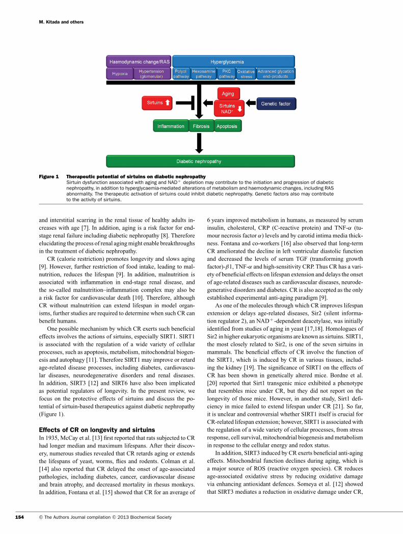
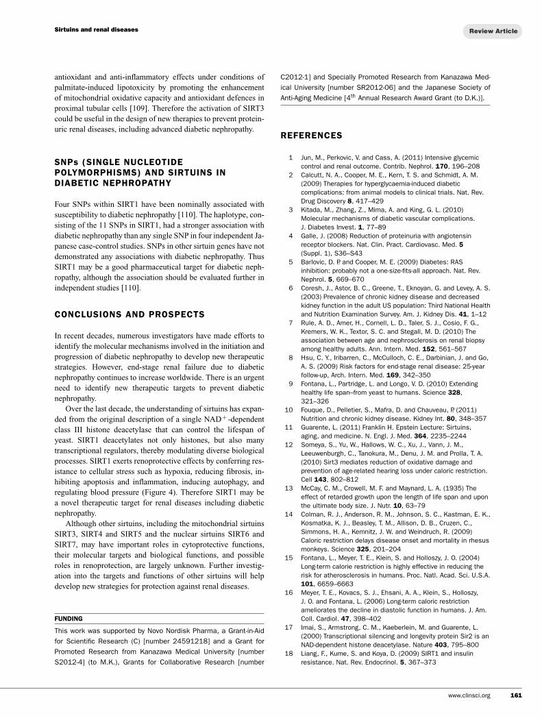
Figure 1 Therapeutic potential of sirtuins on diabetic nephropathySirtuin dysfunction associated with aging and NAD+ depletion may contribute to the initiation and progression of diabeticnephropathy, in addition to hyperglycaemia-mediated alterations of metabolism and haemodynamic changes, including RASabnormality. The therapeutic activation of sirtuins could inhibit diabetic nephropathy. Genetic factors also may contributeto the activity of sirtuins.
and interstitial scarring in the renal tissue of healthy adults in-creases with age [7]. In addition, aging is a risk factor for end-stage renal failure including diabetic nephropathy [8]. Thereforeelucidating the process of renal aging might enable breakthroughsin the treatment of diabetic nephropathy.
CR (calorie restriction) promotes longevity and slows aging[9]. However, further restriction of food intake, leading to mal-nutrition, reduces the lifespan [9]. In addition, malnutrition isassociated with inflammation in end-stage renal disease, andthe so-called malnutrition–inflammation complex may also bea risk factor for cardiovascular death [10]. Therefore, althoughCR without malnutrition can extend lifespan in model organ-isms, further studies are required to determine when such CR canbenefit humans.
One possible mechanism by which CR exerts such beneficialeffects involves the actions of sirtuins, especially SIRT1. SIRT1is associated with the regulation of a wide variety of cellularprocesses, such as apoptosis, metabolism, mitochondrial biogen-esis and autophagy [11]. Therefore SIRT1 may improve or retardage-related disease processes, including diabetes, cardiovascu-lar diseases, neurodegenerative disorders and renal diseases.In addition, SIRT3 [12] and SIRT6 have also been implicatedas potential regulators of longevity. In the present review, wefocus on the protective effects of sirtuins and discuss the po-tential of sirtuin-based therapeutics against diabetic nephropathy(Figure 1).
Effects of CR on longevity and sirtuinsIn 1935, McCay et al. [13] first reported that rats subjected to CRhad longer median and maximum lifespans. After their discov-ery, numerous studies revealed that CR retards aging or extendsthe lifespans of yeast, worms, flies and rodents. Colman et al.[14] also reported that CR delayed the onset of age-associatedpathologies, including diabetes, cancer, cardiovascular diseaseand brain atrophy, and decreased mortality in rhesus monkeys.In addition, Fontana et al. [15] showed that CR for an average of
6 years improved metabolism in humans, as measured by seruminsulin, cholesterol, CRP (C-reactive protein) and TNF-α (tu-mour necrosis factor α) levels and by carotid intima media thick-ness. Fontana and co-workers [16] also observed that long-termCR ameliorated the decline in left ventricular diastolic functionand decreased the levels of serum TGF (transforming growthfactor)-β1, TNF-α and high-sensitivity CRP. Thus CR has a vari-ety of beneficial effects on lifespan extension and delays the onsetof age-related diseases such as cardiovascular diseases, neurode-generative disorders and diabetes. CR is also accepted as the onlyestablished experimental anti-aging paradigm [9].
As one of the molecules through which CR improves lifespanextension or delays age-related diseases, Sir2 (silent informa-tion regulator 2), an NAD+ -dependent deacetylase, was initiallyidentified from studies of aging in yeast [17,18]. Homologues ofSir2 in higher eukaryotic organisms are known as sirtuins. SIRT1,the most closely related to Sir2, is one of the seven sirtuins inmammals. The beneficial effects of CR involve the function ofthe SIRT1, which is induced by CR in various tissues, includ-ing the kidney [19]. The significance of SIRT1 on the effects ofCR has been shown in genetically altered mice. Bordne et al.[20] reported that Sirt1 transgenic mice exhibited a phenotypethat resembles mice under CR, but they did not report on thelongevity of those mice. However, in another study, Sirt1 defi-ciency in mice failed to extend lifespan under CR [21]. So far,it is unclear and controversial whether SIRT1 itself is crucial forCR-related lifespan extension; however, SIRT1 is associated withthe regulation of a wide variety of cellular processes, from stressresponse, cell survival, mitochondrial biogenesis and metabolismin response to the cellular energy and redox status.
In addition, SIRT3 induced by CR exerts beneficial anti-agingeffects. Mitochondrial function declines during aging, which isa major source of ROS (reactive oxygen species). CR reducesage-associated oxidative stress by reducing oxidative damagevia enhancing antioxidant defences. Someya et al. [12] showedthat SIRT3 mediates a reduction in oxidative damage under CR,
154 C© The Authors Journal compilation C© 2013 Biochemical Society
Sirtuins and renal diseases
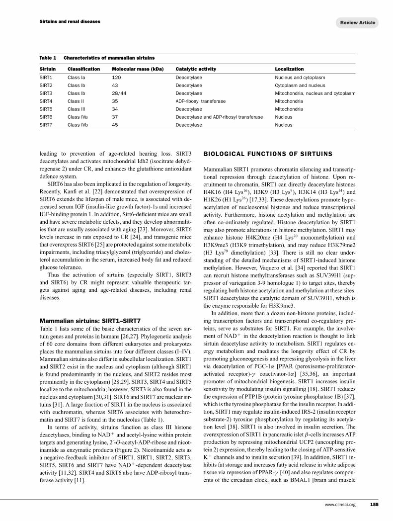
Table 1 Characteristics of mammalian sirtuins
Sirtuin Classification Molecular mass (kDa) Catalytic activity Localization
SIRT1 Class Ia 120 Deacetylase Nucleus and cytoplasm
SIRT2 Class Ib 43 Deacetylase Cytoplasm and nucleus
SIRT3 Class Ib 28/44 Deacetylase Mitochondria, nucleus and cytoplasm
SIRT4 Class II 35 ADP-ribosyl transferase Mitochondria
SIRT5 Class III 34 Deacetylase Mitochondria
SIRT6 Class IVa 37 Deacetylase and ADP-ribosyl transferase Nucleus
SIRT7 Class IVb 45 Deacetylase Nucleus
leading to prevention of age-related hearing loss. SIRT3deacetylates and activates mitochondrial Idh2 (isocitrate dehyd-rogenase 2) under CR, and enhances the glutathione antioxidantdefence system.
SIRT6 has also been implicated in the regulation of longevity.Recently, Kanfi et al. [22] demonstrated that overexpression ofSIRT6 extends the lifespan of male mice, is associated with de-creased serum IGF (insulin-like growth factor)-1s and increasedIGF-binding protein 1. In addition, Sirt6-deficient mice are smalland have severe metabolic defects, and they develop abnormalit-ies that are usually associated with aging [23]. Moreover, SIRT6levels increase in rats exposed to CR [24], and transgenic micethat overexpress SIRT6 [25] are protected against some metabolicimpairments, including triacylglycerol (triglyceride) and choles-terol accumulation in the serum, increased body fat and reducedglucose tolerance.
Thus the activation of sirtuins (especially SIRT1, SIRT3and SIRT6) by CR might represent valuable therapeutic tar-gets against aging and age-related diseases, including renaldiseases.
Mammalian sirtuins: SIRT1–SIRT7Table 1 lists some of the basic characteristics of the seven sir-tuin genes and proteins in humans [26,27]. Phylogenetic analysisof 60 core domains from different eukaryotes and prokaryotesplaces the mammalian sirtuins into four different classes (I–IV).Mammalian sirtuins also differ in subcellular localization. SIRT1and SIRT2 exist in the nucleus and cytoplasm (although SIRT1is found predominantly in the nucleus, and SIRT2 resides mostprominently in the cytoplasm) [28,29]. SIRT3, SIRT4 and SIRT5localize to the mitochondria; however, SIRT3 is also found in thenucleus and cytoplasm [30,31]. SIRT6 and SIRT7 are nuclear sir-tuins [31]. A large fraction of SIRT1 in the nucleus is associatedwith euchromatin, whereas SIRT6 associates with heterochro-matin and SIRT7 is found in the nucleolus (Table 1).
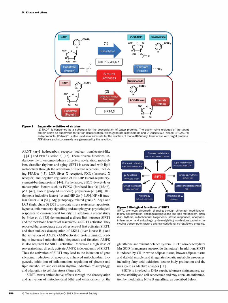
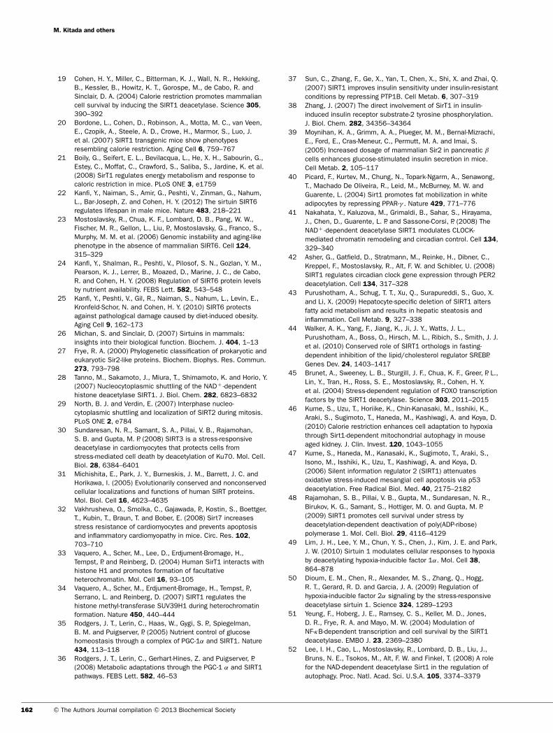
In terms of activity, sirtuins function as class III histonedeacetylases, binding to NAD+ and acetyl-lysine within proteintargets and generating lysine, 2′-O-acetyl-ADP-ribose and nicot-inamide as enzymatic products (Figure 2). Nicotinamide acts asa negative-feedback inhibitor of SIRT1. SIRT1, SIRT2, SIRT3,SIRT5, SIRT6 and SIRT7 have NAD+ -dependent deacetylaseactivity [11,32]. SIRT4 and SIRT6 also have ADP-ribosyl trans-ferase activity [11].
BIOLOGICAL FUNCTIONS OF SIRTUINS
Mammalian SIRT1 promotes chromatin silencing and transcrip-tional repression through deacetylation of histone. Upon re-cruitment to chromatin, SIRT1 can directly deacetylate histonesH4K16 (H4 Lys16), H3K9 (H3 Lys9), H3K14 (H3 Lys14) andH1K26 (H1 Lys26) [17,33]. These deacetylations promote hypo-acetylation of nucleosomal histones and reduce transcriptionalactivity. Furthermore, histone acetylation and methylation areoften co-ordinately regulated. Histone deacetylation by SIRT1may also promote alterations in histone methylation. SIRT1 mayenhance histone H4K20me (H4 Lys20 monomethylation) andH3K9me3 (H3K9 trimethylation), and may reduce H3K79me2(H3 Lys79 dimethylation) [33]. There is still no clear under-standing of the detailed mechanisms of SIRT1-induced histonemethylation. However, Vaquero et al. [34] reported that SIRT1can recruit histone methyltransferases such as SUV39H1 (sup-pressor of variegation 3-9 homologue 1) to target sites, therebyregulating both histone acetylation and methylation at these sites.SIRT1 deacetylates the catalytic domain of SUV39H1, which isthe enzyme responsible for H3K9me3.
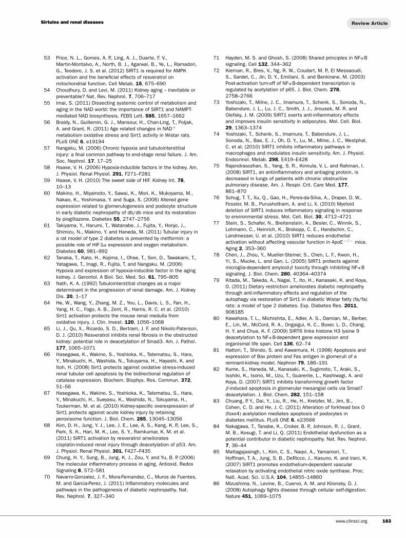
In addition, more than a dozen non-histone proteins, includ-ing transcription factors and transcriptional co-regulatory pro-teins, serve as substrates for SIRT1. For example, the involve-ment of NAD+ in the deacetylation reaction is thought to linksirtuin deacetylase activity to metabolism. SIRT1 regulates en-ergy metabolism and mediates the longevity effect of CR bypromoting gluconeogenesis and repressing glycolysis in the livervia deacetylation of PGC-1α [PPAR (peroxisome-proliferator-activated receptor)-γ coactivator-1α] [35,36], an importantpromoter of mitochondrial biogenesis. SIRT1 increases insulinsensitivity by modulating insulin signalling [18]. SIRT1 reducesthe expression of PTP1B (protein tyrosine phosphatase 1B) [37],which is the tyrosine phosphatase for the insulin receptor. In addi-tion, SIRT1 may regulate insulin-induced IRS-2 (insulin receptorsubstrate-2) tyrosine phosphorylation by regulating its acetyla-tion level [38]. SIRT1 is also involved in insulin secretion. Theoverexpression of SIRT1 in pancreatic islet β-cells increases ATPproduction by repressing mitochondrial UCP2 (uncoupling pro-tein 2) expression, thereby leading to the closing of ATP-sensitiveK+ channels and to insulin secretion [39]. In addition, SIRT1 in-hibits fat storage and increases fatty acid release in white adiposetissue via repression of PPAR-γ [40] and also regulates compon-ents of the circadian clock, such as BMAL1 [brain and muscle
www.clinsci.org 155
M. Kitada and others
Figure 2 Enzymatic activities of sirtuins(1) NAD+ is consumed as a substrate for the deacetylation of target proteins. The acetyl-lysine residues of the targetprotein serve as substrates for sirtuin deacetylation, which generate nicotinamide and 2′ -O-acetyl-ADP-ribose (2′ -OAADPr)as by-products. (2) NAD+ is also used as a substrate for the reaction of mono-ADP-ribosyl transferase with target proteins.ADP-ribose and nicotinamide are generated by the reaction.
ARNT (aryl hydrocarbon receptor nuclear translocator)-like1] [41] and PER2 (Period 2) [42]. These diverse functions un-derscore the interconnectedness of protein acetylation, metabol-ism, circadian rhythms and aging. SIRT1 is associated with lipidmetabolism through the activation of nuclear receptors, includ-ing PPAR-α [43], LXR (liver X receptor), FXR (farnesoid Xreceptor) and negative regulation of SREBP (sterol-regulatory-element-binding protein) [44]. Furthermore, SIRT1 deacetylatestranscription factors such as FOXO (forkhead box O) [45,46],p53 [47], PARP [poly(ADP-ribose) polymerase]-1 [48], HIF(hypoxia-inducible factor)-1α and HIF-2α [49,50], NF-κB (nuc-lear factor κB) [51], Atg (autophagy-related gene) 5, Atg7 andLC3 (light chain 3) [52] to mediate stress resistance, apoptosis,hypoxia, inflammatory signalling and autophagy as physiologicalresponses to environmental toxicity. In addition, a recent studyby Price et al. [53] demonstrated a direct link between SIRT1and the metabolic benefits of resveratrol, a SIRT1 activator. Theyreported that a moderate dose of resveratrol first activates SIRT1,and then induces deacetylation of LKB1 (liver kinase B1) andthe activation of AMPK (AMP-activated protein kinase), lead-ing to increased mitochondrial biogenesis and function. AMPKis also required for SIRT1 activation. Moreover a high dose ofresveratrol may directly activate AMPK independently of SIRT1.Thus the activation of SIRT1 may lead to the induction of genesilencing, reduction of apoptosis, enhanced mitochondrial bio-genesis, inhibition of inflammation, regulation of glucose andlipid metabolism and circadian rhythm, induction of autophagy,and adaptation to cellular stress (Figure 3).
SIRT3 exerts antioxidative effects through the deacetylationand activation of mitochondrial Idh2 and enhancement of the
Figure 3 Biological functions of SIRT1SIRT1 promotes chromatin silencing through chromatin modification,mainly deacetylation, and regulates glucose and lipid metabolism, circa-dian rhythms, mitochondrial biogenesis, stress responses, apoptosis,inflammation and autophagy by deacetylating non-histone proteins, in-cluding transcription factors and transcriptional co-regulatory proteins.
glutathione antioxidant defence system. SIRT3 also deacetylatesMn-SOD (manganese superoxide dismutase). In addition, SIRT3is induced by CR in white adipose tissue, brown adipose tissueand skeletal muscle, and it regulates hepatic metabolic processes,including fatty acid oxidation, ketone body production and theurea cycle as adaptive changes [11].
SIRT6 is involved in DNA repair, telomere maintenance, ge-nomic stability and cell senescence and may attenuate inflamma-tion by modulating NF-κB signalling, as described below.
156 C© The Authors Journal compilation C© 2013 Biochemical Society
Sirtuins and renal diseases
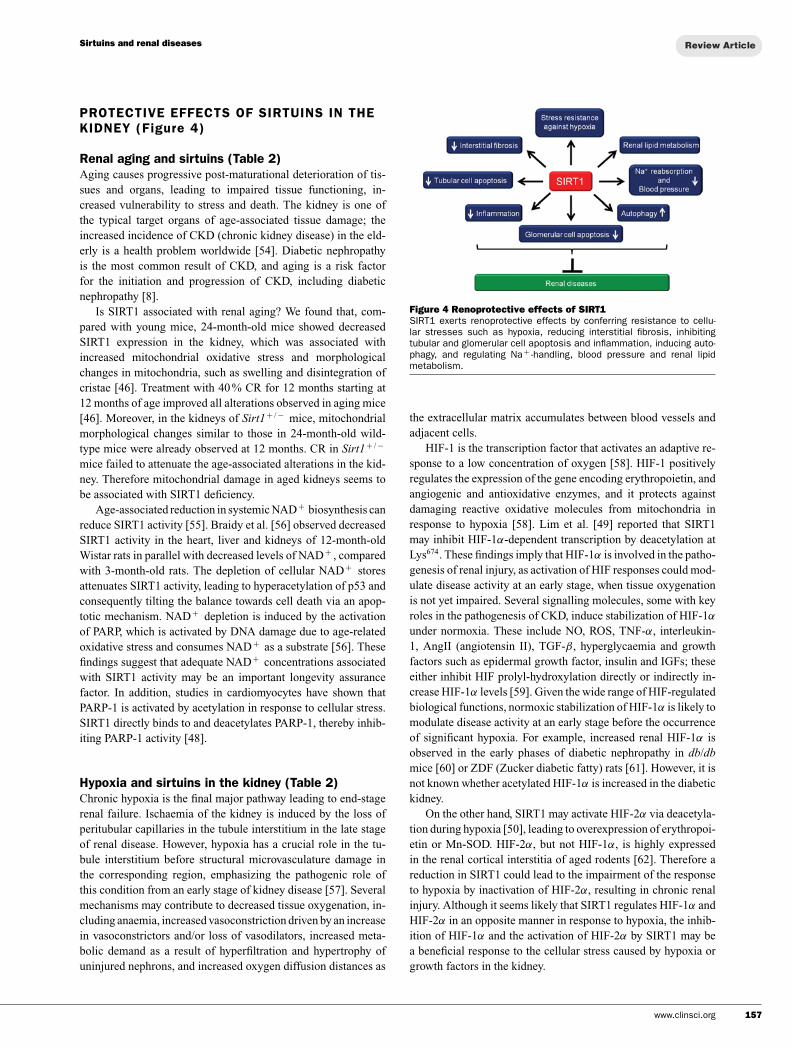
PROTECTIVE EFFECTS OF SIRTUINS IN THEKIDNEY (Figure 4)
Renal aging and sirtuins (Table 2)Aging causes progressive post-maturational deterioration of tis-sues and organs, leading to impaired tissue functioning, in-creased vulnerability to stress and death. The kidney is one ofthe typical target organs of age-associated tissue damage; theincreased incidence of CKD (chronic kidney disease) in the eld-erly is a health problem worldwide [54]. Diabetic nephropathyis the most common result of CKD, and aging is a risk factorfor the initiation and progression of CKD, including diabeticnephropathy [8].
Is SIRT1 associated with renal aging? We found that, com-pared with young mice, 24-month-old mice showed decreasedSIRT1 expression in the kidney, which was associated withincreased mitochondrial oxidative stress and morphologicalchanges in mitochondria, such as swelling and disintegration ofcristae [46]. Treatment with 40 % CR for 12 months starting at12 months of age improved all alterations observed in aging mice[46]. Moreover, in the kidneys of Sirt1+ / − mice, mitochondrialmorphological changes similar to those in 24-month-old wild-type mice were already observed at 12 months. CR in Sirt1+ / −
mice failed to attenuate the age-associated alterations in the kid-ney. Therefore mitochondrial damage in aged kidneys seems tobe associated with SIRT1 deficiency.
Age-associated reduction in systemic NAD+ biosynthesis canreduce SIRT1 activity [55]. Braidy et al. [56] observed decreasedSIRT1 activity in the heart, liver and kidneys of 12-month-oldWistar rats in parallel with decreased levels of NAD+ , comparedwith 3-month-old rats. The depletion of cellular NAD+ storesattenuates SIRT1 activity, leading to hyperacetylation of p53 andconsequently tilting the balance towards cell death via an apop-totic mechanism. NAD+ depletion is induced by the activationof PARP, which is activated by DNA damage due to age-relatedoxidative stress and consumes NAD+ as a substrate [56]. Thesefindings suggest that adequate NAD+ concentrations associatedwith SIRT1 activity may be an important longevity assurancefactor. In addition, studies in cardiomyocytes have shown thatPARP-1 is activated by acetylation in response to cellular stress.SIRT1 directly binds to and deacetylates PARP-1, thereby inhib-iting PARP-1 activity [48].
Hypoxia and sirtuins in the kidney (Table 2)Chronic hypoxia is the final major pathway leading to end-stagerenal failure. Ischaemia of the kidney is induced by the loss ofperitubular capillaries in the tubule interstitium in the late stageof renal disease. However, hypoxia has a crucial role in the tu-bule interstitium before structural microvasculature damage inthe corresponding region, emphasizing the pathogenic role ofthis condition from an early stage of kidney disease [57]. Severalmechanisms may contribute to decreased tissue oxygenation, in-cluding anaemia, increased vasoconstriction driven by an increasein vasoconstrictors and/or loss of vasodilators, increased meta-bolic demand as a result of hyperfiltration and hypertrophy ofuninjured nephrons, and increased oxygen diffusion distances as
Figure 4 Renoprotective effects of SIRT1SIRT1 exerts renoprotective effects by conferring resistance to cellu-lar stresses such as hypoxia, reducing interstitial fibrosis, inhibitingtubular and glomerular cell apoptosis and inflammation, inducing auto-phagy, and regulating Na+ -handling, blood pressure and renal lipidmetabolism.
the extracellular matrix accumulates between blood vessels andadjacent cells.
HIF-1 is the transcription factor that activates an adaptive re-sponse to a low concentration of oxygen [58]. HIF-1 positivelyregulates the expression of the gene encoding erythropoietin, andangiogenic and antioxidative enzymes, and it protects againstdamaging reactive oxidative molecules from mitochondria inresponse to hypoxia [58]. Lim et al. [49] reported that SIRT1may inhibit HIF-1α-dependent transcription by deacetylation atLys674. These findings imply that HIF-1α is involved in the patho-genesis of renal injury, as activation of HIF responses could mod-ulate disease activity at an early stage, when tissue oxygenationis not yet impaired. Several signalling molecules, some with keyroles in the pathogenesis of CKD, induce stabilization of HIF-1α
under normoxia. These include NO, ROS, TNF-α, interleukin-1, AngII (angiotensin II), TGF-β, hyperglycaemia and growthfactors such as epidermal growth factor, insulin and IGFs; theseeither inhibit HIF prolyl-hydroxylation directly or indirectly in-crease HIF-1α levels [59]. Given the wide range of HIF-regulatedbiological functions, normoxic stabilization of HIF-1α is likely tomodulate disease activity at an early stage before the occurrenceof significant hypoxia. For example, increased renal HIF-1α isobserved in the early phases of diabetic nephropathy in db/dbmice [60] or ZDF (Zucker diabetic fatty) rats [61]. However, it isnot known whether acetylated HIF-1α is increased in the diabetickidney.
On the other hand, SIRT1 may activate HIF-2α via deacetyla-tion during hypoxia [50], leading to overexpression of erythropoi-etin or Mn-SOD. HIF-2α, but not HIF-1α, is highly expressedin the renal cortical interstitia of aged rodents [62]. Therefore areduction in SIRT1 could lead to the impairment of the responseto hypoxia by inactivation of HIF-2α, resulting in chronic renalinjury. Although it seems likely that SIRT1 regulates HIF-1α andHIF-2α in an opposite manner in response to hypoxia, the inhib-ition of HIF-1α and the activation of HIF-2α by SIRT1 may bea beneficial response to the cellular stress caused by hypoxia orgrowth factors in the kidney.
www.clinsci.org 157
M. Kitada and others
Further studies are needed to determine the relationshipbetween HIF-1α and HIF-2α under tissue hypoxia or relativehypoxia in various renal injuries.
Interstitial fibrosis, tubular cell apoptosis andsirtuins in the kidney (Table 2)Tubulointerstitial fibrosis is considered a central event in theprogression of CKD, independent of aetiology. Even in glomer-ulopathies, tubulointerstitial fibrosis correlates better than glom-erular injury with the evolution and prognosis of the disease [63].Renal tubular cell apoptosis is implicated in the progression ofrenal injuries.
He et al. [64] found that SIRT1 is abundantly expressed inmouse medullary interstitial cells, where it increases cell resist-ance to oxidative stress. In Sirt1+ / − mice, the decreased SIRT1concentration is associated with increased apoptosis and fibrosisafter UUO (unilateral ureteral obstruction), whereas its activa-tion by treatment with resveratrol or SRT2183 in wild-type micereduced apoptotic and fibrotic changes in the UUO. In addi-tion, SIRT1 deficiency decreases the COX2 (cyclo-oxygenase 2)induction in medullary interstitial cells under oxidative stress,whereas exogenous PGE2 (prostaglandin E2) reduces apoptosisin oxidatively stressed SIRT1-deficient cells. These findingsnot only indicate the protective function of SIRT1, but alsoidentify COX2 as one of its targets. It remains to be estab-lished whether this effect of SIRT1 is due to its deacetylationof COX2 or is mediated via FOXO or other established targets ofSIRT1.
The TGF-β1/Smad3 signalling pathway plays a central role inthe pathogenesis of tissue fibrosis in the kidney. Li et al. [65] re-ported that SIRT1 activation by resveratrol inhibits the acetylationof Smad3, resulting in reduction of TGF-β1-induced collagen IVand fibronectin expression in a UUO animal model and in cul-tured cells (rat fibroblasts, NRK49F; rat proximal tubular cells,NRK52E).
SIRT1 also protects against renal tubular cell apoptosis.Hasegawa et al. [66] found that SIRT1 protects against oxidative-stress-induced apoptosis by inducing catalase via deacetylation ofFOXO3 in cultured proximal tubular cells. Moreover, Hasegawaet al. [67] also reported that renal proximal tubular cell-specificSIRT1 transgenic mice showed resistance to cisplatin-inducedrenal tubular cell injuries, such as apoptosis, by maintainingperoxisome number and function, concomitant up-regulation ofcatalase and elimination of renal ROS. In addition, SIRT1 activ-ation by resveratrol reduces cisplatin-induced proximal tubularcell apoptosis through deacetylation of p53 [68].
Inflammation and sirtuins in the kidney (Table 2)The inflammatory process is one of the pivotal mechanisms forthe initiation and progression of age-related diseases, such asdiabetes, cardiovascular diseases, neurodegenerative disorders,pulmonary disease and kidney diseases including diabetic neph-ropathy [69,70]. Therefore the control of the inflammatory pro-cess might be a potential therapeutic target for attenuating theprogression of such age-related diseases.
The NF-κB signalling pathway plays a central role in theregulation of the expression of several inflammation-related pro-
teins, such as MCP-1 (monocyte chemotactic protein-1), ICAM-1 (intercellular adhesion molecule 1) and VCAM-1 (vascularcell adhesion protein 1) [71]. The observation that several lys-ine residues of the NF-κB p65 subunit can be acetylated hashighlighted the potential regulatory role of lysine acetylation inNF-κB function [72]. Among these acetylated lysine residuesin NF-κB p65, acetylation at Lys310 might promote superiortranscriptional activity while also being a substrate for SIRT1[51]. Therefore SIRT1 may act as a negative regulator of NF-κB activity by deacetylating Lys310 of p65. Reduced levels ofSIRT1 result in the up-regulation of acetylated NF-κB, lead-ing to an increase in the inflammatory response in adipocytes[73], monocytes/macrophages [74,75], myeloid cells [76], en-dothelial cells [77] and microglia [78] of several experimentalanimal or cell models. In those studies, SIRT1 up-regulationusing chemical activators of SIRT1 or overexpression of aSIRT1 gene improved the inflammation through deacetylation ofNF-κB.
We found that SIRT1 protein expression is significantly de-creased and that acetylated NF-κB p65 and inflammation-relatedgene expression levels (ICAM-1, VCAM-1 and MCP-1) areclearly increased in the kidneys of WFRs (Wistar fatty diabeticrats) compared with WLRs (Wistar lean non-diabetic rats) [79].The increased levels of acetylated NF-κB and inflammation-related genes in WFRs were decreased by CR, consistent with therestoration of SIRT1 protein expression in the kidney. Thereforerenal inflammation is induced by increased levels of acetylatedNF-κB p65 owing to reduced SIRT1 protein expression, whereasCR exerts anti-inflammatory effects by restoring SIRT1 expres-sion in the kidneys of WFRs [79].
SIRT6 is also a negative regulator of NF-κB signalling. SIRT6attenuates NF-κB signalling by functioning at the chromatinlevel. Although it directly binds to p65 as SIRT1 does, SIRT6deacetylates H3K9 in the promoters of NF-κB target genes todecrease promoter occupancy by p65, rather than directly mod-ulating p65 [80]. So far, however, there are no reports aboutSIRT6 in renal diseases.
Role of sirtuins in glomerular cells (Table 2)Apoptosis is a distinct form of cell death that is observed undervarious physiological and pathological conditions [81]. Glomer-ular cells, including mesangial cells and podocytes, exhibit up-regulated apoptosis in human and experimental kidney diseases,such as diabetic nephropathy, hypertensive nephrosclerosis andglomerulonephritis. This apoptosis is considered to be involvedin the progression of these diseases. Therefore preventing glom-erular cell apoptosis may help prevent various kidney diseases.
Interestingly, SIRT1 diminishes mesangial cell apoptosis in-duced by oxidative stress by reducing p53 activity [47], andattenuates TGF-β-induced apoptotic signalling mediated by theeffector molecule Smad7 [82]. SIRT1-dependent deacetylation ofSmad7 at Lys60 and Lys70 also enhances the ubiquitin-dependentproteasomal degradation of this effector by Smurf1 (Smad ubi-quitination regulatory factor 1). Glomerular mesangial cells arethus protected from not only oxidative stress, but also TGF-β-dependent apoptosis. Both oxidative stress and TGF-β accelerateand contribute to renal diseases, implying that SIRT1 could be a
158 C© The Authors Journal compilation C© 2013 Biochemical Society
Sirtuins and renal diseases
therapeutic target for preventing glomerular diseases, includingdiabetic nephropathy.
Chuang et al. [83] found that AGE-BSA (AGE-modified BSA)increased FOXO4 acetylation and suppressed the expression ofthe SIRT1 protein in glomerular podocytes of db/db diabetic miceand diabetic patients. Acetylated FOXO4 promotes the expres-sion of the pro-apoptosis gene Bcl2l11 (also known as Bim) andleads to podocyte apoptosis.
NO is a protective factor in vascular tissues, including thekidneys. eNOS (endothelial NO synthase) deficiency due to en-dothelial cell dysfunction plays an important role in the patho-physiologies of cardiovascular diseases (hypertension and ath-erosclerosis) and renal injuries, including diabetic nephropathy[84]. SIRT1 promotes vasodilation and protects vascular tissuesthrough increased NO production by deacetylating eNOS in en-dothelial cells [85].
Autophagy and sirtuins in renal diseases (Table 2)Autophagy is a lysosomal degradation pathway that plays a cru-cial role in removing protein aggregates and damaged or excessorganelles, such as mitochondria, leading to the maintenance ofintracellular homoeostasis and promoting cellular health undervarious stress conditions, including hypoxia, ER (endoplasmicreticulum) stress or oxidative stress [86,87]. Autophagy plays acrucial role in several organs, especially metabolic organs, andits alteration is involved in the pathogenesis of metabolicand age-related diseases, including renal diseases [88]. Accord-ing to experiments in renal injury models, the autophagy system isimportant in renal tubular cells and podocytes. Furthermore, auto-phagy is regulated by nutrition-sensing signals such as SIRT1,mTOR (mammalian target of rapamycin) and AMPK. Results thatdemonstrate the role of SIRT1 in autophagy are still sparse com-pared with those for mTOR and AMPK, but they have been ac-cumulating. SIRT1 can deacetylate essential autophagic factors,such as Atg5, Atg7 and LC3, leading to the induction of auto-phagy. Furthermore, SIRT1 deacetylates the transcription factorFOXO3a, which leads to enhanced expression of proautophagicBnip3 (Bcl-2/adenovirus E1V 19-kDa interacting protein 3).
Hypoxia can cause renal tubular damage in kidney diseases, asdescribed below, and it also stimulates autophagy [87]. Hypoxia-induced autophagy largely depends on HIF-1α, which activatesthe transcription of Bnip3 and Bnip3L (Bnip3-like), which inturn induce autophagy. Normally, Beclin 1 interacts with Bcl-2proteins. Bnip3 can disrupt this interaction, liberating Beclin 1from Bcl-2 and leading to autophagy [89]. Thus HIF-1α-inducedBnip3 overexpression promotes autophagy. We have shown thathypoxia-induced autophagy activity declines with age in mice,which leads to accumulations of damaged mitochondria and mi-tochondrial ROS in the kidney [46]. Interestingly, long-term CRrestores autophagy activity, even in aged kidneys. When we in-vestigated the mechanism of this effect, we found that SIRT1-mediated autophagy is essential in the CR-mediated protectionof aged kidneys [46]. Bnip3 expression is essential for inducingautophagy under hypoxic conditions and is positively regulated byFOXO3a rather than HIF-1α. This regulation is altered in agedkidneys. However, CR-mediated SIRT1 activation deacetylatesand activates FOXO3a transcriptional activity and subsequent
Bnip3-mediated autophagy, even in aged kidneys. Furthermore,the kidneys of Sirt1+ / − mice exhibit lower autophagy activityand decreased Bnip3 expression; thus they are resistant to CR-mediated anti-aging effects. These findings suggest that SIRT1is essential for CR-mediated renoprotection. Chronic hypoxiacauses damage to the mitochondria and promotes the intracellu-lar accumulation of ROS. Removing the damaged mitochondriaunder hypoxic conditions is also an important role of Bnip3-mediated autophagy. Thus restoring autophagic activity even un-der diabetic conditions should be valuable for protecting the kid-ney from hypoxia.
In addition, we observed that mitochondrial morphologicaldamages in the proximal tubular cells and p62/Sqstm1 accumu-lation in kidneys of WFRs, suggesting that an impairment ofthe autophagy system can induce mitochondrial damage. Be-cause SIRT1 is a positive regulator of autophagy, the decreasein SIRT1 expression observed in the kidneys of WFRs may leadto the dysregulation of autophagy. CR improves the function ofthe autophagy system, which resulted in normalization of mito-chondrial morphological changes and p62/Sqstm1 accumulation,which was accompanied by the restoration of SIRT1 expression[79].
On the other hand, Hartleben et al. [90] have demonstrated animportant role for the autophagy system in podocytes under basalconditions and under several renal injury conditions using GFP(green fluorescent protein)–LC3 transgenic mice and podocyte-specific Atg5-knockout mice. They [90] showed that podocytesexhibit an unusually high level of constitutive autophagy andthat podocyte-specific deletion of Atg5 leads to glomerulopathyin aging mice that is accompanied by an accumulation of oxid-ized and ubiquitinated proteins, ER stress and proteinuria. Thesechanges ultimately result in podocyte loss and histological glom-erulosclerosis. Moreover, autophagy is substantially increased inglomeruli from mice with protein-overload-induced renal injuryand in glomeruli from patients with glomerular diseases, suchas focal glomerulosclerosis or membranoproliferative glomer-ulonephritis. Furthermore, mice lacking Atg5 in podocytes ex-hibit strongly increased susceptibility to drug-induced glomer-ular injuries (i.e. induced by puromycin aminonucleoside andadriamycin). These findings highlight the importance of inducedautophagy as a key homoeostatic mechanism for maintainingpodocyte integrity.
Thus it is evident that autophagy deficiency is associated withpodocyte and renal tubular cell injuries. These findings indicatethat autophagy deficiency should contribute to the pathogenesisof renal diseases such as diabetic nephropathy. Therefore dysreg-ulation of SIRT1 in the kidney could result in the accumulationof mitochondrial ROS via suppression of autophagy, which maybe associated with the initiation of the early stages of diabeticnephropathy. Both hypoxia- and proteinuria-induced ER stressmay also contribute to proximal tubular cell damage in the pro-gressive stages of diabetic nephropathy. Autophagy deficiency inkidneys from diabetic animals may make tubular cells fragile un-der hypoxic and ER stress and may lead to progression of diabeticnephropathy. Therefore activation of autophagy may be a thera-peutic option for the advanced stages of diabetic nephropathy[88].
www.clinsci.org 159
M. Kitada and others
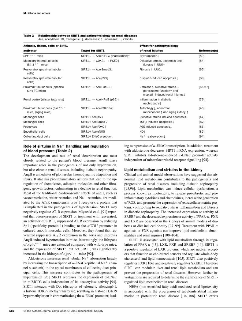
Table 2 Relationship between SIRT1 and pathophysiology on renal diseasesAce, acetylated; TG, transgenic; ↓, decreases; ↑, increases; �, inhibits.
Animals, tissue, cells or SIRT1 Effect for pathophysiology
activator Target for SIRT1 of renal injuries Reference(s)
Sirt1+ / − mice SIRT1↓ → Ace-HIF-2α (inactivation)↑ Erythropoetin↓ [50]
Medullary interstitial cells(Sirt1+ / − mice)
SIRT1↓ → COX2↓ → PGE2↓ Oxidative stress, apoptosis andfibrosis in UUO↑
[64]
Resveratrol (proximal tubularcells)
SIRT1↑ → Ace-Smad3↓ Fibrosis in UUO↓ [65]
Resveratrol (proximal tubularcells)
SIRT1↑ → Ace-p53↓ Cisplatin-induced apoptosis↓ [68]
Proximal tubular cells (specificSirt1-TG mice)
SIRT1↑ → Ace-FOXO3↓ Catalase↑, oxidative stress↓,peroxisome function↑ andcisplatin-induced renal injuries↓
[66,67]
Renal cortex (Wistar fatty rats) SIRT1↓ → Ace-NF-κB (p65)↑ Inflammation in diabeticnephropathy↑
[79]
Proximal tubular cells (Sirt1+ / −
mice) (aging mice)SIRT1↓ → Ace-FOXO3a↑ Autophagy↓, abnormal
mitochondria↑ and aging kidney ↑[46]
Mesangial cells SIRT1� Ace-p53 Oxidative stress-induced apoptosis↓ [47]
Mesangial cells SIRT1� Ace-Smad 7 TGF-β -induced apoptosis↓ [82]
Podocytes SIRT1� Ace-FOXO4 AGE-induced apoptosis↓ [83]
Endothelial cells SIRT1� Ace-eNOS NO↑ [85]
Collecting duct cells SIRT1� ENaC α-subunit Na+ reabsorption↓ [94]
Role of sirtuins in Na+ handling and regulationof blood pressure (Table 2)The development and rate of renal deterioration are mostclosely related to the patient’s blood pressure. AngII playsimportant roles in the pathogenesis of not only hypertension,but also chronic renal diseases, including diabetic nephropathy.AngII is a mediator of glomerular haemodynamic adaptation andinjury. It also has pro-inflammatory actions that lead to the up-regulation of chemokines, adhesion molecules and other fibro-genic growth factors, culminating in a decline in renal function.Most of the traditional cardiovascular effects of angII, such asvasoconstriction, water retention and Na+ retention, are medi-ated by the AT1R (angiotensin type 1 receptor), a protein thatis implicated in the pathogenesis of hypertension. SIRT1 maynegatively regulate AT1R expression. Miyazaki et al. [91] repor-ted that overexpression of SIRT1 or treatment with resveratrol,an activator of SIRT1, suppressed AT1R expression by reducingSp1 (specificity protein 1) binding to the AGTR1 promoter incultured smooth muscular cells. Moreover, they found that res-veratrol suppresses AT1R expression in the aorta and improvesAngII-induced hypertension in mice. Interestingly, the lifespansof Agtr1− / − mice are extended compared with wild-type mice,and the expression of SIRT3, but not SIRT1, was significantlyincreased in the kidneys of Agtr1− / − mice [92].
Aldosterone increases renal tubular Na+ absorption largelyby increasing the transcription of α-ENaC (epithelial Na+ chan-nel α-subunit) in the apical membranes of collecting duct prin-cipal cells. This increase contributes to the pathogenesis ofhypertension [93]. SIRT1 represses the expression of α-ENaCin mIMCD3 cells independent of its deacetylase activity [94].SIRT1 interacts with Dot (disruptor of telomeric silencing)-1,a histone H3K79 methyltransferase, resulting in histone H3K79hypermethylation in chromatin along the α-ENaC promoter, lead-
ing to repression of α-ENaC transcription. In addition, treatmentwith aldosterone decreases SIRT1 mRNA expression, whereasSIRT1 inhibits aldosterone-induced α-ENaC promoter activityindependent of mineralocorticoid receptor signalling [94].
Lipid metabolism and sirtuins in the kidneyClinical and animal model observations have suggested that ab-normal lipid metabolism contributes to the pathogenesis andprogression of renal diseases, including diabetic nephropathy[95,96]. Lipid metabolites can induce cellular dysfunction, aprocess known as lipotoxicity, to release pro-fibrotic and pro-inflammatory cytokines and chemokines, increase the generationof ROS, and promote the expression of extracellular matrix pro-teins, contributing to oxidative stress, inflammation and fibrosisin diabetic nephropathy. The increased expression or activity ofSREBP and the decreased expression or activity of PPAR-α, FXRand LXR are observed in the kidneys of animal models of dia-betes or diet-induced obesity [97–99]. Treatment with PPAR-αagonists or FXR agonists can improve lipid metabolism abnor-malities and renal injuries [100–104].
SIRT1 is associated with lipid metabolism through its regu-lation of PPAR-α [43], LXR, FXR and SREBP [44]. SIRT1 isa positive regulator of LXR proteins, which are nuclear recept-ors that function as cholesterol sensors and regulate whole-bodycholesterol and lipid homoeostasis [105]. SIRT1 also positivelyregulates FXR [106] and negatively regulates SREBP. ThereforeSIRT1 can modulate liver and renal lipid metabolism and canprevent the progression of renal diseases. However, further in-vestigations are required to determine the significance of SIRT1-regulated lipid metabolism in renal diseases.
NEFA (non-esterified fatty acid)-mediated renal lipotoxicityis associated with the progression of tubulointerstitial inflam-mation in proteinuric renal disease [107,108]. SIRT3 exerts
160 C© The Authors Journal compilation C© 2013 Biochemical Society
Sirtuins and renal diseases
antioxidant and anti-inflammatory effects under conditions ofpalmitate-induced lipotoxicity by promoting the enhancementof mitochondrial oxidative capacity and antioxidant defences inproximal tubular cells [109]. Therefore the activation of SIRT3could be useful in the design of new therapies to prevent protein-uric renal diseases, including advanced diabetic nephropathy.
SNPs (SINGLE NUCLEOTIDEPOLYMORPHISMS) AND SIRTUINS INDIABETIC NEPHROPATHY
Four SNPs within SIRT1 have been nominally associated withsusceptibility to diabetic nephropathy [110]. The haplotype, con-sisting of the 11 SNPs in SIRT1, had a stronger association withdiabetic nephropathy than any single SNP in four independent Ja-panese case-control studies. SNPs in other sirtuin genes have notdemonstrated any associations with diabetic nephropathy. ThusSIRT1 may be a good pharmaceutical target for diabetic neph-ropathy, although the association should be evaluated further inindependent studies [110].
CONCLUSIONS AND PROSPECTS
In recent decades, numerous investigators have made efforts toidentify the molecular mechanisms involved in the initiation andprogression of diabetic nephropathy to develop new therapeuticstrategies. However, end-stage renal failure due to diabeticnephropathy continues to increase worldwide. There is an urgentneed to identify new therapeutic targets to prevent diabeticnephropathy.
Over the last decade, the understanding of sirtuins has expan-ded from the original description of a single NAD+ -dependentclass III histone deacetylase that can control the lifespan ofyeast. SIRT1 deacetylates not only histones, but also manytranscriptional regulators, thereby modulating diverse biologicalprocesses. SIRT1 exerts renoprotective effects by conferring res-istance to cellular stress such as hypoxia, reducing fibrosis, in-hibiting apoptosis and inflammation, inducing autophagy, andregulating blood pressure (Figure 4). Therefore SIRT1 may bea novel therapeutic target for renal diseases including diabeticnephropathy.
Although other sirtuins, including the mitochondrial sirtuinsSIRT3, SIRT4 and SIRT5 and the nuclear sirtuins SIRT6 andSIRT7, may have important roles in cytoprotective functions,their molecular targets and biological functions, and possibleroles in renoprotection, are largely unknown. Further investig-ation into the targets and functions of other sirtuins will helpdevelop new strategies for protection against renal diseases.
FUNDING
This work was supported by Novo Nordisk Pharma, a Grant-in-Aidfor Scientific Research (C) [number 24591218] and a Grant forPromoted Research from Kanazawa Medical University [numberS2012-4] (to M.K.), Grants for Collaborative Research [number
C2012-1] and Specially Promoted Research from Kanazawa Med-ical University [number SR2012-06] and the Japanese Society ofAnti-Aging Medicine [4th Annual Research Award Grant (to D.K.)].
REFERENCES
1 Jun, M., Perkovic, V. and Cass, A. (2011) Intensive glycemiccontrol and renal outcome. Contrib. Nephrol. 170, 196–208
2 Calcutt, N. A., Cooper, M. E., Kern, T. S. and Schmidt, A. M.(2009) Therapies for hyperglycaemia-induced diabeticcomplications: from animal models to clinical trials. Nat. Rev.Drug Discovery 8, 417–429
3 Kitada, M., Zhang, Z., Mima, A. and King, G. L. (2010)Molecular mechanisms of diabetic vascular complications.J. Diabetes Invest. 1, 77–89
4 Galle, J. (2008) Reduction of proteinuria with angiotensinreceptor blockers. Nat. Clin. Pract. Cardiovasc. Med. 5(Suppl. 1), S36–S43
5 Barlovic, D. P. and Cooper, M. E. (2009) Diabetes: RASinhibition: probably not a one-size-fits-all approach. Nat. Rev.Nephrol. 5, 669–670
6 Coresh, J., Astor, B. C., Greene, T., Eknoyan, G. and Levey, A. S.(2003) Prevalence of chronic kidney disease and decreasedkidney function in the adult US population: Third National Healthand Nutrition Examination Survey. Am. J. Kidney Dis. 41, 1–12
7 Rule, A. D., Amer, H., Cornell, L. D., Taler, S. J., Cosio, F. G.,Kremers, W. K., Textor, S. C. and Stegall, M. D. (2010) Theassociation between age and nephrosclerosis on renal biopsyamong healthy adults. Ann. Intern. Med. 152, 561–567
8 Hsu, C. Y., Iribarren, C., McCulloch, C. E., Darbinian, J. and Go,A. S. (2009) Risk factors for end-stage renal disease: 25-yearfollow-up, Arch. Intern. Med. 169, 342–350
9 Fontana, L., Partridge, L. and Longo, V. D. (2010) Extendinghealthy life span–from yeast to humans. Science 328,321–326
10 Fouque, D., Pelletier, S., Mafra, D. and Chauveau, P. (2011)Nutrition and chronic kidney disease. Kidney Int. 80, 348–357
11 Guarente, L. (2011) Franklin H. Epstein Lecture: Sirtuins,aging, and medicine. N. Engl. J. Med. 364, 2235–2244
12 Someya, S., Yu, W., Hallows, W. C., Xu, J., Vann, J. M.,Leeuwenburgh, C., Tanokura, M., Denu, J. M. and Prolla, T. A.(2010) Sirt3 mediates reduction of oxidative damage andprevention of age-related hearing loss under caloric restriction.Cell 143, 802–812
13 McCay, C. M., Crowell, M. F. and Maynard, L. A. (1935) Theeffect of retarded growth upon the length of life span and uponthe ultimate body size. J. Nutr. 10, 63–79
14 Colman, R. J., Anderson, R. M., Johnson, S. C., Kastman, E. K.,Kosmatka, K. J., Beasley, T. M., Allison, D. B., Cruzen, C.,Simmons, H. A., Kemnitz, J. W. and Weindruch, R. (2009)Caloric restriction delays disease onset and mortality in rhesusmonkeys. Science 325, 201–204
15 Fontana, L., Meyer, T. E., Klein, S. and Holloszy, J. O. (2004)Long-term calorie restriction is highly effective in reducing therisk for atherosclerosis in humans. Proc. Natl. Acad. Sci. U.S.A.101, 6659–6663
16 Meyer, T. E., Kovacs, S. J., Ehsani, A. A., Klein, S., Holloszy,J. O. and Fontana, L. (2006) Long-term caloric restrictionameliorates the decline in diastolic function in humans. J. Am.Coll. Cardiol. 47, 398–402
17 Imai, S., Armstrong, C. M., Kaeberlein, M. and Guarente, L.(2000) Transcriptional silencing and longevity protein Sir2 is anNAD-dependent histone deacetylase. Nature 403, 795–800
18 Liang, F., Kume, S. and Koya, D. (2009) SIRT1 and insulinresistance. Nat. Rev. Endocrinol. 5, 367–373
www.clinsci.org 161
M. Kitada and others
19 Cohen, H. Y., Miller, C., Bitterman, K. J., Wall, N. R., Hekking,B., Kessler, B., Howitz, K. T., Gorospe, M., de Cabo, R. andSinclair, D. A. (2004) Calorie restriction promotes mammaliancell survival by inducing the SIRT1 deacetylase. Science 305,390–392
20 Bordone, L., Cohen, D., Robinson, A., Motta, M. C., van Veen,E., Czopik, A., Steele, A. D., Crowe, H., Marmor, S., Luo, J.et al. (2007) SIRT1 transgenic mice show phenotypesresembling calorie restriction. Aging Cell 6, 759–767
21 Boily, G., Seifert, E. L., Bevilacqua, L., He, X. H., Sabourin, G.,Estey, C., Moffat, C., Crawford, S., Saliba, S., Jardine, K. et al.(2008) SirT1 regulates energy metabolism and response tocaloric restriction in mice. PLoS ONE 3, e1759
22 Kanfi, Y., Naiman, S., Amir, G., Peshti, V., Zinman, G., Nahum,L., Bar-Joseph, Z. and Cohen, H. Y. (2012) The sirtuin SIRT6regulates lifespan in male mice. Nature 483, 218–221
23 Mostoslavsky, R., Chua, K. F., Lombard, D. B., Pang, W. W.,Fischer, M. R., Gellon, L., Liu, P., Mostoslavsky, G., Franco, S.,Murphy, M. M. et al. (2006) Genomic instability and aging-likephenotype in the absence of mammalian SIRT6. Cell 124,315–329
24 Kanfi, Y., Shalman, R., Peshti, V., Pilosof, S. N., Gozlan, Y. M.,Pearson, K. J., Lerrer, B., Moazed, D., Marine, J. C., de Cabo,R. and Cohen, H. Y. (2008) Regulation of SIRT6 protein levelsby nutrient availability. FEBS Lett. 582, 543–548
25 Kanfi, Y., Peshti, V., Gil, R., Naiman, S., Nahum, L., Levin, E.,Kronfeld-Schor, N. and Cohen, H. Y. (2010) SIRT6 protectsagainst pathological damage caused by diet-induced obesity.Aging Cell 9, 162–173
26 Michan, S. and Sinclair, D. (2007) Sirtuins in mammals:insights into their biological function. Biochem. J. 404, 1–13
27 Frye, R. A. (2000) Phylogenetic classification of prokaryotic andeukaryotic Sir2-like proteins. Biochem. Biophys. Res. Commun.273, 793–798
28 Tanno, M., Sakamoto, J., Miura, T., Shimamoto, K. and Horio, Y.(2007) Nucleocytoplasmic shuttling of the NAD+ -dependenthistone deacetylase SIRT1. J. Biol. Chem. 282, 6823–6832
29 North, B. J. and Verdin, E. (2007) Interphase nucleo-cytoplasmic shuttling and localization of SIRT2 during mitosis.PLoS ONE 2, e784
30 Sundaresan, N. R., Samant, S. A., Pillai, V. B., Rajamohan,S. B. and Gupta, M. P. (2008) SIRT3 is a stress-responsivedeacetylase in cardiomyocytes that protects cells fromstress-mediated cell death by deacetylation of Ku70. Mol. Cell.Biol. 28, 6384–6401
31 Michishita, E., Park, J. Y., Burneskis, J. M., Barrett, J. C. andHorikawa, I. (2005) Evolutionarily conserved and nonconservedcellular localizations and functions of human SIRT proteins.Mol. Biol. Cell 16, 4623–4635
32 Vakhrusheva, O., Smolka, C., Gajawada, P., Kostin, S., Boettger,T., Kubin, T., Braun, T. and Bober, E. (2008) Sirt7 increasesstress resistance of cardiomyocytes and prevents apoptosisand inflammatory cardiomyopathy in mice. Circ. Res. 102,703–710
33 Vaquero, A., Scher, M., Lee, D., Erdjument-Bromage, H.,Tempst, P. and Reinberg, D. (2004) Human SirT1 interacts withhistone H1 and promotes formation of facultativeheterochromatin. Mol. Cell 16, 93–105
34 Vaquero, A., Scher, M., Erdjument-Bromage, H., Tempst, P.,Serrano, L. and Reinberg, D. (2007) SIRT1 regulates thehistone methyl-transferase SUV39H1 during heterochromatinformation. Nature 450, 440–444
35 Rodgers, J. T., Lerin, C., Haas, W., Gygi, S. P., Spiegelman,B. M. and Puigserver, P. (2005) Nutrient control of glucosehomeostasis through a complex of PGC-1α and SIRT1. Nature434, 113–118
36 Rodgers, J. T., Lerin, C., Gerhart-Hines, Z. and Puigserver, P.(2008) Metabolic adaptations through the PGC-1 α and SIRT1pathways. FEBS Lett. 582, 46–53
37 Sun, C., Zhang, F., Ge, X., Yan, T., Chen, X., Shi, X. and Zhai, Q.(2007) SIRT1 improves insulin sensitivity under insulin-resistantconditions by repressing PTP1B. Cell Metab. 6, 307–319
38 Zhang, J. (2007) The direct involvement of SirT1 in insulin-induced insulin receptor substrate-2 tyrosine phosphorylation.J. Biol. Chem. 282, 34356–34364
39 Moynihan, K. A., Grimm, A. A., Plueger, M. M., Bernal-Mizrachi,E., Ford, E., Cras-Meneur, C., Permutt, M. A. and Imai, S.(2005) Increased dosage of mammalian Sir2 in pancreatic β
cells enhances glucose-stimulated insulin secretion in mice.Cell Metab. 2, 105–117
40 Picard, F., Kurtev, M., Chung, N., Topark-Ngarm, A., Senawong,T., Machado De Oliveira, R., Leid, M., McBurney, M. W. andGuarente, L. (2004) Sirt1 promotes fat mobilization in whiteadipocytes by repressing PPAR-γ . Nature 429, 771–776
41 Nakahata, Y., Kaluzova, M., Grimaldi, B., Sahar, S., Hirayama,J., Chen, D., Guarente, L. P. and Sassone-Corsi, P. (2008) TheNAD+ -dependent deacetylase SIRT1 modulates CLOCK-mediated chromatin remodeling and circadian control. Cell 134,329–340
42 Asher, G., Gatfield, D., Stratmann, M., Reinke, H., Dibner, C.,Kreppel, F., Mostoslavsky, R., Alt, F. W. and Schibler, U. (2008)SIRT1 regulates circadian clock gene expression through PER2deacetylation. Cell 134, 317–328
43 Purushotham, A., Schug, T. T., Xu, Q., Surapureddi, S., Guo, X.and Li, X. (2009) Hepatocyte-specific deletion of SIRT1 altersfatty acid metabolism and results in hepatic steatosis andinflammation. Cell Metab. 9, 327–338
44 Walker, A. K., Yang, F., Jiang, K., Ji, J. Y., Watts, J. L.,Purushotham, A., Boss, O., Hirsch, M. L., Ribich, S., Smith, J. J.et al. (2010) Conserved role of SIRT1 orthologs in fasting-dependent inhibition of the lipid/cholesterol regulator SREBP.Genes Dev. 24, 1403–1417
45 Brunet, A., Sweeney, L. B., Sturgill, J. F., Chua, K. F., Greer, P. L.,Lin, Y., Tran, H., Ross, S. E., Mostoslavsky, R., Cohen, H. Y.et al. (2004) Stress-dependent regulation of FOXO transcriptionfactors by the SIRT1 deacetylase. Science 303, 2011–2015
46 Kume, S., Uzu, T., Horiike, K., Chin-Kanasaki, M., Isshiki, K.,Araki, S., Sugimoto, T., Haneda, M., Kashiwagi, A. and Koya, D.(2010) Calorie restriction enhances cell adaptation to hypoxiathrough Sirt1-dependent mitochondrial autophagy in mouseaged kidney. J. Clin. Invest. 120, 1043–1055
47 Kume, S., Haneda, M., Kanasaki, K., Sugimoto, T., Araki, S.,Isono, M., Isshiki, K., Uzu, T., Kashiwagi, A. and Koya, D.(2006) Silent information regulator 2 (SIRT1) attenuatesoxidative stress-induced mesangial cell apoptosis via p53deacetylation. Free Radical Biol. Med. 40, 2175–2182
48 Rajamohan, S. B., Pillai, V. B., Gupta, M., Sundaresan, N. R.,Birukov, K. G., Samant, S., Hottiger, M. O. and Gupta, M. P.(2009) SIRT1 promotes cell survival under stress bydeacetylation-dependent deactivation of poly(ADP-ribose)polymerase 1. Mol. Cell. Biol. 29, 4116–4129
49 Lim, J. H., Lee, Y. M., Chun, Y. S., Chen, J., Kim, J. E. and Park,J. W. (2010) Sirtuin 1 modulates cellular responses to hypoxiaby deacetylating hypoxia-inducible factor 1α. Mol. Cell 38,864–878
50 Dioum, E. M., Chen, R., Alexander, M. S., Zhang, Q., Hogg,R. T., Gerard, R. D. and Garcia, J. A. (2009) Regulation ofhypoxia-inducible factor 2α signaling by the stress-responsivedeacetylase sirtuin 1. Science 324, 1289–1293
51 Yeung, F., Hoberg, J. E., Ramsey, C. S., Keller, M. D., Jones,D. R., Frye, R. A. and Mayo, M. W. (2004) Modulation ofNF-κB-dependent transcription and cell survival by the SIRT1deacetylase. EMBO J. 23, 2369–2380
52 Lee, I. H., Cao, L., Mostoslavsky, R., Lombard, D. B., Liu, J.,Bruns, N. E., Tsokos, M., Alt, F. W. and Finkel, T. (2008) A rolefor the NAD-dependent deacetylase Sirt1 in the regulation ofautophagy. Proc. Natl. Acad. Sci. U.S.A. 105, 3374–3379
162 C© The Authors Journal compilation C© 2013 Biochemical Society
Sirtuins and renal diseases
53 Price, N. L., Gomes, A. P., Ling, A. J., Duarte, F. V.,Martin-Montalvo, A., North, B. J., Agarwal, B., Ye, L., Ramadori,G., Teodoro, J. S. et al. (2012) SIRT1 is required for AMPKactivation and the beneficial effects of resveratrol onmitochondrial function. Cell Metab. 15, 675–690
54 Choudhury, D. and Levi, M. (2011) Kidney aging – inevitable orpreventable? Nat. Rev. Nephrol. 7, 706–717
55 Imai, S. (2011) Dissecting systemic control of metabolism andaging in the NAD world: the importance of SIRT1 and NAMPT-mediated NAD biosynthesis. FEBS Lett. 585, 1657–1662
56 Braidy, N., Guillemin, G. J., Mansour, H., Chan-Ling, T., Poljak,A. and Grant, R. (2011) Age related changes in NAD+
metabolism oxidative stress and Sirt1 activity in Wistar rats.PLoS ONE 6, e19194
57 Nangaku, M. (2006) Chronic hypoxia and tubulointerstitialinjury: a final common pathway to end-stage renal failure. J. Am.Soc. Nephrol. 17, 17–25
58 Haase, V. H. (2006) Hypoxia-inducible factors in the kidney. Am.J. Physiol. Renal Physiol. 291, F271–F281
59 Haase, V. H. (2010) The sweet side of HIF. Kidney Int. 78,10–13
60 Makino, H., Miyamoto, Y., Sawai, K., Mori, K., Mukoyama, M.,Nakao, K., Yoshimasa, Y. and Suga, S. (2006) Altered geneexpression related to glomerulogenesis and podocyte structurein early diabetic nephropathy of db/db mice and its restorationby pioglitazone. Diabetes 55, 2747–2756
61 Takiyama, Y., Harumi, T., Watanabe, J., Fujita, Y., Honjo, J.,Shimizu, N., Makino, Y. and Haneda, M. (2011) Tubular injury ina rat model of type 2 diabetes is prevented by metformin: apossible role of HIF-1α expression and oxygen metabolism.Diabetes 60, 981–992
62 Tanaka, T., Kato, H., Kojima, I., Ohse, T., Son, D., Tawakami, T.,Yatagawa, T., Inagi, R., Fujita, T. and Nangaku, M. (2006)Hypoxia and expression of hypoxia-inducible factor in the agingkidney. J. Gerontol. A Biol. Sci. Med. Sci. 61, 795–805
63 Nath, K. A. (1992) Tubulointerstitial changes as a majordeterminant in the progression of renal damage. Am. J. KidneyDis. 20, 1–17
64 He, W., Wang, Y., Zhang, M. Z., You, L., Davis, L. S., Fan, H.,Yang, H. C., Fogo, A. B., Zent, R., Harris, R. C. et al. (2010)Sirt1 activation protects the mouse renal medulla fromoxidative injury. J. Clin. Invest. 120, 1056–1068
65 Li, J., Qu, X., Ricardo, S. D., Bertram, J. F. and Nikolic-Paterson,D. J. (2010) Resveratrol inhibits renal fibrosis in the obstructedkidney: potential role in deacetylation of Smad3. Am. J. Pathol.177, 1065–1071
66 Hasegawa, K., Wakino, S., Yoshioka, K., Tatematsu, S., Hara,Y., Minakuchi, H., Washida, N., Tokuyama, H., Hayashi, K. andItoh, H. (2008) Sirt1 protects against oxidative stress-inducedrenal tubular cell apoptosis by the bidirectional regulation ofcatalase expression. Biochem. Biophys. Res. Commun. 372,51–56
67 Hasegawa, K., Wakino, S., Yoshioka, K., Tatematsu, S., Hara,Y., Minakuchi, H., Sueyasu, K., Washida, N., Tokuyama, H.,Tzukerman, M. et al. (2010) Kidney-specific overexpression ofSirt1 protects against acute kidney injury by retainingperoxisome function. J. Biol. Chem. 285, 13045–13056
68 Kim, D. H., Jung, Y. J., Lee, J. E., Lee, A. S., Kang, K. P., Lee, S.,Park, S. K., Han, M. K., Lee, S. Y., Ramkumar, K. M. et al.(2011) SIRT1 activation by resveratrol amelioratescisplatin-induced renal injury through deacetylation of p53. Am.J. Physiol. Renal Physiol. 301, F427–F435
69 Chung, H. Y., Sung, B., Jung, K. J., Zou, Y. and Yu, B. P. (2006)The molecular inflammatory process in aging. Antioxid. RedoxSignaling 8, 572–581
70 Navarro-Gonzalez, J. F., Mora-Fernandez, C., Muros de Fuentes,M. and Garcia-Perez, J. (2011) Inflammatory molecules andpathways in the pathogenesis of diabetic nephropathy. Nat.Rev. Nephrol. 7, 327–340
71 Hayden, M. S. and Ghosh, S. (2008) Shared principles in NF-κBsignaling. Cell 132, 344–362
72 Kiernan, R., Bres, V., Ng, R. W., Coudart, M. P., El Messaoudi,S., Sardet, C., Jin, D. Y., Emiliani, S. and Benkirane, M. (2003)Post-activation turn-off of NF-κB-dependent transcription isregulated by acetylation of p65. J. Biol. Chem. 278,2758–2766
73 Yoshizaki, T., Milne, J. C., Imamura, T., Schenk, S., Sonoda, N.,Babendure, J. L., Lu, J. C., Smith, J. J., Jirousek, M. R. andOlefsky, J. M. (2009) SIRT1 exerts anti-inflammatory effectsand improves insulin sensitivity in adipocytes. Mol. Cell. Biol.29, 1363–1374
74 Yoshizaki, T., Schenk, S., Imamura, T., Babendure, J. L.,Sonoda, N., Bae, E. J., Oh, D. Y., Lu, M., Milne, J. C., Westphal,C. et al. (2010) SIRT1 inhibits inflammatory pathways inmacrophages and modulates insulin sensitivity. Am. J. Physiol.Endocrinol. Metab. 298, E419–E428
75 Rajendrasozhan, S., Yang, S. R., Kinnula, V. L. and Rahman, I.(2008) SIRT1, an antiinflammatory and antiaging protein, isdecreased in lungs of patients with chronic obstructivepulmonary disease. Am. J. Respir. Crit. Care Med. 177,861–870
76 Schug, T. T., Xu, Q., Gao, H., Peres-da-Silva, A., Draper, D. W.,Fessler, M. B., Purushotham, A. and Li, X. (2010) Myeloiddeletion of SIRT1 induces inflammatory signaling in responseto environmental stress. Mol. Cell. Biol. 30, 4712–4721
77 Stein, S., Schafer, N., Breitenstein, A., Besler, C., Winnik, S.,Lohmann, C., Heinrich, K., Brokopp, C. E., Handschin, C.,Landmesser, U. et al. (2010) SIRT1 reduces endothelialactivation without affecting vascular function in ApoE− / − mice.Aging 2, 353–360
78 Chen, J., Zhou, Y., Mueller-Steiner, S., Chen, L. F., Kwon, H.,Yi, S., Mucke, L. and Gan, L. (2005) SIRT1 protects againstmicroglia-dependent amyloid-β toxicity through inhibiting NF-κBsignaling. J. Biol. Chem. 280, 40364–40374
79 Kitada, M., Takeda, A., Nagai, T., Ito, H., Kanasaki, K. and Koya,D. (2011) Dietary restriction ameliorates diabetic nephropathythrough anti-inflammatory effects and regulation of theautophagy via restoration of Sirt1 in diabetic Wistar fatty (fa/fa)rats: a model of type 2 diabetes. Exp. Diabetes Res. 2011,908185
80 Kawahara, T. L., Michishita, E., Adler, A. S., Damian, M., Berber,E., Lin, M., McCord, R. A., Ongaigui, K. C., Boxer, L. D., Chang,H. Y. and Chua, K. F. (2009) SIRT6 links histone H3 lysine 9deacetylation to NF-κB-dependent gene expression andorganismal life span. Cell 136, 62–74
81 Hattori, T., Shindo, S. and Kawamura, H. (1998) Apoptosis andexpression of Bax protein and Fas antigen in glomeruli of aremnant-kidney model. Nephron 79, 186–191
82 Kume, S., Haneda, M., Kanasaki, K., Sugimoto, T., Araki, S.,Isshiki, K., Isono, M., Uzu, T., Guarente, L., Kashiwagi, A. andKoya, D. (2007) SIRT1 inhibits transforming growth factorβ -induced apoptosis in glomerular mesangial cells via Smad7deacetylation. J. Biol. Chem. 282, 151–158
83 Chuang, P. Y., Dai, Y., Liu, R., He, H., Kretzler, M., Jim, B.,Cohen, C. D. and He, J. C. (2011) Alteration of forkhead box O(foxo4) acetylation mediates apoptosis of podocytes indiabetes mellitus. PLoS ONE 6, e23566
84 Nakagawa, T., Tanabe, K., Croker, B. P., Johnson, R. J., Grant,M. B., Kosugi, T. and Li, Q. (2011) Endothelial dysfunction as apotential contributor in diabetic nephropathy. Nat. Rev. Nephrol.7, 36–44
85 Mattagajasingh, I., Kim, C. S., Naqvi, A., Yamamori, T.,Hoffman, T. A., Jung, S. B., DeRicco, J., Kasuno, K. and Irani, K.(2007) SIRT1 promotes endothelium-dependent vascularrelaxation by activating endothelial nitric oxide synthase. Proc.Natl. Acad. Sci. U.S.A. 104, 14855–14860
86 Mizushima, N., Levine, B., Cuervo, A. M. and Klionsky, D. J.(2008) Autophagy fights disease through cellular self-digestion.Nature 451, 1069–1075
www.clinsci.org 163
M. Kitada and others
87 Kroemer, G., Marino, G. and Levine, B. (2010) Autophagy andthe integrated stress response. Mol. Cell 40, 280–293
88 Tanaka, Y., Kume, S., Kitada, M., Kanasaki, K., Uzu, T.,Maegawa, H. and Koya, D. (2012) Autophagy as a therapeutictarget in diabetic nephropathy. Exp. Diabetes Res. 2012,628978
89 Bellot, G., Garcia-Medina, R., Gounon, P., Chiche, J., Roux, D.,Pouyssegur, J. and Mazure, N. M. (2009) Hypoxia-inducedautophagy is mediated through hypoxia-inducible factorinduction of BNIP3 and BNIP3L via their BH3 domains. Mol.Cell. Biol. 29, 2570–2581
90 Hartleben, B., Godel, M., Meyer-Schwesinger, C., Liu, S., Ulrich,T., Kobler, S., Wiech, T., Grahammer, F., Arnold, S. J.,Lindenmeyer, M. T. et al. (2010) Autophagy influencesglomerular disease susceptibility and maintains podocytehomeostasis in aging mice. J. Clin. Invest. 120, 1084–1096
91 Miyazaki, R., Ichiki, T., Hashimoto, T., Inanaga, K., Imayama, I.,Sadoshima, J. and Sunagawa, K. (2008) SIRT1, a longevitygene, downregulates angiotensin II type 1 receptor expressionin vascular smooth muscle cells. Arterioscler. Thromb. Vasc.Biol. 28, 1263–1269
92 Benigni, A., Corna, D., Zoja, C., Sonzogni, A., Latini, R., Salio,M., Conti, S., Rottoli, D., Longaretti, L., Cassis, P. et al. (2009)Disruption of the Ang II type 1 receptor promotes longevity inmice. J. Clin. Invest. 119, 524–530
93 Rossier, B. C., Pradervand, S., Schild, L. and Hummler, E.(2002) Epithelial sodium channel and the control of sodiumbalance: interaction between genetic and environmentalfactors. Annu. Rev. Physiol. 64, 877–897
94 Zhang, D., Li, S., Cruz, P. and Kone, B. C. (2009) Sirtuin 1functionally and physically interacts with disruptor of telomericsilencing-1 to regulate α-ENaC transcription in collecting duct.J. Biol. Chem. 284, 20917–20926
95 Moorhead, J. F., Chan, M. K., El-Nahas, M. and Varghese, Z.(1982) Lipid nephrotoxicity in chronic progressive glomerularand tubulo-interstitial disease. Lancet ii, 1309–1311
96 Ruan, X. Z., Varghese, Z. and Moorhead, J. F. (2009) An updateon the lipid nephrotoxicity hypothesis. Nat. Rev. Nephrol. 5,713–721
97 Sun, L., Halaihel, N., Zhang, W., Rogers, T. and Levi, M. (2002)Role of sterol regulatory element-binding protein 1 in regulationof renal lipid metabolism and glomerulosclerosis in diabetesmellitus. J. Biol. Chem. 277, 18919–18927
98 Proctor, G., Jiang, T., Iwahashi, M., Wang, Z., Li, J. and Levi, M.(2006) Regulation of renal fatty acid and cholesterolmetabolism, inflammation, and fibrosis in Akita and OVE26mice with type 1 diabetes. Diabetes 55, 2502–2509
99 Wang, Z., Jiang, T., Li, J., Proctor, G., McManaman, J. L., Lucia,S., Chua, S. and Levi, M. (2005) Regulation of renal lipidmetabolism, lipid accumulation, and glomerulosclerosis inFVBdb/db mice with type 2 diabetes. Diabetes 54, 2328–2335
100 Wang, X. X., Jiang, T., Shen, Y., Caldas, Y., Miyazaki-Anzai, S.,Santamaria, H., Urbanek, C., Solis, N., Scherzer, P., Lewis, L.et al. (2010) Diabetic nephropathy is accelerated by farnesoidX receptor deficiency and inhibited by farnesoid X receptoractivation in a type 1 diabetes model. Diabetes 59,2916–2927
101 Jiang, T., Wang, X. X., Scherzer, P., Wilson, P., Tallman, J.,Takahashi, H., Li, J., Iwahashi, M., Sutherland, E., Arend, L. andLevi, M. (2007) Farnesoid X receptor modulates renal lipidmetabolism, fibrosis, and diabetic nephropathy. Diabetes 56,2485–2493
102 Wang, X. X., Jiang, T., Shen, Y., Adorini, L., Pruzanski, M.,Gonzalez, F. J., Scherzer, P., Lewis, L., Miyazaki-Anzai, S. andLevi, M. (2009) The farnesoid X receptor modulates renal lipidmetabolism and diet-induced renal inflammation, fibrosis,and proteinuria. Am. J. Physiol. Renal Physiol. 297,F1587–F1596
103 Wang, X. X., Jiang, T. and Levi, M. (2010) Nuclear hormonereceptors in diabetic nephropathy. Nat. Rev. Nephrol. 6,342–351
104 Tanaka, Y., Kume, S., Araki, S., Isshiki, K., Chin-Kanasaki, M.,Sakaguchi, M., Sugimoto, T., Koya, D., Haneda, M., Kashiwagi,A. et al. (2011) Fenofibrate, a PPARα agonist, hasrenoprotective effects in mice by enhancing renal lipolysis.Kidney Int. 79, 871–882
105 Li, X., Zhang, S., Blander, G., Tse, J. G., Krieger, M. andGuarente, L. (2007) SIRT1 deacetylates and positivelyregulates the nuclear receptor LXR. Mol. Cell 28, 91–106
106 Kemper, J. K., Xiao, Z., Ponugoti, B., Miao, J., Fang, S.,Kanamaluru, D., Tsang, S., Wu, S. Y., Chiang, C. M. andVeenstra, T. D. (2009) FXR acetylation is normally dynamicallyregulated by p300 and SIRT1 but constitutively elevated inmetabolic disease states. Cell Metab. 10, 392–404
107 Burton, C. and Harris, K. P. (1996) The role of proteinuria in theprogression of chronic renal failure. Am. J. Kidney Dis. 27,765–775
108 Thomas, M. E. and Schreiner, G. F. (1993) Contribution ofproteinuria to progressive renal injury: consequences of tubularuptake of fatty acid bearing albumin. Am. J. Nephrol. 13,385–398
109 Koyama, T., Kume, S., Koya, D., Araki, S., Isshiki, K.,Chin-Kanasaki, M., Sugimoto, T., Haneda, M., Sugaya, T.,Kashiwagi, A. et al. (2011) SIRT3 attenuates palmitate-inducedROS production and inflammation in proximal tubular cells. FreeRadical Biol. Med. 51, 1258–1267
110 Maeda, S., Koya, D., Araki, S., Babazono, T., Umezono, T.,Toyoda, M., Kawai, K., Imanishi, M., Uzu, T., Suzuki, D. et al.(2011) Association between single nucleotide polymorphismswithin genes encoding sirtuin families and diabetic nephropathyin Japanese subjects with type 2 diabetes. Clin. Exp. Nephrol.15, 381–390
Received 17 April 2012/3 August 2012; accepted 28 August 2012
Published on the Internet 5 October 2012, doi: 10.1042/CS20120190
164 C© The Authors Journal compilation C© 2013 Biochemical Society


![Activation of Nrf2 Pathway Contributes to …...NF-κB,p53andNrf2inbraincells[14,15].Amongtheseven sirtuins,Sirtuin1 (SIRT1)hasbeenreportedtobeinvolvedin promoting longevity in various](https://static.fdocuments.net/doc/165x107/5fc15a05ed4fb114a500e06b/activation-of-nrf2-pathway-contributes-to-nf-bp53andnrf2inbraincells1415amongtheseven.jpg)


