
Sindbis Expression System - Thermo Fisher...
54
Sindbis Expression System Catalog no. K750-01 Sindbis Expression System Version E 180402 25-0121 U.S. Headquarters: European Headquarters: Invitrogen Corporation Invitrogen BV 1600 Faraday Avenue De Schelp 12, 9351 NV Leek Carlsbad, CA 92008 The Netherlands Tel: (800) 955-6288 Tel: +31 (0) 594 515 175 Fax: (760) 603-7201 Fax: +31 (0) 594 515 312 E-mail: [email protected] E-mail: [email protected]
Transcript of Sindbis Expression System - Thermo Fisher...
SindbisExpression System
Catalog no. K750-01
SindbisExpression System
Version E18040225-0121
U.S. Headquarters: European Headquarters:Invitrogen Corporation Invitrogen BV1600 Faraday Avenue De Schelp 12, 9351 NV LeekCarlsbad, CA 92008 The NetherlandsTel: (800) 955-6288 Tel: +31 (0) 594 515 175Fax: (760) 603-7201 Fax: +31 (0) 594 515 312E-mail: [email protected] E-mail: [email protected]

ii
iii
Important Information
Shipping andStorage
The Sindbis Expression System is shipped on dry ice. Please store the BHK cells in liquidnitrogen. The aMEM medium should be stored at +4¡C. All other components (seebelow) are stored at -20¡C.
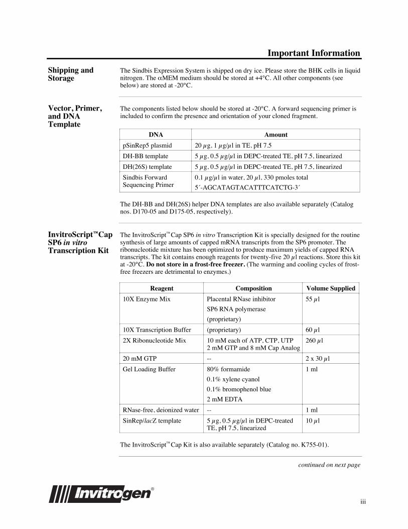
Vector, Primer,and DNATemplate
The components listed below should be stored at -20¡C. A forward sequencing primer isincluded to confirm the presence and orientation of your cloned fragment.
DNA Amount
pSinRep5 plasmid 20ʵg, 1ʵg/µl in TE, pH 7.5
DH-BB template 5ʵg, 0.5ʵg/µl in DEPC-treated TE, pH 7.5, linearized
DH(26S) template 5ʵg, 0.5ʵg/µl in DEPC-treated TE, pH 7.5, linearized
Sindbis ForwardSequencing Primer
0.1ʵg/µl in water, 20ʵl, 330Êpmoles total
5«-AGCATAGTACATTTCATCTG-3«
The DH-BB and DH(26S) helper DNA templates are also available separately (Catalognos. D170-05 and D175-05, respectively).
InvitroScriptªCapSP6 in vitroTranscription Kit
The InvitroScriptªCap SP6 in vitro Transcription Kit is specially designed for the routinesynthesis of large amounts of capped mRNA transcripts from the SP6 promoter. Theribonucleotide mixture has been optimized to produce maximum yields of capped RNAtranscripts. The kit contains enough reagents for twenty-five 20ʵl reactions. Store this kitat -20¡C. Do not store in a frost-free freezer. (The warming and cooling cycles of frost-free freezers are detrimental to enzymes.)
Reagent Composition Volume Supplied
10X Enzyme Mix Placental RNase inhibitor
SP6 RNA polymerase
(proprietary)
55ʵl
10X Transcription Buffer (proprietary) 60ʵl
2X Ribonucleotide Mix 10ÊmM each of ATP, CTP, UTP2ÊmM GTP and 8ÊmM Cap Analog
260ʵl
20ÊmM GTP -- 2 x 30ʵl
Gel Loading Buffer 80% formamide
0.1% xylene cyanol
0.1% bromophenol blue
2ÊmM EDTA
1Êml
RNase-free, deionized water -- 1Êml
SinRep/lacZ template 5ʵg, 0.5ʵg/µl in DEPC-treatedTE, pH 7.5, linearized
10ʵl
The InvitroScriptªCap Kit is also available separately (Catalog no. K755-01).
continued on next page

iv
Important Information, continued
Baby HamsterKidney Cells
One vial of 3 x 10⁶ baby hamster kidney (BHK) cells are supplied in 1Êml of aMEM,10% fetal bovine serum, 10% DMSO. Store in liquid nitrogen upon receipt. BHK cellsare also available separately (Catalog no. R700-01).
aMEM Mediumwith L-glutamine
500Êml aMEM medium with 2mM L-glutamine is supplied with the Sindbis ExpressionSystem. It is recommended for the growth of BHK cells. Store aMEM medium with2mM L-glutamine at +4¡C. Additional aMEM with 2mM L-glutamine is available fromInvitrogen in 500 ml bottles (Catalog no. Q400-01).
pSinHis pSinHis (Catalog no. V970-20) is available for use with the Sindbis Expression System.This vector contains an N-terminal Xpressª tag that allows simple purification and rapiddetection of fusion proteins. The vector is provided in three reading frames to facilitatecloning.
Materials Suppliedby the User
The following materials are required for use with this kit. Other materials may benecessary depending on the particular experiment. See the experiment of interest for thematerials required.
¥ CO2 incubator, 37¡C, 5% CO2
¥ aMEM medium
¥ Fetal bovine serum (FBS)
¥ Tissue culture grade 200ÊmM L-glutamine
¥ Phosphate-buffered saline (PBS) with divalent cations (see Recipes, page 34)
¥ Phosphate-buffered saline, divalent cation free, RNase-free (see Recipes, page 34)
¥ Tissue culture flasks and plates (75Êcm2 flasks, 175Êcm2 flasks, 35Êmm plates)
¥ Electroporator or liposomes for transfection
¥ Electroporation cuvettes, sterile (0.4Êcm)
¥ microcentrifuge tubes, RNase-free
Technical Service For Technical Service, please call, write, fax or E-mail:
U.S. Headquarters: European Headquarters:Invitrogen Corporation Invitrogen BV1600 Faraday Avenue De Schelp 12, 9351 NV LeekCarlsbad, CA 92008 The NetherlandsTel: (800) 955-6288 Tel: +31 (0) 594 515 175Fax: (760) 603-7201 Fax: +31 (0) 594 515 312E-mail: [email protected] E-mail: [email protected]
v
License Agreement
Terms of theLicense
Invitrogen Corporation ("Invitrogen") grants you a non-exclusive license to use theenclosed Sindbis Expression Kit for laboratory research purposes only. The SindbisExpression Kit is being transferred to you in furtherance of, and reliance on, such license.You may not use the Sindbis Expression Kit or the materials in the Kit for any otherpurpose without first obtaining a license for such purpose from Washington University,which owns all rights and titles to these materials.
Licensing Contact Dr. E. J. Brandt, DirectorTechnology ManagementWashington UniversityCampus Box 8013724 South Euclid AvenueSt. Louis, MO 63110Phone: 1-314-747-0922
YourResponsibilities
Access to the Sindbis Expression Kit must be limited solely to those officers, employees,and students of your institution who need access in order to perform the above-describedresearch. You must inform each officer, employee, and student of the provisions of thisAgreement and require them to agree, in writing, to be bound by the provisions of thisAgreement. You may not distribute the Sindbis Expression Kit or any component thereofto any other, except those within your own laboratory facility. You may not assign,sublicense, rent, lease, or otherwise transfer this License or any of the rights or obligationthereunder, except with prior written permission from Invitrogen.
Termination ofLicense
This License is effective until terminated. You may terminate it at any time or it willterminate automatically if you fail to comply with the terms and conditions of theAgreement. You shall, upon termination of the License, destroy all Sindbis ExpressionKits in your possession or control, and so notify Invitrogen in writing.
This License shall be governed in its interpretation and enforcement by the laws of theState of California.
User RegistrationCard
Please complete and return the enclosed User Registration Card for each SindbisExpression Kit that you purchase. Registration allows Invitrogen to provide you withvaluable information regarding kit upgrades and new products and services.
Technical Service If you need help with your Sindbis Expression Kit, please call our Technical ServicesDepartment at 1-800-955-6288 (U. S.) or +31 (0) 594 515 175 (Europe).
Thank You Invitrogen thanks you for your purchase of the Sindbis Expression Kit. We hope thisfurthers your research goals and allows you to obtain your results. Please call us if wemay be of service.

vi

vii
Safety Issues
Safety of SindbisVirus
The low level of pathogenicity of Sindbis virus in humans has allowed it to be classifiedas a Biosafety Level-2 (BL-2) agent by the NIH Recombinant DNA AdvisoryCommittee. Before synthesizing any constructs, experiments should be cleared throughyour institutional biosafety committee. All personnel working with the SindbisExpression System should be properly trained to work with BL-2 level organisms. BL-2precautions include the use of laminar flow hoods, laboratory coats, gloves, anddecontamination of infectious wastes. Sindbis virus can be inactivated by organicsolvents, bleach, or autoclaving. In addition, the components of the Sindbis ExpressionSystem have been designed to guard against any potential health threats (see RiskAssessment, below).
Risk Assessment Experiments involving transfection of the recombinant RNA (replicon RNA) pose nohealth risk since replication occurs only within the cells receiving the RNA with noconcomitant release of recombinant particles. Co-transfection of the replicon RNA withthe helper RNAs, DH-BB or DH(26S), results in the release of particles which can infectnew cells. Particles produced by these transfections have little or no plaque forming unit(pfu) capability associated with them. Although the replicon RNA is the predominantRNA found in these particles, the helper RNAs themselves are packaged at low levels(Bredenbeek et al., 1993). Therefore, the replicon particles that are thought to be virusfree have the potential for being infectious and appropriate caution should be used (seeSafety of Sindbis Virus, above). Recombination between Sindbis RNA molecules hasbeen reported (Ausubel, et al., 1994); however, recombination when using DH-BB orDH(26S) in transfected BHK cells occurs very rarely and only when the transfectionefficiency is poor. The production of recombinant virus is probably suppressed underconditions in which most cells are transfected with the recombinant RNA (Bredenbeek etal., 1993).
NOTE
A handbook of safety guidelines, Biosafety in Microbiological and BiomedicalLaboratories (stock number 017-040-00523-7) is available through the U.S. GovernmentPrinting Office at (202) 512-2356, or write to: Superintendent of Documents, U.S. GPO,Washington, D. C. 20402.

viii

ix
Table of Contents
Introduction............................................................................................................................................................... 1Overview.......................................................................................................................................................... 1
pSinRep5.......................................................................................................................................................... 5
DH-BB Template ............................................................................................................................................. 7
DH(26S) Template ........................................................................................................................................... 9
SinRep/lacZ Template ................................................................................................................................... 11
Methods .................................................................................................................................................................. 12Culturing BHK Cells ..................................................................................................................................... 12
Cloning into pSinRep5................................................................................................................................... 14
In vitro Transcription of DNA Templates ..................................................................................................... 16
Transfection ................................................................................................................................................... 19
Production of Recombinant Sindbis Pseudovirions....................................................................................... 23
Analysis of Protein Expression...................................................................................................................... 28
Appendix ................................................................................................................................................................ 31Sindbis Virus Life Cycle ............................................................................................................................... 31
Recipes ........................................................................................................................................................... 34
Analysis of RNA Quantity ............................................................................................................................. 37
Staining Cells for b-galactosidase Expression............................................................................................... 38
b-galactosidase Assay.................................................................................................................................... 39
Miniprep Plasmid Preparation ....................................................................................................................... 40
Internet Access ............................................................................................................................................... 42
References...................................................................................................................................................... 43
Sequence of pSinRep5 .................................................................................................................. after page 44

x
1
Introduction
Overview
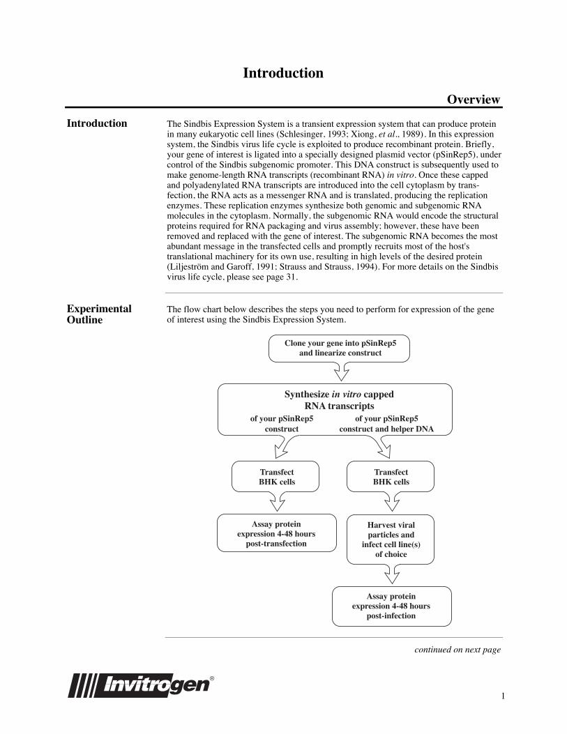
Introduction The Sindbis Expression System is a transient expression system that can produce proteinin many eukaryotic cell lines (Schlesinger, 1993; Xiong, et al., 1989). In this expressionsystem, the Sindbis virus life cycle is exploited to produce recombinant protein. Briefly,your gene of interest is ligated into a specially designed plasmid vector (pSinRep5), undercontrol of the Sindbis subgenomic promoter. This DNA construct is subsequently used tomake genome-length RNA transcripts (recombinant RNA) in vitro. Once these cappedand polyadenylated RNA transcripts are introduced into the cell cytoplasm by trans-fection, the RNA acts as a messenger RNA and is translated, producing the replicationenzymes. These replication enzymes synthesize both genomic and subgenomic RNAmolecules in the cytoplasm. Normally, the subgenomic RNA would encode the structuralproteins required for RNA packaging and virus assembly; however, these have beenremoved and replaced with the gene of interest. The subgenomic RNA becomes the mostabundant message in the transfected cells and promptly recruits most of the host'stranslational machinery for its own use, resulting in high levels of the desired protein(Liljestr�m and Garoff, 1991; Strauss and Strauss, 1994). For more details on the Sindbisvirus life cycle, please see page 31.
ExperimentalOutline
The flow chart below describes the steps you need to perform for expression of the geneof interest using the Sindbis Expression System.
Synthesize in vitro cappedRNA transcripts
of your pSinRep5construct
of your pSinRep5construct and helper DNA
TransfectBHK cells
Assay proteinexpression 4-48 hours
post-transfection
TransfectBHK cells
Harvest viralparticles and
infect cell line(s)of choice
Assay proteinexpression 4-48 hours
post-infection
Clone your gene into pSinRep5and linearize construct
continued on next page
2
Overview, continued
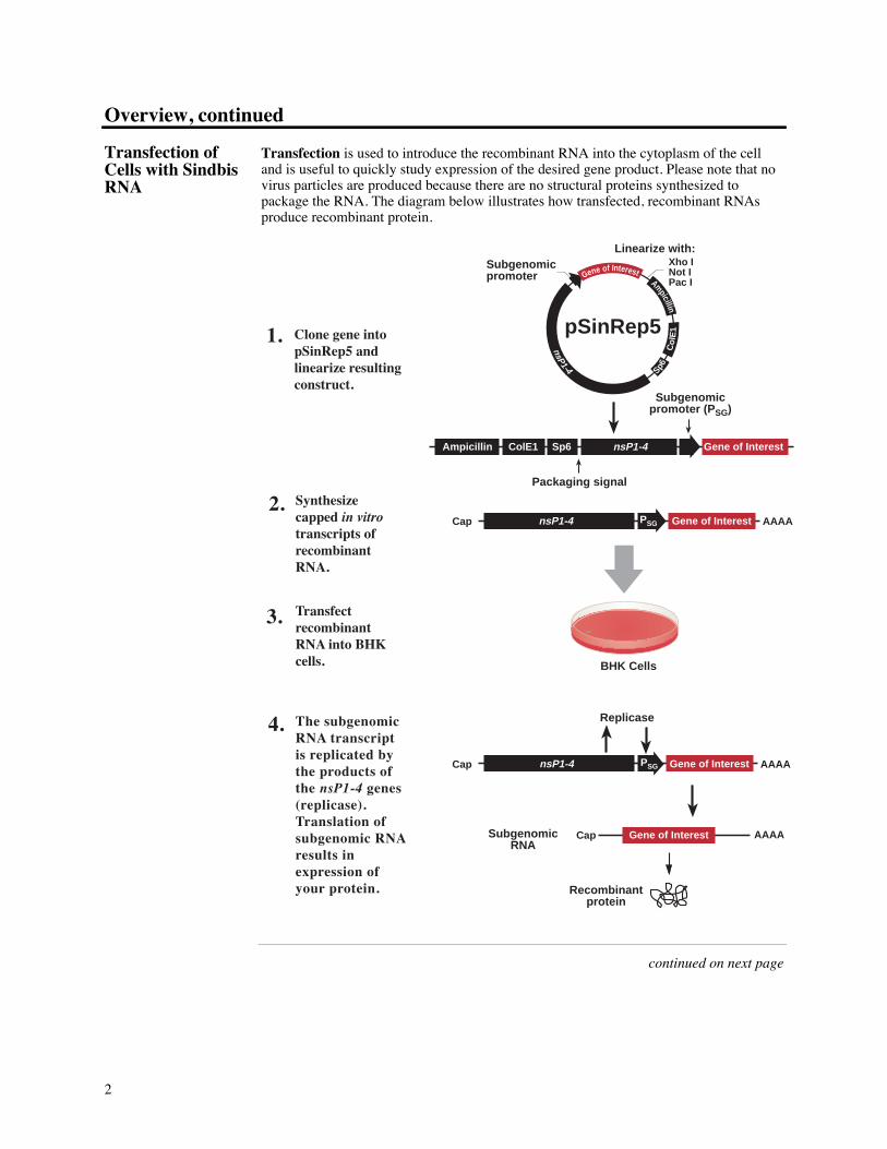
Transfection ofCells with SindbisRNA
Transfection is used to introduce the recombinant RNA into the cytoplasm of the celland is useful to quickly study expression of the desired gene product. Please note that novirus particles are produced because there are no structural proteins synthesized topackage the RNA. The diagram below illustrates how transfected, recombinant RNAsproduce recombinant protein.
nsP1-4 Gene of InterestCap AAAAPSG
Clone gene into pSinRep5 and linearize resulting construct.
Ampicillin
Co
lE1
Sp6
nsP1-4pSinRep5
Subgenomicpromoter
nsP1-4 Gene of InterestCap AAAA
Replicase
Gene of Interest AAAA
Synthesize capped in vitro transcripts of recombinant RNA.
Transfect recombinant RNA into BHK cells.
The subgenomic RNA transcript is replicated by the products of the nsP1-4 genes (replicase). Translation of subgenomic RNA results in expression of your protein.
1.
2.
3.
4.
Xho INot IPac I
Sp6 nsP1-4 Gene of Interest
Subgenomicpromoter (P SG)
ColE1Ampicillin
BHK Cells
Recombinantprotein
Subgenomic RNA
Cap
Linearize with:
Packaging signal
Gene of Interest
PSG
continued on next page

3
Overview, continued
Production ofSindbisPseudovirions
Infection (transduction) uses virus-like particles (pseudovirions) to deliver therecombinant RNA into the cytoplasm. Infection using Sindbis virus is a simple and veryefficient method to deliver the recombinant RNA molecules to a variety of cell types thatmay be difficult to transfect using standard techniques.
Production of pseudovirions is accomplished by transfecting cells with the recombinantRNA and a helper RNA that provides the Sindbis structural proteins in trans. Particlesreleased by the transfected cells contain only the recombinant RNA and are ready toinfect new cells for expression studies. These virions will undergo only one round ofinfection as they do not contain the helper RNA which encodes the structural proteins.The diagram below illustrates how recombinant viral particles are produced.
Cap AAAA Structural ProteinsCap AAAA
AAAA
E1, E2
Replicase
nsP1-4Cap AAAA
Packaging signal
Synthesize capped in vitro transcripts of recombinant RNA and helper RNA.
1.
Subgenomic promoter (P SG)
Structural ProteinsCap AAAA
Recombinant RNA Helper RNA
BHK Cells
Capsid
RecombinantRNA Capsid
Nucleocapsid
Gene of Interest
Gene of Interest
E2
E1
PlasmidMembrane
Recombinantprotein
Cytoplasm
Extracellular space
Transfect recombinant RNA and helper RNA into BHK cells.
2.
Recombinant RNA is packaged by the capsid protein to make the nucleocapsid.
4.
Gene of InterestnsP1-4
RNA transcripts are replicated by products of the nsP1-4 genes. Recombinant protein and structural proteins are expressed (capsid, E1, and E2).
3.
Viral glycoproteins E1 and E2 are processed and transported to the plasma membrane.
5.
Nucleocapsid associates with the E1 and E2 glycoproteins embedded in the plasma membrane. Viral particles bud into the medium.
6.
PSG
Cap
continued on next page
4
Overview, continued
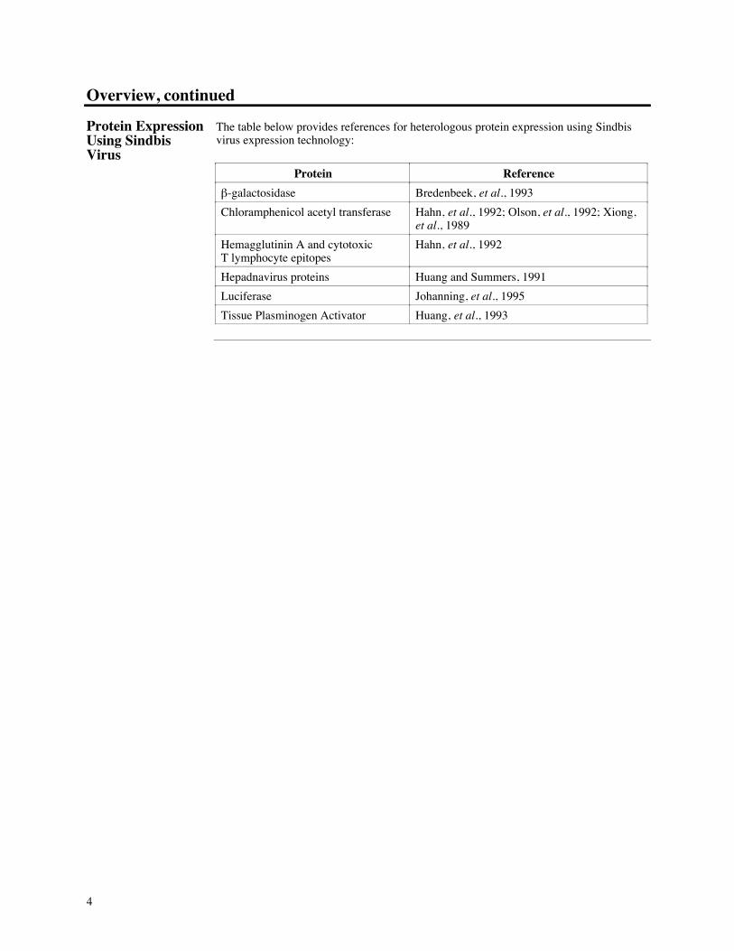
Protein ExpressionUsing SindbisVirus
The table below provides references for heterologous protein expression using Sindbisvirus expression technology:
Protein Reference
b-galactosidase Bredenbeek, et al., 1993
Chloramphenicol acetyl transferase Hahn, et al., 1992; Olson, et al., 1992; Xiong,et al., 1989
Hemagglutinin A and cytotoxicT lymphocyte epitopes
Hahn, et al., 1992
Hepadnavirus proteins Huang and Summers, 1991
Luciferase Johanning, et al., 1995
Tissue Plasminogen Activator Huang, et al., 1993
5
pSinRep5
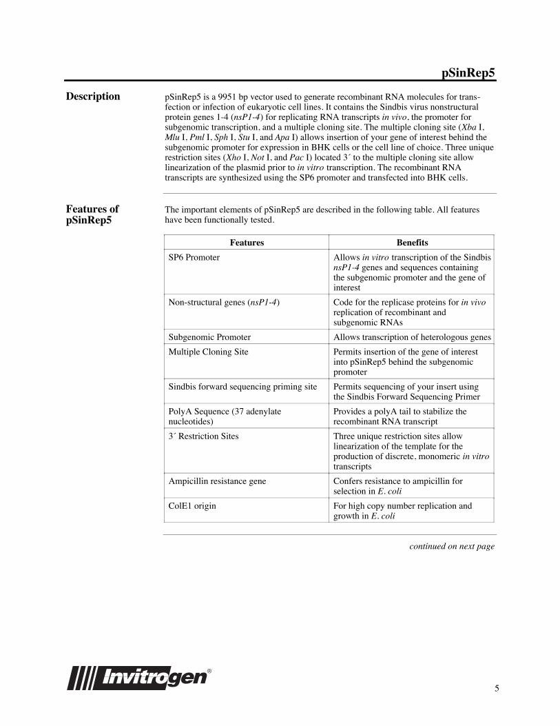
Description pSinRep5 is a 9951 bp vector used to generate recombinant RNA molecules for trans-fection or infection of eukaryotic cell lines. It contains the Sindbis virus nonstructuralprotein genes 1-4 (nsP1-4) for replicating RNA transcripts in vivo, the promoter forsubgenomic transcription, and a multiple cloning site. The multiple cloning site (XbaÊI,MluÊI, PmlÊI, SphÊI, StuÊI, and ApaÊI) allows insertion of your gene of interest behind thesubgenomic promoter for expression in BHK cells or the cell line of choice. Three uniquerestriction sites (XhoÊI, NotÊI, and PacÊI) located 3« to the multiple cloning site allowlinearization of the plasmid prior to in vitro transcription. The recombinant RNAtranscripts are synthesized using the SP6 promoter and transfected into BHK cells.
Features ofpSinRep5
The important elements of pSinRep5 are described in the following table. All featureshave been functionally tested.
Features Benefits
SP6 Promoter Allows in vitro transcription of the SindbisnsP1-4 genes and sequences containingthe subgenomic promoter and the gene ofinterest
Non-structural genes (nsP1-4) Code for the replicase proteins for in vivoreplication of recombinant andsubgenomic RNAs
Subgenomic Promoter Allows transcription of heterologous genes
Multiple Cloning Site Permits insertion of the gene of interestinto pSinRep5 behind the subgenomicpromoter
Sindbis forward sequencing priming site Permits sequencing of your insert usingthe Sindbis Forward Sequencing Primer
PolyA Sequence (37 adenylatenucleotides)
Provides a polyA tail to stabilize therecombinant RNA transcript
3« Restriction Sites Three unique restriction sites allowlinearization of the template for theproduction of discrete, monomeric in vitrotranscripts
Ampicillin resistance gene Confers resistance to ampicillin forselection in E. coli
ColE1 origin For high copy number replication andgrowth in E. coli
continued on next page
6
pSinRep5, continued
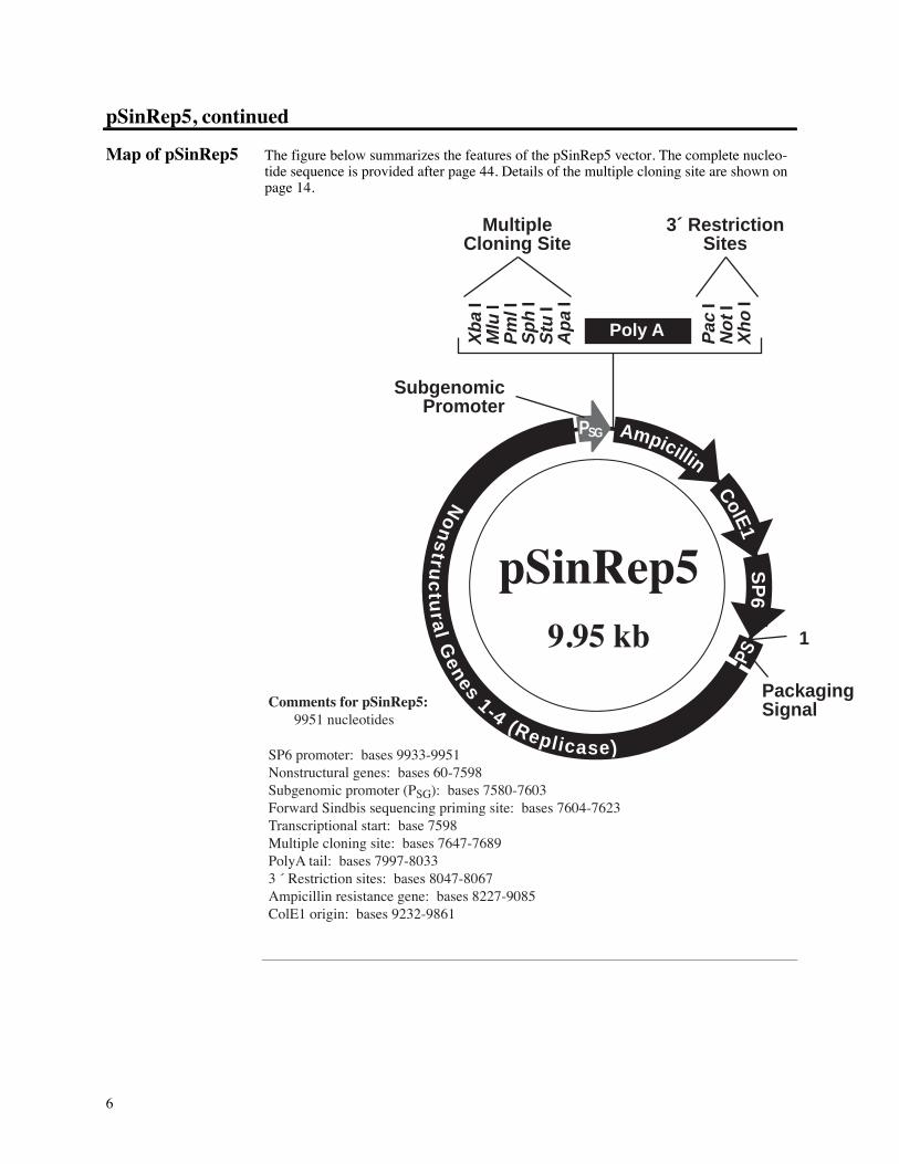
Map of pSinRep5 The figure below summarizes the features of the pSinRep5 vector. The complete nucleo-tide sequence is provided after page 44. Details of the multiple cloning site are shown onpage 14.
Xba
IM
lu I
Pm
l I
Sph
IS
tu I
Apa
I
Comments for pSinRep5: 9951 nucleotides
SP6 promoter: bases 9933-9951Nonstructural genes: bases 60-7598Subgenomic promoter (PSG): bases 7580-7603Forward Sindbis sequencing priming site: bases 7604-7623Transcriptional start: base 7598Multiple cloning site: bases 7647-7689PolyA tail: bases 7997-80333 « Restriction sites: bases 8047-8067Ampicillin resistance gene: bases 8227-9085ColE1 origin: bases 9232-9861
pSinRep59.95 kb
No
ns
truc
tur a
l Genes 1-4 (Rep l i case)
SP
6
1
ColE1
Ampicillin
PSG
PS
Pac
IN
ot I
Xho
I
Poly A
PackagingSignal
SubgenomicPromoter
MultipleCloning Site
3´ RestrictionSites
7
DH-BB Template
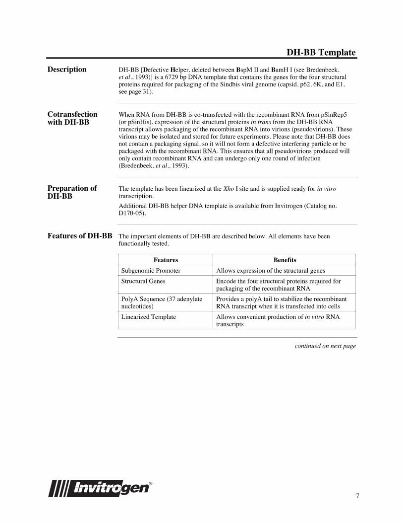
Description DH-BB [Defective Helper, deleted between BspMÊII and BamHÊI (see Bredenbeek,etÊal., 1993)] is a 6729 bp DNA template that contains the genes for the four structuralproteins required for packaging of the Sindbis viral genome (capsid, p62, 6K, and E1,see page 31).
Cotransfectionwith DH-BB
When RNA from DH-BB is co-transfected with the recombinant RNA from pSinRep5(or pSinHis), expression of the structural proteins in trans from the DH-BB RNAtranscript allows packaging of the recombinant RNA into virions (pseudovirions). Thesevirions may be isolated and stored for future experiments. Please note that DH-BB doesnot contain a packaging signal, so it will not form a defective interfering particle or bepackaged with the recombinant RNA. This ensures that all pseudovirions produced willonly contain recombinant RNA and can undergo only one round of infection(Bredenbeek, et al., 1993).
Preparation ofDH-BB
The template has been linearized at the Xho I site and is supplied ready for inÊvitrotranscription.
Additional DH-BB helper DNA template is available from Invitrogen (Catalog no.D170-05).
Features of DH-BB The important elements of DH-BB are described below. All elements have beenfunctionally tested.
Features Benefits
Subgenomic Promoter Allows expression of the structural genes
Structural Genes Encode the four structural proteins required forpackaging of the recombinant RNA
PolyA Sequence (37 adenylatenucleotides)
Provides a polyA tail to stabilize the recombinantRNA transcript when it is transfected into cells
Linearized Template Allows convenient production of in vitro RNAtranscripts
continued on next page

8
DH-BB Template, continued
Map of DH-BB The figure below summarizes the features of the DH-BB template.
Structural Genes
Comments for DH-BB: 6729 nucleotides
Ampicillin resistance gene: bases 5005-5863ColE1 origin: bases 6010-6639SP6 promoter: bases 6711-6729Subgenomic promoter (PSG): bases 671-694Structural genes: bases 738-4472
Poly A sequence: bases 4795-4831
Ampicillin ColE1 PSGSp6
1
Capsid:p62 (E2 & E3):
6K:E1:
bases 738-1529bases 1530-2990bases 2991-3155bases 3456-4472
9
DH(26S) Template
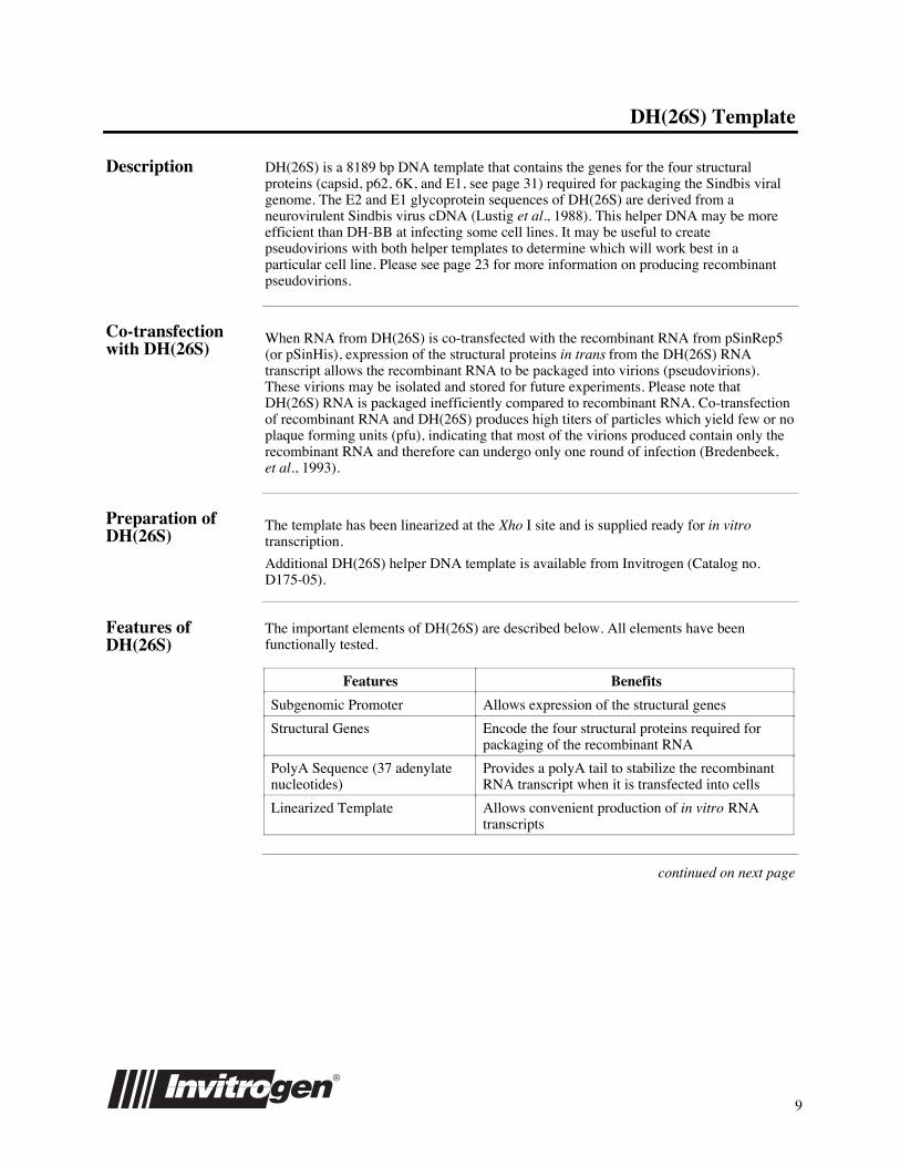
Description DH(26S) is a 8189 bp DNA template that contains the genes for the four structuralproteins (capsid, p62, 6K, and E1, see page 31) required for packaging the Sindbis viralgenome. The E2 and E1 glycoprotein sequences of DH(26S) are derived from aneurovirulent Sindbis virus cDNA (Lustig et al., 1988). This helper DNA may be moreefficient than DH-BB at infecting some cell lines. It may be useful to createpseudovirions with both helper templates to determine which will work best in aparticular cell line. Please see page 23 for more information on producing recombinantpseudovirions.
Co-transfectionwith DH(26S)
When RNA from DH(26S) is co-transfected with the recombinant RNA from pSinRep5(or pSinHis), expression of the structural proteins in trans from the DH(26S) RNAtranscript allows the recombinant RNA to be packaged into virions (pseudovirions).These virions may be isolated and stored for future experiments. Please note thatDH(26S) RNA is packaged inefficiently compared to recombinant RNA. Co-transfectionof recombinant RNA and DH(26S) produces high titers of particles which yield few or noplaque forming units (pfu), indicating that most of the virions produced contain only therecombinant RNA and therefore can undergo only one round of infection (Bredenbeek,etÊal., 1993).
Preparation ofDH(26S)
The template has been linearized at the Xho I site and is supplied ready for in vitrotranscription.
Additional DH(26S) helper DNA template is available from Invitrogen (Catalog no.D175-05).
Features ofDH(26S)
The important elements of DH(26S) are described below. All elements have beenfunctionally tested.
Features Benefits
Subgenomic Promoter Allows expression of the structural genes
Structural Genes Encode the four structural proteins required forpackaging of the recombinant RNA
PolyA Sequence (37 adenylatenucleotides)
Provides a polyA tail to stabilize the recombinantRNA transcript when it is transfected into cells
Linearized Template Allows convenient production of in vitro RNAtranscripts
continued on next page
10
DH(26S) Template, continued
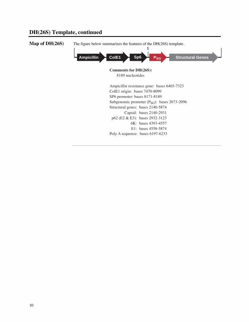
Map of DH(26S) The figure below summarizes the features of the DH(26S) template.
Structural Genes
Comments for DH(26S): 8189 nucleotides
Ampicillin resistance gene: bases 6465-7323ColE1 origin: bases 7470-8099SP6 promoter: bases 8171-8189Subgenomic promoter (PSG): bases 2073-2096Structural genes: bases 2140-5874
Poly A sequence: bases 6197-6233
Ampicillin ColE1 PSGSp6
1
Capsid:p62 (E2 & E3):
6K:E1:
bases 2140-2931bases 2932-3123bases 4393-4557bases 4558-5874
11
SinRep/lacZ
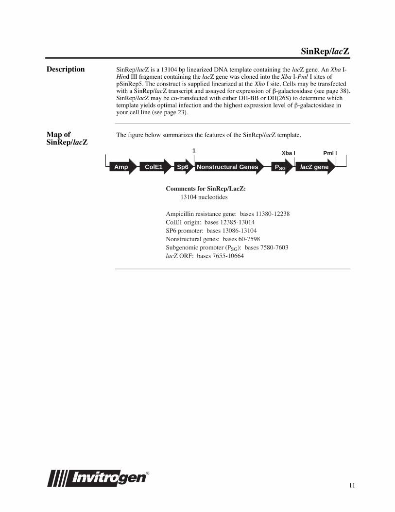
Description SinRep/lacZ is a 13104 bp linearized DNA template containing the lacZ gene. An XbaÊI-HindÊIII fragment containing the lacZ gene was cloned into the XbaÊI-PmlÊI sites ofpSinRep5. The construct is supplied linearized at the XhoÊI site. Cells may be transfectedwith a SinRep/lacZ transcript and assayed for expression of b-galactosidase (see page 38).SinRep/lacZ may be co-transfected with either DH-BB or DH(26S) to determine whichtemplate yields optimal infection and the highest expression level of b-galactosidase inyour cell line (see page 23).
Map ofSinRep/lacZ
The figure below summarizes the features of the SinRep/lacZ template.
Amp ColE1 PSG
Pml IXba I
Comments for SinRep/LacZ: 13104 nucleotides
Ampicillin resistance gene: bases 11380-12238ColE1 origin: bases 12385-13014SP6 promoter: bases 13086-13104Nonstructural genes: bases 60-7598Subgenomic promoter (PSG): bases 7580-7603lacZ ORF: bases 7655-10664
1
Sp6 Nonstructural Genes lacZ gene
12
Methods
Culturing BHK Cells
Introduction Use the procedures below to initiate and maintain a culture of BHK cells. These cells areprovided to get you started with the Sindbis Expression System. Other cell lines may beinfected with Sindbis pseudovirions.
Description ofBHK Cells
The BHK cell line was derived from baby hamster kidney. The cells have a tendency toclump in complete medium. In general, this is not a problem except when preparing thecells for electroporation (see page 19). In this case, care must be taken to avoid clumps.PBS is required to keep the cells from clumping during electroporation. Additional BHKcells are available from Invitrogen (Catalog no. R700-01).
General CellHandling
¥ All solutions and equipment that come in contact with the cells must be sterile.
¥ Always use proper sterile technique in a laminar flow hood.
¥ All incubations are performed in a humidified, 37¡C, 5% CO2 incubator.
¥ The medium for BHK cells is aMEM.
¥ Complete medium for BHK cells is aMEM + 2ÊmM L-glutamine + 5% fetal bovineserum (see page 34). FBS does not need to be heat inactivated for use with BHK cells.
Note: 10% FBS makes the cells grow too fast resulting in complete lysis and celldeath when infected with Sindbis virus.
¥ Use cells that are 80-90% confluent for all experiments.
¥ Before starting experiments, be sure to have cells established and also have somefrozen stocks on hand.
¥ For general maintenance of cells, pass BHK cells when they are 80-90% confluent(1-2Êdays) and split at a 1:5 dilution. For example, transfer 2Êml of a 10Êml cellsuspension to a new 175 cm2 flask.
¥ Cells may be passaged 60-70 times before re-starting a culture from frozen stocks.
Before Starting Be sure to have the following solutions and supplies available:
¥ 15Êml sterile, conical tubes
¥ 5, 10, and 25Êml sterile pipettes
¥ Cryovials
¥ Hemacytometer
¥ aMEM medium
¥ Tissue culture grade 200 mM L-glutamine
¥ Complete aMEM medium (aMEM + 2ÊmM L-glutamine + 5% FBS)
¥ aMEM medium + 2ÊmM L-glutamine + 10% FBS + 10% DMSO (freezing medium)
¥ Table-top centrifuge (+4¡C)
¥ 175 cm2 flasks and 35Êmm plates (other flasks and plates may be used)
¥ PBS with cations
¥ Trypsin/versene (EDTA) solution (BioWhittaker) or other trypsin solution
continued on next page
13
Culturing BHK Cells, continued
Initiating CellCulture fromFrozen Stock
The following protocol is designed to help you initiate a cell culture from a frozen stock.Note that the vial of BHK cells supplied with the kit contains 3 x 10⁶ cells.
1. Remove the vial of cells from the liquid nitrogen and thaw quickly at 37¡C.
2. Just before the cells are completely thawed, decontaminate the outside of the vialwith 70% ethanol, and transfer the cells to a 15Êml sterile, conical tube.
3. Add 9Êml of prewarmed (37¡C) complete aMEM medium dropwise to cells.
4. Centrifuge in a table-top centrifuge at 250 x g for 5 minutes at +4¡C. Decant themedium. (This removes the DMSO from the cells.)
5. Resuspend the cells in 10Êml of complete aMEM, transfer to a 75Êcm2 flask, andincubate at 37¡C. Incubation for 1-2 days should yield an 80-90% confluentmonolayer.
Passaging the BHKCells
1. When cells are ~80-90% confluent, remove all medium from the flask.
2. Wash cells once with 10Êml PBS with cations (see Recipes, page 34) to removemedium. Serum contains inhibitors of trypsin.
3. Add 5Êml of trypsin/versene (EDTA) solution to the monolayer and incubate 1-2minutes at room temperature until cells detach. Check the cells under a microscopeand confirm that most of the cells have detached. If cells are still attached, incubatea little longer until most of the cells have detached.
4. Once the cells have detached, briefly pipet the solution up and down to break upclumps of cells.
5. Add 5Êml of complete aMEM to stop trypsinization.
6. Transfer 2Êml of the cell suspension in Step 5 to a 175Êcm2 flask. Add completeaMEM medium to 30Êml total volume for each new 175Êcm2 flask and incubate in ahumidified, 37¡C, 5% CO2 incubator.
Repeat Steps 1-6 as necessary to expand cells.
Freezing theBHK Cells
Before starting, label cryovials and place on ice. Prepare freezing medium: aMEMcontaining 2 mM L-glutamine, 10% FBS, and 10% DMSO.
1. When cells are ~80% confluent in a 175Êcm2 flask, remove the medium and washthe cells one time with 10Êml PBS with cations.
2. Add 5 ml of trypsin/versene (EDTA) solution and incubate 1-2 minutes until cellsdetach.
3. Once cells have detached, briefly pipet solution up and down to break up clumps ofcells.
4. Add 5Êml of complete aMEM to stop trypsinization. Count a sample of cells in ahemacytometer.
5. Pellet all of the cells at 250 x g for 5Êminutes in a table top centrifuge at +4¡C.
6. Resuspend the cells at a density of 3 x 10⁶ cells/ml in freezing medium (see above).
7. Aliquot 1Êml of the cell suspension per vial. Place vials at -20¡C for 2-3Êhours.
8. Transfer vials to a -70 or -80¡C freezer and hold overnight.
9. Transfer vials to liquid nitrogen for long term storage.

14
Cloning into pSinRep5
General MolecularBiologyTechniques
For help with DNA ligations, E.Êcoli transformations, restriction enzyme analysis, DNAsequencing, and DNA biochemistry, please see Molecular Cloning: A LaboratoryManual (Sambrook, et al., 1989) or Current Protocols in Molecular Biology (Ausubel,etÊal., 1994).
Maintenance ofpSinRep5
In order to propagate and maintain pSinRep5, we recommend that you transform thevector into a recA, endA E. coli strain like TOP10 (Catalog no. C664-55), DH5a, orequivalent. Select on LB plates containing 50-100ʵg/ml ampicillin.
Cloning intopSinRep5
The subgenomic promoter region and multiple cloning site of pSinRep5 is includedbelow to help you ligate your gene into pSinRep5. The entire subgenomic promoter ispresent in pSinRep5 (Raju and Huang, 1991). It is recommended to include a Kozakconsensus sequence in your insert for proper initiation of translation of your gene (Kozak,1990) and a stop codon for termination of the recombinant protein.
TTCCAAGCCA TCAGAGGGGA AATAAAGCAT CTCTACGGTG GTCCTAAATA
GTCAGCATAG TACATTTCAT CTGACTAATA CTACAACACC ACCACCTCTA
GACGCGTAGA TCTCACGTGA GCATGCAGGC CTTGGGCCCA ATGATCCGAC
CAGCAAAACT CGATGTACTT CCGAGGAACT GATGTGCATA ATGCATCAGG
CTGGTACATT AGATCCCCGC TTACCGCGGG CAATATAGCA ACACTAAAAA
CTCGATGTAC TTCCGAGGAA GCGCAGTGCA TAATGCTGCG CAGTGTTGCC
ACATAACCAC TATATTAACC ATTTATCTAG CGGACGCCAA AAACTCAATG
TATTTCTGAG GAAGCGTGGT GCATAATGCC ACGCAGCGTC TGCATAACTT
TTATTATTTC TTTTATTAAT CAACAAAATT TTGTTTTTAA CATTTCAAAA
AAAAAAAAAA AAAAAAAAAA AAAAAAAAAA AAAGGGAATT CCTCGATTAA
TTAAGCGGCC GCTCGAGGGG AATTAATTCT TGAAGACGAA
Transcriptional start (7598)
7551
7601
7651
7701
7751
7801
7851
7901
7951
8001
8051
5« end of subgenomic promoter
PolyA tail (7997-8033)
3« end of subgenomic promoter
Pac I Not I Xho I
Mlu I Pml I Sph I
Sindbis forward sequencing priming site (7604-7623)
Apa I
Xba I
Stu I
continued on next page
15
Cloning into pSinRep5, continued
E.ÊcoliTransformation
Transform your ligation mixtures into a competent recA, endA E. coli strain (e.g. TOP10,DH5a) and select on LB plates containing 50-100ʵg/ml ampicillin. Select 10-20 clonesand analyze for the presence and orientation of your insert.
RE
CO
MMENDAT
ION
We recommend that you sequence your construct with the Sindbis Forward SequencingPrimer to confirm that your gene is in the correct orientation for expression and has aKozak translation initiation sequence and a stop codon.
ImportantThe plasmid DNA must be free of RNA and RNase before performing in vitrotranscription. RNA will inhibit transcription and may lower transfection efficiencies.RNase will degrade your transcripts.
PlasmidPreparation
Once you have your recombinant construct, you will need to prepare plasmid for in vitrotranscription. You will need at least 1ʵg of DNA for each in vitro transcription reaction.We recommend purifying your DNA using CsCl gradient centrifugation or using theplasmid miniprep procedure on page 40. This method uses RNase to eliminate RNA anda Proteinase K digest to remove the RNase. It is very important to eliminate RNase toavoid degradation of your transcripts. After isolation of plasmid DNA, be sure todetermine the concentration of your sample using UV absorbance, fluorescence, or theDNA DipStickª Kit (Catalog no. K5632-01).

16
In vitro Transcription of DNA Templates
Purpose The purpose of these procedures is to generate recombinant RNA (and helper RNA) fortransfection of BHK cells. You will use purified, linearized pSinRep5 (or pSinHis)containing your gene as the DNA template to produce recombinant RNA with theInvitroScriptªCap SP6 in vitro Transcription Kit. The recombinant RNA produced willbe capped and have a polyA tail, so when it is transfected into the cells, it will be treatedas messenger RNA.
InvitroScriptªCapSP6 in vitroTranscription
The InvitroScriptªCap SP6 in vitro Transcription Kit produces large amounts ofpolyadenylated RNA transcripts from the SP6 promoter using linearized DNA templates.The kit contains a GTP cap analog to produce capped RNA transcripts. The reactionconditions have been optimized to yield high levels of capped RNA in the presence ofhigh nucleotide concentrations. Additional InvitroScriptªCap Kits are available fromInvitrogen (Catalog no. K755-01).
Helper RNA If you wish to package your recombinant RNA to produce pseudovirions, you will needto supply the structural proteins in trans. These proteins are encoded on the DH-BB andDH(26S) templates. These templates are supplied linearized and can be used directly inthe in vitro transcription reaction with no further preparation. The helper RNA fromthese templates and your recombinant RNA may be co-transfected into BHK cells toproduce pseudovirions (see page 23). These pseudovirions may be used to infect BHKor other cell lines.
Control RNA Linearized SinRep/lacZ template is included in the InvitroScriptªCap SP6 in vitroTranscription Kit as a transcription control and a positive control for expression. Thistemplate contains the lacZ gene which expresses b-galactosidase. The template can beused directly in the in vitro transcription reaction with no further preparation.
ExperimentalOutline
The table below outlines the steps needed to prepare recombinant RNA.
Step Action
1 Digest 2-20ʵg of pSinRep5 containing your gene of interest with theappropriate 3« restriction enzyme to linearize the plasmid.
2 Phenol extract and ethanol precipitate the DNA template. Resuspend DNAat 0.5ʵg/µl in RNase free TE.
3 Set up the in vitro transcription reaction with recombinant pSinRep5,helper RNA, or SinRep/lacZ (control template). Incubate the reactionfor 2Êhours at 37¡C.
4 Analyze the resulting RNA on a 1% agarose gel.
General MolecularBiologyTechniques
For help with restriction enzyme digests, please see Molecular Cloning: A LaboratoryManual (Sambrook, et al., 1989) or Current Protocols in Molecular Biology (Ausubel,etÊal., 1994).
continued on next page

17
In vitro Transcription of DNA Templates, continued
ImportantBecause you will be generating RNA, it is very important that any solution that comes incontact with the RNA be made with diethylpyrocarbonate-treated (DEPC) water andautoclaved (Sambrook, et al., 1989). All plasticware should be sterile. Be sure to weargloves .
Before Starting You will need to have available the following solutions and supplies:
¥ Restriction enzymes and buffers
¥ Phenol/chloroform (1:1)
¥ 0.5ÊM EDTA
¥ RNase-free 5ÊM ammonium acetate
¥ 100% ethanol
¥ RNase-free water or TE buffer
¥ 37¡C incubator
In vitroTranscription
Linearization of the DNA template prior to in vitro transcription is necessary to create ahomogeneous population of RNA molecules. There are three enzymes that you can use tolinearize the DNA: XhoÊI, NotÊI, and PacÊI. Choose the enzyme that does not cut in yourgene to linearize the DNA template. Each in vitro transcription reaction requires 1 µg oflinearized template. Note: The DH-BB, DH(26S) and SinRep/lacZ templates are providedlinearized. These templates may be added directly to the transcription reaction at Step 5.
1. Linearize 2-20ʵg of your pSinRep5 construct by restriction digestion using Xho I,Not I, or Pac I.
2. Terminate the restriction digest with 1/20 volume of 0.5ÊM EDTA.
3. Phenol extract the restriction digest and ethanol precipitate the DNA with 1/10volume of 5ÊM ammonium acetate and 2 volumes of 100% ethanol.
4. Resuspend the DNA to a concentration of 0.5 µg/µl in RNase-free TE or water. Youmay wish to check an aliquot of your linearized template on an agarose gel toanalyze quantity and quality.
5. Set up the in vitro transcription reaction at room temperature in a microcentrifugetube by mixing the following reagents. If you wish to quantitate the transcriptionreaction, refer to page 37.
RNase-free water 2 µl
Linearized DNA template at 0.5ʵg/µl (1ʵg) 2 µl
2X Ribonucleotide Mix 10 µl
20ÊmM GTP* 2ʵl
10X Transcription Buffer 2 µl
10X SP6 Enzyme Mix 2 µl
Final Volume 20 µl
*Inclusion of extra GTP is needed to ensure full-length transcript (see next page) Do not set up the reaction on ice as the 10X Transcription Buffer will precipitate.
6. Mix the reaction gently and incubate for 2 hours at 37¡C. A typical reaction shouldyield 10-20 µg of RNA from 1ʵg of linearized template. Termination of the reactionis not required for transfection. If you wish to store the RNA, see Step 8, next page.
continued on next page
18
In vitro Transcription of DNA Templates, continued
In vitroTranscription,continued
7. Check the quality of the RNA by mixing 1ʵl of the transcription reaction with 3ʵlof the gel loading dye, heating for 3-5Êminutes at 80¡C-90¡C, and running on a 1%agarose gel.
Note: RNA run on the agarose gel can be visualized by staining with ethidiumbromide. Since this gel is nondenaturing, this procedure will only reflect the qualityand quantity of the RNA. A nondenaturing gel cannot be used to accuratelydetermine the size of the RNA. You may wish to compare the quality of the RNAband with DNA standards such as digested lDNA. The RNA band should bediscreet and relatively thick in comparison to the DNA bands. Please note that youmay see dimers of single-stranded RNA.
8. Proceed to Transfection, next page. Alternatively, you may aliquot the RNA in10ʵl samples and freeze at -80¡C for storage.
ImportantSince the Sindbis RNA transcript is greater than 10Êkb, you must add 1-2ʵl of 20ÊmMGTP in the transcription reaction as the concentration of GTP will become limitingduring the reaction. Adding GTP will decrease the fraction of transcripts containing thecap (since it decreases the ratio of the cap analog to GTP), but will increase the yield offull-length product.

19
Transfection
Purpose The purpose of this section is to supply guidelines for transfection of RNA into BHK cells andother eukaryotic cells. Electroporation is strongly recommended as conditions may beoptimized to obtain nearly 100% transfection frequency. We use electroporation exclusivelyat Invitrogen to transfect cells with recombinant Sindbis RNA.
Transfection UsingLiposomes
It is possible to transfect cells using liposome-mediated transfection. Please refer to CurrentProtocols in Molecular Biology, pages 16.20.5-16.20.6 for a protocol or Johanning, et al.,1995. You will need 9-20ʵg of liposomes per each 35Êmm plate of cells to transform. Pleasenote that the transfection efficiency may be much lower than with electroporation.
Before Starting You will need the following solutions and equipment:
¥ 175Êcm2 flasks
¥ aMEM medium + 2ÊmM L-glutamine (no FBS)
¥ complete aMEM medium (see Recipes, page 34)
¥ PBS with cations (see Recipes, page 34)
¥ RNase-free PBS without cations (see Recipes, page 34)
¥ Trypsin/versene (EDTA) solution
¥ Tabletop centrifuge
¥ Hemacytometer
¥ Electroporation device (be sure to use a device that can transform eukaryotic cells and notjust bacteria. You will need settings to give a field strength of 2125ÊV/cm with a capaci-tance of 50ʵF)
¥ 0.4Êcm, sterile electroporation cuvettes
NOTE
If you wish to transfect cells with your recombinant RNA and the helper RNA to producepseudovirions, please see page 23 before continuing.
Preparation ofCells
We generally use 175Êcm2 flasks to grow cells for electroporation. One to two flasks provideenough BHK cells for two electroporations. It is very important to keep the BHK cells fromclumping as this will prevent RNA from entering cells and lowering your transfectionefficiency.
1. Grow BHK cells in a 175 cm2 tissue culture flask containing 30Êml complete aMEMand grow until the monolayer is approximately 90% confluent and composed ofapproximately 107 cells. This should take one to three days.
2. Aspirate medium and wash the cells once with room temperature PBS with cations.
3. Add 5Êml of the trypsin/versene (EDTA) solution and incubate 1-2 minutes until cellsdetach. Briefly pipet the solution to obtain a single-cell suspension. Monitor under amicroscope until a single-cell suspension has been obtained.
4. Add 5Êml complete aMEM medium to stop trypsinization. Transfer cells to a sterile15Êml conical tube.
continued on next page
20
Transfection, continued
Preparation ofCells, continued
5. Centrifuge the cells at 400 x g for 5Êminutes at room temperature or +4¡C andaspirate supernatant. Resuspend cells in 10Êml aMEM containing 2ÊmM L-glu-tamine (no serum).
If you find that there are still some clumps, keep the tube upright and let the clumpssettle to the bottom of the tube (1-2Êminutes). Carefully remove the cell suspensionfrom the clumps of cells.
6. Centrifuge the cells at 400 x g for 5Êminutes at room temperature or +4¡C andaspirate supernatant. Resuspend cells in 10Êml RNase-free PBS without cations.Determine the number of cells with a counting chamber.
7. Centrifuge cells at 400 x g for 5Êminutes at room temperature or +4¡C and aspiratesupernatant. Resuspend cells in RNase-free PBS without cations at a concentrationof 107 cells/ml. Proceed immediately to Electroporation of Cells, below. BHKcells prepared for electroporation cannot be stored; they must be used immediately.
ImportantCells must be resuspended in PBS that is
¥ RNase-free to prevent RNA degradation¥ Without cations to prevent arcing during electroporation
Guidelines forElectroporation
Actual settings are provided below for Invitrogen's Electroporator II. If you are usinganother electroporation device, please consult the manufacturer's instructions. If you areusing another cell line, you will have to optimize electroporation conditions to achieve hightransfection efficiencies. Please see Electroporation Parameters to Optimize, next page.
Controls We recommend transforming BHK cells with SinRep/lacZ control RNA as a positivecontrol for expression. Remember to also include a "no RNA" control.
Electroporation ofCells
1. Place 0.5Êml (~107 cells/ml) of cell suspension from Step 7, above, into a 0.4Êcmelectroporation cuvette.
2. Add 5-10ʵl of the transcription reaction (5-10 µg of RNA, from Step 8, page 18) tothe cell suspension, place cap on cuvette and mix thoroughly by inverting.
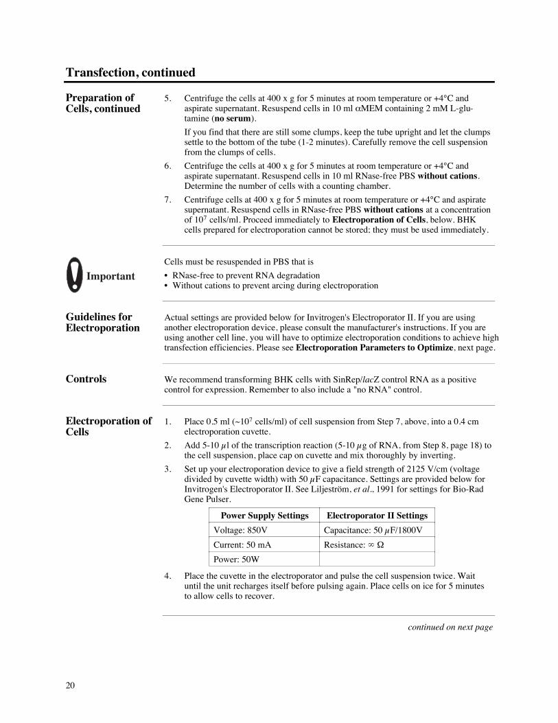
3. Set up your electroporation device to give a field strength of 2125ÊV/cm (voltagedivided by cuvette width) with 50ʵF capacitance. Settings are provided below forInvitrogen's Electroporator II. See Liljestr�m, et al., 1991 for settings for Bio-RadGene Pulser.
Power Supply Settings Electroporator II Settings
Voltage: 850V Capacitance: 50ʵF/1800V
Current: 50ÊmA Resistance: ° ½
Power: 50W
4. Place the cuvette in the electroporator and pulse the cell suspension twice. Waituntil the unit recharges itself before pulsing again. Place cells on ice for 5Êminutesto allow cells to recover.
continued on next page
21
Transfection, continued
Electroporation ofcells, continued
5. Transfer electroporated cells (0.5Êml) to 9.5Êml of complete aMEM medium. Rinse the cuvette with the cell suspension to collect all the cells.
6. Plate the cells (2Êml on a 35Êmm plate, 5Êml on a 60Êmm plate, or 10Êml on a100Êmm plate). Expression can be observed as early as 4 hours and as late as72Êhours post-transfection. Proceed to Analysis of Protein Expression page 24.
Assay forb-galactosidase
If you transfected the positive control RNA derived from SinRep/lacZ, you may stainyour transfected cells in situ for b-galactosidase activity (page 38). Cell-free lysates mayalso be prepared and assayed for b-galactosidase activity using orthonitrophenyl-b-D-galactoside (ONPG), page 39.
Expression in CellsOther Than BHKCells
The Sindbis Expression System can be used to express recombinant protein in a variety ofcell lines. BHK cells, however, have been shown to yield the maximum levels of protein.To study the effects of your gene in another cell line, we recommend that you:
¥ Optimize transfection to maximize the number of cells expressing your recombinantRNA
¥ Perform a time course of expression as host cell functions affect the Sindbis life cycle
¥ Determine which helper DNA template will yield the most efficient expression
¥ Use viral particles to infect cell line
ElectroporationParameters toOptimize
All mammalian cell electroporations are generally performed using 0.4Êcm cuvettes. Thevoltage and capacitance must be optimized for each cell line used. The resistance isdetermined by the electroporation buffer and the volume in which the cells are suspended.Note that the conditions to electroporate RNA are much different than those for DNA.Other parameters to optimize are:
Electroporation buffer
¥ Use "High salt" (sterile PBS without cations) when electroporating cells with RNA
Voltage
¥ For high salt buffer, use 200-1200ÊV. We recommend varying the voltage first whenoptimizing conditions for transfection of RNA
Capacitance
¥ For high salt buffer, start with 50 µF and increase to lengthen pulse
Volume
¥ Start with 500ʵl and decrease to 250ʵl to increase resistance or increase to 800ʵl toreduce resistance.
continued on next page
22
Transfection, continued
Electroporator IIand Cuvettes
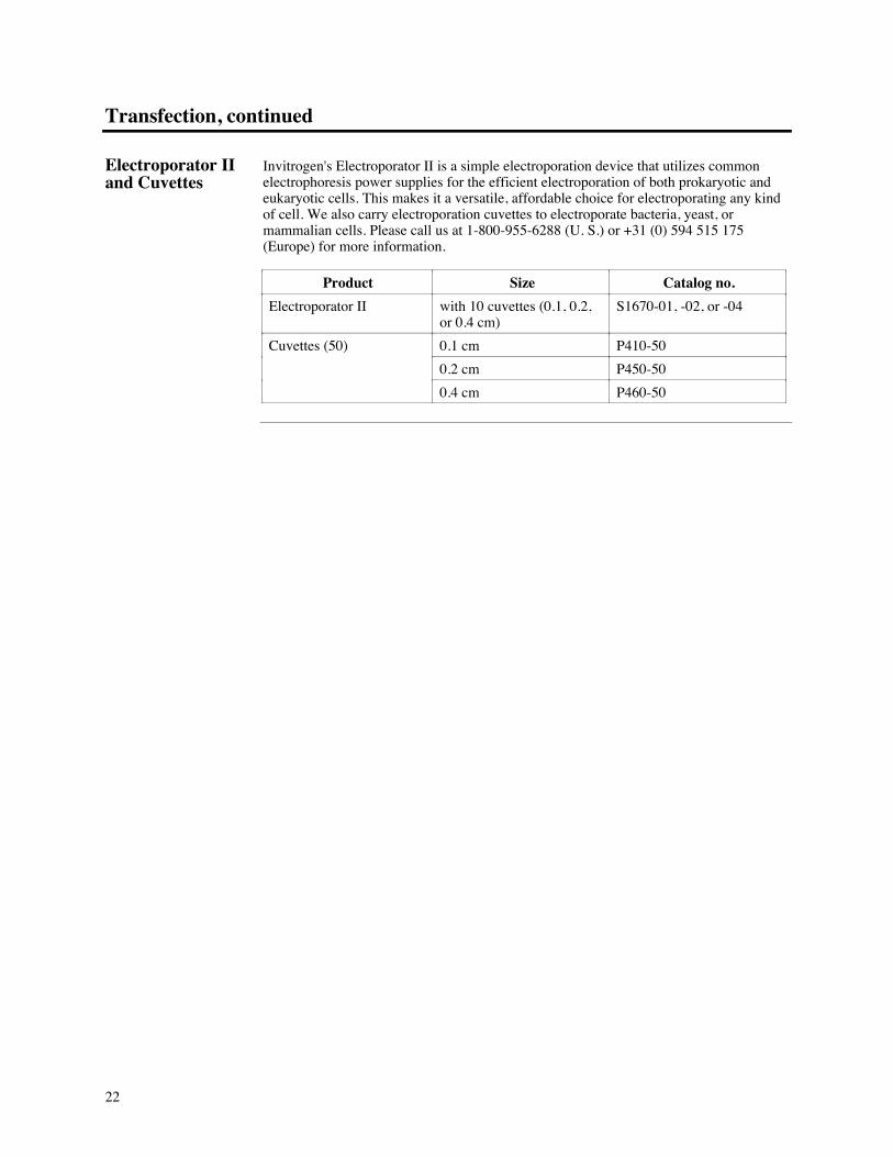
Invitrogen's Electroporator II is a simple electroporation device that utilizes commonelectrophoresis power supplies for the efficient electroporation of both prokaryotic andeukaryotic cells. This makes it a versatile, affordable choice for electroporating any kindof cell. We also carry electroporation cuvettes to electroporate bacteria, yeast, ormammalian cells. Please call us at 1-800-955-6288 (U. S.) or +31 (0) 594 515 175(Europe) for more information.
Product Size Catalog no.
Electroporator II with 10 cuvettes (0.1, 0.2,or 0.4 cm)
S1670-01, -02, or -04
Cuvettes (50) 0.1Êcm P410-50
0.2Êcm P450-50
0.4Êcm P460-50
23
Production of Recombinant Sindbis Pseudovirions
Purpose You may wish to produce pseudovirions containing your recombinant RNA. Pseudo-virions may be used to infect a variety of different cell lines to optimize expression in thecell line of choice or to infect large scale cultures for production and purification of yourprotein. To package your recombinant RNA, you will first need to co-transfect yourrecombinant RNA with the helper RNA made from either the DH-BB or DH(26S)template. If you do not have RNA from any of these three plasmids, you will need tosynthesize RNA using the InvitroScriptªCap SP6 in vitro Transcription Kit (seepageÊ16).
Once you have obtained pseudovirions, you will need to determine the optimal amount ofvirus needed to infect cells. This may vary between cell lines and needs to be optimized.
ExperimentalOutline
The table below outlines the steps needed to produce pseudovirions.
Steps Description
1 Prepare RNA transcripts from pSinRep5 containing your gene (orSinRep/lacZ) and the DH-BB or DH(26S) template (structural genes) usingthe InvitroScriptªCap SP6 in vitro Transcription Kit
2 Transfect a 1:1 v/v mixture of the two RNAs into BHK cells
3 24-36 hours posttransfection, harvest the medium containing thepseudovirions
4 Serially dilute the viral solution and infect cells to empirically determine theoptimal dilution needed for expression of your protein
5 Virus may be frozen in 0.5Êml aliquots for future use
Positive Control You may wish to produce pseudovirions containing the SinRep/lacZ recombinant RNAas a positive control.
Helper DNATemplates
Both DH-BB and DH(26S) helper DNA templates are included in the Sindbis ExpressionSystem. Each DNA template encodes different E2 and E1 glycoproteins which affect theability of the virus to infect a given cell line. Some cell types may be infected better withpseudovirions produced from one helper DNA over the other. We recommend producingpseudovirions of the SinRep/lacZ control with both the DH-BB and DH(26S) templatesto determine which helper DNA yields optimal infection and the highest expression of b-galactosidase in your cell line. In general, DH(26S) is more efficient at infection and,therefore, yields higher expression levels than DH-BB.
continued on next page

24
Production of Recombinant Sindbis Pseudovirions, continued
Before Starting Be sure to have on hand the following reagents:
¥ RNA transcript of pSinRep5 plasmid containing desired insert (see page 17, steps 1-8)
¥ SinRep/lacZ RNA (see page 17, steps 1-8)
¥ DH-BB or DH(26S) Helper RNA (see page 17, steps 1-8)
¥ BHK cells, 80-90% confluent, ready for electroporation (see page 19, steps 1-7)
¥ Complete aMEM (containing 2ÊmM L-glutamine and 5% FBS)
¥ aMEM (or PBS) + 2ÊmM L-glutamine + 1% FBS
¥ PBS with cations
¥ 35 mm tissue culture plates
¥ If you wish to use liposomes, please see page 19
NOTE
All solutions and equipment coming into contact with cells must be sterile. Always useproper sterile technique. All incubations are performed in a humidified, 37¡C, 5% CO2
incubator unless otherwise noted.
ObtainingPseudovirions
You should have capped RNA transcripts of your construct and helper DNA, and BHKcells ready for transfection before starting this procedure.
1. Mix the RNAs in a 1:1 v/v ratio. Typically, equal amounts of the in vitrotranscription reactions (usually 5-10 µl of each ) result in the highest yield ofpseudovirions.
2. Place 0.5Êml (~107 cells/ml) of the cell suspension into a 0.4Êcm electroporationcuvette.
3. Add 10-20ʵl of the RNA mixture (10-20ʵg of RNA) to the cell suspension, placecap on cuvette and mix thoroughly by inverting.
4. Set up your electroporation device to give a field strength of 2125ÊV/cm (voltagedivided by cuvette width) and a capacitance of 50ʵF. See page 20 for moreinformation.
5. Place the cuvette in the electroporator and pulse the cell suspension twice. Waituntil the unit recharges itself before pulsing again.
6. Transfer electroporated cells (0.5Êml) to 9.5Êml of complete aMEM medium. Rinsethe cuvette with the cell suspension to collect all the cells.
7. Plate the cells (2Êml on a 35Êmm plate, 5Êml on a 60Êmm plate, or 10Êml on a100Êmm plate). Plate out all of the transfected cells. This will ensure the maximumamount of pseudovirion containing medium.
8. 24-36 hours posttransfection, remove the medium from the cells. Remove any loosecells by centrifuging the medium for 10Êminutes at 2000 x g in a table top centrifugeat +4¡C. Save the supernatant as this contains the pseudovirions.
9. Take 0.5Êml aliquots of the supernatant and freeze in an ethanol/dry ice bath. Storeat -80¡C. Freezing the virus in aliquots is recommended since repeated freeze-thawing reduces virus infectivity. Proceed to Infecting Cells with SindbisPseudovirions, next page.
continued on next page
25
Production of Recombinant Sindbis Pseudovirions, continued
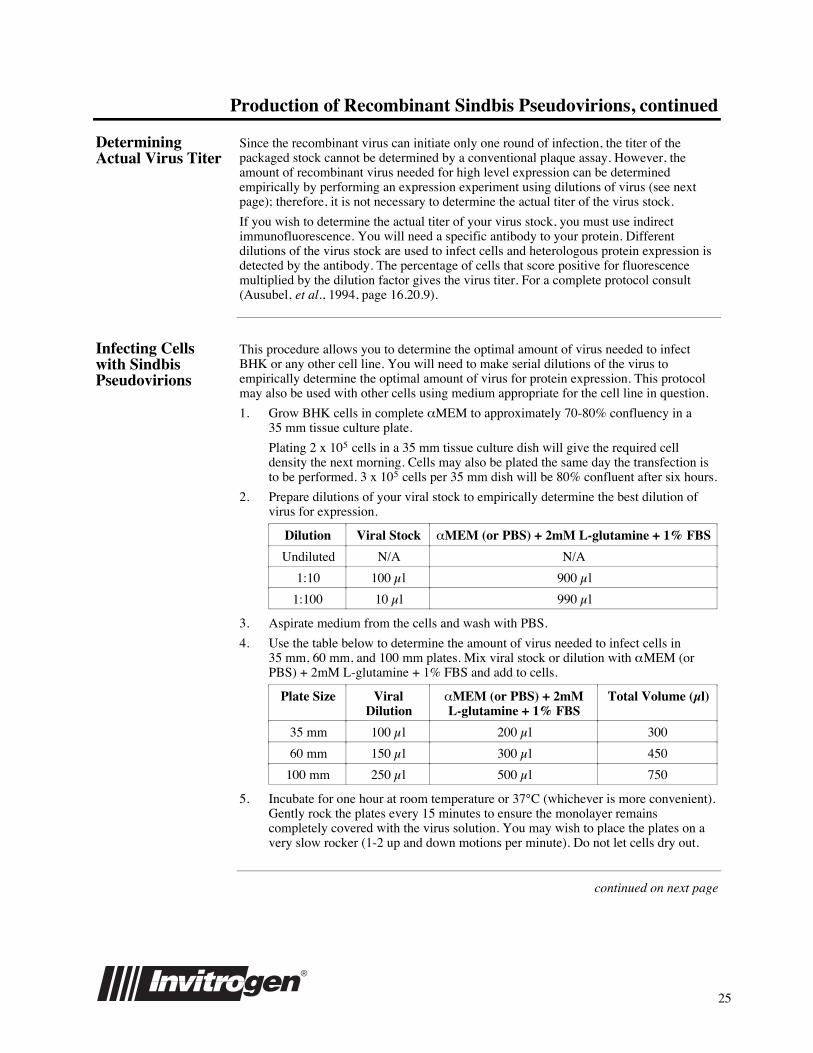
DeterminingActual Virus Titer
Since the recombinant virus can initiate only one round of infection, the titer of thepackaged stock cannot be determined by a conventional plaque assay. However, theamount of recombinant virus needed for high level expression can be determinedempirically by performing an expression experiment using dilutions of virus (see nextpage); therefore, it is not necessary to determine the actual titer of the virus stock.
If you wish to determine the actual titer of your virus stock, you must use indirectimmunofluorescence. You will need a specific antibody to your protein. Differentdilutions of the virus stock are used to infect cells and heterologous protein expression isdetected by the antibody. The percentage of cells that score positive for fluorescencemultiplied by the dilution factor gives the virus titer. For a complete protocol consult(Ausubel, et al., 1994, page 16.20.9).
Infecting Cellswith SindbisPseudovirions
This procedure allows you to determine the optimal amount of virus needed to infectBHK or any other cell line. You will need to make serial dilutions of the virus toempirically determine the optimal amount of virus for protein expression. This protocolmay also be used with other cells using medium appropriate for the cell line in question.
1. Grow BHK cells in complete aMEM to approximately 70-80% confluency in a35Êmm tissue culture plate.
Plating 2 x 10⁵ cells in a 35Êmm tissue culture dish will give the required celldensity the next morning. Cells may also be plated the same day the transfection isto be performed. 3 x 10⁵ cells per 35Êmm dish will be 80% confluent after six hours.
2. Prepare dilutions of your viral stock to empirically determine the best dilution ofvirus for expression.
Dilution Viral Stock aMEM (or PBS) + 2mM L-glutamine + 1% FBS
Undiluted N/A N/A
1:10 100ʵl 900ʵl
1:100 10ʵl 990ʵl
3. Aspirate medium from the cells and wash with PBS.
4. Use the table below to determine the amount of virus needed to infect cells in35Êmm, 60Êmm, and 100Êmm plates. Mix viral stock or dilution with aMEM (orPBS) + 2mM L-glutamine + 1% FBS and add to cells.
Plate Size ViralDilution
aMEM (or PBS) + 2mML-glutamine + 1% FBS
Total Volume (µl)
35 mm 100ʵl 200ʵl 300
60 mm 150ʵl 300ʵl 450
100Êmm 250ʵl 500ʵl 750
5. Incubate for one hour at room temperature or 37¡C (whichever is more convenient).Gently rock the plates every 15 minutes to ensure the monolayer remainscompletely covered with the virus solution. You may wish to place the plates on avery slow rocker (1-2 up and down motions per minute). Do not let cells dry out.
continued on next page
26
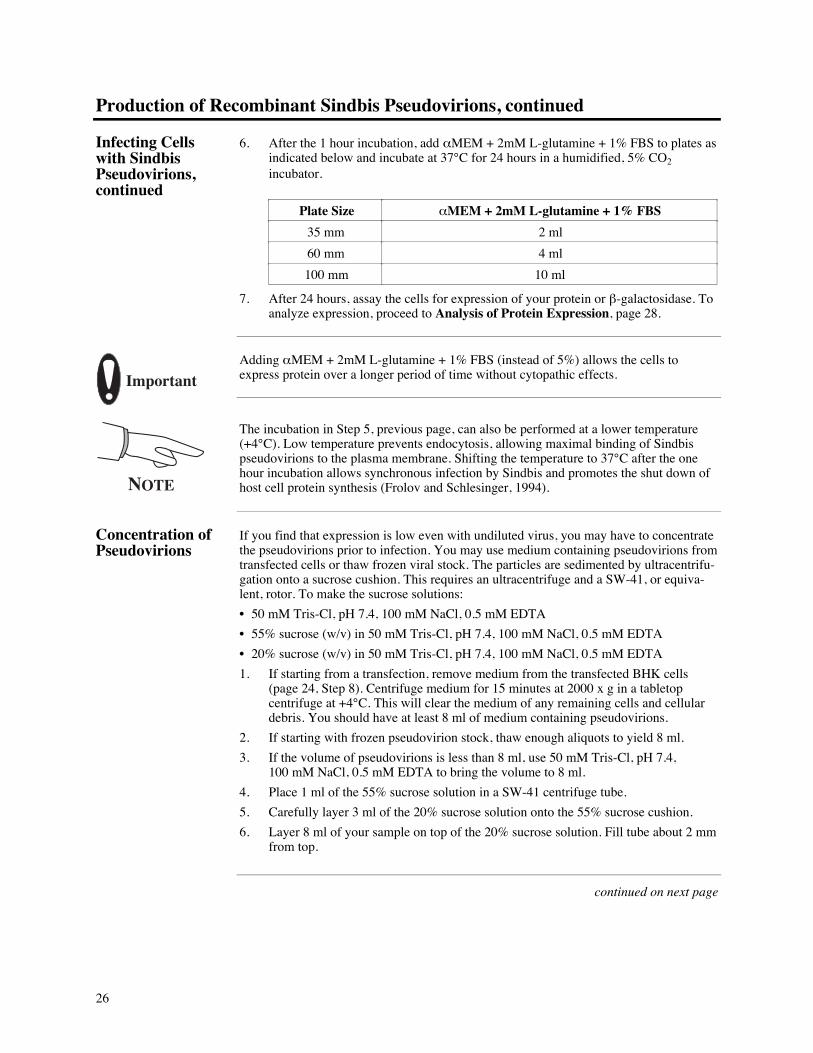
Production of Recombinant Sindbis Pseudovirions, continued
Infecting Cellswith SindbisPseudovirions,continued
6. After the 1Êhour incubation, add aMEM + 2mM L-glutamine + 1% FBS to plates asindicated below and incubate at 37¡C for 24 hours in a humidified, 5% CO2
incubator.
Plate Size aMEM + 2mM L-glutamine + 1% FBS
35 mm 2Êml
60Êmm 4Êml
100Êmm 10Êml
7. After 24 hours, assay the cells for expression of your protein or b-galactosidase. Toanalyze expression, proceed to Analysis of Protein Expression, page 28.
ImportantAdding aMEM + 2mM L-glutamine + 1% FBS (instead of 5%) allows the cells toexpress protein over a longer period of time without cytopathic effects.
NOTE
The incubation in Step 5, previous page, can also be performed at a lower temperature(+4¡C). Low temperature prevents endocytosis, allowing maximal binding of Sindbispseudovirions to the plasma membrane. Shifting the temperature to 37¡C after the onehour incubation allows synchronous infection by Sindbis and promotes the shut down ofhost cell protein synthesis (Frolov and Schlesinger, 1994).
Concentration ofPseudovirions
If you find that expression is low even with undiluted virus, you may have to concentratethe pseudovirions prior to infection. You may use medium containing pseudovirions fromtransfected cells or thaw frozen viral stock. The particles are sedimented by ultracentrifu-gation onto a sucrose cushion. This requires an ultracentrifuge and a SW-41, or equiva-lent, rotor. To make the sucrose solutions:
¥ 50ÊmM Tris-Cl, pH 7.4, 100ÊmM NaCl, 0.5ÊmM EDTA
¥ 55% sucrose (w/v) in 50ÊmM Tris-Cl, pH 7.4, 100ÊmM NaCl, 0.5ÊmM EDTA
¥ 20% sucrose (w/v) in 50ÊmM Tris-Cl, pH 7.4, 100ÊmM NaCl, 0.5ÊmM EDTA
1. If starting from a transfection, remove medium from the transfected BHK cells(page 24, StepÊ8). Centrifuge medium for 15Êminutes at 2000 x g in a tabletopcentrifuge at +4¡C. This will clear the medium of any remaining cells and cellulardebris. You should have at least 8Êml of medium containing pseudovirions.
2. If starting with frozen pseudovirion stock, thaw enough aliquots to yield 8Êml.
3. If the volume of pseudovirions is less than 8Êml, use 50ÊmM Tris-Cl, pH 7.4,100ÊmM NaCl, 0.5ÊmM EDTA to bring the volume to 8Êml.
4. Place 1 ml of the 55% sucrose solution in a SW-41 centrifuge tube.
5. Carefully layer 3Êml of the 20% sucrose solution onto the 55% sucrose cushion.
6. Layer 8Êml of your sample on top of the 20% sucrose solution. Fill tube about 2Êmmfrom top.
continued on next page
27
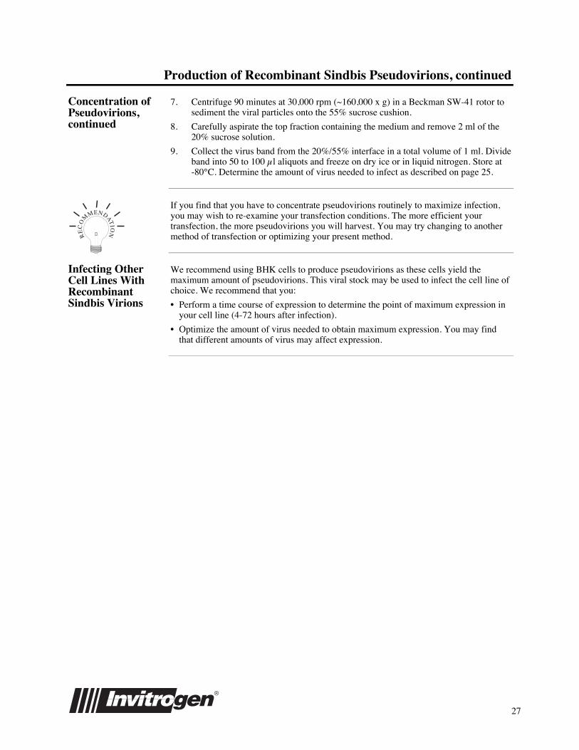
Production of Recombinant Sindbis Pseudovirions, continued
Concentration ofPseudovirions,continued
7. Centrifuge 90 minutes at 30,000 rpm (~160,000 x g) in a Beckman SW-41 rotor tosediment the viral particles onto the 55% sucrose cushion.
8. Carefully aspirate the top fraction containing the medium and remove 2Êml of the20% sucrose solution.
9. Collect the virus band from the 20%/55% interface in a total volume of 1Êml. Divideband into 50 to 100ʵl aliquots and freeze on dry ice or in liquid nitrogen. Store at-80¡C. Determine the amount of virus needed to infect as described on pageÊ25.
RE
CO
MMENDAT
ION
If you find that you have to concentrate pseudovirions routinely to maximize infection,you may wish to re-examine your transfection conditions. The more efficient yourtransfection, the more pseudovirions you will harvest. You may try changing to anothermethod of transfection or optimizing your present method.
Infecting OtherCell Lines WithRecombinantSindbis Virions
We recommend using BHK cells to produce pseudovirions as these cells yield themaximum amount of pseudovirions. This viral stock may be used to infect the cell line ofchoice. We recommend that you:
¥ Perform a time course of expression to determine the point of maximum expression inyour cell line (4-72 hours after infection).
¥ Optimize the amount of virus needed to obtain maximum expression. You may findthat different amounts of virus may affect expression.
28
Analysis of Protein Expression
Introduction For most proteins expressed in the Sindbis Expression System, expression levels are in therange of microgram of recombinant protein per mg total cell protein. For CAT expressionin C6/36 mosquito cells and BHK cells, levels of CAT reached 3% of the total cellularprotein (Olson, et al., 1992; Xiong, et al., 1989). In some cases, it is possible to visualizeyour protein using SDS-PAGE and Coomassie blue staining. Comparison of thetransfected cell lysate with an untransfected lysate facilitates identification of your protein.
Alternatively, western blotting (if you have antibody) or metabolic labeling using aradioisotope such as [35S]-methionine can be used to visualize your protein (Frolov andSchlesinger, 1994). Since the Sindbis replication cycle inhibits host protein synthesis, theexpressed protein should be the prominent band on the resulting autoradiogram. Purifica-tion protocols such as immunoprecipitations should not be necessary to identify theexpressed protein.
Before Starting You will need the following solutions and equipment:
¥ 35Êmm plate with transfected BHK cells, 80-90% confluent
¥ PBS with cations
¥ 1.5Êml microcentrifuge tubes
¥ Microcentrifuge
¥ 250ÊmM Tris-Cl, pH 8.0
¥ Nonidet P-40 detergent (10% stock)
¥ 250ÊmM Tris-Cl, pH 8.0, 1% NP-40, 150ÊmM NaCl
¥ Dry ice/ethanol bath
¥ 37¡C temperature block or water bath
¥ SDS-PAGE sample buffer, solutions, and apparatus (prepare an SDS-polyacrylamidegel that will resolve your protein)
Preparation of CellLysates
There are a number of ways to prepare cell lysates for analysis on SDS-PAGE. Thefollowing two procedures (with and without detergent) are included for yourconvenience. One 35Êmm plate (~5 x 10⁵ cells) is sufficient for each sample to be tested.It is important to include a plate of untransfected (uninfected) cells as a negative controland a plate of cells transfected (infected) with recombinant RNA made from SinRep/lacZas a positive control.
1. Wash cells two times with half the initial culture volume of PBS.
2. Scrape cells using a rubber policeman into 1Êml PBS with cations. Transfer to a1.5Êml microcentrifuge tube.
continued on next page

29
Analysis of Protein Expression, continued
Preparation of CellLysates, continued
3. Centrifuge 1-2Êminutes at maximum speed in a microcentrifuge. Decant thesupernatant. At this point you may proceed to the left column for cell lysis withoutdetergent or to the right column for cell lysis with detergent.
Step Without Detergent With Detergent
4. Resuspend cell pellet in 30-50ʵl250ÊmM Tris-Cl, pH 8.0
Resuspend cell pellet in 30-50ʵl250ÊmM Tris-Cl, pH 8.0, 1% NP-40,150ÊmM NaCl.
5. Freeze cell suspension in a dryice/ethanol bath and then thaw at 37¡C.Repeat two more times.
Incubate at 37¡C for 10Êminutes, vortex,and then place on ice.
6. Pellet the cell debris by centrifuging 1-2 minutes at maximum speed in amicrocentrifuge. Transfer supernatant to a new tube.
7. Add your SDS-PAGE sample buffer of choice and boil 5 minutes. Load 20-25% ofyour sample onto an SDS-PAGE and process according to your own procedure.
8. If solution is too viscous to load samples, shear DNA by passing through a 22 gaugeneedle 5-10 times.
SDS-PAGEAnalysis
Compare the lane containing the sample from the untransfected (uninfected) cells withthe lane containing the sample from cells expressing the recombinant protein. Forexpression in BHK cells, you may be able to see a discrete, overexpressed band at thecorrect molecular weight for your protein. However, in other cell lines, it may benecessary to use a western blot or metabolic labeling (see below) to visualize therecombinant protein.
MetabolicLabeling
This procedure is designed to quickly determine whether your protein is expressed. Youmay assay cells starting 4 hours postinfection or posttransfection; however, you may wishto wait until 24 hours after infection or transfection to ensure expression of yourconstruct. You will need the following reagents:
¥ Methionine-free MEM (not aMEM), available from ICN or LTI¥ aMEM containing 2ÊmM L-glutamine only (no FBS)¥ [35S]-methionine, 10ʵCi/µl¥ PBS¥ SDS-PAGE loading buffer, solutions, and apparatus¥ Film for autoradiography
To label cells in a 35Êmm plate, use the following protocol.
1. Wash the cells two times with half the initial culture volume of PBS.
2. Incubate the monolayer at 37¡C in 400ʵl methionine-free medium for 20-30 minutes.
3. Add 5-10ʵCi [35S]-methionine and incubate the cells for 20-30Êminutes at 37¡C.
4. Add 1Êml of aMEM containing 2ÊmM L-glutamine (no FBS) to dilute isotope.Incubate for 10Êminutes.
5. After the 10Êminute incubation, wash the monolayer three times with PBS andcollect the monolayer in 1 ml PBS using a rubber policeman to scrape cells.
6. Pellet the cells by centrifugation and dissolve the cell pellet in 25-50ʵl SDS-PAGEloading buffer.
7. Analyze one-fifth of the sample (5-10ʵl) on an SDS-PAGE and autoradiograph.
continued on next page
30
Analysis of Protein Expression, continued
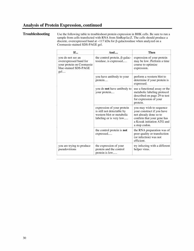
Troubleshooting Use the following table to troubleshoot protein expression in BHK cells. Be sure to run asample from cells transfected with RNA from SinRep/lacZ. The cells should produce adiscrete, overexpressed band at ~117ÊkDa for b-galactosidase when analyzed on aCoomassie-stained SDS-PAGE gel.
If... And.... Then
you do not see anoverexpressed band foryour protein on Coomassieblue-stained SDS-PAGEgel....
the control protein, b-galac-tosidase, is expressed,....
expression of your proteinmay be low. Perform a timecourse to optimizeexpression.
you have antibody to yourprotein....
perform a western blot todetermine if your protein isexpressed.
you do not have antibody toyour protein,...
use a functional assay or themetabolic labeling protocoldescribed on page 29 to testfor expression of yourprotein.
expression of your proteinis still not detectable bywestern blot or metaboliclabeling or is very low,....
you may wish to sequenceyour construct if you havenot already done so toconfirm that your gene hasa Kozak initiation ATG anda stop codon.
the control protein is notexpressed,....
the RNA preparation was ofpoor quality or transfection(or infection) was notefficient.
you are trying to producepseudovirions
the expression of yourprotein and the controlprotein is low,....
try infecting with a differenthelper virus.

31
Appendix
Sindbis Virus Life Cycle
Alphaviruses The Sindbis virus is a member of the alphavirus family. These viruses are small-enveloped viruses with single-stranded RNA genomes. The RNA genome has a positivepolarity and replicates in virtually all eukaryotic cells. A quick review of the Sindbis lifecycle is provided below for your convenience (Schlesinger and Schlesinger, 1990). Formore information about Sindbis replication, please refer to (Peters and Dalrymple, 1990;Schlesinger, 1993; Strauss and Strauss, 1994).
Sindbis Genomeand Expression ofViral Proteins
The complete sequence of the Sindbis genome has been determined and is approximately11,700 nucleotides (Rice and Strauss, 1981; Strauss, et al., 1984). During replication twomRNA species are produced, the full-length genomic RNA and the smaller subgenomicRNA. The 5« ends of both transcripts are capped with 7-methylguanosine and the 3« endsare polyadenylated. The genomic RNA transcript codes for the replication, or non-structural, proteins (nsP1-4) while the subgenomic transcript codes for the structuralproteins required for assembly and budding of the virus (capsid, p62, 6K, and E1).
CAP poly ANSP-1 1 2 3
CAP poly ACapsid P62 6K E1
Subgenomic mRNA
264aa
E3° E2*423aa64aa
Start sites for translationStop sites for translationVirion Structural ProteinsOccasionally found in virions°
*
Capsid*
4
P62° 6K°264aa
E1*439aa
Infection Virus particles bind to the cell membrane through glycoprotein spikes (E1 and E2) in theviral membrane. Once bound to the cell surface, Sindbis virions are thought to beendocytosed into the cell cytoplasm by coated vesicles. These vesicles become uncoatedand acidify, promoting fusion of the viral membrane with the vesicle membrane andreleasing the nucleocapsid into the cytoplasm. Endocytosis may not be the only mode ofentry. Sindbis virus may also fuse directly with the plasma membrane to release thenucleocapsid into the cytoplasm. (Fan and Sefton, 1978).
continued on next page
32
Sindbis Virus Life Cycle, continued
Translation andReplication
The nucleocapsid is released from the viral RNA genome by an as yet undefinedmechanism, and the RNA genome functions as a mRNA in the cytoplasm of the cell.Ribosomes bind to the genomic RNA (the plus-strand) and produce the replicationenzymes necessary for the production of minus-strand RNA, additional genomic, plus-strand RNA, and subgenomic, plus-strand RNA. The minus strand is used as a templateto make more genomic-length RNA (plus-strand). Once enough genomic-length RNA hasbeen synthesized, the minus-strand is used to generate the subgenomic plus-strand RNA.
Genomic RNA
Ribosomes
Replication enzymes
Genomic RNA
Genomic RNA Subgenomic RNA
Structural Proteins
Nucleocapsids
(+)
(-)
(+)
Ribosomes
TemporalRegulation
Production of plus- and minus-strand genomic RNA occurs during the first 3-3.5 hoursafter infection. Minus-strand synthesis then stops while plus-strand genomic andsubgenomic RNA synthesis continues. Most of the genomic, plus-strand RNA moleculesbecome packaged into nucleocapsids, so there is an excess of subgenomic RNAmolecules over genomic molecules (10:1). The subgenomic RNA molecules recruitnearly all the host's ribosomes to produce the viral structural proteins (Strauss andStrauss, 1994).
Production ofStructural Proteins
The structural polypeptides result from several proteolytic cleavages that occur as thenascent polyprotein is translated. The capsid protein is the first protein released, and itrapidly interacts with genomic, plus-strand RNA to form the nucleocapsid. The nextprotein, p62 is translated and translocated into the endoplasmic reticulum (ER). Thepolyprotein is again cleaved, releasing p62 into the endoplasmic reticulum. Furthertranslation creates another signal for membrane insertion to direct the E1 protein into theendoplasmic reticulum. E1 and p62 quickly form a heterodimer in the ER and areprocessed further (see next page).
continued on next page
33
Sindbis Virus Life Cycle, continued
Maturation andTransport
As p62 and E1 proteins move through the ER and the Golgi apparatus, the followingmodifications occur:
¥ Glycosylation of p62 and E1 in the ER
¥ Acylation of both p62 and E1 in the ER
¥ Proteolysis of p62 to yield E2 and E3 in the Golgi
It is not known what role these modifications play in the viral life cycle. They do notseem to function as signals for sorting or plasma membrane localization. These post-translational modifications may have some role in promoting fusion of the virus with hostmembranes and promoting interactions with the host lipids or the viral nucleocapsids. E1and E2 become the glycoprotein spikes in mature virus while the E3 protein is rarelyfound in Sindbis virions.
Assembly andBudding
Assembly of the virion occurs at the plasma membrane (Scheefers, et al., 1980; Simonsand Garoff, 1980; Smith and Brown, 1977). The heterodimer of E1 and E2 inserts into theplasma membrane. From electron micrographs, almost all of the E1/E2 glycoprotein isimmobilized in patches of budding virus. It is thought that the E2 cytoplasmic tailprovides the binding site for the nucleocapsid. This interaction between E2 and thenucleocapsid is thought to initiate the actual budding and release of the virion.
34
Recipes
Complete aMEMMedium
aMEM medium with 2mM L-glutamine is available from Invitrogen in 500Êml bottles(Catalog no. Q400-01). To make complete aMEM medium, add FBS to a finalconcentration of 5%.
PhosphateBuffered SalineWith Cations
For washing cells only. The solution does not need to be RNase-free.
137ÊmM NaCl2.7ÊmM KCl10ÊmM Na2HPO4
1.8ÊmM KH2PO4
0.9ÊmM CaCl2-2H2O0.5ÊmM MgCl2-6H2O
1. Dissolve: 8Êg NaCl0.2Êg KCl1.44Êg Na2HPO4
0.24Êg KH2PO4
0.13Êg CaCl2-2H2O0.10Êg MgCl2-6H2O
in 800Êml deionized water.
2. Adjust pH to 7.4 with concentrated HCl.
3. Bring the volume to 1Êliter and autoclave for 20 minutes on liquid cycle.
4. Store at +4¡C or room temperature.
PhosphateBuffered SalineWithout Cations,RNase-free
For electroporation of cells:
137ÊmM NaCl2.7ÊmM KCl10ÊmM Na2HPO4
1.8ÊmM KH2PO4
1. Dissolve: 8Êg NaCl0.2Êg KCl1.44Êg Na2HPO4
0.24Êg KH2PO4
in 800Êml deionized water.
2. Adjust pH to 7.4 with concentrated HCl.
3. Bring the volume to 1Êliter.
4. Add 1 ml diethylpyrocarbonate (DEPC) and stir overnight to make the solutionRNase-free.
5. Autoclave for 20 minutes on liquid cycle to sterilize and inactivate the DEPC.
6. Store at +4¡C or room temperature.
continued on next page

35
Recipes, continued
Formaldehyde/GlutaraldehydeSolution
2% formaldehyde0.2% glutaraldehydein PBS without cations, pHÊ7.4
1. Make fresh and prepare only what you need. Use 1Êml of reagent per 35Êmm plate,2Êml per 60Êmm plate, and 5Êml per 100Êmm plate.
2. For 10Êml of reagent, mix
0.54Êml 37% formaldehyde40ʵl 50% glutaraldehyde9.42Êml PBS without cations, pH 7.4
3. Use immediately.
X-Gal Reagent forStaining Cells
1 mg/ml X-Gal4 mM potassium ferricyanide (K3Fe(CN)6)4 mM potassium ferrocyanide (K4Fe(CN)6-3H2O)2 mM magnesium chloride, hexahydrate
in PBS (without cations), pH 7.4
Prepare just enough solution to stain the number of plates you desire. You will use 1Êmlof reagent to stain a 35Êmm plate, 2Êml for a 60Êmm plate, and 5Êml for a 100Êmm plate.Multiply by the number of plates you are staining to determine the total volume ofreagent needed.
1. Prepare 10Êml each of the following stock solutions. Solutions are stable indefinitelyif stored as indicated.
¥ X-gal: 20 mg/ml in dimethylformamide (DMF). Dissolve 200 mg in 10 ml DMFand store at -20¡C.
¥ Potassium Ferricyanide and Potassium Ferrocyanide: 0.4ÊM each in deionizedwater. Dissolve 1.32Êg of potassium ferricyanide and 1.69Êg of potassiumferrocyanide in 10Êml deionized water. Store at -20¡C.
¥ Magnesium Chloride: 200ÊmM in deionized water. Dissolve 0.4Êg in 10Êmldeionized water and store at room temperature or -20¡C.
2. For 10Êml of reagent, mix together:
0.5Êml of 20Êmg/ml X-Gal stock solution0.1Êml of the potassium ferricyanide/ferrocyanide stock solution0.1Êml of the magnesium chloride stock solution9.3Êml of PBS without cations
3. Solution is ready for use.
continued on next page
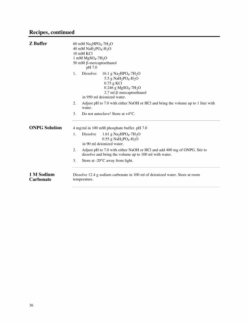
36
Recipes, continued
Z Buffer 60ÊmM Na2HPO4-7H2O40ÊmM NaH2PO4-H2O10ÊmM KCl1ÊmM MgSO4-7H2O50ÊmM b-mercaptoethanol pHÊ7.0
1. Dissolve: 16.1Êg Na2HPO4-7H2O 5.5Êg NaH2PO4-H2O 0.75Êg KCl 0.246Êg MgSO4-7H2O 2.7Êml b-mercaptoethanol
in 950Êml deionized water.
2. Adjust pH to 7.0 with either NaOH or HCl and bring the volume up to 1 liter withwater.
3. Do not autoclave! Store at +4¡C.
ONPG Solution 4Êmg/ml in 100ÊmM phosphate buffer, pH 7.0
1. Dissolve 1.61Êg Na2HPO4-7H2O0.55Êg NaH2PO4-H2O
in 90Êml deionized water.
2. Adjust pH to 7.0 with either NaOH or HCl and add 400Êmg of ONPG. Stir todissolve and bring the volume up to 100Êml with water.
3. Store at -20¡C away from light.
1ÊM SodiumCarbonate
Dissolve 12.4Êg sodium carbonate in 100Êml of deionized water. Store at roomtemperature.
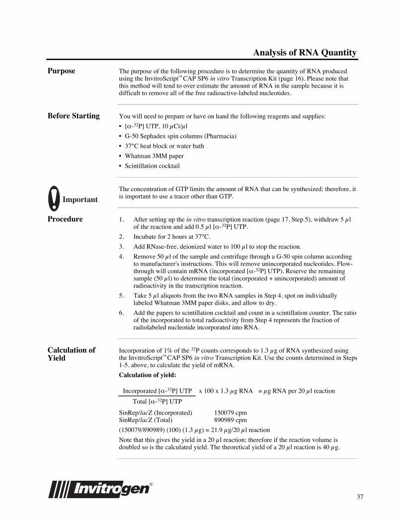
37
Analysis of RNA Quantity
Purpose The purpose of the following procedure is to determine the quantity of RNA producedusing the InvitroScriptªCAP SP6 in vitro Transcription Kit (page 16). Please note thatthis method will tend to over estimate the amount of RNA in the sample because it isdifficult to remove all of the free radioactive-labeled nucleotides.
Before Starting You will need to prepare or have on hand the following reagents and supplies:
¥ [a-32P] UTP, 10ʵCi/µl
¥ G-50 Sephadex spin columns (Pharmacia)
¥ 37¡C heat block or water bath
¥ Whatman 3MM paper
¥ Scintillation cocktail
ImportantThe concentration of GTP limits the amount of RNA that can be synthesized; therefore, itis important to use a tracer other than GTP.
Procedure 1. After setting up the in vitro transcription reaction (page 17, Step 5), withdraw 5ʵlof the reaction and add 0.5ʵl [a-32P] UTP.
2. Incubate for 2Êhours at 37¡C.
3. Add RNase-free, deionized water to 100ʵl to stop the reaction.
4. Remove 50ʵl of the sample and centrifuge through a G-50 spin column accordingto manufacturer's instructions. This will remove unincorporated nucleotides. Flow-through will contain mRNA (incorporated [a-32P] UTP). Reserve the remainingsample (50ʵl) to determine the total (incorporated + unincorporated) amount ofradioactivity in the transcription reaction.
5. Take 5ʵl aliquots from the two RNA samples in Step 4, spot on individuallylabeled Whatman 3MM paper disks, and allow to dry.
6. Add the papers to scintillation cocktail and count in a scintillation counter. The ratioof the incorporated to total radioactivity from Step 4 represents the fraction ofradiolabeled nucleotide incorporated into RNA.
Calculation ofYield
Incorporation of 1% of the 32P counts corresponds to 1.3 µg of RNA synthesized usingthe InvitroScriptªCAP SP6 in vitro Transcription Kit. Use the counts determined in Steps1-5, above, to calculate the yield of mRNA.
Calculation of yield:
Incorporated [a-32P] UTP x 100 x 1.3ʵg RNA = µgÊRNA per 20ʵl reaction
Total [a-32P] UTP
SinRep/lacZ (Incorporated) 150079 cpmSinRep/lacZ (Total) 890989 cpm
(150079/890989) (100) (1.3 µg) = 21.9ʵg/20ʵl reaction
Note that this gives the yield in a 20ʵl reaction; therefore if the reaction volume isdoubled so is the calculated yield. The theoretical yield of a 20ʵl reaction is 40ʵg.
38
Staining Cells for b-galactosidase Expression
Introduction The SinRep/lacZ positive control template can be used to check expression levels andtransfection efficiencies. A quick way to do this is to stain the transfected cells directly inthe tissue culture dish. Comparison of the number of stained cells on a transfected orinfected plate to an untransfected plate can be used to determine the efficiency oftransfection. Alternatively, a transfected or infected cell lysate can be assayed forb-galactosidase activity.
Before Starting You will need the following reagents and equipment:
¥ 35Êmm plates with transfected or infected cells, 7-16 hours posttransfection
¥ PBS with cations
¥ 2% formaldehyde, 0.2% glutaraldehyde in PBS, pH 7.3 (See Recipes, page 34)
¥ X-gal Reagent (see Recipes, page 34)
Staining Cells forb-galactosidaseActivity
At any point 4 hours posttransfection, you may stain the cells for b-galactosidase activity.Please note that this procedure will kill the cells. Also, to detect b-galactosidase activity atearly time points, it may be necessary to incubate the cells in the X-gal reagent for longerperiods to detect color development (Steps 6 and 7). Optimal expression may not occuruntil later time points.
1. Aspirate the tissue culture medium from the dish containing the cells to be stained.
2. Rinse twice with PBS using half the volume of the initial culture medium.
3. Fix the cells in 2% formaldehyde, 0.2% glutaraldehyde in PBS, pH 7.3 for 10minutes at room temperature.
4. Rinse twice with half the volume of the initial culture medium of PBS.
5. Stain with the X-gal reagent using half the volume of the initial culture medium at37¡C and incubate for 15 minutes.
6. After incubating for 15 minutes, check the cells under the microscope for thedevelopment of blue color in the cells.
7. If cells are not blue, return the cells to 37¡C and keep checking until you see a bluecolor develop. Color development should be visible within 30-60Êminutes afteraddition of the X-gal reagent.
NOTE
For your convenience, the b-Gal Staining Kit (Catalog no. K1465-01) is availablefrom Invitrogen. The kit contains all the reagents described above to stain whole cellsfor b-galactosidase expression.
39
b-galactosidase Assay
Introduction In the b-galactosidase assay, the lactose analog orthonitrophenyl-b-D-galactoside (ONPG),is added to the cell lysate (Miller, 1972, p. 403). ONPG is cleaved by b-galactosidase toproduce the ONP anion which produces a yellow color under basic conditions. The assay ismonitored by observing the change in absorbance at 420Ênm using a UV-VISspectrophotometer. A protein assay is also performed for each sample in order to normalizethe number of b-galactosidase units per mg total protein (Bollag and Edelstein, 1991).
Before Starting The solutions for the assay are found in the Recipe section, page 34. You will need tomake the following solutions:
¥ 4Êmg/ml ONPG
¥ "Z" Buffer
¥ 1ÊM Sodium carbonate
Assay forb-galactosidaseProtein
1. To test each lysate (see Analysis of Protein Expression, page 28), dilute 1:100with Z buffer. Take 1-10ʵl and transfer to a fresh microcentrifuge tube.
2. Add distilled, deionized water to a final volume of 30ʵl.
3. Add 66ʵl of 4Êmg/ml ONPG and 204ʵl "Z" buffer. Mix by gently flicking the tubeand centrifuging briefly.
4. Incubate the tube at 37¡C for 30 minutes. You should see a faint yellow colordevelop if b-galactosidase is present.
5. To stop reaction, add 500ʵl of 1ÊM sodium carbonate.
6. Read the absorbance at 420Ênm against a blank without lysate. Be sure to assay asample of the untransfected cell lysate as a control.
7. Be sure to assay at least three different volumes of lysate. Changes in absorbanceshould be linear with respect to the amount of lysate assayed. If it is not, you willnot get an accurate determination of expression. The most common error is usingtoo much lysate which will cause you to overestimate the expression levels.Decrease the amount of lysate until absorbance is linear with the amount of lysate.
8. Once you have determined you are assaying the lysate accurately, determine theprotein concentration of the lysate, and calculate the activity of the lysate using thefollowing formula:
b-galactosidase Units = (OD420 x 380)/ t / mg protein assayed
where t = the time of incubation in minutes at 37¡C, 380 is a conversion factor toconvert absorbance to µmoles of ONPG, and mg protein is the amount of proteinassayed. Be sure to subtract the background activity of the untransfected cell lysate.
NOTE
For your convenience, the b-Gal Assay Kit (Catalog no. K1455-01) is available fromInvitrogen. The kit contains all the reagents described above to assay cell lysates forb-galactosidase expression.

40
Miniprep Plasmid Preparation
Introduction Generally, the cleaner the template DNA, the greater the yield from the in vitrotranscription reaction. Template DNA purified on a CsCl gradient gives the highestyields. However, the miniprep procedure below yields DNA template of sufficient qualityfor in vitro transcription. The procedure uses a Proteinase K digestion step to ensurethat all ribonuclease is degraded prior to the in vitro transcription reaction.
Before Starting You will need the following reagents and equipment:
¥ ~1.5Êml of bacterial culture (E. coli transformed with pSinRep5 containing your gene)
¥ Sterile 50ÊmM glucose, 10ÊmM EDTA, pH 8, 25ÊmM Tris-HCl, pH 8.0
¥ 0.2ÊN NaOH, 1% SDS (make fresh)
¥ 3ÊM potassium/5ÊM acetate (for 100Êml, mix 60Êml 5ÊM potassium acetate and 11.5Êmlglacial acetic acid with 28.5Êml deionized water)
¥ 100% ethanol
¥ 10Êmg/ml RNase
¥ TE with 20ʵg/ml RNase (Add 1 µl of 10Êmg/ml RNase to 500ʵl TE)
¥ XhoÊI, NotÊI, or PacÊI restriction enzyme and appropriate buffer
¥ 20Êmg/ml Proteinase K
¥ Phenol/chloroform
¥ 5ÊM ammonium acetate (RNase-free)
¥ RNase-free water
Isolation of NucleicAcid and RNaseDigestion
The procedure below starts with the basic alkaline miniprep procedure to isolate nucleicacid.
1. Centrifuge 1.5Êml of the bacterial culture at room temperature for 30Êseconds.Remove all the medium.
2. Add 110ʵl of 50ÊmM glucose, 10ÊmM EDTA, pH 8, 25ÊmM Tris-HCl, pH 8.0 tothe pellet and vortex vigorously to fully resuspend the bacterial pellet.
3. Add 220ʵl of 0.2ÊN NaOH, 1% SDS, invert the tube 10 times to mix, and incubateon ice for 1Êminute (or longer).
4. Add 165ʵl 3ÊM potassium/5ÊM acetate, vortex for 10Êseconds, and incubate for5Êminutes on ice.
5. Centrifuge at +4¡C for 5 minutes at maximum speed in a microcentrifuge.
6. Transfer the supernatant to a tube containing 1Êml ethanol, invert several times tomix, and incubate on ice for 5Êminutes.
7. Centrifuge at +4¡C for 5Êminutes at maximum speed. Decant supernatant, centrifugebriefly, and aspirate off residual supernatant.
8. Resuspend nucleic acid pellet in 50ʵl TE buffer containing 20ʵg/ml RNase.Vortex vigorously and incubate for 5Êminutes at 37-42¡C. Make sure pellet issolubilized.
continued on next page
41
Miniprep Plasmid Preparation, continued
Restriction-Proteinase KDigestion
After removing RNA, the plasmid is ready to be linearized prior to in vitro transcription.Choose a restriction enzyme that will linearize the plasmid without cutting your gene.After restriction digestion, Proteinase K is added to remove all enzymes. The digest isthen extracted with phenol/chloroform and the DNA ethanol precipitated. The volume ofthe restriction digest should be at least 2-3 times the volume of the miniprep used. Forexample, it your miniprep volume is 10ʵl, perform the restriction digest in a 20-30ʵlvolume.
1. Digest miniprep DNA (all or part) with XhoÊI, NotÊI, or PacÊI. Digest at 37¡C for atleast 1 hour, using 0.3-0.8 units of restriction enzyme per microliter of miniprepDNA.
2. Add a 1/10 volume of Proteinase K (20Êmg/ml), mix by pipetting, and incubate at50¡C for at least 30Êminutes.
3. Extract the digest once with phenol/chloroform and remove the top, aqueous phaseto a new tube.
4. Add 1/10 volume of 5ÊM ammonium acetate (RNase-free) and 2 volumes ofethanol. Incubate at -20¡C for at least 15Êminutes.
5. Centrifuge the solution at maximum speed in a microcentrifuge for 15Êminutes at+4¡C. Decant supernatant, centrifuge briefly, and remove all the ethanol.
6. Resuspend DNA pellet in 10-20ʵl RNase-free water per 50ʵl of miniprep nucleicacid per 1.5Êml bacterial culture.
7. Determine concentration of the DNA. You will need 0.5-1.0ʵg of template DNAper a 20ʵl in vitro transcription reaction. This will be about 1-3ʵl of the 10-20ʵlpreparation in Step 6.
42
Technical Service
World Wide Web Visit the Invitrogen Web Resource using your World Wide Web browser. At the site,you can:
¥ Get the scoop on our hot new products and special product offers¥ View and download vector maps and sequences¥ Download manuals in Adobeª Acrobatª (PDF) format¥ Explore our Catalog with full color graphics¥ Obtain citations for Invitrogen products¥ Post a question at one of our many user forums¥ Request catalog and product literature
Once connected to the Internet, launch your web browser (Netscape 3.0 or newer), thenenter the following location (or URL):
http://www.invitrogen.com
...and the program will connect directly. Click on underlined text or outlined graphics toexplore. Don't forget to put a bookmark at our site for easy reference!
Technical Service If you need technical information or help, please email, call, or fax us:
Location Email Telephone/Fax
United States
Canada
[email protected] Voice: 1-800-955-6288
Fax: 1-760-603-7201
Mexico
South America
[email protected] Voice: 01-760-603-7200
Fax: 01-760-603-7201
Europe
Africa
[email protected] Voice: +31 (0) 594 515 175
Fax: +31 (0) 594 515 312
Asia
Australia
India
[email protected] Voice: 01-760-603-7200, x250
Fax: 01-760-603-7201
Addresses If you want to write to us, here are our addresses:
U.S. Headquarters: European Headquarters:Invitrogen Corporation Invitrogen BV1600 Faraday Avenue De Schelp 12, 9351 NV LeekCarlsbad, CA 92008 USA The Netherlands
Limited Warranty Invitrogen is committed to providing our customers with high-quality goods and services. Our goal is to ensure that every customer is 100% satisfiedwith our products and our service. If you should have any questions or concerns about an Invitrogen product or service, please contact our TechnicalService Representatives at 800-955-6288 extension 2.
Invitrogen warrants that all of its products will perform according to the specifications stated on the certificate of analysis. The company will replace,free of charge, any product that does not meet those specifications. This warranty limits Invitrogen CorporationÕs liability only to the cost of theproduct. No warranty is granted for products beyond their listed expiration date. No warranty is applicable unless all product components are stored inaccordance with instructions. Invitrogen reserves the right to select the method(s) used to analyze a product unless Invitrogen agrees to a specifiedmethod in writing prior to acceptance of the order.
Invitrogen makes every effort to ensure the accuracy of its publications, but realizes that the occasional typographical or other error is inevitable.Therefore Invitrogen makes no warranty of any kind regarding the contents of any publications or documentation. If you discover an error in any ofour publications, please report it to our Technical Service Representatives.
Invitrogen assumes no responsibility or liability for any special, incidental, indirect or consequential loss or damage whatsoever. The abovelimited warranty is sole and exclusive. No other warranty is made, whether expressed or implied, including any warranty of merchantabilityor fitness for a particular purpose.
43
References
Citations Ausubel, F. M., Brent, R., Kingston, R. E., Moore, D. D., Seidman, J. G., Smith, J. A. andStruhl, K., eds (1994) Current Protocols in Molecular Biology. Greene PublishingAssociates and Wiley-Interscience. New York
Bollag, D. M. and Edelstein, S. J. (1991) Protein Methods, Wiley-Liss. New York.
Bredenbeek, P. J., Frolov, I., Rice, C. M. and Schlesinger, S. (1993) Sindbis VirusExpression Vectors: Packaging of RNA Replicons by Using Defective HelperRNAs. J. Virology 67: 6439-6446.
Fan, D. P. and Sefton, B. M. (1978) The Entry into Host Cells of Sindbis Virus, VesicularStomatitis Virus and Sendai Virus. Cell 15: 985-992.
Frolov, I. and Schlesinger, S. (1994) Comparison of the Effects of Sindbis Virus andSindbis Virus Replicons on Host Cell Protein Synthesis and Cytopathogenicity inBHK Cells. J. Virology 68: 1721-1727.
Hahn, C. S., Hahn, Y. S., Braciale, T. J. and Rice, C. M. (1992) Infectious Sindbis VirusTransient Expression Vectors for Studying Antigen Processing and Presentation.Proc. Natl. Acad. Sci. USA 89: 2679-2683.
Huang, H. V., Levis, R., Rice, C. M., Schlesinger, S., Shen, P. and Xiong, C. (1993)United States, Patent No. 5,217,879
Huang, M. and Summers, J. (1991) Infection Initiated by the RNA Pregenome of a DNAVirus. J. Virology 65: 5435-5439.
Johanning, F. W., Conroy, R. M., LoBuglio, A. F., Wright, M., Sumerel, L. A., Pike, M.J. and Curiel, D. T. (1995) A Sindbis Virus mRNA Polynucleotide Vector AchievesProlonged and High Level Heterologous Gene Expression In Vivo. Nucleic AcidsRes. 23: 1495-1501.
Kozak, M. (1990) Downstream Secondary Structure Facilitates Recognition of InitiatorCodons by Eukaryotic Ribosomes. Proc. Natl. Acad. Sci. USA 87: 8301-8305.
Liljestr�m, P. and Garoff, H. (1991) A New Generation of Animal Cell ExpressionVectors Based on the Semliki Forest Virus Replicon. Bio/technology 9: 1356-1361.
Lustig, S., A. C. Jackson, C. S. Hahn, D. E. Griffinn, E. G. Strauss, and J. H. Strauss(1988) Molecular Basis of Sindbis Virus Neurovirulnce in Mice. J. Virology62: 2329-2336.
Olson, K. E., Carlson, J. O. and Beaty, B. J. (1992) Expression of the ChloramphenicolAcetyltransferase Gene in Aedes albopictus (C6/36) Cells Using a Non-infectiousSindbis Virus Expression Vector. Insect Molec. Biol. 1: 49-52.
Peters, C. J. and Dalrymple, J. M. (1990) Alphaviruses. In: B. N. Fields and D. M. Knipe(ed) Vol. 1. Fields Virology. Raven Press. New York. pp. 713-762.
Rice, C. M. and Strauss, J. H. (1981) Nucleotide Sequence of the 26S mRNA of SindbisVirus and Deduced Sequence of the Encoded Virus Structural Proteins. Proc. Natl.Acad. Sci. USA 78: 2062-2066.
Sambrook, J., Fritsch, E. F. and Maniatis, T. (1989) Molecular Cloning: A LaboratoryManual, Second. Cold Spring Harbor Laboratory Press. Plainview, New York.
Scheefers, H., Scheefers-Borchel, U., Edwards, J. and Brown, D. T. (1980) Distributionof Virus Structural Proteins and Protein-Protein Interactions in Plasma Membraneof Baby Hamster Kidney Cells Infected with Sindbis or Vesicular Stomatitis Virus.Proc. Natl. Acad. Sci. USA 77: 7277-7281.
Schlesinger, S. (1993) Alphaviruses-Vectors for Expression of Heterologous Genes.Trends in Biotechnology 11: 18-22.
continued on next page
44
References, continued
Citations,continued
Schlesinger, S. and Schlesinger, M. J. (1990) Replication of Togaviridae andFlaviviridae. In: B. N. Fields and D. M. Knipe (ed) Vol. 1. Fields Virology. RavenPress. New York. pp. 697-712.
Simons, K. and Garoff, H. (1980) The Budding Mechanisms of Enveloped AnimalViruses. J. Gen. Virol. 50: 1-21.
Smith, J. F. and Brown, D. T. (1977) Envelopments of Sindbis Virus: Synthesis andOrganization of Proteins in Cells Infected with Wild Type and Maturation DefectiveMutants. J. Virology 22: 662-678.
Strauss, E. G., Rice, C. M. and Strauss, J. H. (1984) Complete Nucleotide Sequence ofthe Genomic RNA of Sindbis Virus. Virology 133: 92-110.
Strauss, J. H. and Strauss, E. G. (1994) The Alphaviruses: Gene Expression, Replication,and Evolution. Microbiol. Rev. 58: 491-562.
Xiong, C., Levis, R., Shen, P., Schlesinger, S., Rice, C. M. and Huang, H. V. (1989)Sindbis Virus: An Efficient, Broad Host Range Vector for Gene Expression inAnimal Cells. Science 243: 1188-1191.


