SIMULATING STANFORD STUDENTS - Stanford...
4
Case Study 16 SIMULIA Community News September 2016 www.3ds.com/simulia SIMULATING STANFORD STUDENTS Dr. Ellen Kuhl and her Living Matter Laboratory Group uses Abaqus to explore subject-specific changes in the human body Students analyze personalized brain models.
Transcript of SIMULATING STANFORD STUDENTS - Stanford...

Case Study
16 SIMULIA Community News September 2016 www.3ds.com/simulia
SIMULATING STANFORD STUDENTSDr. Ellen Kuhl and her Living Matter Laboratory Group uses Abaqus
to explore subject-specific changes in the human body
Students analyze personalized brain models.

17SIMULIA Community News September 2016www.3ds.com/simulia
Times have changed dramatically in the world of engineering since the baby boomers were in college, especially as
simulation has grown to become such an integral part of the design process. Slide rules, mechanical calculators, even meshing by hand are a thing of the past and current Millennial engineering students are inspired to push the boundaries of innovative technology ever further.
One Stanford University professor, Dr. Ellen Kuhl, is helping drive such innovation alongside her students in the Living Matter Laboratory. Her group works with 10 students using personalized finite element models to predict the interplay of form and function throughout the human body.
Dr. Kuhl has used SIMULIA’s Abaqus finite element analysis tool ever since she took her first finite element course at the Leibnitz University in Hanover, Germany—and her Living Matter Lab at Stanford follows suit. “I’ve been using Abaqus for 20 years now,” she says.
Following her studies of computational, civil, and aerospace engineering in Hanover, Stuttgart, and Delft, Dr. Kuhl became an assistant professor in the department of Mechanical Engineering at the Technical University of Kaiserslautern. In 2007 she moved to Stanford, where she is currently a professor in the department of mechanical engineering with courtesy appointments in bioengineering and cardiothoracic surgery.
With a passion for living-matter physics, Dr. Kuhl focuses her work on the precise prediction of the response of living structures to environmental changes. “My specific interest is the multi-scale modeling of growth and remodeling, the study of how living matter adapts its form and function to changes in mechanical loading, and how this adaption can be traced back to structural alterations on the cellular or molecular levels,” she says.
“When I came to Stanford, a few students who were working in cardiac surgery contacted me to help with their simulation,” says Kuhl. “One student was interested in understanding atrial fibrillation. That’s how we got into modeling the heart. We connected with heart surgeons who had collected great data and we started modeling the electromechanical interaction in the heart.”
After she began her heart simulation work, Dr. Kuhl reached out to SIMULIA and asked if they would be interested in implementing biology into the software—without knowing that the Living Heart Project was already in the works. “As we had already developed similar software, we had some experience with it,” she says, “so the timing couldn’t have been better.”
Since then, the students in the Living Matter Lab have studied different parts of the human body, often using their own medical images. Attracted by the wide range of medical problems, students from multiple disciplines came on board.
“The classes that we offer attract undergraduates who are future medical students, human biologists, and engineers,” says Kuhl. "There’s a broad mix of people and that’s what I like the most. I’ve even had some business and education students; what’s interesting is that when you show them a simulation, they have very different ideas than our engineering students.”
Not surprisingly, Dr. Kuhl is affectionately known by many of her students as “Dr. Cool.”
Below are some highlights of recent work by students within the Living Matter Lab:
TENNIS AND TWISTED BONE GROWTHStudents had heard about a tennis player at the University who was having problems with his shoulder and recognized that he would be an ideal subject to see if they could simulate the physical changes to the bone resulting from overuse of a player’s dominant arm.
The team chose to study the humerus because of its structural simplicity and the convenience of being able to use the non-dominant arm as a control. “With a high-speed video recording of the player’s motions during a serve, and bone density scans available, the students created a finite element model and determined the muscle force vectors, muscle attachment points, and boundary conditions,” explains Kuhl.
The results showed that, due to torqued loadings on the player’s arm during his serve, the bone was growing in a twisted manner—which explained why the Stanford athlete was experiencing pain in his dominant arm. Armed with this insight, the group suggested ways to optimize training strategies so tennis players could avoid such irregular bone growth.
(Left). Variation in humerus density of the left and right arms of a professional tennis player. (Right) Density changes with increasing number of load cycles.
Left arm Right arm
“It’s important that academia and commercial groups work together hand in hand, and I believe that SIMULIA understands that as well.”
—Dr. Ellen Kuhl, associate professor, Department of Mechanical Engineering, Stanford University

right guard left guard punter
change in bone mineral density [g/cm2]
18 SIMULIA Community News September 2016 www.3ds.com/simulia
Case StudyASYMMETRIC LOADING IN A LINEMAN AND A PUNTERTwo students in Dr. Kuhl’s class—both players on the University’s Division 1 football team—decided to model and study their own bodies. The students had very different body types; one was a defensive lineman and one a punter. They wondered whether or not they had asymmetric loading in their bones due to their positions (see Figure 1).
They put themselves in a density scanner and recreated their body geometries. “We then asked them stand on a force plate that measures the forces on the feet when making a specific motion,” says Kuhl. “We used these forces along with the geometries to create finite element models of their femur. We asked ourselves ‘how is the density in the bone distributed in response to loading?’” The results were surprising. The students found that there was indeed a marked asymmetry: the right lineman had a stronger left leg, the punter had a stronger plant leg. (see Figure 2).
HIGH HEELS AND SHORT MUSCLESThe idea that our day-to-day behavior can change the structure of our bodies is not limited to extreme conditions. One of Dr. Kuhl’s students always wore high heels to class and chose to study the effect of doing so on her legs. She had her leg scanned both with and without her high heels on and created finite element models from those scans. The team also performed ultrasound imaging to visualize the muscle fiber orientation and used a new technique, developed by a colleague of Dr. Kuhl, to image the sarcomere microstructure.
Simulations of the calf muscle and the tendon showed that, when wearing high heels, the leg muscle shortens. The body senses this change in length and the muscle actually shrinks over time. When switching back from high heels to flat shoes, the muscle becomes overstretched and triggers pain (see figure 3).
“What’s great about finite element modeling is that it can predict where exactly the deformation is largest,” says Kuhl; all deformation from wearing heels was found in the center region of the muscle, not in the periphery or in the tendon. This insight explains why it is not healthy to wear high heels over long periods of time.
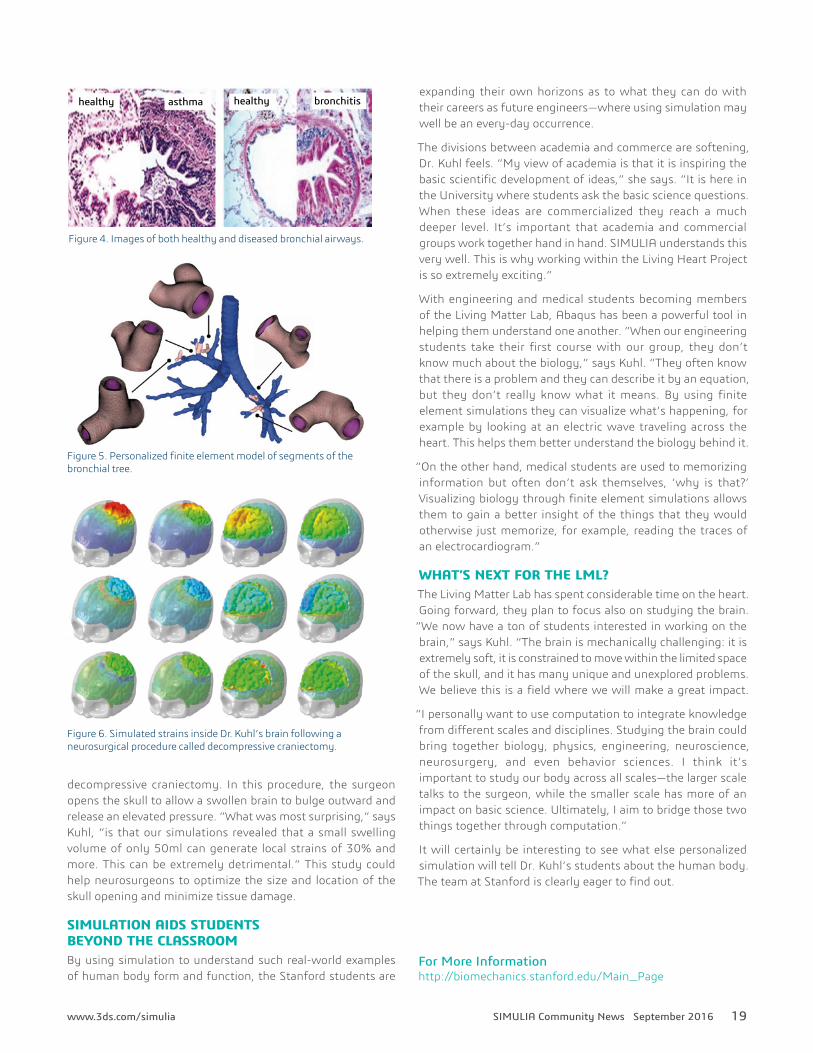
AIRWAY OBSTRUCTION IN THE LUNGS Many people have studied airway obstruction in the lungs, but most studies only look at an idealized circular cross-section of the airway. When a student in the Living Matter Lab at Stanford studied the effects of asthma and bronchitis on the lungs, she instead used the real geometry of an actual bronchial tree. “With this study we were able to show that very small changes in the geometry can have large effects on airway wall collapses,” says Dr. Kuhl. (See Figures 4-5).
The student created an airway modeled from medical scans to show that the real airway actually collapses much earlier than previously predicted. Her study highlights the importance of using anatomically realistic models, something that has
only become feasible recently with the development of high-performance computing.
PERSONALIZED BRAIN FORCESMost recently, Dr. Kuhl’s team started to investigate the role of mechanical forces in human brains. Their work began as a fairly simple class project: magnetic resonance images of their own brains to compare size and shape. But then the project expanded. They created an Abaqus model of a brain—Dr. Kuhl’s own—and one student even 3D printed the brain for use in surgical training.(See Figure 6).
A postdoctoral researcher in the group simulated the brain in different scenarios and clinical procedures including a
Figure 1. Model of a lineman in position.
Figure 2. Change in bone mineral density of both players.
Figure 3. Analysis of strain in the calf muscle over time when wearing high heels.
healthy healthyasthma bronchitis
19SIMULIA Community News September 2016www.3ds.com/simulia
For More Information http://biomechanics.stanford.edu/Main_Page
decompressive craniectomy. In this procedure, the surgeon opens the skull to allow a swollen brain to bulge outward and release an elevated pressure. “What was most surprising,” says Kuhl, “is that our simulations revealed that a small swelling volume of only 50ml can generate local strains of 30% and more. This can be extremely detrimental.” This study could help neurosurgeons to optimize the size and location of the skull opening and minimize tissue damage.
SIMULATION AIDS STUDENTS BEYOND THE CLASSROOMBy using simulation to understand such real-world examples of human body form and function, the Stanford students are
expanding their own horizons as to what they can do with their careers as future engineers—where using simulation may well be an every-day occurrence.
The divisions between academia and commerce are softening, Dr. Kuhl feels. “My view of academia is that it is inspiring the basic scientific development of ideas,” she says. “It is here in the University where students ask the basic science questions. When these ideas are commercialized they reach a much deeper level. It’s important that academia and commercial groups work together hand in hand. SIMULIA understands this very well. This is why working within the Living Heart Project is so extremely exciting.”
With engineering and medical students becoming members of the Living Matter Lab, Abaqus has been a powerful tool in helping them understand one another. “When our engineering students take their first course with our group, they don’t know much about the biology,” says Kuhl. “They often know that there is a problem and they can describe it by an equation, but they don’t really know what it means. By using finite element simulations they can visualize what’s happening, for example by looking at an electric wave traveling across the heart. This helps them better understand the biology behind it.
“On the other hand, medical students are used to memorizing information but often don’t ask themselves, ‘why is that?’ Visualizing biology through finite element simulations allows them to gain a better insight of the things that they would otherwise just memorize, for example, reading the traces of an electrocardiogram.”
WHAT’S NEXT FOR THE LML?The Living Matter Lab has spent considerable time on the heart. Going forward, they plan to focus also on studying the brain.
“We now have a ton of students interested in working on the brain,” says Kuhl. “The brain is mechanically challenging: it is extremely soft, it is constrained to move within the limited space of the skull, and it has many unique and unexplored problems. We believe this is a field where we will make a great impact.
“I personally want to use computation to integrate knowledge from different scales and disciplines. Studying the brain could bring together biology, physics, engineering, neuroscience, neurosurgery, and even behavior sciences. I think it’s important to study our body across all scales—the larger scale talks to the surgeon, while the smaller scale has more of an impact on basic science. Ultimately, I aim to bridge those two things together through computation.”
It will certainly be interesting to see what else personalized simulation will tell Dr. Kuhl’s students about the human body. The team at Stanford is clearly eager to find out.
Figure 6. Simulated strains inside Dr. Kuhl’s brain following a neurosurgical procedure called decompressive craniectomy.
Figure 5. Personalized finite element model of segments of the bronchial tree.
Figure 4. Images of both healthy and diseased bronchial airways.