![Assistive Planning in Complex, Dynamic Environments: a ... · of use cases: assistive wheelchair technology [12], assistive au-tomobile driving, and assistive manufacturing vehicle](https://static.fdocuments.net/doc/165x107/6055985ff7e719060567e863/assistive-planning-in-complex-dynamic-environments-a-of-use-cases-assistive.jpg)
Simulating ideal assistive devices to reduce the...
25
RESEARCH ARTICLE Simulating ideal assistive devices to reduce the metabolic cost of walking with heavy loads Christopher L. Dembia 1 *, Amy Silder 2 , Thomas K. Uchida 2 , Jennifer L. Hicks 2 , Scott L. Delp 1,2,3 1 Department of Mechanical Engineering, Stanford University, Stanford, California, United States of America, 2 Department of Bioengineering, Stanford University, Stanford, California, United States of America, 3 Department of Orthopaedic Surgery, Stanford University, Stanford, California, United States of America * [email protected] Abstract Wearable robotic devices can restore and enhance mobility. There is growing interest in designing devices that reduce the metabolic cost of walking; however, designers lack guide- lines for which joints to assist and when to provide the assistance. To help address this prob- lem, we used musculoskeletal simulation to predict how hypothetical devices affect muscle activity and metabolic cost when walking with heavy loads. We explored 7 massless devices, each providing unrestricted torque at one degree of freedom in one direction (hip abduction, hip flexion, hip extension, knee flexion, knee extension, ankle plantarflexion, or ankle dorsiflexion). We used the Computed Muscle Control algorithm in OpenSim to find device torque profiles that minimized the sum of squared muscle activations while tracking measured kinematics of loaded walking without assistance. We then examined the meta- bolic savings provided by each device, the corresponding device torque profiles, and the resulting changes in muscle activity. We found that the hip flexion, knee flexion, and hip abduction devices provided greater metabolic savings than the ankle plantarflexion device. The hip abduction device had the greatest ratio of metabolic savings to peak instantaneous positive device power, suggesting that frontal-plane hip assistance may be an efficient way to reduce metabolic cost. Overall, the device torque profiles generally differed from the cor- responding net joint moment generated by muscles without assistance, and occasionally exceeded the net joint moment to reduce muscle activity at other degrees of freedom. Many devices affected the activity of muscles elsewhere in the limb; for example, the hip flexion device affected muscles that span the ankle joint. Our results may help experimentalists decide which joint motions to target when building devices and can provide intuition for how devices may interact with the musculoskeletal system. The simulations are freely available online, allowing others to reproduce and extend our work. Introduction Wearable robotic devices are currently used to help restore mobility to individuals following a stroke, a spinal cord injury, or the loss of a limb [1–4]. Other potential uses for assistive devices PLOS ONE | https://doi.org/10.1371/journal.pone.0180320 July 12, 2017 1 / 25 a1111111111 a1111111111 a1111111111 a1111111111 a1111111111 OPEN ACCESS Citation: Dembia CL, Silder A, Uchida TK, Hicks JL, Delp SL (2017) Simulating ideal assistive devices to reduce the metabolic cost of walking with heavy loads. PLoS ONE 12(7): e0180320. https://doi.org/ 10.1371/journal.pone.0180320 Editor: Øyvind Sandbakk, Norwegian University of Science and Technology, NORWAY Received: February 7, 2017 Accepted: June 14, 2017 Published: July 12, 2017 Copyright: © 2017 Dembia et al. This is an open access article distributed under the terms of the Creative Commons Attribution License, which permits unrestricted use, distribution, and reproduction in any medium, provided the original author and source are credited. Data Availability Statement: We performed this study using the OpenSim software package, which is open-source and freely available at https:// opensim.stanford.edu. The data and scripts required for reproducing the results are available at https://simtk.org/projects/assistloadwalk. Funding: All authors were supported by Defense Advanced Research Projects Agency (www.darpa. mil) contract W911QX-12-C-0018 (Warrior Web) and by National Institutes of Health (www.nih.gov) grants U54 EB020405 (Mobilize Center), U54 GM072970 (Simulation of Biological Structures),
Transcript of Simulating ideal assistive devices to reduce the...
RESEARCH ARTICLE
Simulating ideal assistive devices to reduce
the metabolic cost of walking with heavy loads
Christopher L. Dembia1*, Amy Silder2, Thomas K. Uchida2, Jennifer L. Hicks2, Scott
L. Delp1,2,3
1 Department of Mechanical Engineering, Stanford University, Stanford, California, United States of America,
2 Department of Bioengineering, Stanford University, Stanford, California, United States of America,
3 Department of Orthopaedic Surgery, Stanford University, Stanford, California, United States of America
Abstract
Wearable robotic devices can restore and enhance mobility. There is growing interest in
designing devices that reduce the metabolic cost of walking; however, designers lack guide-
lines for which joints to assist and when to provide the assistance. To help address this prob-
lem, we used musculoskeletal simulation to predict how hypothetical devices affect muscle
activity and metabolic cost when walking with heavy loads. We explored 7 massless
devices, each providing unrestricted torque at one degree of freedom in one direction (hip
abduction, hip flexion, hip extension, knee flexion, knee extension, ankle plantarflexion, or
ankle dorsiflexion). We used the Computed Muscle Control algorithm in OpenSim to find
device torque profiles that minimized the sum of squared muscle activations while tracking
measured kinematics of loaded walking without assistance. We then examined the meta-
bolic savings provided by each device, the corresponding device torque profiles, and the
resulting changes in muscle activity. We found that the hip flexion, knee flexion, and hip
abduction devices provided greater metabolic savings than the ankle plantarflexion device.
The hip abduction device had the greatest ratio of metabolic savings to peak instantaneous
positive device power, suggesting that frontal-plane hip assistance may be an efficient way
to reduce metabolic cost. Overall, the device torque profiles generally differed from the cor-
responding net joint moment generated by muscles without assistance, and occasionally
exceeded the net joint moment to reduce muscle activity at other degrees of freedom. Many
devices affected the activity of muscles elsewhere in the limb; for example, the hip flexion
device affected muscles that span the ankle joint. Our results may help experimentalists
decide which joint motions to target when building devices and can provide intuition for how
devices may interact with the musculoskeletal system. The simulations are freely available
online, allowing others to reproduce and extend our work.
Introduction
Wearable robotic devices are currently used to help restore mobility to individuals following a
stroke, a spinal cord injury, or the loss of a limb [1–4]. Other potential uses for assistive devices
PLOS ONE | https://doi.org/10.1371/journal.pone.0180320 July 12, 2017 1 / 25
a1111111111
a1111111111
a1111111111
a1111111111
a1111111111
OPENACCESS
Citation: Dembia CL, Silder A, Uchida TK, Hicks JL,
Delp SL (2017) Simulating ideal assistive devices
to reduce the metabolic cost of walking with heavy
loads. PLoS ONE 12(7): e0180320. https://doi.org/
10.1371/journal.pone.0180320
Editor: Øyvind Sandbakk, Norwegian University of
Science and Technology, NORWAY
Received: February 7, 2017
Accepted: June 14, 2017
Published: July 12, 2017
Copyright: © 2017 Dembia et al. This is an open
access article distributed under the terms of the
Creative Commons Attribution License, which
permits unrestricted use, distribution, and
reproduction in any medium, provided the original
author and source are credited.
Data Availability Statement: We performed this
study using the OpenSim software package, which
is open-source and freely available at https://
opensim.stanford.edu. The data and scripts
required for reproducing the results are available at
https://simtk.org/projects/assistloadwalk.
Funding: All authors were supported by Defense
Advanced Research Projects Agency (www.darpa.
mil) contract W911QX-12-C-0018 (Warrior Web)
and by National Institutes of Health (www.nih.gov)
grants U54 EB020405 (Mobilize Center), U54
GM072970 (Simulation of Biological Structures),
are to reduce injury risk for those carrying heavy loads, such as firefighters [5], laborers [6],
and soldiers [7]. A common goal of assistive device designers is to reduce the metabolic cost
of walking. Yet, reducing the metabolic cost of walking using a device is difficult—despite
decades of effort [8–11], this has been accomplished only recently [9,12–19]. Designers are
making progress on overcoming the challenges of large subject-to-subject variability in perfor-
mance [14], minimizing the metabolic penalty of carrying a device [9], and designing effective
training protocols [20,21]. One of the largest challenges is understanding the complex neuro-
musculoskeletal adaptations (short- and long-term) that occur when the body is augmented
with assistive devices. For example, even if a device reduces muscle activity, the device may not
reduce metabolic cost or muscle fiber power [22,23].
Using experiments, researchers have learned much about providing assistance during gait.
For example, metabolic cost can be sensitive to actuation timing [13,24], metabolic cost can be
reduced with a strictly passive device [9], and unilateral assistance (on one leg only) can affect
the activity of muscles on the unassisted leg [25]. Nevertheless, there is still much to be learned.
In particular, discovering the metabolic effect of separately assisting each joint of the leg during
walking would be a significant milestone in understanding human–device interaction [26].
Achieving this milestone through experiments alone is currently impractical, as device devel-
opers would need to invest substantial time and money into designing and refining a device
for each joint [9]. Further, an experimental approach provides limited ability to evaluate how
the timing and magnitude of applied torques affect the performance of a device, independent
of the device’s mass, its comfort, the subject’s adaptations, and other practical factors.
Musculoskeletal simulations can complement experiments in designing assistive devices.
Simulations have revealed the breakdown of energy consumption during walking into stance
and swing costs [27], have shown that wearable robots can negatively affect muscle fiber
mechanics [22,28], and have suggested that asymmetric gaits are metabolically optimal for uni-
lateral amputees with prostheses [29]. Using simulations to accurately predict the effects of an
assistive device is a substantial challenge, as modeling the effects of device mass, device com-
fort, and training protocols is very difficult. However, a strength of simulation is that, rather
than mathematically characterizing all features of a device, we can explore hypothetical ideal
devices—devices that are massless, provide lossless transmission of torque to the limb, and
have no torque or power limits—and thus compare different types of devices (e.g., hip vs.
knee) independent of other practical challenges. We can also optimize the devices for a specific
objective, such as minimizing muscle activation. Moreover, we can perform such simulations
over many subjects and scenarios [30–34]. When paired with a model of muscle energy con-
sumption [27,32,35,36], simulations can also predict how devices might affect the energy con-
sumed by individual muscles. Our simulations and energy estimates can be used to inform
decisions about which joints to target with devices and to estimate how real-world devices
affect muscle activity. In turn, the results of experimental studies can be used to further vali-
date and improve the predictive capability of simulations.
Study objectives
In this study, we examined how ideal (massless with no torque or power limits) assistive
devices affect metabolic cost when walking with heavy loads. We used musculoskeletal simula-
tion to evaluate 7 ideal bilateral assistive devices that each provided one joint moment (uniarti-
cular) in one direction (unidirectional): hip abduction, hip flexion, hip extension, knee flexion,
knee extension, ankle plantarflexion, or ankle dorsiflexion. The simulations tracked motion
capture data of loaded walking. We used an optimization procedure to simultaneously opti-
mize the behavior of the devices and predict changes in muscle activity in response to applied
Simulated assistive devices for loaded walking
PLOS ONE | https://doi.org/10.1371/journal.pone.0180320 July 12, 2017 2 / 25
R24 HD065690, and P2C HD065690 (Simulation in
Rehabilitation Research). CLD also received
funding from National Science Foundation (www.
nsf.gov) Graduate Research Fellowship DGE-
114747 and a Stanford Bio-X (biox.stanford.edu)
Bowes Graduate Student Fellowship. The funders
had no role in study design, data collection and
analysis, decision to publish, or preparation of the
manuscript.
Competing interests: The authors have declared
that no competing interests exist.
device torques. Specifically, at regular intervals throughout the motion, we solved for the
device torques that minimized the sum of squared muscle activations while tracking measured
kinematics of loaded walking without assistance. We did not optimize any attributes of the
device other than the torque profiles of its two actuators. We repeated these simulations for 7
subjects and multiple gait cycles. This study is methodologically similar to a recent study from
our research group that also examined hypothetical assistive devices, but the devices were bidi-
rectional, multi-joint, and assisted running [33].
We had three specific aims. First, we sought to determine which of the 7 devices had the
highest ratio of metabolic savings to device power (on average across subjects); we used this
ratio as an “efficiency” metric to identify devices that provide relatively large metabolic savings
with a small power requirement. Second, we aimed to compare features between the optimal
torque profiles of the devices and the corresponding net joint moments produced by muscles
during loaded walking. Third, we sought to assess how each device might change lower-limb
muscle activity.
Our study presents the first muscle-actuated simulations of heavily loaded walking. We per-
formed the simulations with the open-source OpenSim software package (version 3.3) [37,38],
which was also used for much of the simulation work described earlier [22,30,32,33] and con-
tains validated muscle [39] and metabolics [27,32,35] models. The data and code required to
reproduce the results are available at https://simtk.org/home/assistloadwalk.
Methods
Experiments
We collected motion capture data from 7 male individuals (age 25 ± 5 years, height 1.86 ± 0.04
m, mass 84 ± 15 kg; mean ± standard deviation; S1 Table). Subjects were recruited between
May and September of 2013 by word of mouth from the Stanford University campus and the
surrounding communities. Data were collected for four conditions:
1. without load at a freely selected speed (1.46 ± 0.15 m/s; referred to as the no loadcondition),
2. without load at approximately 80% of the speed from the subject’s no load trials (1.20 ± 0.10
m/s),
3. while carrying 38 kg on the torso at a freely selected speed (1.27 ± 0.09 m/s; loaded), and
4. while carrying 38 kg on the torso at approximately the same speed as in the subject’s noload trials (1.48 ± 0.09 m/s).
All subjects performed these conditions in the order listed above, and all subjects who
enrolled in the study completed all conditions.
For each condition, the subjects completed at least 3 overground trials and a single 7-min-
ute treadmill trial (no incline). During the overground trials, we measured optical marker tra-
jectories, ground reaction forces and moments, and muscle activity. The trajectories of 41
markers were collected at 100 Hz with an 8-camera optical motion capture system (Motion
Analysis Corp., Santa Rosa, CA, USA). Ground reaction forces and moments were collected at
2000 Hz from 3 floor-mounted force plates (Bertec Corp., Columbus, OH, USA). Synchro-
nously, we collected the activity of 10 lower-limb muscles at 2000 Hz using surface electromy-
ography (EMG) sensors (Trigno™; Delsys Inc., Boston, MA, USA). Each overground trial
captured approximately one gait cycle. We used the treadmill trial (Woodway Pro XL; Wood-
way Inc., Waukesha, WI, USA) to estimate whole-body metabolic energy consumption using
indirect calorimetry (Quark b2; COSMED, Rome, Italy). We analyzed the final minute of data
Simulated assistive devices for loaded walking
PLOS ONE | https://doi.org/10.1371/journal.pone.0180320 July 12, 2017 3 / 25
to estimate metabolic rate [40], using the percent change over the last 3 minutes to verify that
steady-state was reached.
The added mass in the loaded conditions was split between a backpack (8 kg, including the
backpack itself) and 3 weight vests containing lead (30 kg, including the vests; Hyperware,
Austin, TX, USA). The backpack did not have a hip belt but was worn tightly, and the weight
in each weight vest was evenly distributed between the front and back of the vest.
Only the loaded condition was used to study assistive devices. The no load and loaded con-
ditions were used to validate the metabolics model. The remaining two conditions were used
only for normalizing EMG sensor readings; because subjects did not perform maximum vol-
untary contractions, the fourth condition listed above—our most intensive—typically pro-
vided the highest measured muscle activity.
The Stanford University Institutional Review Board approved our experimental protocol
and all subjects provided written informed consent.
Simulations of experiments
We generated simulations of the no load and loaded conditions. We refer to the simulations of
the loaded condition as no assistance because the no assistance simulations are the baseline for
the assisted simulations (described in the “Simulations of assisted loaded walking” section,
below).
We used a three-dimensional musculoskeletal model that is based on 21 cadavers and 24
young healthy humans [41]. The model contains 39 degrees of freedom, though we locked 8 of
them that we deemed nonessential for our study (bilateral ankle eversion, toe flexion, wrist
flexion, and wrist deviation). For our simulations of the loaded condition, we modeled the load
as a hollow cylindrical channel (height 40 cm, inner radius 13 cm, outer radius 15 cm) with
uniform density, welded to the torso.
Our simulation workflow began with scaling the geometry of the generic musculoskeletal
model to match the anthropometry of each of our subjects, using the OpenSim Scale Tool.
Additionally, we scaled the maximum isometric forces of the muscles according to a regression
equation based on subject mass and height [42]. For each subject and condition, we simulated
3 of the overground trials. For each of these trials, we generated joint angle trajectories using
OpenSim’s Inverse Kinematics (IK) Tool. We assigned greater tracking weights to anatomical
markers than to tracking markers, the latter of which were attached to marker plates on the
thigh and shank.
We used OpenSim’s Residual Reduction Algorithm (RRA) Tool to reduce the residual
forces (applied to the pelvis) resulting from inconsistencies between force plate data, marker
data, and the musculoskeletal model [37]. We ran RRA twice for each trial: first, to generate an
adjusted model (RRA-model), and then to generate adjusted kinematics (RRA-kinematics). We
combined all adjusted models from each run of RRA-model for the same subject and condition
(by averaging the suggested mass adjustments) to create a single adjusted model for each sub-
ject and condition. This strategy helps to avoid overfitting the model to the experimental data
from any particular trial, which may occur when using a separate adjusted model for each
trial. For the loaded condition, we used RRA-model to adjust the mass and location of the load.
We then produced adjusted kinematics for each trial by running RRA-kinematics, using the
single adjusted model and the kinematics from IK as input. Finally, we generated muscle-
driven simulations of the overground trials with OpenSim’s Computed Muscle Control
(CMC) Tool [43], using the single adjusted model and the adjusted kinematics.
Objective function in Computed Muscle Control. CMC solves for muscle excitations
that can produce the observed walking motion while minimizing the sum of squared muscle
Simulated assistive devices for loaded walking
PLOS ONE | https://doi.org/10.1371/journal.pone.0180320 July 12, 2017 4 / 25
activations at regular intervals in the motion. Specifically, CMC’s objective function, J, consists
of an effort term, Jeffort, and a term associated with modeling and measurement error, Jerror:
J ¼ Jeffort þ Jerror;# ð1Þ
Jeffort ¼X
i2M
a2
i ;# ð2Þ
Jerror ¼X
i2R
fiwf ;i
!2
:# ð3Þ
The effort term (Eq 2) depends only on the activation a of the set of muscles M in the
model. The error term (Eq 3) penalizes the force or moment f applied by the set of reserve and
residual actuators R in the model. Reserve actuators apply small joint moments to compensate
for unmodeled passive structures (e.g., ligaments) and potential muscle weakness, and residual
actuators apply the residual forces explained above. The weighting factor wf is adjusted to
make the reserves and residuals much more costly to use compared to the muscles; in Open-
Sim, this factor is the actuators’ “optimal force” property.
We generated 21 no load simulations and 21 no assistance (loaded) simulations (7 subjects,
3 trials per condition).
Simulations of assisted loaded walking
Assistive devices. We also used the CMC Tool to design and predict the effect of 7 hypo-
thetical assistive devices. The new simulations built upon the no assistance simulations
described above. Each device was uniarticular (acted at a single degree of freedom), unidirec-
tional (acted in only one direction), and added bilaterally (to both legs). We considered 6 sagit-
tal-plane devices: hip flexion, hip extension, knee flexion, knee extension, ankle plantarflexion,
and ankle dorsiflexion. We also considered a hip abduction device because a preliminary anal-
ysis showed that carrying a load can substantially increase the activity of the hip abductors
[44]. We chose unidirectional devices because they are common in the literature and provide a
clear picture of how the devices affect muscle activity.
We were interested in the maximum possible benefit that each of these devices could pro-
vide. As such, we modeled the devices as massless, with lossless transmission of force to the
limb, and as not having any limits on the torques or mechanical power they provide. The
devices were implemented in OpenSim as CoordinateActuators, using control bounds to
define their unidirectionality. Each device was bilateral and thus consisted of two Coordina-
teActuators, one on each leg. The two actuators were controlled independently.
Objective function in Computed Muscle Control. In the assisted simulations, the CMC
algorithm controlled both the muscles and the device. As a result, the objective function
included the torques τ applied by the two actuators (left and right legs) of the device:
Jeffort ¼X
i2M
a2
i þtleft
wt;left
!2
þtright
wt;right
!2
:# ð4Þ
To maximize the use of the device in place of muscles, we set the weighting factors wτ to a
large value (1000 N-m) so that using the device had a negligible penalty. The CMC optimiza-
tion played the two roles of finding the optimal device behavior and predicting changes in
muscle activity. The assisted simulations tracked the same kinematics (and used the same
ground reaction forces) as the no assistance simulations on which they were based, so the net
Simulated assistive devices for loaded walking
PLOS ONE | https://doi.org/10.1371/journal.pone.0180320 July 12, 2017 5 / 25
joint moments throughout the motion were conserved for all degrees of freedom. With the aid
of the device to achieve those same net joint moments, overall muscle coordination could
change to arrive at a lower Jeffort.
From each of the 21 no assistance simulations, we generated 7 simulations of assistance
(one per device), giving a total of 147 simulations of assistance.
Validation of simulations
Joint angles and net joint moments (S1 Fig) for the no load and loaded simulations were quali-
tatively similar to those from other studies of loaded walking [45,46]. The primary exception
to this was the hip flexion moment reported by Huang and Kuo [46], which we might expect
to be different because the load they used was different from the one we used: in their study,
the entire load was in a backpack, which had a hip belt.
The timing of muscle activity (S2 Fig) was similar between simulations and EMG measure-
ments for the gluteus maximus, gluteus medius, vastus lateralis, vastus medialis, gastrocne-
mius, soleus, and tibialis anterior. The discrepancy in timing for the medial hamstrings, biceps
femoris, and gastrocnemius resulted from excessive passive knee force. The magnitude of mus-
cle activity was similar between simulations and EMG for all but a few recorded muscles.
Large tibialis anterior activity during swing resulted from excessive soleus force (passive force
in dorsiflexion, and lingering activity during deactivation; in our model, the soleus can exert
much larger ankle moments than the tibialis anterior). The simulated anterior gluteus medius
activity was greater than EMG likely because the gluteus medius is a fan-shaped muscle and it
is difficult to experimentally measure the activity of the entire muscle. The simulated vasti
activity was lower than EMG because EMG was normalized by the greatest activity we
observed across conditions and it is unlikely the vasti were maximally activated in any of our
conditions. The implications of these discrepancies on our results are mentioned in the
Discussion.
The next three subsections present a set of error metrics we computed based on sugges-
tions by Hicks et al. (page 20 in [47]). These metrics were computed over all simulations we
performed.
Kinematics errors. The simulations tracked lower-limb joint angles with a root-mean-
square (RMS) error of 0.3 degrees, averaged across lower-limb degrees of freedom and simula-
tions; the maximum error was 2.2 degrees. The RMS error in marker trajectories between
CMC simulations and experimental data had a mean value of 2.1 cm across lower-limb mark-
ers and simulations; the maximum error across time, lower-limb markers, and simulations
was 9.9 cm. Excluding the two distal toe markers, the maximum error was 6.9 cm; we expected
larger errors for the distal toe markers (mostly in swing) because we locked the ankle eversion
degree of freedom. We believe these marker errors are sufficiently small, given that this study’s
conclusions are largely qualitative.
Residual errors. The RMS magnitudes of the residual force and moment had mean values
of 12 N and 19 N-m, respectively, across simulations. The maximum magnitudes of the resid-
ual force and moment over time and simulations were 49 N and 59 N-m, respectively. Exp-
ressed as a percentage of the peak ground reaction force (GRF) magnitude, the RMS residual
force magnitude had a mean value of 0.9% across simulations; the peak residual force magni-
tude had a maximum value of 4.2%. These percentages are within the guideline of 5% provided
by Hicks et al. [47]. The residual moments, however, were greater than the guideline of 1%
suggested by Hicks et al.: expressed as a percentage of the product of average COM height and
peak GRF magnitude, the RMS residual moment had a mean value of 1.4%; the peak residual
moment magnitude had a maximum value of 4.8%. Despite exceeding the residual moment
Simulated assistive devices for loaded walking
PLOS ONE | https://doi.org/10.1371/journal.pone.0180320 July 12, 2017 6 / 25
guideline, we do not expect the magnitudes of our residual moments to affect the main conclu-
sions of our study: having accurate net joint moments for the muscle-actuated degrees of free-
dom is more important for generating realistic muscle behavior, and our net joint moments
compared favorably with those from the literature [45,46].
Reserve errors. The RMS error between generated (from muscles and the device) and
tracked net joint moments (i.e., the reservemoment in OpenSim terminology) had a mean
value of 0.14 N-m across degrees of freedom and simulations; the maximum error across time,
degrees of freedom, and simulations was 21 N-m. The ratio of the RMS error to the maximum
absolute tracked net joint moment had a mean value of 0.2% over degrees of freedom and sim-
ulations; this meets the guideline of 5% provided by Hicks et al. [47] and indicates that muscles
and devices supplied nearly all the required net joint moments. The ratio of the peak error to
the maximum absolute tracked joint moment had a maximum value of 19%; large peaks of this
magnitude occurred for only two trials of a single subject, and spanned less than 10% of the
gait cycle (providing limited opportunity to affect metabolic cost estimates), and therefore do
not affect the study’s conclusions.
Metabolics model
To estimate metabolic energy consumption from the simulations, we used a metabolics model
developed by Umberger et al. [27,35] with some modifications [32]. To employ this metabolics
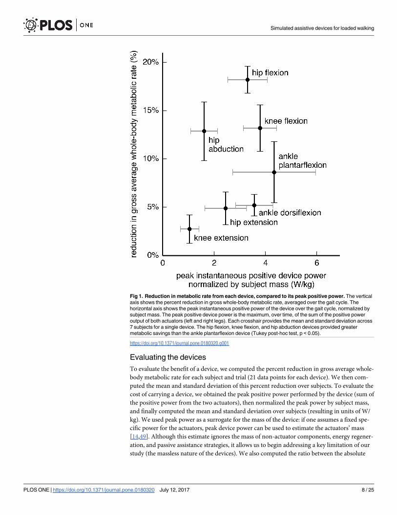
model, we used the Umberger2010MuscleMetabolicsProbe in OpenSim 3.3. To compute grossaverage whole-body metabolic rate (Fig 1), we used the following procedure: we summed the
rate of energy consumption of all muscles, added a basal rate (1.2 W/kg [35]), then integrated
the resulting whole-body rate over the gait cycle and divided by the duration of the gait cycle.
The preferred speed and stride length of some subjects caused us to have insufficient
force plate data to simulate a complete gait cycle for several trials (16 no load trials and 6
loaded trials, affecting 29% of the simulations of assistance). To estimate average whole-
body metabolic rate for simulations derived from these trials, we averaged the instantaneous
whole-body rate over half a gait cycle (exploiting the approximate mediolateral symmetry of
walking). To assess the effect of this approximation on our results, we also computed an
average rate for 5 equally spaced half-gait cycles throughout the available data. We found
negligible difference in our results between using the mean over those 5 average rates and
using a single half-gait cycle.
Validation of the metabolics model. We validated the metabolics model by comparing
its estimates of the gross average whole-body metabolic rate for the no load and loaded condi-
tions to the respective estimates from indirect calorimetry (S3 Fig). We normalized both esti-
mates by subject mass and walking speed; because the treadmill speed could only be set in
increments of 0.1 mph (0.045 m/s), subjects walked at slightly different speeds between the
overground and treadmill trials. This normalization yielded a quantity known as metabolic costof transport [48], which describes the energy required to travel a unit distance (1 W/kg/(m/s) =
1 J/m/kg). The simulations estimated the normalized gross average metabolic rate for the
loaded condition to be 5.77 W/kg/(m/s); indirect calorimetry estimated this quantity to be 5.83
W/kg/(m/s). The relative error in normalized gross average whole-body metabolic rate
between simulation and indirect calorimetry had a mean value of 11% across subjects and
both conditions. In this study, we were primarily interested in predicting the percent change
in metabolic rate between conditions and across subjects. The simulations estimated a 40%
increase in metabolic rate from the no load to the loaded condition, on average across subjects;
indirect calorimetry estimated a 48% increase. This discrepancy suggests the simulations also
underestimated the change in metabolic rate with assistance.
Simulated assistive devices for loaded walking
PLOS ONE | https://doi.org/10.1371/journal.pone.0180320 July 12, 2017 7 / 25
Evaluating the devices
To evaluate the benefit of a device, we computed the percent reduction in gross average whole-
body metabolic rate for each subject and trial (21 data points for each device). We then com-
puted the mean and standard deviation of this percent reduction over subjects. To evaluate the
cost of carrying a device, we obtained the peak positive power performed by the device (sum of
the positive power from the two actuators), then normalized the peak power by subject mass,
and finally computed the mean and standard deviation over subjects (resulting in units of W/
kg). We used peak power as a surrogate for the mass of the device: if one assumes a fixed spe-
cific power for the actuators, peak device power can be used to estimate the actuators’ mass
[14,49]. Although this estimate ignores the mass of non-actuator components, energy regener-
ation, and passive assistance strategies, it allows us to begin addressing a key limitation of our
study (the massless nature of the devices). We also computed the ratio between the absolute
Fig 1. Reduction in metabolic rate from each device, compared to its peak positive power. The vertical
axis shows the percent reduction in gross whole-body metabolic rate, averaged over the gait cycle. The
horizontal axis shows the peak instantaneous positive power of the device over the gait cycle, normalized by
subject mass. The peak positive device power is the maximum, over time, of the sum of the positive power
output of both actuators (left and right legs). Each crosshair provides the mean and standard deviation across
7 subjects for a single device. The hip flexion, knee flexion, and hip abduction devices provided greater
metabolic savings than the ankle plantarflexion device (Tukey post-hoc test, p < 0.05).
https://doi.org/10.1371/journal.pone.0180320.g001
Simulated assistive devices for loaded walking
PLOS ONE | https://doi.org/10.1371/journal.pone.0180320 July 12, 2017 8 / 25

reduction in metabolic rate (normalized by subject mass; units of W/kg) and the peak device
power; this unitless “efficiency” metric attempts to capture the preference for devices that pro-
vide large metabolic savings with a small power requirement.
In addition, we computed the average positive and average negative device power (using
the sum of power output from the two actuators, normalized by subject mass); average positive
device power can be used to estimate the battery life of untethered devices. Lastly, we com-
puted the ratio between the absolute reduction in metabolic rate and average positive device
power. This (unitless) ratio provided a metric similar to the “performance index” from Sawicki
and Ferris [50] and the “muscle–tendon efficiency” from Mooney et al. [14].
Statistical testing. To compare the devices for the metrics listed above, we employed a lin-
ear mixed model (fixed effect: device; random effect: subject) with analysis of variance (ANOVA)
tests and Tukey post-hoc pairwise tests [51]. We used a significance level of α = 0.05. The data for
the statistical analyses consisted of 49 observations (7 subjects and 7 devices); we averaged over
the 3 trials for each subject–device pair to remove hierarchical structure from our data [52]. The
statistical tests were performed with R [53–55].
Metabolic rate attributed to joint motions
To understand how the devices affected metabolic cost, we estimated the metabolic cost of
actuating individual joint motions. We define a joint motion as one direction of a degree of
freedom (e.g., hip flexion and hip extension are two joint motions). Uchida et al. [32] pre-
sented a method for partitioning the metabolic rate of biarticular muscles in the sagittal plane;
here, we generalize that method to muscles that actuate more than two degrees of freedom.
We first partitioned the instantaneous metabolic rate of each muscle i, Ėi(t), across the joint
motions g that the muscle actuated in proportion to its moment arms ri,g(t)� 0 about those
joint motions:
_Ei;gðtÞ ¼ri;gðtÞ
Pk2Gri;kðtÞ
_EiðtÞ;# ð5Þ
where Ėi,g(t) is the metabolic rate of muscle i attributed to joint motion g, and G is the set of
joint motions in the model. We obtained the instantaneous metabolic rate for a joint motion,
Ėg(t), by summing the contributions from all muscles M:
_EgðtÞ ¼X
i2M
_Ei;gðtÞ:# ð6Þ
One can recover the whole-body metabolic rate by summing Ėg(t) across all joint motions
(and adding the basal rate), though we report results for only a subset of joint motions. The set
of joint motions G did not include those from constrained degrees of freedom such as ankle
eversion.
We summed Ėg(t) over the same joint motion for both legs, then averaged this sum over the
gait cycle (using only half a gait cycle when necessary; see the “Metabolics model” section,
above), and normalized this average rate by the subject’s mass and walking speed. We then
found the mean and standard deviation of the normalized average joint motion metabolic rate
over subjects, for the no assistance simulations and for each device.
Other methods for apportioning metabolic cost to joint motions, such as using the product
of moment arm and angular velocity [33], may be equally valid. We drew qualitative conclu-
sions from this analysis that we believe would hold under different apportioning methods.
Simulated assistive devices for loaded walking
PLOS ONE | https://doi.org/10.1371/journal.pone.0180320 July 12, 2017 9 / 25

Results
Device performance
All 7 of our ideal devices significantly decreased average whole-body metabolic rate from that
of walking without assistance (Fig 1, vertical axis; p< 0.05). The hip flexion (18.2% reduction),
knee flexion (13.2%), and hip abduction (12.9%) devices provided greater savings than the
other devices tested, including the ankle plantarflexion device (8.6%; Tukey post-hoc test,
p< 0.05). The remaining 3 devices had smaller effects on metabolic rate—the knee extension
(2.8%), hip extension (4.9%), and ankle dorsiflexion (5.2%) devices.
The peak instantaneous positive power (Fig 1, horizontal axis; Table 1, column c) for the
ankle plantarflexion device (4.34 W/kg) was significantly greater than that for all other devices
except the knee flexion device (Tukey post-hoc test, p< 0.05). The hip abduction device had
the greatest ratio of metabolic savings to peak positive device power (0.63; Table 1, column f)
and the greatest ratio of metabolic savings to average positive device power (2.37; Table 1, col-
umn g).
By partitioning whole-body metabolic cost into the metabolic cost of actuating individual
joint motions (S4 Fig), we arrived at two key insights. First, most devices only partially reduced
the metabolic rate of its associated joint motion: the reduction was less than half for the hip
extension and knee extension devices. Second, many devices affected the metabolic rate attrib-
uted to joint motions other than the one actuated by the device; for example, the hip abduction
and knee flexion devices both reduced the metabolic rate attributed to hip flexion.
Optimal device torques and powers
The optimal torque for most devices differed substantially from the net joint moment of the
assisted degree of freedom (Fig 2). In some cases, the device torque exceeded the net joint
moment; this was evident for the hip flexion (Fig 2B), knee flexion (Fig 2D), and ankle dorsi-
flexion (Fig 2G) devices. In these cases, the device torque and net muscle moment opposed
each other; the “Muscle-generated joint moments” section, below, explains why this behavior
was optimal. The hip flexion and knee flexion devices were active primarily during late stance
Table 1. Device performance and power.
device reduction in gross average
whole-body metabolic rate
device power (W/kg) ratio of reduction in metabolic
rate (b) to positive device power
(a) relative
(%)
(b) absolute (W/
kg)
(c) peak
positive
(d) average
positive
(e) average
negative
(f) peak (b)/
(c)
(g) average (b)/
(d)
hip abduction 12.9 ± 3.0 0.93 ± 0.18 1.61 ± 0.52 0.42 ± 0.11 −0.27 ± 0.09 0.63 ± 0.14 2.37 ± 0.58
hip flexion 18.2 ± 1.4 1.33 ± 0.14 3.30 ± 0.77 1.04 ± 0.14 −0.29 ± 0.13 0.42 ± 0.09 1.29 ± 0.12
hip extension 4.9 ± 1.7 0.36 ± 0.14 2.45 ± 0.81 0.57 ± 0.18 −0.00 ± 0.00 0.15 ± 0.05 0.62 ± 0.10
knee flexion 13.2 ± 2.4 0.96 ± 0.18 3.79 ± 0.64 1.10 ± 0.25 −0.37 ± 0.10 0.26 ± 0.03 0.89 ± 0.10
knee extension 2.8 ± 1.5 0.21 ± 0.13 1.05 ± 0.36 0.17 ± 0.08 −0.17 ± 0.08 0.19 ± 0.07 1.18 ± 0.21
ankle
plantarflexion
8.6 ± 3.2 0.65 ± 0.28 4.34 ± 1.61 0.51 ± 0.21 −0.18 ± 0.07 0.15 ± 0.01 1.26 ± 0.08
ankle dorsiflexion 5.2 ± 1.1 0.38 ± 0.10 3.56 ± 0.73 0.60 ± 0.11 −0.25 ± 0.03 0.11 ± 0.02 0.64 ± 0.11
This table shows the (a) relative and (b) absolute reduction in gross average whole-body metabolic rate achieved by each assistive device, and each
device’s (c) peak positive, (d) average positive, and (e) average negative power. Device power quantities are evaluated over the sum of the power output of
both actuators (left and right legs). Quantities in columns (b)–(e) are normalized by subject mass. Column (f) shows the ratio of the relative reduction in
average whole-body metabolic rate to peak positive device power (i.e., column (b) over column (c)); column (g) is similar but uses average positive device
power (i.e., column (d)) in the denominator. All columns are reported as mean ± standard deviation across 7 subjects.
https://doi.org/10.1371/journal.pone.0180320.t001
Simulated assistive devices for loaded walking
PLOS ONE | https://doi.org/10.1371/journal.pone.0180320 July 12, 2017 10 / 25
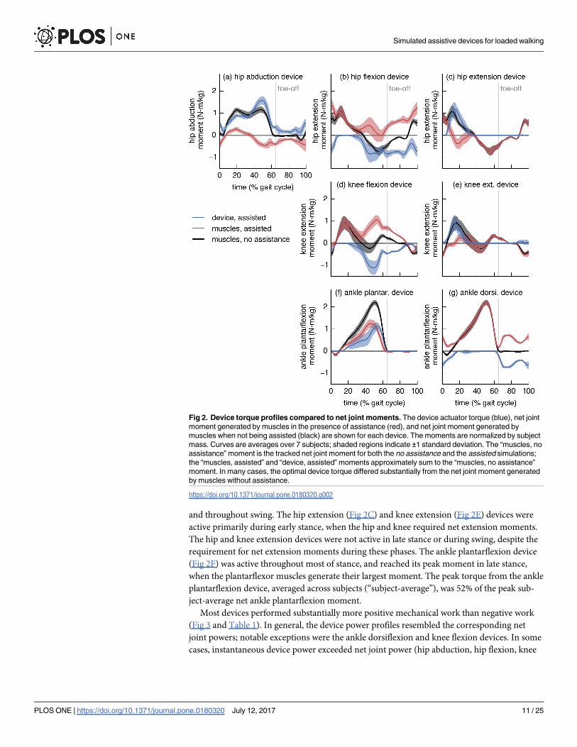
and throughout swing. The hip extension (Fig 2C) and knee extension (Fig 2E) devices were
active primarily during early stance, when the hip and knee required net extension moments.
The hip and knee extension devices were not active in late stance or during swing, despite the
requirement for net extension moments during these phases. The ankle plantarflexion device
(Fig 2F) was active throughout most of stance, and reached its peak moment in late stance,
when the plantarflexor muscles generate their largest moment. The peak torque from the ankle
plantarflexion device, averaged across subjects (“subject-average”), was 52% of the peak sub-
ject-average net ankle plantarflexion moment.
Most devices performed substantially more positive mechanical work than negative work
(Fig 3 and Table 1). In general, the device power profiles resembled the corresponding net
joint powers; notable exceptions were the ankle dorsiflexion and knee flexion devices. In some
cases, instantaneous device power exceeded net joint power (hip abduction, hip flexion, knee
Fig 2. Device torque profiles compared to net joint moments. The device actuator torque (blue), net joint
moment generated by muscles in the presence of assistance (red), and net joint moment generated by
muscles when not being assisted (black) are shown for each device. The moments are normalized by subject
mass. Curves are averages over 7 subjects; shaded regions indicate ±1 standard deviation. The “muscles, no
assistance” moment is the tracked net joint moment for both the no assistance and the assisted simulations;
the “muscles, assisted” and “device, assisted” moments approximately sum to the “muscles, no assistance”
moment. In many cases, the optimal device torque differed substantially from the net joint moment generated
by muscles without assistance.
https://doi.org/10.1371/journal.pone.0180320.g002
Simulated assistive devices for loaded walking
PLOS ONE | https://doi.org/10.1371/journal.pone.0180320 July 12, 2017 11 / 25
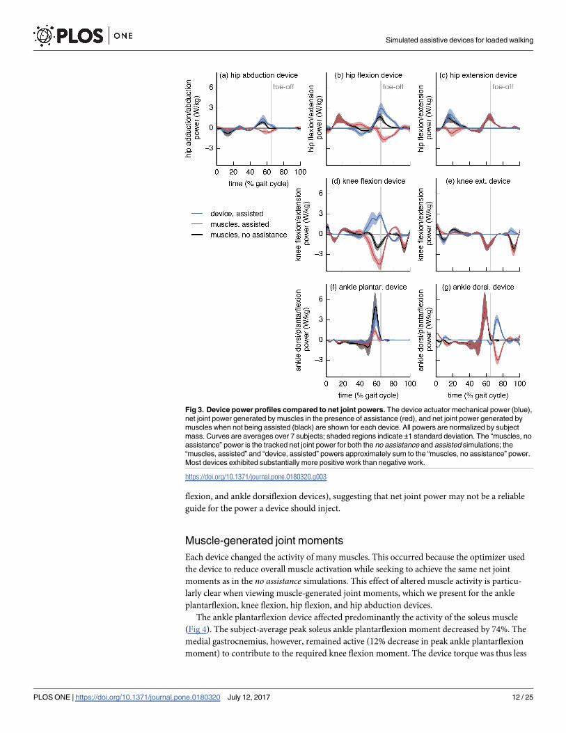
flexion, and ankle dorsiflexion devices), suggesting that net joint power may not be a reliable
guide for the power a device should inject.
Muscle-generated joint moments
Each device changed the activity of many muscles. This occurred because the optimizer used
the device to reduce overall muscle activation while seeking to achieve the same net joint
moments as in the no assistance simulations. This effect of altered muscle activity is particu-
larly clear when viewing muscle-generated joint moments, which we present for the ankle
plantarflexion, knee flexion, hip flexion, and hip abduction devices.
The ankle plantarflexion device affected predominantly the activity of the soleus muscle
(Fig 4). The subject-average peak soleus ankle plantarflexion moment decreased by 74%. The
medial gastrocnemius, however, remained active (12% decrease in peak ankle plantarflexion
moment) to contribute to the required knee flexion moment. The device torque was thus less
Fig 3. Device power profiles compared to net joint powers. The device actuator mechanical power (blue),
net joint power generated by muscles in the presence of assistance (red), and net joint power generated by
muscles when not being assisted (black) are shown for each device. All powers are normalized by subject
mass. Curves are averages over 7 subjects; shaded regions indicate ±1 standard deviation. The “muscles, no
assistance” power is the tracked net joint power for both the no assistance and assisted simulations; the
“muscles, assisted” and “device, assisted” powers approximately sum to the “muscles, no assistance” power.
Most devices exhibited substantially more positive work than negative work.
https://doi.org/10.1371/journal.pone.0180320.g003
Simulated assistive devices for loaded walking
PLOS ONE | https://doi.org/10.1371/journal.pone.0180320 July 12, 2017 12 / 25
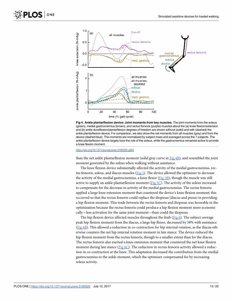
than the net ankle plantarflexion moment (solid gray curve in Fig 4B), and resembled the joint
moment generated by the soleus when walking without assistance.
The knee flexion device substantially affected the activity of the medial gastrocnemius, rec-
tus femoris, soleus, and iliacus muscles (Fig 5). The device allowed the optimizer to decrease
the activity of the medial gastrocnemius, a knee flexor (Fig 5B), though the muscle was still
active to supply an ankle plantarflexion moment (Fig 5C). The activity of the soleus increased
to compensate for the decrease in activity of the medial gastrocnemius. The rectus femoris
applied a large knee extension moment that countered the device’s knee flexion moment; this
occurred so that the rectus femoris could replace the iliopsoas (iliacus and psoas) in providing
a hip flexion moment. This trade between the rectus femoris and iliopsoas was favorable in the
optimization because the rectus femoris could produce a hip flexion moment more economi-
cally—less activation for the same joint moment—than could the iliopsoas.
The hip flexion device affected muscles throughout the limb (Fig 6). The subject-average
peak hip flexion moment from the iliacus, a large hip flexor, decreased by 58% with assistance
(Fig 6B). This allowed a reduction in co-contraction for hip internal rotation, as the iliacus oth-
erwise counters the net hip internal rotation moment in late stance. The device reduced the
hip flexion moment from the rectus femoris, though to a smaller extent than for the iliacus.
The rectus femoris also exerted a knee extension moment that countered the net knee flexion
moment during late stance (Fig 6C). The reduction in rectus femoris activity allowed a reduc-
tion in co-contraction at the knee. This adaptation decreased the contribution from the medial
gastrocnemius to the ankle moment, which the optimizer compensated for by increasing
soleus activity.
Fig 4. Ankle plantarflexion device: Joint moments from key muscles. The joint moments from the soleus
(green), medial gastrocnemius (brown), and rectus femoris (purple) muscles about the (a) knee flexion/extension
and (b) ankle dorsiflexion/plantarflexion degrees of freedom are shown without (solid) and with (dashed) the
ankle plantarflexion device. For comparison, we also show the net moments from all muscles (gray) and from the
device (dashed blue). The moments are normalized by subject mass and averaged across the 7 subjects. The
ankle plantarflexion device largely bore the role of the soleus, while the gastrocnemius remained active to provide
a knee flexion moment.
https://doi.org/10.1371/journal.pone.0180320.g004
Simulated assistive devices for loaded walking
PLOS ONE | https://doi.org/10.1371/journal.pone.0180320 July 12, 2017 13 / 25

The hip abduction device, like the hip flexion device, affected muscles throughout the limb
(Fig 7). Without assistance, a large fraction of the hip abduction moment came from the ante-
rior portion of the gluteus medius (Fig 7B). The device allowed the optimizer to reduce the glu-
teus medius hip abduction moment, though the reduction was larger in late stance than in
early stance, as the muscle was still needed to generate a hip internal rotation moment in early
stance (Fig 7A). In late stance, the adductor longus countered the device to provide a hip flex-
ion moment (Fig 7C) more economically than could the iliopsoas. This adaptation allowed the
iliacus to reduce its opposition to the net hip internal rotation moment (Fig 7A), resulting in
an overall decrease in muscle activation despite the increase in adductor longus activity. To
achieve the tracked net hip abduction moment in late stance, the device torque exceeded the
net hip abduction moment. The hip abduction device allowed the adductor longus to replace
some of the rectus femoris’ hip flexion moment, and thus also decreased co-contraction at the
Fig 5. Knee flexion device: Joint moments from key muscles. The moments from the soleus (green),
medial gastrocnemius (brown), rectus femoris (purple), and iliacus (pink) muscles about the (a) hip flexion/
extension, (b) knee flexion/extension, and (c) ankle dorsiflexion/plantarflexion degrees of freedom. Muscle
moments are shown without (solid) and with (dashed) the knee flexion device. For comparison, we also show
the net moments from all muscles (gray) and from the device (dashed blue). The moments are normalized by
subject mass and averaged across the 7 subjects. The device replaced much of the knee moment ordinarily
generated by the gastrocnemius, which resulted in a decrease in gastrocnemius activity but an increase in the
demand on the soleus to generate the plantarflexion moment. The rectus femoris countered the device to
replace the iliacus and psoas (not shown) in providing a hip flexion moment.
https://doi.org/10.1371/journal.pone.0180320.g005
Simulated assistive devices for loaded walking
PLOS ONE | https://doi.org/10.1371/journal.pone.0180320 July 12, 2017 14 / 25
knee. As with the hip flexion device, this decrease in co-contraction at the knee resulted in an
increase in soleus activity to achieve the necessary net ankle moment.
Fig 6. Hip flexion device: Joint moments from key muscles. The moments from the soleus (green),
medial gastrocnemius (brown), rectus femoris (purple), and iliacus (pink) muscles about the (a) hip external/
internal rotation, (b) hip flexion/extension, (c) knee flexion/extension, and (d) ankle dorsiflexion/plantarflexion
degrees of freedom are shown without (solid) and with (dashed) the hip flexion device. For comparison, we
also show the net moments from all muscles (gray) and from the device (dashed blue). The moments are
normalized by subject mass and averaged across the 7 subjects. The device mostly replaced the iliacus—
which normally counters the net hip internal rotation moment—and psoas (not shown). The device also
partially replaced the rectus femoris, allowing for a decrease in co-contraction at the knee. The soleus
replaced some of the ankle moment that had been provided by the gastrocnemius.
https://doi.org/10.1371/journal.pone.0180320.g006
Simulated assistive devices for loaded walking
PLOS ONE | https://doi.org/10.1371/journal.pone.0180320 July 12, 2017 15 / 25
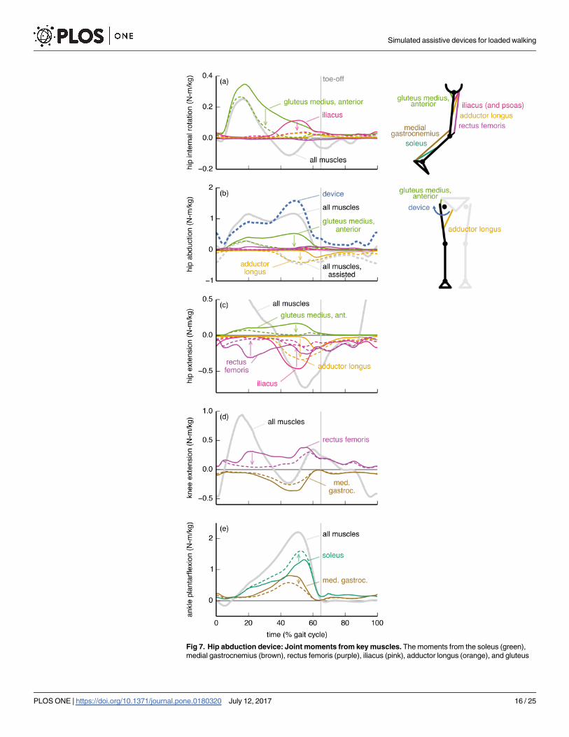
Fig 7. Hip abduction device: Joint moments from key muscles. The moments from the soleus (green),
medial gastrocnemius (brown), rectus femoris (purple), iliacus (pink), adductor longus (orange), and gluteus
Simulated assistive devices for loaded walking
PLOS ONE | https://doi.org/10.1371/journal.pone.0180320 July 12, 2017 16 / 25
Discussion
We simulated 7 hypothetical ideal devices and found that three of them yielded greater meta-
bolic savings than our simulated ankle plantarflexion device (Fig 1). This is noteworthy given
the current popularity of experimental ankle plantarflexion devices [9,13,14,22,50,56]. Because
we directly estimated the metabolic savings achieved with different device locations, our study
is an important step away from relying on indirect and coarse measures like positive joint
power to decide where to assist. Part of the focus on ankle devices comes from the ankle’s large
share of positive power output in walking [46,57,58]. Our results suggest that a device at a joint
with high positive work, such as the ankle in loaded walking [46], does not necessarily yield
the highest metabolic savings [19]. Nevertheless, all the devices we explored warrant consider-
ation from device designers: reducing metabolic rate by even 5%—as is possible with a hip
extension device [59]—is tantamount to removing 4 kg from a torso load [45] and would
markedly help load carriers.
Hip abduction device
The metabolic savings from the hip abduction device were surprisingly large, given that walk-
ing is a predominantly sagittal-plane motion. Hip abduction has a low net joint power (Fig 3),
so it is unlikely that a joint-level power analysis would produce interest in assisting hip abduc-
tion. However, muscles consume energy even when not performing work (e.g., when contract-
ing isometrically), and hip abduction has a substantial metabolic rate during loaded walking
(S4 Fig).
The hip abduction device is even more attractive when considering its relatively low power
requirements: this device had the greatest ratio of metabolic savings to peak instantaneous posi-
tive device power and the greatest ratio of metabolic savings to average positive device power
(Table 1). These metrics can be used to estimate, respectively, the increase in metabolic cost from
carrying the mass of a device and the duration for which an untethered device could operate (that
is, battery life); see the “Evaluating the devices” section, above, for an explanation. Notably, the
hip abduction device performed similar amounts of positive and negative mechanical work (the
ratio of average positive to average negative power was 1.6; Table 1), suggesting that a hip abduc-
tion device could incorporate passive components to reduce its power consumption. Thus, a hip
abduction device could weigh less and operate for longer than devices placed elsewhere, especially
since its mass could be located more proximally than that of devices that assist the ankle or knee
[49]. Given these benefits, we suggest that experimentalists further pursue the feasibility and per-
formance of hip abduction devices for reducing metabolic cost.
Optimal device torques and the underlying musculature
To maximize metabolic savings, device torque profiles should generally differ from the net
joint moments; this is because human joints are driven by muscles and not simple torque actu-
ators. In particular, the optimal device torque profiles and the corresponding changes in
medius (anterior portion; light green) muscles about the (a) hip external/internal rotation, (b) hip adduction/
abduction, (c) hip flexion/extension, (d) knee flexion/extension, and (e) ankle dorsiflexion/plantarflexion
degrees of freedom are shown without (solid) and with (dashed) the hip flexion device. For comparison, we
also show the net moments from all muscles (gray) and from the device (dashed blue). The moments are
normalized by subject mass and averaged across the 7 subjects. Without assistance, a large fraction of the
hip abduction moment was generated by the gluteus medius. The device replaced the gluteus medius in late
stance, but the gluteus medius remained active in early stance to provide a hip internal rotation moment. In
late stance, the adductor longus countered the device to provide hip flexion more economically than could the
iliacus or psoas (not shown).
https://doi.org/10.1371/journal.pone.0180320.g007
Simulated assistive devices for loaded walking
PLOS ONE | https://doi.org/10.1371/journal.pone.0180320 July 12, 2017 17 / 25
muscle activity we observed were shaped by the presence of muscles that actuate multiple
degrees of freedom (this includes biarticular muscles as well as muscles crossing a single joint
that has multiple degrees of freedom). We found that a device torque may be less than the cor-
responding net joint moment if an assisted muscle also contributes to the required net joint
moment about another joint motion (e.g., ankle plantarflexion device and medial gastrocne-
mius [60,61]; Fig 4A). Conversely, if a muscle crossing an assisted joint motion generates an
undesired moment about another joint motion, then the device can largely take over for this
muscle (e.g., hip flexion device and iliacus; Fig 6A). Surprisingly, device torques may also
exceed net joint moments to allow antagonistic muscles to take over for less economical mus-
cles (in terms of joint moment per unit activation) at other degrees of freedom (e.g., knee flex-
ion device and rectus femoris; Fig 5B). Designers can use our optimal device torque profiles as
guidelines for choosing the timing and magnitude of assistive torques that take into account
musculotendon dynamics, musculoskeletal geometry, and muscle energetics. Additionally,
simulated optimal torque profiles can narrow the range of experimental conditions required to
find a device’s optimal performance [62]. Our results may also help understand how and why
muscle activation patterns change in response to applied torques.
Devices can affect the activity of muscles that do not span the assisted degree of freedom—for
example, the knee flexion, hip flexion, and hip abduction devices all affected soleus activity. There
is experimental support for this observation: Lenzi et al. [63] created a hip flexion/extension device
that reduced medial gastrocnemius activity in walking. To exploit coupling across degrees of free-
dom, we suggest that experimentalists devote more attention to devices that actuate multiple
degrees of freedom [17,18]. For example, unlike the strictly ankle plantarflexion device we pre-
sented (Fig 4), a device that provides both ankle plantarflexion and knee flexion moments may
decrease activity of the gastrocnemius substantially. Indeed, Quinlivan et al. [18] have shown large
metabolic savings with a device that simultaneously assists ankle plantarflexion and hip flexion.
Study limitations
This study has a number of limitations that require consideration when interpreting our
results. We assumed that subjects walked with the same kinematics (and ground reaction
forces) when assisted. Some experimental exoskeleton studies report relatively small changes
in kinematics or joint moments with assistance [9,17,20,25,64,65], while others report much
larger changes [12,18,61,63]. Our simulations also do not capture the effects of training proto-
cols or long-term adaptation to the devices, which are important considerations during experi-
mental testing of devices [20,21].
We were interested in devices that minimize metabolic cost, yet we minimized the sum of
squared muscle activations. We chose this objective function because minimizing activations is
more computationally tractable, activation is a dominant variable in the metabolics model [35],
and muscle activity correlates well with metabolic cost [25,32,66]. Simulations that allow kine-
matics to adapt and that minimize metabolic cost directly may reveal assistance strategies that
are even more metabolically beneficial than those we presented, and could alter the ranking of
the devices’ metabolic savings. On the other hand, it may be desirable in some situations to
retain normal kinematics, as altered kinematics could have negative side effects (e.g., increased
joint loading).
Our simulations produced muscle activity in the medial hamstrings, biceps femoris short
head, and gastrocnemius (early swing) that was not present in our electromyography measure-
ments (S2 Fig). This excess activity resulted from excessive passive force generated by the knee
extensor muscles, a known problem with some musculoskeletal models [47]. We found that
removing the knee extensor passive force in the model reduced the metabolic savings of the
Simulated assistive devices for loaded walking
PLOS ONE | https://doi.org/10.1371/journal.pone.0180320 July 12, 2017 18 / 25
hip flexion device (from 18 to 15%) and knee flexion device (from 13 to 8%). Removing passive
knee extensor force did not substantially affect the metabolic savings of the hip abduction or
ankle plantarflexion devices, did not change the overall nature of the muscle adaptations, and
does not affect the main conclusions of our study. Our simulations also produced excessive
tibialis anterior activity in swing; as a result, the reported metabolic savings for the ankle dorsi-
flexion device are likely inflated.
Several factors influence the uncertainty in our predictions for changes in metabolic cost.
Our simulations estimated a 40% increase in cost with load while indirect calorimetry during
our experiments estimated a 48% increase, suggesting that our simulations also underesti-
mated reductions in cost with assistance. Another potential source of underestimation in met-
abolic rate is our constraint that kinematics could not change between the no assistance and
assisted simulations. Still, our predicted metabolic savings seem reasonable in comparison to
published experimental studies. Our ideal massless ankle plantarflexion device resulted in an
8.7% reduction in gross metabolic rate (approx. 11% in net metabolic rate), which is greater
than the 8.0% reduction in net metabolic rate reported when using an ankle plantarflexion
device during a loaded walking experiment [14]. Ding et al. [59] achieved a 5.7–8.5% reduction
in net metabolic rate (compared to wearing the device unpowered) with a hip extension device
for loaded walking, which is similar to our reduction of 4.9% in gross metabolic rate. However,
our simulations produced device torques and changes in muscle activity that were much
greater than what experimentalists have observed [9,12,16,17,59], so it is expected that our pre-
dicted reductions should exceed experimental reductions.
Considering these limitations, the value of this study is in the ranking of the metabolic sav-
ings and power requirements of the devices, and the qualitative insights we obtained about
how muscle activity may change with assistance. Without accounting for kinematic adapta-
tion, neural constraints, training protocols, and other practical matters, it is unreasonable to
expect a close quantitative match in metabolic reductions, device torques, and muscle activity
adaptations between experiments and our simulations.
Summary of insights
Our experience using muscle-driven simulations to study uniarticular assistance strategies
have led to the following qualitative insights:
1. Most experimentalists have focused on assisting the ankle, yet assisting the hip or knee has
the potential to lead to greater metabolic savings than assisting the ankle.
2. Assisting hip abduction may be an effective strategy to reduce metabolic cost, yet this strat-
egy is largely unexplored.
3. Devices that assist one joint can affect the activity of muscles that do not span that joint.
4. The activity of an assisted muscle may remain if the muscle provides a beneficial action at
an unassisted degree of freedom.
5. Joint-level moment and power analyses may not sufficiently explain the relative perfor-
mance of a device because optimal device torques sometimes differ from the net joint
moments and device performance is sensitive to details of the device torque profiles.
Future work
In light of the study limitations, future studies should employ simulation approaches that
examine how changes in kinematics may affect the performance of a device (“predictive
Simulated assistive devices for loaded walking
PLOS ONE | https://doi.org/10.1371/journal.pone.0180320 July 12, 2017 19 / 25

simulation”) [29,67]. To obtain more realistic results, predictive simulations could model non-
ideal aspects such as device mass and actuator torque and power limits. To make stronger con-
clusions from simulations (e.g., discovering the maximum achievable savings), studies should
be performed with larger sample sizes. Simulation experts and experimentalists should work
together to improve the accuracy of simulated exoskeletal assistance through comparison of
simulated muscle activations with experimental recordings of muscle activity and further test-
ing of changes in metabolic cost with load and assistance. Device designers must tackle issues
we could not; for example, a hip abduction device will require effective means of transmitting
forces to the skeleton.
Future studies should look beyond metabolic cost, as optimizing a device to minimize solely
metabolic cost may worsen muscle fatigue, joint loading, and joint stability [10]. For example,
our knee flexion device caused very high rectus femoris activation during lengthening, which
is likely to cause fatigue [68]. Our hip flexion device decreased co-contraction at the knee,
which could decrease knee stability [69]. Furthermore, devices that explicitly optimize the
ratio of metabolic savings to device power may achieve greater values for this ratio than did
our hip abduction device. Novel methods that allow flexible objective functions could discover
such devices, and could also optimize other metrics that would improve safety, comfort, and
performance.
Conclusions
In this study, we used musculoskeletal simulation to evaluate how 7 hypothetical, ideal, bilat-
eral assistive devices affected muscle activity and metabolic cost when walking with heavy
loads. This work provides a foundation for understanding the musculoskeletal factors that
may affect device performance. We also provided suggestions to device designers, which can
serve as a springboard for deciding which devices to create next. In particular, we are excited
for designers to create hip abduction devices that incorporate passive components, and to
explore devices that actuate multiple degrees of freedom.
The insights we gained in this study relied on the use of musculoskeletal and metabolics
models. These models can reveal insights that are difficult to discover via experiments alone;
for example, we found that devices may substantially affect the metabolic rate of joint motions
other than the one being assisted. Our findings support use of musculoskeletal modeling and
simulation to predict how hypothetical devices may perform and to understand the perfor-
mance of actual devices [22,28,60]. This work complements experiments, which are necessary
to test the accuracy of the predictions made by simulations, improve musculoskeletal and
metabolics models, and solve the practical challenges we ignored. We invite other researchers
to use our data and code (freely available at https://simtk.org/home/assistloadwalk) to build
upon our work.
Supporting information
S1 Table. Demographics of subjects.
(PDF)
S1 Fig. Joint angles and net joint moments for the no load and loaded conditions. Joint
angles (left) and net joint moments from muscles (normalized by subject mass; right) are
shown for the simulations of the no load (green) and loaded (black) conditions for the hip
adduction/abduction (top), hip flexion/extension, knee flexion/extension, and ankle dorsiflex-
ion/plantarflexion (bottom) degrees of freedom. Curves are averages over 7 subjects; shaded
regions indicate ±1 standard deviation. The vertical lines indicate average toe-off time for the
Simulated assistive devices for loaded walking
PLOS ONE | https://doi.org/10.1371/journal.pone.0180320 July 12, 2017 20 / 25

two conditions.
(TIF)
S2 Fig. Simulated and measured muscle activity for the no load and loaded conditions.
Each graph compares the electromyography measurements (black) of the muscle listed on the
left to the simulated activation (unitless, between 0 and 1; blue, green, red) of the relevant mus-
cles in the model. Electromyography measurements were first band-pass filtered (50–500 Hz),
then rectified, and finally low-pass filtered (7.5 Hz). We normalized the electromyography
data by the maximum value observed across all four experimental conditions for a given sub-
ject and sensor. Curves are averages over 7 subjects; shaded regions indicate ±1 standard devi-
ation. Electromyography data were collected on the right leg, but activation is averaged over
both the left and right legs of each subject. (posterior, intermed., and anterior correspond to
muscle–tendon units 3, 2 and 1, respectively, in the model; semimem.: semimembranosus;
semiten.: semitendinosus).
(TIF)
S3 Fig. Validation of metabolics estimates. Graph (a) shows gross average whole-body meta-
bolic rate normalized by subject mass and walking speed (W/kg/(m/s)) for the no load and
loaded conditions, obtained with indirect calorimetry (triangles, dashed lines) and with the
simulations (circles, solid lines). Each triangle comes from the last minute of 7 minutes of
treadmill walking for a single subject and condition. Each circle is obtained by averaging across
3 trials for a single subject and condition; error bars provide the standard deviation across
these 3 trials. Each color represents a single subject. Graph (b) shows the same data as (a) but
displayed as simulation versus indirect calorimetry, with a linear regression fit (dashed gray)
and a y = x line (solid gray). We refer to the simulations of the loaded condition as no assis-tance, as they are the baseline for the simulations of assistance. The simulations appear to
underestimate the change in metabolic rate between conditions.
(TIF)
S4 Fig. Metabolic rate attributed to joint motions, without and with assistance, for each
device. Most devices only partially reduced the metabolic rate of its associated joint motion.
Each graph shows the metabolic rate (horizontal axis) for a single device that we attributed to 8
joint motions (one direction of a degree of freedom; vertical axis), summed over the same joint
motion for both legs and averaged over the gait cycle, without (white) and with (gray) assis-
tance. The metabolic rate we attributed to a joint motion comes from all the muscles that actu-
ate the joint motion, apportioned according to the muscles’ moment arms; see Eq (6). Dots to
the left of the bars denote the joint motion being assisted by the device. Not all joint motions
are shown (namely, hip rotation). The length of each bar indicates an average over 7 subjects;
whiskers indicate ±1 standard deviation.
(TIF)
Acknowledgments
We thank Ben Rhyne for helping with the motion capture experiments, as well as the subjects
who volunteered to participate. All graphs were generated with the python package matplotlib[70]. To carry out the OpenSim workflow over subjects and trials, we employed the python
package doit (pydoit.org).
Author Contributions
Conceptualization: CLD AS TKU JLH SLD.
Simulated assistive devices for loaded walking
PLOS ONE | https://doi.org/10.1371/journal.pone.0180320 July 12, 2017 21 / 25
Data curation: CLD AS.
Formal analysis: CLD.
Funding acquisition: SLD.
Investigation: CLD AS.
Methodology: CLD TKU.
Software: CLD TKU.
Supervision: JLH SLD.
Validation: CLD AS TKU JLH.
Visualization: CLD AS TKU JLH SLD.
Writing – original draft: CLD.
Writing – review & editing: CLD AS TKU JLH SLD.
References1. Au S, Herr H. Powered ankle-foot prosthesis. IEEE Robot Autom Mag. 2008; 15: 52–59. https://doi.org/
10.1109/MRA.2008.927697
2. Ferris DP. The exoskeletons are here. J Neuroeng Rehabil. 2009; 6: 17. https://doi.org/10.1186/1743-
0003-6-17 PMID: 19508711
3. Strickland E. Good-bye, wheelchair. IEEE Spectr. 2012; 49: 30–32. https://doi.org/10.1109/MSPEC.
2012.6117830
4. Riener R, Lunenburger L, Maier I, Colombo G, Dietz V. Locomotor training in subjects with sensori-
motor deficits: an overview of the robotic gait orthosis lokomat. J Healthc Eng. 2010; 1: 197–216.
https://doi.org/10.1260/2040-2295.1.2.197
5. Ruby BC, Leadbetter GW III, Armstrong DW, Gaskill SE. Wildland firefighter load carriage: effects on
transit time and physiological responses during simulated escape to safety zone. Int J Wildl Fire. 2003;
12: 111–116. https://doi.org/10.1071/WF02025
6. van Vuuren BJ, Becker PJ, van Heerden HJ, Zinzen E, Meeusen R. Lower back problems and occupa-
tional risk factors in a South African steel industry. Am J Ind Med. 2005; 47: 451–457. https://doi.org/10.
1002/ajim.20164 PMID: 15828071
7. Knapik JJ, Reynolds KL, Harman E. Soldier load carriage: historical, physiological, biomechanical, and
medical aspects. Mil Med. 2004; 169: 45–56. PMID: 14964502
8. Yagn N. Apparatus for facilitating walking, running, and jumping. United States; 440684, 1890.
9. Collins SH, Wiggin MB, Sawicki GS. Reducing the energy cost of human walking using an unpowered
exoskeleton. Nature. 2015; 522: 212–215. https://doi.org/10.1038/nature14288 PMID: 25830889
10. Dollar AM, Herr H. Lower extremity exoskeletons and active orthoses: challenges and state-of-the-art.
IEEE Trans Robot. 2008; 24: 144–158. https://doi.org/10.1109/TRO.2008.915453
11. Gregorczyk KN, Hasselquist L, Schiffman JM, Bensel CK, Obusek JP, Gutekunst DJ. Effects of a
lower-body exoskeleton device on metabolic cost and gait biomechanics during load carriage. Ergo-
nomics. 2010; 53: 1263–1275. https://doi.org/10.1080/00140139.2010.512982 PMID: 20865609
12. Galle S, Malcolm P, Derave W, De Clercq D. Adaptation to walking with an exoskeleton that assists
ankle extension. Gait Posture. 2013; 38: 495–499. https://doi.org/10.1016/j.gaitpost.2013.01.029
PMID: 23465319
13. Malcolm P, Derave W, Galle S, De Clercq D. A simple exoskeleton that assists plantarflexion can
reduce the metabolic cost of human walking. PLoS ONE. 2013; 8: e56137. https://doi.org/10.1371/
journal.pone.0056137 PMID: 23418524
14. Mooney LM, Rouse EJ, Herr HM. Autonomous exoskeleton reduces metabolic cost of human walking
during load carriage. J Neuroeng Rehabil. 2014; 11: 80. https://doi.org/10.1186/1743-0003-11-80
PMID: 24885527
15. Mooney LM, Rouse EJ, Herr HM. Autonomous exoskeleton reduces metabolic cost of human walking.
J Neuroeng Rehabil. 2014; 11: 151. https://doi.org/10.1186/1743-0003-11-151 PMID: 25367552
Simulated assistive devices for loaded walking
PLOS ONE | https://doi.org/10.1371/journal.pone.0180320 July 12, 2017 22 / 25
16. Mooney LM, Herr HM. Biomechanical walking mechanisms underlying the metabolic reduction caused
by an autonomous exoskeleton. J Neuroeng Rehabil. 2016; 13: 4. https://doi.org/10.1186/s12984-016-
0111-3 PMID: 26817449
17. Panizzolo FA, Galiana I, Asbeck AT, Siviy C, Schmidt K, Holt KG, et al. A biologically-inspired multi-joint
soft exosuit that can reduce the energy cost of loaded walking. J Neuroeng Rehabil. 2016; 13: 43.
https://doi.org/10.1186/s12984-016-0150-9 PMID: 27169361
18. Quinlivan BT, Sangjun L, Malcolm P, Rossi DM, Grimmer M, Siviy C, et al. Assistance magnitude ver-
sus metabolic cost reductions for a tethered multiarticular soft exosuit. Sci Robot. 2017; 2: eaah4416.
https://doi.org/10.1126/scirobotics.aah4416
19. Seo K, Lee J, Lee Y, Ha T, Shim Y. Fully autonomous hip exoskeleton saves metabolic cost of walking.
Proceedings of the 2016 IEEE International Conference on Robotics and Automation. Stockholm, Swe-
den; 2016. pp. 4628–4635. 10.1109/ICRA.2016.7487663
20. Gordon KE, Ferris DP. Learning to walk with a robotic ankle exoskeleton. J Biomech. 2007; 40: 2636–
2644. https://doi.org/10.1016/j.jbiomech.2006.12.006 PMID: 17275829
21. Selinger JC, O’Connor SM, Wong JD, Donelan JM. Humans can continuously optimize energetic cost
during walking. Curr Biol. 2015; 25: 1–5. https://doi.org/10.1016/j.cub.2014.10.064 PMID: 25532895
22. Farris DJ, Hicks JL, Delp SL, Sawicki GS. Musculoskeletal modelling deconstructs the paradoxical
effects of elastic ankle exoskeletons on plantar-flexor mechanics and energetics during hopping. J Exp
Biol. 2014; 217: 4018–4028. https://doi.org/10.1242/jeb.107656 PMID: 25278469
23. Jackson RW, Dembia CL, Delp SL, Collins SH. Muscle-tendon mechanics explain unexpected effects
of exoskeleton assistance on metabolic rate during walking. J Exp Biol. 2017; https://doi.org/10.1242/
jeb.150011 PMID: 28341663
24. Malcolm P, Quesada RE, Caputo JM, Collins SH. The influence of push-off timing in a robotic ankle-foot
prosthesis on the energetics and mechanics of walking. J Neuroeng Rehabil. 2015; 12: 21. https://doi.
org/10.1186/s12984-015-0014-8 PMID: 25889201
25. Jackson RW, Collins SH. An experimental comparison of the relative benefits of work and torque assis-
tance in ankle exoskeletons. J Appl Physiol. 2015; 119: 541–557. https://doi.org/10.1152/japplphysiol.
01133.2014 PMID: 26159764
26. Ferris DP, Sawicki GS, Daley MA. A physiologist’s perspective on robotic exoskeletons for human loco-
motion. Int J Humanoid Robot. 2007; 4: 507–528. https://doi.org/10.1142/S0219843607001138 PMID:
18185840
27. Umberger BR. Stance and swing phase costs in human walking. J R Soc Interface. 2010; 7: 1329–
1340. https://doi.org/10.1098/rsif.2010.0084 PMID: 20356877
28. Sawicki GS, Khan NS. A simple model to estimate plantarflexor muscle-tendon mechanics and energet-
ics during walking with elastic ankle exoskeletons. IEEE Trans Biomed Eng. 2016; 63: 914–923. https://
doi.org/10.1109/TBME.2015.2491224 PMID: 26485350
29. Handford ML, Srinivasan M. Robotic lower limb prosthesis design through simultaneous computer opti-
mizations of human and prosthesis costs. Sci Rep. 2016; 6: 19983. https://doi.org/10.1038/srep19983
PMID: 26857747
30. Steele KM, van der Krogt MM, Schwartz MH, Delp SL. How much muscle strength is required to walk in
a crouch gait? J Biomech. 2012; 45: 2564–2569. https://doi.org/10.1016/j.jbiomech.2012.07.028 PMID:
22959837
31. Afschrift M, De Groote F, De Schutter J, Jonkers I. The effect of muscle weakness on the capability gap
during gross motor function: a simulation study supporting design criteria for exoskeletons of the lower
limb. Biomed Eng Online. 2014; 13: 111. https://doi.org/10.1186/1475-925X-13-111 PMID: 25092209
32. Uchida TK, Hicks JL, Dembia CL, Delp SL. Stretching your energetic budget: how tendon compliance
affects the metabolic cost of running. PLoS ONE. 2016; 11: e0150378. https://doi.org/10.1371/journal.
pone.0150378 PMID: 26930416
33. Uchida TK, Seth A, Pouya S, Dembia CL, Hicks JL, Delp SL. Simulating ideal assistive devices to
reduce the metabolic cost of running. PLoS ONE. 2016; 11: e0163417. https://doi.org/10.1371/journal.
pone.0163417 PMID: 27656901
34. Markowitz J, Herr H. Human leg model predicts muscle forces, states, and energetics during walking.
PLoS Comput Biol. 2016; 12: e1004912. https://doi.org/10.1371/journal.pcbi.1004912 PMID: 27175486
35. Umberger BR, Gerritsen KGM, Martin PE. A model of human muscle energy expenditure. Comput
Methods Biomech Biomed Engin. 2003; 6: 99–111. https://doi.org/10.1080/1025584031000091678
PMID: 12745424
36. Miller RH. A comparison of muscle energy models for simulating human walking in three dimensions. J
Biomech. 2014; 47: 1373–1381. https://doi.org/10.1016/j.jbiomech.2014.01.049 PMID: 24581797
Simulated assistive devices for loaded walking
PLOS ONE | https://doi.org/10.1371/journal.pone.0180320 July 12, 2017 23 / 25
37. Delp SL, Anderson FC, Arnold AS, Loan P, Habib A, John CT, et al. OpenSim: open-source software to
create and analyze dynamic simulations of movement. IEEE Trans Biomed Eng. 2007; 54: 1940–1950.
https://doi.org/10.1109/TBME.2007.901024 PMID: 18018689
38. Seth A, Sherman M, Reinbolt JA, Delp SL. OpenSim: a musculoskeletal modeling and simulation frame-
work for in silico investigations and exchange. Procedia IUTAM. 2011; 2: 212–232. https://doi.org/10.
1016/j.piutam.2011.04.021 PMID: 25893160
39. Millard M, Uchida T, Seth A, Delp SL. Flexing computational muscle: modeling and simulation of muscu-
lotendon dynamics. J Biomech Eng. 2013; 135: 021005. https://doi.org/10.1115/1.4023390 PMID:
23445050
40. Lusk G. Animal calorimetry, the twenty-fourth paper, analysis of the oxidation of mixtures of carbohy-
drate and fat, a correction. J Biol Chem. 1924; 59: 41–42.
41. Rajagopal A, Dembia C, DeMers M, Delp D, Hicks J, Delp S. Full body musculoskeletal model for mus-
cle-driven simulation of human gait. IEEE Trans Biomed Eng. 2016; 63: 2068–2079. https://doi.org/10.
1109/TBME.2016.2586891 PMID: 27392337
42. Handsfield GG, Meyer CH, Hart JM, Abel MF, Blemker SS. Relationships of 35 lower limb muscles to
height and body mass quantified using MRI. J Biomech. 2014; 47: 631–638. https://doi.org/10.1016/j.
jbiomech.2013.12.002 PMID: 24368144
43. Thelen DG, Anderson FC, Delp SL. Generating dynamic simulations of movement using computed
muscle control. J Biomech. 2003; 36: 321–328. https://doi.org/10.1016/S0021-9290(02)00432-3 PMID:
12594980
44. Neumann DA, Hase AD. An electromyographic analysis of the hip abductors during load carriage: impli-
cations for hip joint protection. J Orthop Sport Phys Ther. 1994; 19: 296–304. https://doi.org/10.2519/
jospt.1994.19.5.296 PMID: 8199623
45. Silder A, Delp SL, Besier T. Men and women adopt similar walking mechanics and muscle activation
patterns during load carriage. J Biomech. 2013; 46: 2522–2528. https://doi.org/10.1016/j.jbiomech.
2013.06.020 PMID: 23968555
46. Huang T-WP, Kuo AD. Mechanics and energetics of load carriage during human walking. J Exp Biol.
2014; 217: 605–613. https://doi.org/10.1242/jeb.091587 PMID: 24198268
47. Hicks JL, Uchida TK, Seth A, Rajagopal A, Delp SL. Is my model good enough? Best practices for verifi-
cation and validation of musculoskeletal models and simulations of human movement. J Biomech Eng.
2015; 137: 020905. https://doi.org/10.1115/1.4029304 PMID: 25474098
48. Alexander RM. Models and the scaling of energy costs for locomotion. J Exp Biol. 2005; 208: 1645–
1652. https://doi.org/10.1242/jeb.01484 PMID: 15855396
49. Browning RC, Modica JR, Kram R, Goswami A. The effects of adding mass to the legs on the energetics
and biomechanics of walking. Med Sci Sport Exerc. 2007; 39: 515–525. https://doi.org/10.1249/mss.
0b013e31802b3562 PMID: 17473778
50. Sawicki GS, Ferris DP. Mechanics and energetics of level walking with powered ankle exoskeletons. J
Exp Biol. 2008; 211: 1402–1413. https://doi.org/10.1242/jeb.009241 PMID: 18424674
51. Bretz F, Hothorn T, Westfall P. Multiple comparisons using R. Boca Raton, FL: CRC Press; 2011.
52. Samuels ML, Witmer JA. Statistics for the Life Sciences. 3rd ed. Upper Saddle River, NJ: Pearson
Education, Inc.; 1999.
53. Core Team R. R: A language and environment for statistical computing. R Foundation for Statistical
Computing, Vienna, Austria. 2016. https://doi.org/10.1038/sj.hdy.6800737 PMID: 16106260
54. Bates D, Machler M, Bolker BM, Walker SC. Fitting linear mixed-effects models using lme4. J Stat
Softw. 2015; 67: 1–48. https://doi.org/10.18637/jss.v067.i01
55. Hothorn T, Bretz F, Westfall P. Simultaneous inference in general parametric models. Biom J. 2008; 50:
346–363. https://doi.org/10.1002/bimj.200810425 PMID: 18481363
56. Galle S, Malcolm P, Derave W, De Clercq D. Uphill walking with a simple exoskeleton: Plantarflexion
assistance leads to proximal adaptations. Gait Posture. 2015; 41: 246–251. https://doi.org/10.1016/j.
gaitpost.2014.10.015 PMID: 25455436
57. Farris DJ, Sawicki GS. The mechanics and energetics of human walking and running: a joint level per-
spective. J R Soc Interface. 2012; 9: 110–118. https://doi.org/10.1098/rsif.2011.0182 PMID: 21613286
58. Winter DA. Energy generation and absorption at the ankle and knee during fast, natural, and slow
cadences. Clin Orthop Relat Res. 1983; 175: 147–154.
59. Ding Y, Panizzolo FA, Siviy C, Malcolm P, Galiana I, Holt KG, et al. Effect of timing of hip extension
assistance during loaded walking with a soft exosuit. J Neuroeng Rehabil. 2016; 13: 87. https://doi.org/
10.1186/s12984-016-0196-8 PMID: 27716439
Simulated assistive devices for loaded walking
PLOS ONE | https://doi.org/10.1371/journal.pone.0180320 July 12, 2017 24 / 25
60. Arch ES, Stanhope SJ, Higginson JS. Passive-dynamic ankle-foot orthosis replicates soleus but not
gastrocnemius muscle function during stance in gait: Insights for orthosis prescription. Prosthet Orthot
Int. 2015; 40: 606–616. https://doi.org/10.1177/0309364615592693 PMID: 26209424
61. Koller JR, Jacobs DA, Ferris DP, Remy CD. Learning to walk with an adaptive gain proportional myo-
electric controller for a robotic ankle exoskeleton. J Neuroeng Rehabil. 2015; 12: 97. https://doi.org/10.
1186/s12984-015-0086-5 PMID: 26536868
62. Selinger JC, Donelan JM. Myoelectric control for adaptable biomechanical energy harvesting. IEEE
Trans Neural Syst Rehabil Eng. 2016; 24: 364–373. https://doi.org/10.1109/TNSRE.2015.2510546
PMID: 26841402
63. Lenzi T, Carrozza MC, Agrawal SK. Powered hip exoskeletons can reduce the user’s hip and ankle
muscle activations during walking. IEEE Trans Neural Syst Rehabil Eng. 2013; 21: 938–948. https://doi.
org/10.1109/TNSRE.2013.2248749 PMID: 23529105
64. Kao P-C, Lewis CL, Ferris DP. Invariant ankle moment patterns when walking with and without a robotic
ankle exoskeleton. J Biomech. 2010; 43: 203–209. https://doi.org/10.1016/j.jbiomech.2009.09.030
PMID: 19878952
65. Lewis CL, Ferris DP. Invariant hip moment pattern while walking with a robotic hip exoskeleton. J Bio-
mech. 2011; 44: 789–793. https://doi.org/10.1016/j.jbiomech.2011.01.030 PMID: 21333995
66. Silder A, Besier T, Delp SL. Predicting the metabolic cost of incline walking from muscle activity and
walking mechanics. J Biomech. 2012; 45: 1842–1849. https://doi.org/10.1016/j.jbiomech.2012.03.032
PMID: 22578744
67. Dorn TW, Wang JM, Hicks JL, Delp SL. Predictive simulation generates human adaptations during
loaded and inclined walking. PLoS ONE. 2015; 10: e0121407. https://doi.org/10.1371/journal.pone.
0121407 PMID: 25830913
68. Newham DJ, Mills KR, Quigley BM, Edwards RH. Pain and fatigue after concentric and eccentric mus-
cle contractions. Clin Sci. 1983; 64: 55–62. PMID: 6822050
69. Baratta R, Solomonow M, Zhou BH, Letson D, Chuinard R, D’Ambrosia R. Muscular coactivation. The
role of the antagonist musculature in maintaining knee stability. Am J Sports Med. 1988; 16: 113–122.
https://doi.org/10.1177/036354658801600205 PMID: 3377094
70. Hunter JD. Matplotlib: A 2D graphics environment. Comput Sci Eng. 2007; 9: 99–104. https://doi.org/
10.1109/MCSE.2007.55
Simulated assistive devices for loaded walking
PLOS ONE | https://doi.org/10.1371/journal.pone.0180320 July 12, 2017 25 / 25


![RESEARCH ARTICLE Simulating Ideal Assistive Devices to ...[30] designed an exoskeleton incorporating a carbon composite leaf spring spanning the ankle, knee, and hip, but reported](https://static.fdocuments.net/doc/165x107/5e7f4cf2ad3838026d7a10e3/research-article-simulating-ideal-assistive-devices-to-30-designed-an-exoskeleton.jpg)