Silver Nanoparticles: Real Antibacterial Bullets
16
20 Silver Nanoparticles: Real Antibacterial Bullets G. Thirumurugan and M. D. Dhanaraju Research Lab, GIET School of Pharmacy, Chaitanya Nagar, Rajahmundry, AP India 1. Introduction One of the first and most natural questions to ask when starting to deal with nanoparticles is: “why are nanoparticles so interesting”? Why even bother to work with these extremely small structures when handling and synthesis is much more complicated than that of their macroscopic counterparts. The answer lies in the nature of and unique properties possessed by nanostructures. Nanoparticles possess a very high surface to volume ratio. This can be utilized in areas where high surface areas are critical for success. Over the past few decades, Metal nanoparticles, whose structures exhibit significantly novel and distinct physical, chemical, and biological properties, and functionality due to their nanoscale size, have elicited much interest. Especially in biological and pharmaceutical sector nanostructure materials are attracting a great deal of attention because of their potential for achieving specific processes and selectivity. Decreasing the dimension of nanoparticles has pronounced effect on the physical properties that significantly differ from the bulk material. Moreover, there are several reasons for the use of silver nanoparticles in nanotechnology as well as in medical and pharmaceutical field. (i) First of all, silver compounds have been used in medicine throughout the history of civilization. (Patra, 2008; Klasen, 2000; Lansdown, 2002) (ii) It is easy to synthesize silver nanoparticles by several simple, economically cheap, safe and reliable methods such as wet chemical, physical and biological; (iii) it can be synthesized from sizes of 2–500 nm by changing the reaction parameters; (iv) it can be easily synthesized with different shapes (spheres, rods, tubes, wires, ribbons, plate, cubic, hexagonal, triangular) using templates and changing reaction conditions; (v) due to the presence of a negative charge on the surface, they are highly reactive, which helps to modify the surface of silver nanoparticles using several biomolecules. Due to the strong interaction between the metal surface and thiol/amine containing molecules (organic molecules, DNA, protein, enzyme etc.) the surface of SNPs can be easily modified; (Bhattacharya, 2007) (vi) SNPs can be easily characterized due to the presence of the characteristic surface plasmon resonance (SPR) bands; (Daniel and Astruc, 2004) due to the presence of a unique optical as well as electronic behavior, these metal particles can be used in biosensors and molecular imaging; (Oghabian, 2010) due to its strong antimicrobial activity, it has found variety of application in different fields (Fig. 1).
Transcript of Silver Nanoparticles: Real Antibacterial Bullets
20
Silver Nanoparticles: Real Antibacterial Bullets G. Thirumurugan and M. D. Dhanaraju
Research Lab, GIET School of Pharmacy, Chaitanya Nagar, Rajahmundry, AP India
1. Introduction One of the first and most natural questions to ask when starting to deal with nanoparticles is: “why are nanoparticles so interesting”? Why even bother to work with these extremely small structures when handling and synthesis is much more complicated than that of their macroscopic counterparts. The answer lies in the nature of and unique properties possessed by nanostructures. Nanoparticles possess a very high surface to volume ratio. This can be utilized in areas where high surface areas are critical for success. Over the past few decades, Metal nanoparticles, whose structures exhibit significantly novel and distinct physical, chemical, and biological properties, and functionality due to their nanoscale size, have elicited much interest. Especially in biological and pharmaceutical sector nanostructure materials are attracting a great deal of attention because of their potential for achieving specific processes and selectivity. Decreasing the dimension of nanoparticles has pronounced effect on the physical properties that significantly differ from the bulk material. Moreover, there are several reasons for the use of silver nanoparticles in nanotechnology as well as in medical and pharmaceutical field. (i) First of all, silver compounds have been used in medicine throughout the history of civilization. (Patra, 2008; Klasen, 2000; Lansdown, 2002) (ii) It is easy to synthesize silver nanoparticles by several simple, economically cheap, safe and reliable methods such as wet chemical, physical and biological; (iii) it can be synthesized from sizes of 2–500 nm by changing the reaction parameters; (iv) it can be easily synthesized with different shapes (spheres, rods, tubes, wires, ribbons, plate, cubic, hexagonal, triangular) using templates and changing reaction conditions; (v) due to the presence of a negative charge on the surface, they are highly reactive, which helps to modify the surface of silver nanoparticles using several biomolecules. Due to the strong interaction between the metal surface and thiol/amine containing molecules (organic molecules, DNA, protein, enzyme etc.) the surface of SNPs can be easily modified; (Bhattacharya, 2007) (vi) SNPs can be easily characterized due to the presence of the characteristic surface plasmon resonance (SPR) bands; (Daniel and Astruc, 2004) due to the presence of a unique optical as well as electronic behavior, these metal particles can be used in biosensors and molecular imaging; (Oghabian, 2010) due to its strong antimicrobial activity, it has found variety of application in different fields (Fig. 1).

Antimicrobial Agents
408
Fig. 1. Applications of metal nanoparticles in various fields (G.Thirumurugan, 2011).
The synthesis of monodispersed metal nanoparticles with different size and shape has been challenge in nanotechnology. Although various physical and chemical methods are extensively used to produce monodispersed nanoparticles, the stability and the use of toxic chemicals is the subject of paramount concern. Moreover, the use of toxic chemicals on the surface of nanoparticles and non-polar solvents in the synthesis procedure limits their applications in clinical and pharmaceutical field (Oghabian, 2010) Therefore, development of clean, biocompatible, non-toxic and eco-friendly methods for silver nanoparticles synthesis deserves merit.
In this chapter, we will discuss an overview of silver nanoparticle preparation involving physical, chemical method and biological method, mechanism and advantages and disadvantages of the above methods. We provide various antibacterial mechanisms of silver nanoparticles to reduce antibiotic resistance and incorporation of SNPs on cotton fabrics and conjugation of SNPs on pharmaceutical compounds. Finally, we will discuss site-specific, antibacterial drug delivery of SNPs due to its unique surface modification, photo-thermal properties.
2. Synthesis of silver nanoparticles 2.1 Physical and chemical method
There are various physical and chemical approach widely used for the synthesis of silver nanoparticles, such as reduction in solution, (Goia, 1998) chemical and photochemical reactions in reverse micelles, (Taleb, 1997) thermal decomposition of metal compounds, (Esumi, 1990) radiation assisted, (Henglein, 2001) electrochemical, (Rodriguez-Sanchez, 2000) sonochemical, (Zhu, 2000) microwave assisted process (Isabel Pastoriza-Santos, 2002).

Silver Nanoparticles: Real Antibacterial Bullets
409
Most frequently preparation of silver nanoparticles is carried out by chemical reduction method. Borohydrate, citrate, ascorbate, and elemental hydrogen are commonly used reductants for the synthesis of silver nanoparticles. The reduction of metal ions (Me+) like silver (Ag+ or gold (Au+) in aqueous solution generally yields colloidal metal with particle diameters of several nanometers (Wiley, 2005). Initially, the reduction of various complexes with metal (Ag+) ions leads to the formation of metal atoms (Ag0), which is followed by agglomeration into oligomeric clusters (Kapoor, 2002). These clusters eventually lead to the formation of colloidal Metal particles (Kapoor, 2002). For example, while formation of colloidal silver particles, when the colloidal particles are much smaller than the wavelength of visible light, the solutions have a yellow color with an intense band in the 380–400 nm range and other less intense or smaller bands at longer wavelength in the absorption spectrum (Cao, 2002). This band is attributed to collective excitation of the electron gas in the particles, with a periodic change in electron density at the surface (surface Plasmon absorption), (Gutiérrez, 1993).
The synthesis of silver nanoparticles in this project will be based on a wet chemical method. The starting point of the synthesis is the production of a silver nitrate (AgNO3) solution. When silver nitrate is dissolved it splits into a positive silver ion (Ag+) and a negative nitrate ion (NO3-). In order to turn the silver ions into solid silver, the ions have to be reduced by receiving an electron from a donator. A flowchart illustrating the reduction of the silver ions by addition of an electron can be seen in Equation 1. The flowchart of Equation 2 illustrates the reduction of (Ag+) in a solution of ethanol. After the silver germ has been formed it starts to grow and continue the growth until the equilibrium between the final nanoparticles and the (Ag+) of the solution is reached (Chou, 2005).
(aq) (s)Ag e Ag+ −+ → (1)
(aq) 2 5 2 (s) 2 5 22Ag C H OH+H O 2Ag C H O 3H+ − ++ → + + (2)
The chemical reaction is the sodium borohydride reduction of silver nitrate:
3 4 2 2 6 31 1AgNO NaBH Ag+ H B H NaNO2 2+ → + +
(3)
The preparation of silver nanoparticles in briefly, A 10-mL volume of 1.0 mM silver nitrate was added dropwise (about 1 dropsecond) to 30 mL of 2.0 mM sodium borohydride solution that had been chilled in an ice bath. The reaction mixture was stirred vigorously on a magnetic stir plate. The solution turned light yellow after the addition of 2 mL of silver nitrate and a brighter yellow, when all of the silver nitrate had been added. The entire addition took about three minutes, after which the stirring was stopped and the stir bar removed. The clear yellow colloidal silver is stable at room temperature stored in a transparent vial for as long as several weeks or months. Reaction conditions including stirring time and relative quantities of reagents (both the absolute number of moles of each reactant as well as their relative molarities) must be carefully controlled to obtain stable yellow colloidal silver. A large excess of sodium borohydride is needed both to reduce the ionic silver and to stabilize the silver nanoparticles. The possibility of aggregation during the synthesis, colloidal silver solution turns darker
Antimicrobial Agents
410
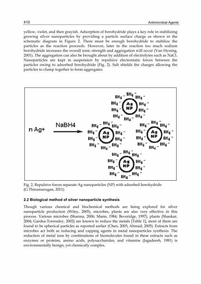
yellow, violet, and then grayish. Adsorption of borohydride plays a key role in stabilizing growing silver nanoparticles by providing a particle surface charge as shown in the schematic diagram in Figure 2. There must be enough borohydride to stabilize the particles as the reaction proceeds. However, later in the reaction too much sodium borohydride increases the overall ionic strength and aggregation will occur (Van Hyning, 2001). The aggregation can also be brought about by addition of electrolytes such as NaCl. Nanoparticles are kept in suspension by repulsive electrostatic forces between the particles owing to adsorbed borohydride (Fig. 2). Salt shields the charges allowing the particles to clump together to form aggregates.
Fig. 2. Repulsive forces separate Ag nanoparticles (NP) with adsorbed borohydride (G.Thirumurugan, 2011).
2.2 Biological method of silver nanoparticle synthesis
Though various chemical and biochemical methods are being explored for silver nanoparticle production (Wiley, 2005), microbes, plants are also very effective in this process. Various microbes (Sharma, 2006; Mann, 1984; Beveridge, 1997), plants (Shankar, 2004; Gardea-Torresdey, 2002) are known to reduce the metals [Table 1], most of them are found to be spherical particles as reported earlier (Chen, 2003; Ahmad, 2005). Extracts from microbes act both as reducing and capping agents in metal nanoparticles synthesis. The reduction of metal ions by combinations of biomolecules found in these extracts such as enzymes or proteins, amino acids, polysaccharides, and vitamins (Jagadeesh, 1981) is environmentally benign, yet chemically complex.

Silver Nanoparticles: Real Antibacterial Bullets
411
Bio-recovery of silver metals from solution, a process referred to as “biosorption”, occurs by either active or passive mechanisms. Active metal transformation processes require viable microbes, enzymatically catalyzing the alteration of the metal, leading to sequestration or concentration. One possible (passive) role of the microorganisms is in providing a multitude of nucleation centers; establishing conditions for obtaining highly disperse nanoparticle systems. In addition, they slow down aggregation, or entirely prevent it by immobilizing the particles, and providing viscous medium (Sun, 2002). Thus produced nanoparticles have highly intricate architectures and are ordered during assembly. In some cases, the particles have a well-defined shape formed within an arrow size range and have orientational and geometrical symmetry (Sarikaya, 1999). In case of silver nanoparticle production, the resistance conferred by bacteria to silver is determined by the ‘sil’ gene in plasmids (Silver, 2003) while a nitrate-dependent reductase and a shuttle quinine extracellular process were reported for the reduction of silver ions by several Fusarium oxysporum strains (Duran, 2005). The extract of unicellular green algae Chlorella vulgaris was used to synthesize single-crystalline Ag nanoplates at room temperature (Jianping Xie, 2007). Proteins in the extract provide dual function of Ag+ reduction and shape-control in the nano silver synthesis. The carboxyl groups in aspartic and or glutamine residues and the hydroxyl groups in tyrosine residues of the proteins were suggested to be responsible for the Ag+ ion reduction (Jianping Xie, 2007). Carrying out the reduction process by a simple bifunctional tripeptide Asp-Asp-Tyr-OMe further identified the involvement of these residues. This synthesis process gave small Ag nanoplates with low polydispersity in good yield (>55%) (Gole, 2001). Balaji, 2009 reported FTIR spectroscopic studies on silver nanoparticles obtained from the fungus, Cladosporium cladosporioides. Their study confirmed that the carbonyl groups from the amino acid residues and peptides of proteins have strong ability to bind silver. The proteins could possibly forma coat covering the metal nanoparticles to prevent their agglomeration and aid in its stabilization in the medium. Hence, the biological molecules could possibly function in the formation and stabilization of the silver nanoparticles in aqueous medium. Gole, 2001 reported that proteins can bind to silver nanoparticles either through free amine groups in the proteins and possibly play a role in stabilization of the silver nanoparticles by surface-bound proteins. Moreover, Vigneshwaran, 2006 explained the synthesis of metal nanoparticles by using fungal mycelium, in the a rotary shaker, metal ions in solution were adsorbed on the surface of the mycelia through interactions with chemical functional groups such as carboxylate anion, carboxyl and peptide bond of proteins, and hydroxyl of saccharides (Lin, 2005) found on the mycelia. The mycelia, matted together, was more immobile, and more capable of binding Me+ than that of the external cellular substances that distributed in the inter-mycelial space, then most of the Me+ was in situ reduced to Me0 by reducing sugars from the saccharides (Gole, 2001) on the mycelia. In the mean time, stronger adsorptive groups such as the carbonyl group on the extracellular substances could further adsorb the particles located on the surface of the mycelia, resulting in capping these nanoparticles, while rocking. When other Me+ in the solution was rocked on to this overlay and was bound and reduced to Me0 on the surface of the layer and these Me0 might be possibly further coated with the other extra cellular substances; this process was repeated continuously until these substances distributing in the inter-mycelial space was used up.
Plant extracts from live alfalfa, the broths of lemon grass, geranium leaves and others have served as green reactants in silver nanoparticle synthesis (Shankar, 2004; Gardea-Torresdey,

Antimicrobial Agents
412
2002). Using plants for nanoparticle synthesis can be advantageous over other biological processes because it eliminates the elaborate process of maintaining cell cultures and can also be suitably scaled up for large-scale nanoparticle synthesis (Shenton, 1999)
3. Anti bacterial effect of silver nanoparticles Due to the outbreak of the infectious diseases caused by different pathogenic bacteria and the development of antibiotic resistance the pharmaceutical companies and the researchers are searching for new antibacterial agents free of resistance and cost. In the present scenario silver nanoparticles have emerged up as novel antimicrobial agents owing to their high surface area to volume ratio and its unique chemical and physical properties. The use of silver nanoparticles can be exploited in various fields, particularly medical and pharmaceutical due to their low toxicity to human cells, high thermal stability and low volatility (Silver, 2003). This has resulted in a broad array of studies in which silver nanoparticles have played a role as drug and as well as superior anti bacterial agent. The highest synergistic antibacterial activity was observed with silver nanoparticles combined antibiotics (Raymond Wai-Yin Sun, 2005). silver nanoparticle incorporated cotton fabrics showed antibacterial activity (Shahverdi, 2007), and silver nanoparticle containing poly vinyl nano- fibres shows efficient antibacterial property (Duran, 2007), it can be used in silver dressings, creams, gel effectively reduce the bacterial infections in chronic wound (Jun, 2007; Richard, 2002; Leaper, 2006). Siver nanoparticles are reported to show better wound healing capacity, better cosmetic appearance and scarless healing when tested using an animal model (Ip, 2006). Kumar et al., 2008 investigated an eco-friendly method for synthesis of metal nanoparticles embedded paint from using vegetable oil. The paint depicts excellent antibacterial activity and in future this paint can be used for efficient antimicrobial coating agent to coat various surfaces such as wood, glass, walls. Silver has been used in water and air filtration to eliminate microorganisms, additionally the Fe3 O4 attached Ag nanoparticles can be used for the treatment of water and easily removed using magnetic field to avoid contamination in the environment (Kumar et al., 2008).
4. Silver nanoparticles and antibiotic resistance Antibiotic resistance is a type of drug resistance where a microorganism has developed the ability to survive exposure to an antibiotic. The volume of antibiotic prescribed is the major factor in increasing rates of bacterial resistance rather than compliance with antibiotics. The four main mechanisms by which microorganisms exhibit resistance to antimicrobials are: Drug inactivation or modification ( e.g. enzymatic deactivation of Penicillin G in some penicillin-resistant bacteria through the production of β-lactamases) and .alteration of target site( e.g. alteration of Penicillin-binding proteins (PBPs) —the binding target site of penicillins—in Methicillin-resistant Staphylococcus aureus (MRSA) and other penicillin-resistant bacteria). Alteration of metabolic pathway( e.g. some sulfonamide-resistant bacteria do not require para-aminobenzoic acid (PABA), an important precursor for the synthesis of folic acid and nucleic acids in bacteria inhibited by sulfonamides. Instead, like mammalian cells, they turn to utilizing preformed folic acid) and reduced drug accumulation: by decreasing drug permeability and/or increasing active efflux (pumping out) of the drugs across the cell surface [Figure 3].

Silver Nanoparticles: Real Antibacterial Bullets
413
Fig. 3. Various mechanisms of bacterial resistance against antibacterials (G. Thirumurugan, 2011).
Therefore, an alternative way to overcome the antibiotic and drug resistance of various micro organisms is needed desperately, especially in medical devices, pharmaceutical etc. The nano size allowed expansion of the contact surface of silver with the microorganisms, and this nano scale has applicability for medical devices and pharmaceutical by surface coating agents. Kim et al., 2007 studied antibacterial mechanism of silver nanoparticles for certain microbial species. The peptidoglycan layer is a specific membrane feature of bacterial species and not mammalian cells. Therefore, if the antibacterial effect of silver nanoparticles is associated with the peptidoglycan layer, it will be easier and more specific to use silver nanoparticles as an antibacterial agent. Sondi and Salopek-Sondi, 2007 reported that the antibacterial activity of silver nanoparticles on Gram-negative bacteria was dependent on the concentration of Ag nanoparticle, and was closely associated with the formation of ‘pits’ in the cell wall of bacteria. Then, Ag nanoparticles accumulated in the bacterial membrane caused the permeability, resulting in cell death and they reported degradation of the membrane structure of micro organism with silver nanoparticles. Kim et al., 2007 suggested that the antimicrobial mechanism of Ag nanoparticles is related to the formation of free radicals and subsequent free radical–induced membrane damage. The free radicals may be derived from the surface of silver nanoparticles and be responsible for the antibacterial activity. In proteomic and biochemical studies, nano molar concentrations of AgNPs have killed E.coli cells within minutes possibly due to immediate dissipation of the proton motive force (Lok, 2006). This action is similar to that found for antibacterial activities of Ag+ ions (Dibrov, 2002). For example, low concentrations of Ag+ ion result in massive proton leakage through the Vibrio cholerae membrane (Dibrov, 2002). This proton leak might be happening from either any Ag+ -modified membrane protein or any Ag+-modified phospholipids

Antimicrobial Agents
414
bilayer. The phenomenon causes deenergization of the membrane and consequently cell death (Dibrov, 2002). Shahverdi et al., 2007 studied the combined effect of silver nanoparticles with different antibiotics against S.aureus and E.coli using the disk diffusion method. The antibacterial activities of penicillin G, amoxicillin, erythromycin, clindamycin, and vancomycin increased in the presence of Ag-NPs against both test strains.
5. Silver nanoparticles incorporated cotton fabrics The current interest is to development of efficient, non-toxic, durable and cost effective antibacterial finishing textiles with increased application in medical, healthcare, hygienic products as well as protective textiles materials. However the ability of cotton fibres to absorb large amount of moisture makes them more prone to microbial attack under certain conditions of humidity and temperature. Cotton may act as a nutrient, becoming suitable medium for bacterial and fungal growth (Rosemary, 2006). Therefore, cotton fibres are treated with numerous chemicals to get better antibacterial cotton textiles. Among the various antibacterial agents, silver nanoparticles have shown strong inhibitory and antibacterial activity, has no negative effect on the human body (Gao, 2008). These particles can be incorporated in several kinds of materials such as clothes. These clothes with silver nanoparticles are sterile and can be used to prevent or to minimize infection with pathogenic bacteria. Nowadays, metal based topical dressings have been widely used as a treatment for infections in burns, open wounds, and chronic ulcers (Panyala, 2008). Incorporation of silver naoparticles was carried out by physical means, before being used; cotton fabrics were washed, sterilized and dried. These were submerged in an Erlenmeyer flask containing silver nanoparticles and agitated at 600 rpm for 24 hrs and dried at 70º C followed by curing at 150 º C. The schematic representation of the formation of silver nanoparticles on cotton fabrics is presented in Scheme 1.
Scheme 1. Incorporation of silver nanoparticles on cotton fabrics (G.Thirumurugan, 2011).

Silver Nanoparticles: Real Antibacterial Bullets
415
The antibacterial properties and the toxicity of metals to micro-organisms is well known, thus, now a days, silver is used in different kinds of formulations like surface coating agents, wound dressing, etc., (Shahverdi, 2007). The silver dressings make use of delivery systems that release silver in different concentrations. But different factors like the distribution of silver in the dressing, its chemical and physical form, affinity of dressing to moisture also influence the killing of micro organisms (Lansdown, 2002). In this direction, metal nanocomposite fibres were prepared containing silver nanoparticles incorporated inside the fabric but from the scanning electron microscopic study it was concluded that the silver nanoparticles incorporated in the sheath part of fabrics possessed significant antibacterial property compared to the fabrics incorporated with silver nanoparticles in the core part (Chopra, 2007). Similar results were obtained by using silver nanoparticles on polyester nonwovens. It is also reported that silver nanoparticles coated textile fabrics possess antibacterialactivity against S.aureus (Shahverdi, 2007).
6. Silver nanoparticles in antibacterial drug delivery Drug delivery system provide useful adjuncts for therapeutics including drugs, nucleic acids and proteins, with variety or roles like improving poor solubility, enhancing invivo stability, optimizing the biodistribution and pharmacokinetics of drugs. In recent years, interest has been stimulated by capability of the metal nanoparticles like AgNPs to bind a wide range of organic molecules, their low toxicity, and their strong and tunable optical absorption. This has resulted in a broad array of studies in which silver nanoparticles have played a role as drug and vaccine carriers into target cells or specific tissues. Furthermore, the unique chemical, physical, and photo-physical properties of silver nanoparticles can be exploited in innovative ways to control the transport and controlled release of pharmaceutical compounds (Skirtach, 2006). Generally, this has been achieved by modifying the surface of the silver nanoparticles so that they can bind to the specific targeting drugs or other biomolecules. But direct conjucation of metal nanoparticle with drugs also possible, it has been shown that conjugates of metal nanoparticles with antibiotics provide promising results in the treatment of intracellular infections (Skirtach, 2006). The conjugation of silver nanoparticles in antibiotic can increase the effectiveness of drug delivery to target some cases. Generally, exact dose is required to kill the pathogens but the amount of antibiotic used in therapy is much higher than the actual dose required. The excess amount of antibiotic can cause adverse effects. Therefore, this conjugation of antibiotic with silver nanopaticles would be helpful to improve antibiotic efficacy. Silver nanoparticles can be directly conjugated with antibiotics or other drug molecules via ionic or covalent bonding, or by physical absorption. For example, Drug has been conjugated to silver nanoparticles [Figure 4]. The cytotoxic effect of free drug is about seven times lower than that of drug conjugated silver nanoparticles. Saha et al., 2007, conjugated directly different type of antibiotic to non-functionalized spherical metal nanoparticles, conjugated form showed greater degree of antibacterial activity with stability than free antibiotics. However, the conjugated form showed some aggregation after conjugation, a situation that other workers consider very deleterious. Therefore, it is likely that modification of the surface of the metal nanoparticles to prevent aggregation would improve the efficacy of such drug delivery systems further.
Surface chemistry of nanomaterial plays an important role, to improve the stability of metal nanoparticles and prevent their aggregation during the conjugation process between biomolecules and nanoparticles. Compared with other drug release materials, the unique

Antimicrobial Agents
416
surface plasmonic properties of the silver particles make it possible to observe the drug release process in living cells by surface enhanced Raman scattering (SERS) method. Jing Yang et al., 2009, found that silver nanoparticles can be used to control the release of drug in living cells. The reason may be that silver nanoparticles can hold the surrounding drug molecules to its surface until a monolayer is formed. The way drug absorbing on the silver surfaces plays an important role in their drug delivery effect in living cells. Figure 5, depicts the SERS spectra of drug in (a) solution and (b) living cells and viability of cells treated with different concentrations of drug and silver nanoparticles. (a) Pure drug; (b) Drug and silver nanoparticle complex; (c) pure silver nanoparticles.
Fig. 4. Cytotoxic effect of drug conjugated silver nanoparticles (G.Thirumurugan, 2011).
Fig. 5. Left side shows SERS spectra of drug in (a) solution and (b) living cells and right side shows viability of cells treated with different concentrations of drug and silver nanoparticles. (a) Pure drug; (b) Drug and silver nanoparticle complex; (c) pure silver nanoparticles.

Silver Nanoparticles: Real Antibacterial Bullets
417
In addition to the surface chemistry of SNPs, their physical properties could be exploited for delivery applications (Asadishad, 2010). The release of a drug from silver nanoparticles could proceed via internal stimuli (pH or glutathione mediated) or also via external stimuli with the application of light. Silver nanoparticles of various shapes can undergo a strong plasmon resonance with light; therefore light induced plasmonic heating may be exploited to release a chemical payload which had been attached to the silver nanoparticles. This may be provided an interesting approach to deliver pharmaceutical compound directly into the cytoplasm or nucleus of target cells. Due to optical properties silver nanoparticles, light sensitive molecule can be attached onto silver nanoparticles in which SNPs serves as a substrate. In this case, light sensitive and fully reversible conformation-changing molecule had been attached onto the silver naoparticles (Jain, 2007). The conformation has been changed from a closed form to an open form when it is irradiated by UV light and the process can be reversed by the application of visible light or heat. This light mediated metal nanoparticle conjugate system could be very effective in the controlled release to treat selected conditions (Jain, 2007). Cheng, 2008, prepared the microcapsules via layer-by-layer technique to encapsulate fluorescein-labeled dextran. The capsule-shells were doped with metal nanoparticles, which response against near infra red (NIR) light. FITC-dextran released upon laser (1064) treatment due to rupture of the shell [Figure 6].
Fig. 6. Light- mediated drug release (G.Thirumurugan, 2011).
7. Conclusion Increasing awareness towards green chemistry and biological processes has led to a desire to develop an environment-friendly approach for the synthesis of non toxic nanoparticles. Unlike other processes in physical and chemical methods, which involve hazardous chemicals, microbial biosynthesis of nanoparticles is cost-effective and eco friendly approach. Therefore, microbes regarded as potential bio factories for nanoparticles synthesis and serves as a new generation anti bacterial agent with their unique chemical and physical properties. The silver nanoparticle have also found diverse applications in the form of wound dressings, coating medical devices, silver nanoparticles impregnated textile fabrics,

Antimicrobial Agents
418
silver nanoparticls incorporated pharmaceutical compounds, etc. The presence of a negative charge on the surface, they are highly reactive, which helps to modify the surface of silver nanoparticles using several biomolecules that leads to loading of SNPs with drug or genes, offers the targeted and controlled release. In particular, the combination of silver nanopartcilcles and laser irradiation to control the release of drugs and genes has provided useful therapeutic benefits.
8. References Ahmad, A,; Senapati, S,; Khan, M. I,; Kumar, R,; Sastry, M. (2005). Extra/Intracellular
Biosynthesis of Gold Nanoparticles by a Novel Alkalotolerant Fungus, Trichothecium sp, J Biomed Nanotechnol. 1, 47-53.
Asadishad, B,; Vossoughi, M,; Alemzadeh, I. (2010). Folate-Receptor-Targeted Delivery of Doxorubicin Using Polyethylene Glycol-Functionalized Gold Nanoparticles, Industrial & Engineering Chemistry Research. 49(4), 1958–1963.
Balaji, D.S,; Basavaraja, S,; Deshpande, R,; Mahesh, B.D,; Prabhakar, B.K,; Venkataraman, A. (2009). Extracellular biosynthesis of functionalized silver nanoparticles by strains of Cladosporium cladosporioides fungus, Coll Surf B: Bio interf. 68, 88- 92.
Beveridge, T.J,; Hughes, M.N,; Lee, H,; Leung, K.T,; Poole, R.K,; Savvaidis, I,; Silver, S and Trevors, J.T. (1997). Metal-microbe interactions: Contemporary approaches, Advances in Microbial Physiology. 38, 177- 243.
Bhattacharya, R,; Patra C. R,; Verma R,; Kumar S,; Greipp P. R and Mukherjee P. (2007). Gold Nanoparticles Inhibit the Proliferation of Multiple Myeloma Cells. Adv. Mater, 19, 711.
Cao,; YunWei Charles,; Jin,; Rongchao,; Mirkin,; Chad A. (2002). Nanoparticles with Raman Spectroscopic Fingerprints for DNA and RNA Detection, Science, 297, 1536-1540.
Chen, J. C,; Lin Z. H and Ma, X. X. (2003). Evidence of the production of silver nanoparticles via pretreatment of Phoma sp.3.2883 with silver nitrate, Letters in Applied Microbiology. 37, 105–108.
Cheng, Y,; Anna, C.S,; Meyers, J.D,; Panagopoulos, I,; Fei, B,; Burda, C. (2008). Deep Penetration of a PDT Drug into Tumors by Noncovalent Drug-Gold Nanoparticle Conjugates, J.Am.Chem.Soc. 130 (32), 10643–10647.
Chopra. (2007). The increasing use of silver-based products as antimicrobial agents: a useful development or a cause for concern, Antimicrob Chemother. 59, 587–90.
Chou, K-S,; Lu, Y-C,; and Lee, H-H. (2005). Effect of alkaline ion on the mechanism and kinetics of chemical reduction of silver, Materials Chemistry and Physics. 94, 429-433.
Daniel, M.-C. and D. Astruc. (2004). Gold nanoparticles: assembly, supramolecular chemistry, quantum-size-related properties, and applications toward biology, catalysis, and nanotechnology. Chemical reviews, 104(1): p. 293-346.

Silver Nanoparticles: Real Antibacterial Bullets
419
Dibrov, P,; Dzioba, J,; Gosinl, K.K,; Hase, C.C. (2002). Chemiosmotic mechanism of antimicrobial activity of Ag+ in Vibrio cholerae, Antimicrob Agents Chemother. 46, 2668.
Duran, N,; Marcarto, P.D,; DeSouza, G.I.H,; Alves, O.L,; Esposito, E. (2007). Antibacterial Effect of Silver Nanoparticles Produced by Fungal Process on Textile Fabrics and Their Effluent Treatment, J Biomed Nanotechnol. 3, 203–8.
Duran, N,; Marcato, P.D,; Alves, O.L,; DeSouza, G.I.H,; Esposity, E. (2005). Mechanistic aspects of biosynthesis of silver nanoparticles by several Fusarium oxysporum strains, J. Nanobiotech. 3, 8.
Esumi, K,; Tano, T,; Torigoe, K,; Meguro, K. (1990). Preparation and characterization of bimetallic palladium-copper colloids by thermal decomposition of their acetate compounds in organic solvents. Chem. Mater, 2(5), 564- 567.
Gao, Y,; Cranston, R. (2008). Recent Advances in Antimicrobial Treatments of Textile,Text. Res. J. 78, 60–72.
Gardea-Torresdey, Parsons, J.G,; Gomez, E,; Peralta-Videa, J,; Troiani, H.E,; Santiago, P,; JoseYacaman, M. (2002). Formation and Growth of Au Nanoparticles Inside Live Alfalfa Plants, Nano let. 2(4), 397-401.
Goia D. V and Matijevic E. (1998). Preparation of Monodispersed Metal Particles, New J. Chem. 22, 1203-1215.
Gole, A,; Dash, C,; Ramachandran, V,; Sainkar, S.R,; Mandale, A.B,; Rao, M. (2001). Pepsin−Gold Colloid Conjugates: Preparation, Characterization, and Enzymatic Activity, Langmuir. 17, 1674- 9.
Gutiérrez, M. and Henglein A. (1993). Formation of colloidal silver by "push-pull" reduction of Ag+. J. Phys. Chem. 97, 11368.
Henglein, A. (2001). Reduction of Ag (CN)2" on silver and platinum colloidal nanoparticles. Langmuir, 17, 2329-2333.
Ip, M,; Lui, S.L,; Poon, V.K.M,; Lung, I,; Burd, A. (2006). Antimicrobial activities of silver dressings: an in vitro comparison, J Med Microbiob. 55, 59–63.
Isabel Pastoriza-Santos and Luis M. Liz-Marzلn. (2002). Synthesis of Silver Nanoprisms in DMF. Langmuir, 18, 2888.
Jagadeesh M. S and Seehra, M. S. (1981). Principal magnetic susceptibilities of MnO and their temperature dependence, Phys. Rev. B. 23, 1185.
Jain, P.K,; El-Sayed, I.H,; El-Sayed, M.A. (2007). Au nanoparticles target cancer, NanoToday. 2, 18–29.
Jianping Xie,; Jim Yang Lee and Daniel I.C. Wang. (2007). Synthesis of Single-Crystalline Gold Nanoplates in Aqueous Solutions through Biomineralization by Serum Albumin Protein, J. Phys. Chem. C. 111 (28), 10226– 10232.
Jing yang,; Hong Wang,; Zhuyuan Wang,; Xuebin tan,; Chunyuan Song,; Ruohu Zhang,; Jin Li,; Yiping Cui. (2009). Interaction between antitumor drug and silver nanoparticles: combined fluorescence and surface enhanced Raman scattering study, Chinese Optics Letters. 7(10).
Jun, J,; Yuan-Yuan, D,; Shao-hai, W,; Shao-feng, Z,; Zhong-yi, W. (2007). Preparation and characterization of antibacterial silver containing nano fibers for wound dressing applications, JUS-China Med Sci. 4(2), 52– 4.
Antimicrobial Agents
420
Kapoor, M.P.; Yang, Q.; Inagaki, S. (2002). Self-Assembly of Biphenylene-Bridged Hybrid Mesoporous Solid with Molecular-Scale Periodicity in the Pore Walls. J. Am. Chem. Soc, 124, 15176-15177.
Kim, J.S,; Kuk, E,; Yu, K.N,; Kim, J.H,; Park, S.J,; Lee, H.J. (2007). Antimicrobial effects of silver nanoparticles, Nanomed Nanotechnol Biol Med. 3, 95-101.
Klasen, H. J. (2000). Historical review of the use of silver in the treatment of burns. Early uses. Burns, 26, 11730.
Kumar, A,; Vemula, P.K,; Ajayan, P.M,; John, G. (2008). Silver-nanoparticle-embedded antimicrobial paints based on vegetable oil, Nature Materials. 7(3), 236–41.
Lansdown, A.B. (2002). Silver. 2: Toxicity in mammals and how its products aid wound repair, Journal of Wound Care. 11(5), 173–177.
Lansdown, A.B. (2002). Silver: its antibacterial properties and mechanism of action. J. Wound Care. 11, 125.
Leaper, D.L. (2006). Silver dressings: their role in wound management, Int Wound J. 3(4), 282–94.
Lin, Z,; Wu, J,; Xue, R,; Yong, Y. (2005). Spectroscopic characterization of Au3+ biosorption by waste biomass of Saccharomyces cerevisiae, Spectrochim. Acta Part A. 61, 761–765.
Lok, C-N,; Ho, C-M,; Chen, R,; He, Q-Y,; Yu, W-Y,; Sun, H. (2006). Proteomic analysis of the mode of antibacterial action of silver nanoparticles, J Proteome Res. 5, 916.
Mann, S,; Frankel, R.B,; Blakemore, R.P. (1984). Structure, Morphology, and Crystal Growth of Bacterial Magnetite, Nature. 310, 405- 407.
Oghabian, M. A and Farahbakhsh, N. M. (2010). Potential use of nanoparticle based contrast agents in MRI: a molecular imaging perspective, J. Biomed. Nanotechnol, 6, 203.
Panyala, N.R,; Pena-Mendez, E.M,; Havel, J. (2008). Silver or silver nanoparticles: A hazardous threat to the environment and human health? J Applied Biomedicine. 6(3), 117–129.
Patra C. R,; Bhattacharya R,; E. Wang, and Katarya A. (2008).Targeted Delivery of Gemcitabine to Pancreatic Adenocarcinoma Using Cetuximab as a Targeting Agent. Cancer Re, 68, 1970.
Raymond Wai-Yin Sun,; Rong Chen,; Nancy P.-Y.Chung,; Chi-Ming Ho,; Chen-Lung Steve Lin and Chi-Ming Che. (2005). Silver nanoparticles fabricated in Hepes buffer exhibit cytoprotective activities toward HIV-1 infected cells, Chem.Commun. 5059–5061.
Richard, J.W,; Spencer, B.A,; McCoy, L.F,; Carina, E,; Washington, J,; Edgar, P. (2002). J Burns Surg Wound Care. 1, 11–20.
Rodriguez-Sanchez, L,; Blanco, M. C. and Lopez-Quintela, M. A. (2000).Electrochemical Synthesis of Silver Nanoparticles. J. Phys.Chem. B, 104, 9683-9688.
Rosemary, M.J,; Mac Laren, I,; Pradeep, T. (2006). Investigations of the antibacterial properties of ciprofloxacin and SiO2, Langmuir. 22, 10125.
Saha, B,; Bhattacharya, J,; Mukherjee, A,; Ghosh, A.K,; Santra, C.R,; Dasgupta, A.K,; Karmakar, P. (2007). In vitro structural and functional evaluation of gold nanoparticles conjugated antibiotics, Nanoscale Res. Lett. 2, 614–622.

Silver Nanoparticles: Real Antibacterial Bullets
421
Sarikaya, M,; Fong, H,; French, D.W,; Humbert, R. (1999). Biomimetic assembly of nanostructured materials, Mat. Sci. Forum. 293, 83–98.
Shahverdi, A.R,; Fakhimi, A,; Shahverdi, H.R,; Minaian, S. (2007). Synthesis and effect of silver nanoparticles on the antibacterial activity of different antibiotics against Staphylococcus aureus and Escherichia coli, Nanomed: Nanotechnol. Biol Med. 3 (2), 168–71.
Shankar, S.S,; Rai, A,; Ahmad, A,; Sastry, M. J. (2004). Rapid synthesis of Au, Ag, and bimetallic Au core-Ag shell nanoparticles using Neem (Azadirachta indica) leaf broth, J Colloid Interf Sci. 275, 496-502.
Sharma, V. K,; Nevecna, T,; Zboril, R. (2006). Silver colloid nanoparticles: synthesis, characterization, and their antibacterial activity, J. Phys. Chem. B. 110, 16248-16253.
Shenton, W,; Douglas, T,; Young, M,; Stubbs, G,; Mann, S. (1999). Inorganic–Organic Nanotube Composites from Template Mineralization of Tobacco Mosaic Virus, Adv Mater. 11,253.
Silver, S. (2003). Bacterial silver resistance: molecular biology and uses and misuses of silvercompounds, FEMS Microbiol. Rev. 27 (2–3), 341–353.
Skirtach, A.G,; Javier, A.M,; Kreft, O,; Köhler, K,; Alberola, A.P,; Möhwald, H,; Parak, W.J,; Sukhorukov, G.B. (2006). Laser-induced release of encapsulated materials inside living Cells, Angew. Chem. 118(28), 4728–4733.
Sondi, B,; alopek-Sondi. (2007). Silver nanoparticles as antimicrobial agent: a case study on E. coli as a model for Gram-negative bacteria, J Colloid Interface. 275, 177– 182.
Sun Y and Y. Xia. (2002). Shape-Controlled Synthesis of Gold and Silver Nanoparticles, Science. 298, 2176–2179.
Taleb, A,; Petit, C,; Pileni, M. P. (1997). Synthesis of Highly Monodisperse Silver Nanoparticles from AOT Reverse Micelles: A Way to 2D and 3D Self-Organization. Chem. Mater, 9, 950-959.
Thirumurugan, G and Dhanaraju.M.D. (2011). Novel Biogenic Metal Nanoparticles for Pharmaceutical Applications, Adv. Sci. Lett, 4, 339-348.
Thirumurugan, G,; Satya Veni, V,; Ramachandran, S,; Seshagiri Rao, J. V. L. N. and Dhanaraju, M. D. (2011). Superior wound healing effect of topically delivered silver nanoparticle formulation using eco-friendly potato plant pathogenic fungus: synthesis and characterization, J. Biomed. Nanotechnol. 7, 659- 666.
Van Hyning, D. L,; Klemperer, W. G and Zukoski, C. F. (2001). Silver nanoparticle formation: Predictions and verification of the aggregative growth model, Langmuir 17, 3128-3135.
Vigneshwaran, N,; Kathe, A. A,; Varadarajan, P.V,; Nachane, R.P,; Balasubramanya, R.H. (2006). Biomimetics of silver nanoparticles by white rot fungus, Phaenerochaete chrysosporium, Coll Surf B: Interf. 53, 55- 59.
Wiley, B.J,; Sun, Y,; Mayers, B and Xia, Y. (2005). Shape-Controlled Synthesis of Metal Nanostructures: The Case of Silver, Chem. Eur. J. 11, 454.

Antimicrobial Agents
422
Zhu, J. J,; Koltypin, Y and Gedanken, A. (2000). A General Sonochemical Method for the preparation of Nanophased Selenides: the synthesis of ZnSe. Chem. Mater, 12, 73.