Silk fiber — molecular formation mechanism, structure property relationship and advanced...
34
Chapter 3 Silk Fiber — Molecular Formation Mechanism, Structure- Property Relationship and Advanced Applications Xinfang Liu and Ke-Qin Zhang Additional information is available at the end of the chapter http://dx.doi.org/10.5772/57611 1. Introduction Silk fibers spun by several species of arthropods have existed naturally for hundreds of millions of years. The ecological functions of the silk fibers are closely related to their proper‐ ties. For example, orb-weaving spiders produce a variety of different silks with diverse properties, each tailored to achieve a certain task (Figure 1) [1]. Most arthropod species produce silks used for building structures to capture prey and protect their offspring against environ‐ mental hazards [2]. The most investigated categories that have piqued the greatest amount of interest are spider silk and dragline silk in particular, produced by major ampullate glands and the cocoon silk of Bombyx mori (B. mori). The ongoing evolutionary optimization of silks from silkworms and spiders exhibit outstanding mechanical properties, such as strength and extensibility, as well as toughness, which outperform most other natural and man-made silk fibers (Table 1) [3, 4]. Due to its smooth texture, luster and strength, silks from natural silkworms have been extensively used in apparel and fashion applications for thousands of years [5]. Silks from spiders have also been utilized throughout history, such as sutures and fishing equipment in ancient Greece and Australasia. In contrast petrochemical-based synthetic polymers commonly used today, such as polyethy‐ lene, which is formed by polymerization of ethylene at high temperature and pressure, or under the presence of some metal-based catalysis, B. mori and spider spin fibers from a highly concentrated, water-based protein solution under mild conditions [6, 7]. Due to current trends in exploration of natural biological materials and the demand for environmentally friendly (green) materials, investigation of the applications of silk fibers has steadily gained promi‐ nence. Silk fibers are emerging as candidates for applications in even non-apparel areas due in part to recognition for their extraordinary mechanical properties, as well as their biocom‐ patibility and biodegradability. Currently, the promotion of silkworm as bio-factory to © 2014 The Author(s). Licensee InTech. This chapter is distributed under the terms of the Creative Commons Attribution License (http://creativecommons.org/licenses/by/3.0), which permits unrestricted use, distribution, and reproduction in any medium, provided the original work is properly cited.
-
Upload
truong-quang-trung -
Category
Engineering
-
view
77 -
download
1
Transcript of Silk fiber — molecular formation mechanism, structure property relationship and advanced...
Chapter 3
Silk Fiber — Molecular Formation Mechanism, Structure-Property Relationship and Advanced Applications
Xinfang Liu and Ke-Qin Zhang
Additional information is available at the end of the chapter
http://dx.doi.org/10.5772/57611
1. Introduction
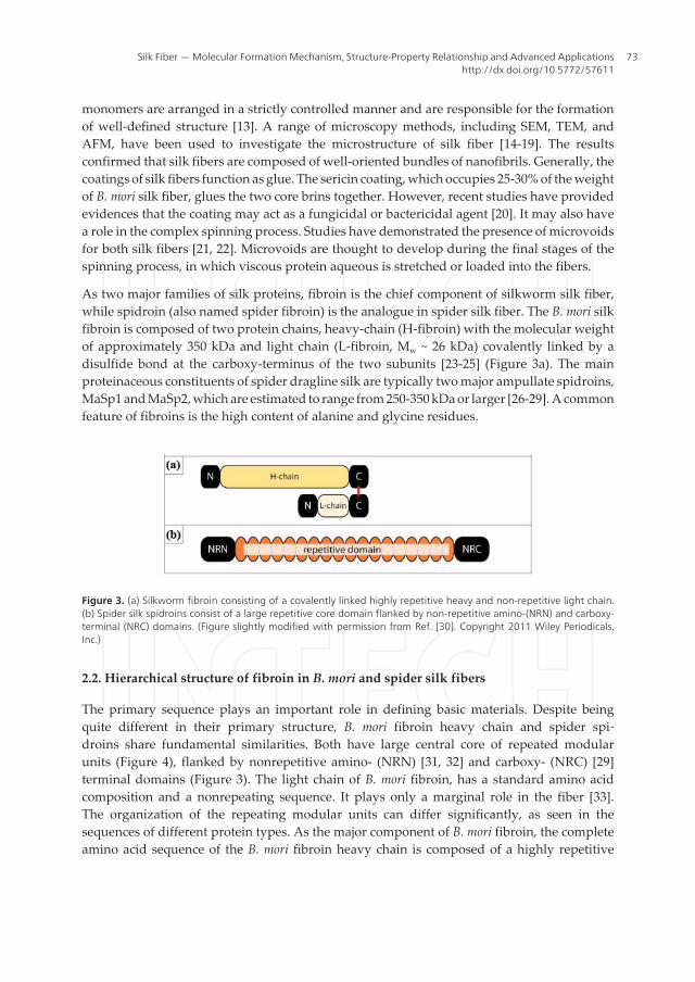
Silk fibers spun by several species of arthropods have existed naturally for hundreds ofmillions of years. The ecological functions of the silk fibers are closely related to their proper‐ties. For example, orb-weaving spiders produce a variety of different silks with diverseproperties, each tailored to achieve a certain task (Figure 1) [1]. Most arthropod species producesilks used for building structures to capture prey and protect their offspring against environ‐mental hazards [2]. The most investigated categories that have piqued the greatest amount ofinterest are spider silk and dragline silk in particular, produced by major ampullate glandsand the cocoon silk of Bombyx mori (B. mori). The ongoing evolutionary optimization of silksfrom silkworms and spiders exhibit outstanding mechanical properties, such as strength andextensibility, as well as toughness, which outperform most other natural and man-made silkfibers (Table 1) [3, 4]. Due to its smooth texture, luster and strength, silks from naturalsilkworms have been extensively used in apparel and fashion applications for thousands ofyears [5]. Silks from spiders have also been utilized throughout history, such as sutures andfishing equipment in ancient Greece and Australasia.
In contrast petrochemical-based synthetic polymers commonly used today, such as polyethy‐lene, which is formed by polymerization of ethylene at high temperature and pressure, orunder the presence of some metal-based catalysis, B. mori and spider spin fibers from a highlyconcentrated, water-based protein solution under mild conditions [6, 7]. Due to current trendsin exploration of natural biological materials and the demand for environmentally friendly(green) materials, investigation of the applications of silk fibers has steadily gained promi‐nence. Silk fibers are emerging as candidates for applications in even non-apparel areas duein part to recognition for their extraordinary mechanical properties, as well as their biocom‐patibility and biodegradability. Currently, the promotion of silkworm as bio-factory to
© 2014 The Author(s). Licensee InTech. This chapter is distributed under the terms of the Creative CommonsAttribution License (http://creativecommons.org/licenses/by/3.0), which permits unrestricted use,distribution, and reproduction in any medium, provided the original work is properly cited.
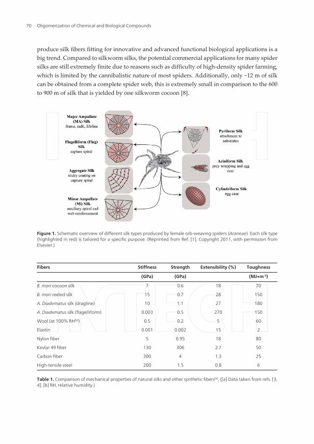
produce silk fibers fitting for innovative and advanced functional biological applications is abig trend. Compared to silkworm silks, the potential commercial applications for many spidersilks are still extremely finite due to reasons such as difficulty of high-density spider farming,which is limited by the cannibalistic nature of most spiders. Additionally, only ~12 m of silkcan be obtained from a complete spider web, this is extremely small in comparison to the 600to 900 m of silk that is yielded by one silkworm cocoon [8].
Figure 1. Schematic overview of different silk types produced by female orb-weaving spiders (Araneae). Each silk type(highlighted in red) is tailored for a specific purpose. (Reprinted from Ref. [1]. Copyright 2011, with permission fromElsevier.)
Fibers Stiffness Strength Extensibility (%) Toughness
(GPa) (GPa) (MJ∙m-3)
B. mori cocoon silk 7 0.6 18 70
B. mori reeled silk 15 0.7 28 150
A. Diadematus silk (dragline) 10 1.1 27 180
A. Diadematus silk (flagelliform) 0.003 0.5 270 150
Wool (at 100% RH[b]) 0.5 0.2 5 60
Elastin 0.001 0.002 15 2
Nylon fiber 5 0.95 18 80
Kevlar 49 fiber 130 306 2.7 50
Carbon fiber 300 4 1.3 25
High-tensile steel 200 1.5 0.8 6
Table 1. Comparison of mechanical properties of natural silks and other synthetic fibers[a]. ([a] Data taken from refs. [3,4]. [b] RH, relative humidity.)
Oligomerization of Chemical and Biological Compounds70

Artificial spinning is the most promising method of promoting the application of silk fibers,as it can output sufficient man-made fibers cost-effectively and with specific tailored proper‐ties. Remarkable efforts for silk fiber reproduction via reconstituted/recombinant silk fibroinare currently underway [9, 10]. Reconstituted silk protein is derived from B. mori cocoons andthe degummed B. mori silk fibers, they are soluble in concentrated LiBr aqueous solutions,yielding reconstituted silk protein solutions after dialysis [9]. Recent advances in transgenictechnology enable the high level expression of recombinant proteins. Spider silk proteins havebeen produced by other organisms so that recombinant spider silk protein might be suitablefor creation of artificial silk threads or other applications. Host organisms include bacteria,yeasts, animal cells and plants [3]. In order to biomimic native silks, the reconstituted orrecombinant proteins used to spin artificial silks should possess amino acid similar to the onesfound in native sites for sequence and composition. However, the properties of synthetic silkfibers currently do not meet the standards of native silks yet, as the composition, hierarchicalstructure and production conditions of natural silks all reportedly affect their mechanicalproperties [11]. Consequently, a profound knowledge of the natural formation process,chemical composition, relationships of structure and properties of silk fibers seems thereforimperative.
Thanks to recent developments in modern analytical techniques, significant progress has beenmade with respect to the structural characterization of silk. These techniques can providemolecular information about silk, including microscopic methods (atomic force microscopy(AFM), scanning and transmission electron microscopy (SEM and TEM), and scanningtransmission x-ray microscopy (STXM)) and synchrotron x-ray diffraction (wide-angle x-raydiffraction (WAXD) and small-angle x-ray scattering (SAXS) combined with synchrotronradiation). Solid-state nuclear magnetic resonance (SS-NMR) is a powerful technique becauseit allows for the study of molecular structure and dynamics of semi-crystalline and amorphousmaterials. Raman and FTIR spectroscopy can provide the dominant conformational contentsof a fiber. Raman microspectroscopy can be used to determine quantitative parameterscharacterizing the molecular structure (orientation and conformation, amino acid composi‐tion) of micrometer-sized biological samples. In this chapter, we will provide an overview ofthe current understanding of the silk fibers’ structure taken advantage of these analyticmethods, then describe in detail the structure-property relationships and the formationprocesses of silk fiber. Additionally, we will explore material morphologies and applicationsof these silk fibers.
2. The structure-property relationship of silk fibers
The structure-property relationship is one of the most intriguing ‘mysteries’ of silk fibers.Various studies have suggested that there is a strong connection between the structures of silkfibers and their physical (e. g., mechanical) properties. An understanding of the structure-property relationship requires background knowledge of local structure, including thecomponent and composition of silk fiber, the conformation and orientation of constitutive unitswith respect to the fiber, and so on.
Silk Fiber — Molecular Formation Mechanism, Structure-Property Relationship and Advanced Applicationshttp://dx.doi.org/10.5772/57611
71

2.1. The structure and composition of B. mori and spider dragline silk fibers
In principle, the full range of properties of silk fibers can be calculated from their structuralmorphology and chemical composition. On the macroscopic level, the morphological structureof B. mori silk and spider dragline silk are very similar, as both possess a core-shell structure(Figure 2) [12]. The silk thread diameter varies across types and species. For example, coatingthe two core brins of B. mori silk fiber with sericin yields fibers about 20 μm width. Spiderdragline silks have a diameter of 3-5 μm and to date, have been described to contain only oneprotein monofilament.
Figure 2. Examples of silk fibers produced by silkworms and spiders and a schematic illustration. (Reprinted from Ref.[12]. Copyright 2008, with permission from Elsevier.)
Silk fibers are normally polyamino acid-based fibrous proteins. In contrast, the syntheticpolymers, which are usually homopolymers or copolymers consisting of one or several simplermonomer, the biopolymers − silk fibers, the primary sequence and linkage between the
Oligomerization of Chemical and Biological Compounds72
monomers are arranged in a strictly controlled manner and are responsible for the formationof well-defined structure [13]. A range of microscopy methods, including SEM, TEM, andAFM, have been used to investigate the microstructure of silk fiber [14-19]. The resultsconfirmed that silk fibers are composed of well-oriented bundles of nanofibrils. Generally, thecoatings of silk fibers function as glue. The sericin coating, which occupies 25-30% of the weightof B. mori silk fiber, glues the two core brins together. However, recent studies have providedevidences that the coating may act as a fungicidal or bactericidal agent [20]. It may also havea role in the complex spinning process. Studies have demonstrated the presence of microvoidsfor both silk fibers [21, 22]. Microvoids are thought to develop during the final stages of thespinning process, in which viscous protein aqueous is stretched or loaded into the fibers.
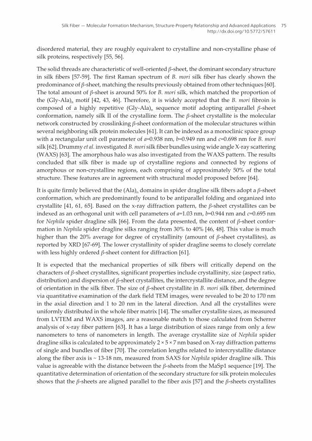
As two major families of silk proteins, fibroin is the chief component of silkworm silk fiber,while spidroin (also named spider fibroin) is the analogue in spider silk fiber. The B. mori silkfibroin is composed of two protein chains, heavy-chain (H-fibroin) with the molecular weightof approximately 350 kDa and light chain (L-fibroin, Mw ~ 26 kDa) covalently linked by adisulfide bond at the carboxy-terminus of the two subunits [23-25] (Figure 3a). The mainproteinaceous constituents of spider dragline silk are typically two major ampullate spidroins,MaSp1 and MaSp2, which are estimated to range from 250-350 kDa or larger [26-29]. A commonfeature of fibroins is the high content of alanine and glycine residues.
Figure 3. (a) Silkworm fibroin consisting of a covalently linked highly repetitive heavy and non-repetitive light chain.(b) Spider silk spidroins consist of a large repetitive core domain flanked by non-repetitive amino-(NRN) and carboxy-terminal (NRC) domains. (Figure slightly modified with permission from Ref. [30]. Copyright 2011 Wiley Periodicals,Inc.)
2.2. Hierarchical structure of fibroin in B. mori and spider silk fibers
The primary sequence plays an important role in defining basic materials. Despite beingquite different in their primary structure, B. mori fibroin heavy chain and spider spi‐droins share fundamental similarities. Both have large central core of repeated modularunits (Figure 4), flanked by nonrepetitive amino- (NRN) [31, 32] and carboxy- (NRC) [29]terminal domains (Figure 3). The light chain of B. mori fibroin, has a standard amino acidcomposition and a nonrepeating sequence. It plays only a marginal role in the fiber [33].The organization of the repeating modular units can differ significantly, as seen in thesequences of different protein types. As the major component of B. mori fibroin, the completeamino acid sequence of the B. mori fibroin heavy chain is composed of a highly repetitive
Silk Fiber — Molecular Formation Mechanism, Structure-Property Relationship and Advanced Applicationshttp://dx.doi.org/10.5772/57611
73

(Gly-Ala)n sequence motif and tyrosine-rich domains [34]. In MaSp1, the modular unitsmainly consist of a subset of the sequence motifs (Ala)n followed by several GGX motifs,with X representing a variable amino acid. In MaSp2, the GGX motif is replaced by theGPGXX motif, which contains more proline residues [26, 27]. The modular units are repeatedup to several hundred times in the central core of B. mori fibroin heavy chain and spiderspidroins such that they largely determine the macroscopic properties of the fibers. Thehighly conserved sequence of nonrepetitive amino- and carboxy- terminal domains areessential for fiber formation and expected to be of functional relevance [35-39]. Moreover,the analysis of the hydropathicity of these fibroins reveals a pair of hydrophobic andhydrophilic counterparts. The central region of the protein is mostly hydrophobic, whilethe nonrepetitive amino- and carboxy- terminal domains are more hydrophilic [40].
Figure 4. Typical amino acid sequences of repetitive core of B. mori fibroin heavy chain, minor ampullate spidroins andmajor ampullate spidroins. The highly repetitive (Gly-Ala)n and (Ala)n sequence motifs are highlighted in red. The ac‐cession numbers for the sequences are P05790, P19837, P46804, AAC47009 and AAC47010 respectively.
The primary structural motifs have a preferred secondary structure and give rise to structureshigher up the hierarchy. NMR, circular dichroism (CD), IR and Raman spectroscopy wereusually used to examine the chemical, conformational, and orientational information ofsecondary structures for silk proteins [41-51]. There are three major conformations of silkproteins: the random coil, the α-helix and the β-sheet [52-54]. Using the approach of Porter,and reducing the complex secondary structure of silk proteins into fractions of ordered and
Oligomerization of Chemical and Biological Compounds74
disordered material, they are roughly equivalent to crystalline and non-crystalline phase ofsilk proteins, respectively [55, 56].
The solid threads are characteristic of well-oriented β-sheet, the dominant secondary structurein silk fibers [57-59]. The first Raman spectrum of B. mori silk fiber has clearly shown thepredominance of β-sheet, matching the results previously obtained from other techniques [60].The total amount of β-sheet is around 50% for B. mori silk, which matched the proportion ofthe (Gly-Ala)n motif [42, 43, 46]. Therefore, it is widely accepted that the B. mori fibroin iscomposed of a highly repetitive (Gly-Ala)n sequence motif adopting antiparallel β-sheetconformation, namely silk II of the crystalline form. The β-sheet crystallite is the molecularnetwork constructed by crosslinking β-sheet conformation of the molecular structures withinseveral neighboring silk protein molecules [61]. It can be indexed as a monoclinic space groupwith a rectangular unit cell parameter of a=0.938 nm, b=0.949 nm and c=0.698 nm for B. morisilk [62]. Drummy et al. investigated B. mori silk fiber bundles using wide angle X-ray scattering(WAXS) [63]. The amorphous halo was also investigated from the WAXS pattern. The resultsconcluded that silk fiber is made up of crystalline regions and connected by regions ofamorphous or non-crystalline regions, each comprising of approximately 50% of the totalstructure. These features are in agreement with structural model proposed before [64].
It is quite firmly believed that the (Ala)n domains in spider dragline silk fibers adopt a β-sheetconformation, which are predominantly found to be antiparallel folding and organized intocrystallite [41, 61, 65]. Based on the x-ray diffraction pattern, the β-sheet crystallites can beindexed as an orthogonal unit with cell parameters of a=1.03 nm, b=0.944 nm and c=0.695 nmfor Nephila spider dragline silk [66]. From the data presented, the content of β-sheet confor‐mation in Nephila spider dragline silks ranging from 30% to 40% [46, 48]. This value is muchhigher than the 20% average for degree of crystallinity (amount of β-sheet crystallites), asreported by XRD [67-69]. The lower crystallinity of spider dragline seems to closely correlatewith less highly ordered β-sheet content for diffraction [61].
It is expected that the mechanical properties of silk fibers will critically depend on thecharacters of β-sheet crystallites, significant properties include crystallinity, size (aspect ratio,distribution) and dispersion of β-sheet crystallites, the intercrystallite distance, and the degreeof orientation in the silk fiber. The size of β-sheet crystallite in B. mori silk fiber, determinedvia quantitative examination of the dark field TEM images, were revealed to be 20 to 170 nmin the axial direction and 1 to 20 nm in the lateral direction. And all the crystallites wereuniformly distributed in the whole fiber matrix [14]. The smaller crystallite sizes, as measuredfrom LVTEM and WAXS images, are a reasonable match to those calculated from Scherreranalysis of x-ray fiber pattern [63]. It has a large distribution of sizes range from only a fewnanometers to tens of nanometers in length. The average crystallite size of Nephila spiderdragline silks is calculated to be approximately 2 × 5 × 7 nm based on X-ray diffraction patternsof single and bundles of fiber [70]. The correlation lengths related to intercrystallite distancealong the fiber axis is ~ 13-18 nm, measured from SAXS for Nephila spider dragline silk. Thisvalue is agreeable with the distance between the β-sheets from the MaSp1 sequence [19]. Thequantitative determination of orientation of the secondary structure for silk protein moleculesshows that the β-sheets are aligned parallel to the fiber axis [57] and the β-sheets crystallites
Silk Fiber — Molecular Formation Mechanism, Structure-Property Relationship and Advanced Applicationshttp://dx.doi.org/10.5772/57611
75

representing the highly ordered fraction are well-oriented along the silk fiber [70-72]. Addi‐tionally, the β-sheets of B. mori silks are slightly better oriented than those of dragline silks,corresponding to the fact that they are more crystalline than spider dragline silk [46].
The non-crystalline regions are often described as amorphous, poorly orientated, or randomlycoiled sections of the peptide. The structural organization in the amorphous phase is not wellunderstood yet. The existence of β-turn or β-spiral and helical conformations has beensuggested for amorphous domains [42, 46, 65, 73, 74]. Tyrosine residue, on average, may formdistorted β-turns and distorted β-sheets, which is characterized by 13C solid-state NMR in theamorphous matrix of B. mori silk [42, 75]. The Gly-rich regions in spider dragline silk have beendescribed as the amorphous rubber based on X-ray diffraction studies [76]. The precisestructure of the GlyGlyX motif in MaSp1 has been somewhat controversial. Recent NMRstudies provided evidence of the presence of less ordered helical type structure or distortedβ-sheets adopted by the GlyGlyX motif [48, 73]. However, for MaSp2, ADF 3 and 4 (the fibroinof major ampullate dragline silk for spider Araneus diadematus), the structure of the GlyPro‐GlyXX repeat has been proposed to be a β-turns or spiral structure. The stability of thesestructures is given by the interchain hydrogen bonding [77]. The molecular chains in theamorphous phase are often considered to be randomly oriented. Studies from Raman [46] andSS-NMR [73] reported that the protein backbones in the amorphous regions of silk fibers arenot randomly oriented but exhibit certain degree of orientation along the fiber axis, albeit muchless oriented than β-sheet crystallites. Meanwhile, the higher level of orientation of theamorphous phase for the spider silks than that for B. mori silk.
Recent computational approaches have been useful in modeling nanostructure of silk.Molecular modeling integrated the information known about the structures, and has been usedto characterize the nanostructure of the silk. Based on a bottom-up molecular computationalapproach using replica exchange molecular dynamic, Keten et al. reported atomic-levelstructures of MaSp1 and MaSp2 proteins from the Nephila Clavipes spider dragline silksequence. It showed that poly-alanine segments in silk have an extremely high propensity forforming distinct and orderly β-sheet crystallites. Previous molecular dynamic simulations onpoly-alanine aggregation also suggested that anti-parallel orientations in the hydrogenbonding direction and parallel stacking in the side – chain direction leads to stable β-sheets[78]. Glycine-rich regions are less orderly, predominantly forming helical type structures andβ-turns in amorphous domains. The density of hydrogen bonds in amorphous regions is lowerthan in β-sheet crystallites [79, 80]. All of the results are excellently consistent with availableexperiment evidence and may contribute towards an improved understanding of the sourceof silk’s strength and toughness.
According to the prevalent characterizations mentioned above, silk fiber is considered asemicrystalline polymer with a hierarchical structure in which highly oriented β-sheetscrystallites connecting with an amorphous matrix are organized in nanofibrils or fibrillarentities [81] (Figure 5). However, it has been proposed that there exists a third phase, orinterphase consisting of weakly oriented β-sheets regions [68, 82-84] or oriented amorphousdomains [85, 86] in silk. Recent NMR and IR studies performed on silk used hydrogen-deuterium (H-D) exchange to differentiate among three structures. The data revealed that the
Oligomerization of Chemical and Biological Compounds76

D2O-inaccessible β-sheets are associated with crystallites, while D2O-accessible ones arecomposed of amorphous domains and interphase β-sheets [82-84]. In the case of spiderdragline silk, crystalline component larger in size and poorer in orientation are detectablebeside the ~ 2 nm sized β-sheets crystallite that are commonly observed [69, 87, 88]. Theseobservations of larger ordered regions have been explained as ‘Non Periodic Lattice (NPL)’crystals which form as a result of statistical matches between compatible sequences on adjacentmolecular chains. It was revealed that the border where β-sheet crystallite regions andamorphous domains do not have any discrete phase boundaries. The presence of the inter‐phase has also been deduced from STXM studies on Nephila dragline silk. It indicates thathighly oriented and unoriented domains are surrounded by a moderately oriented matrix [85].In summary, there is ongoing debate on the molecular structure of silk at the nanoscopic level.
2.3. The physical (mechanical) properties of silk fibers
Spider silk and B. mori silk feature unique physical properties – such as superior mechanicalproperties in terms of toughness (the amount of energy absorbed before breakage) (Table 1).So far, the maximum strength of spider dragline silk (dragline of Caerostris darwini) up to 1.7GPa, which exceeds that of steel (1.5 GPa), is in the range of high-tech materials [1]. Due to itsgreat extensibility, spider dragline silks have three times of toughness of man-made syntheticfibers like Kevlar 49 [3, 89]. Typical B. mori silk is presumed to be weaker and less extensiblethan spider dragline silk. However, when forcibly silking from immobilized silkworms
Figure 5. (a) The hierarchical structure of spider dragline and silkworm silk fiber. Both spider dragline and fibroin arecomposed of numerous minute fibrils, which are separated into crystalline and amorphous segments. (b) The minutefibrils in silkworm B. mori silk as revealed in an AFM image (scale bar: 150 nm). The silk fiber direction is indicated bythe arrow. (Adapted with permission from Ref. [81]. Copyright 2011, WILEY-VCH.)
Silk Fiber — Molecular Formation Mechanism, Structure-Property Relationship and Advanced Applicationshttp://dx.doi.org/10.5772/57611
77
artificially at certain spinning speed, the mechanical properties of the specific B. mori silk havegreatly improved to a level that is comparable the toughest spider silk [4].
The mechanical properties of silk fibers can be described by stress-strain curve profiles, whichare generated by stretching the fibers at a specific strain rate. The stress is expressed as forceper cross-sectional area and the strain is defined as a normalized extensibility. Typical stress-strain curves for B. mori silkworm silk and spider dragline silk show both elastic behaviorfollowed by plastic deformation [90]. The linear portion of the curve, up to the yield point, isthe elastic region. The slope is defined as Young’s modulus [91], a measure of the stiffness ofthe fiber. After the yield point, the fiber buffers the plastic deformation and the stress-strainprofiles are subjected to sudden slope changes. This behavior indicates that major structuraltransition from rubberlike to glassy state occurs in the fiber [92-94]. These characteristics havedriven scientists to explore the structural origin of the high-performance silk fiber, with thegoal of obtaining templates for designing novel materials with comparable properties.
2.4. The structure-property relationship of silk fibers
Evidently, the attractive macroscopic mechanical properties of silk fiber can be ascribed to thestructural effects. Most of the attention has focused on the nanometer scale: predominantly,primary and secondary structure, as well as organization and arrangement of protein mole‐cules. In terms of primary structure of silk proteins, amino acid composition, sequential orderand the number of the motifs in each module are important for the mechanical properties ofthe final fibers. For example, the primary structure of Antheraea pernyi (A. pernyi) silk fibroinproduced by the ‘wild’ silkworm, especially the motif, is more like that of major ampullatespidroins than that of B. mori fibroin [34, 95]. It has been found that such ‘wild’ silkworm silkdisplays similar mechanical properties as spider dragline silk [96]. In addition, six novel silkproteins from Mygalomorphae (terantulas) do not possess high tensile strength and elasticity,due to the absence of the four motifs found in major ampullate spidroins [97]. Several studieshave tried to establish correlations between specific peptide segments and the mechanicalfunctions of the silk fibers [81, 89, 96, 98-100]. For example, glycine and proline play importantspecific roles in silk, as they modulate the backbone hydration and conformational order ofpeptides to govern the behavior of the fibers [101]. The proline-containing motif, GPGXX, washypothesized to account for the elasticity of silk [102]. However, the primary structure of silkproteins alone does not explain the properties of silk fibers. With merely the protein of theright primary structure, the artificial spun silk fiber is far inferior to the native one [103].
The mechanical properties of silk fibers, also depend crucially on spinning conductions, suchas humidity, temperature, and reeling speed, and so on [19, 104]. Variations in crystallinityand alignment can be found within the silk fiber due to variations in reeling speed of thecollected sample. These variations have been mapped to mechanical properties by affectingthe formation of the β-sheet crystals. As reeling speed is increased, the content of β-sheetstructures rise in silk, with increasing orientation of both crystalline and amorphous fractions[105, 106]. Additionally, the tensile properties (the breaking stress and modulus) of silk fibersincrease while breaking strain decreases [4, 19, 107]. It has also shown that reducing the crystalsize by increasing the reeling speed has a significant influence on the toughness and ultimate
Oligomerization of Chemical and Biological Compounds78
strength of the fiber [19]. Recently, Buehler et al. investigated β-sheet nanocrystals using thesequence from B. mori silk as a model system. They examined the key mechanical parametersof the silk β-sheet nanocrystals as a function of size. It concluded that small nanocrystals arepredominantly loaded in uniform shear so that the hydrogen bonds in β-sheet strands breakby means of stick-slip motion with enhanced energy dissipation and leading to greater stiffnessand fracture resistance of silk [108]. Molecular models of silk protein β-sheet crystals withvariation in their β-strand length were mechanically tested in molecular dynamics simulations.It was found that β-strands of around eight residues in length were optimal [109]. In summary,primary structure and spinning conditions both contribute to the observed structures higherup the hierarchy associated with the mechanical properties of the silk fiber.
Modern analytical technologies and tools have steadily contributed to the progress in experi‐mental studies of the structure of silk fibers, as described above. However, it is still noconsensus on the hierarchical structure of silk at the nanometer scale. Some models have beenproposed to interpret the structure-property relationship of silk fibers. The first such modelwas Termonia’s early model [64]. The model hypothesized that silk is a hydrogen-bondedamorphous phase with embedded stiff crystal domains acting as multifunctional cross-linksand creating a thin layer of high modulus in the amorphous regions. The stiff hydrogen bondsare first broken to give the fiber its high initial modulus. Meanwhile, it allows the dynamicrubber phase to redistribute the deformation field for prediction of the nonlinear large straindeformation. The simulated properties based on the theoretical model properly reproduce thecombination of high initial modulus, strength and toughness of dragline silk fiber. However,in this model, a theoretical modulus of 160 GPa for rigid β-sheets crystals, assuming fullyextended crystals, is much higher than the moduli for β-sheets crystals obtained from experi‐ments and molecular dynamic simulation [110-112]. Porter and Vollrath et al. includesmorphological parameters by simplifying complex structural arrangements of silk fiber intoordered and disordered fractions which are best quantified by the number of amide-amidehydrogen bonds between adjacent chains. These fractions can each impart individual attrib‐utes to the property profile (such as stiffness and energy dissipation). This model predicts therange of silk tensile properties in good agreement with the experimental observation [56,113]. Krasnov et al. established a viscoelatic model for B. mori silk in the form of standard three-parameter Maxwell model, where the elastic modulus is split into amorphous and crystallineelastances. Subsequently, the elastic modulus is parallelly connected to the elements standingfor the relaxation processes of the amorphous regions which are observed through cyclictensile stretching measurements on a single silkworm silk fiber. They separated the mechanicalproperties of the crystalline and amorphous phases, as well as the interplay between mechan‐ical properties and morphology, of silk. The model fits well with the reports of testingexperiment [111]. Buehler et al. presented a simple coarse-grained model in which a combina‐tion of β-sheet nanocrystal and semi-amorphous region is modeled by beads connected viamultilinear springs in a serial arrangement, representing the fundamental unit building blockof the silk fiber. The mechanical behavior of these domains was simulated based on this modeland the resulting stress-strain curve displays the characteristic shape observed in silk. Itdevelops a fundamental understanding of silk’s mechanics. In general, amorphous regionscontribute to the elasticity of the material. The amorphous regions unravel first when silk is
Silk Fiber — Molecular Formation Mechanism, Structure-Property Relationship and Advanced Applicationshttp://dx.doi.org/10.5772/57611
79
being stretched, leading to its large extensibility. Conversely, highly ordered, crystallineregions play a major role in determining the strength and stiffness of silks [114].
Multi-scale experimental and simulation analyses are the key to improve our systematicunderstanding of how structure and properties are linked. The mechanical mechanism at themacroscopic scale, namely, the fibril, including morphology and its consequence for mechan‐ical behavior and the mechanistic interplay with nanostructure of silk, has also been elucidated[115-117]. At the same time, many experiments have been employed to assess the effect ofstructural changes on the mechanical deformation of silk [118-121]. When mechanical load areapplied to the fibers, conformation, reorientation, crystallite size, and some other structuralcharacters are monitored to explain the structure-property relationships.
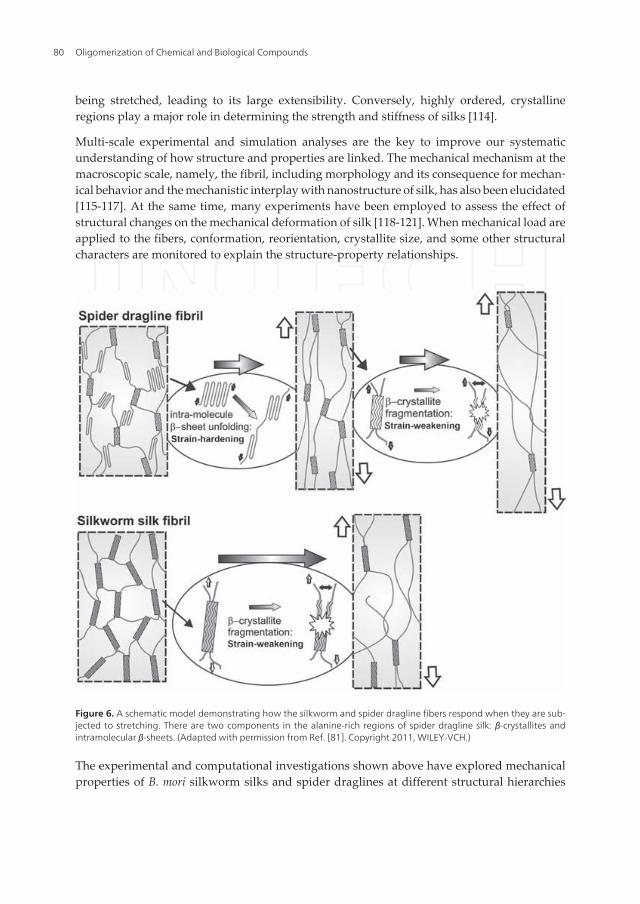
Figure 6. A schematic model demonstrating how the silkworm and spider dragline fibers respond when they are sub‐jected to stretching. There are two components in the alanine-rich regions of spider dragline silk: β-crystallites andintramolecular β-sheets. (Adapted with permission from Ref. [81]. Copyright 2011, WILEY-VCH.)
The experimental and computational investigations shown above have explored mechanicalproperties of B. mori silkworm silks and spider draglines at different structural hierarchies
Oligomerization of Chemical and Biological Compounds80
from sequence to crystallites to fibrils to fibers, as well as the effect of structural changes onthe overall mechanical behavior of silk fibers. In particular, a closer analysis of the mechanicalresponse, spider draglines behave typically the strain-hardening process in the post-yieldregion [81]. Based on the ‘β-sheet splitting’ mechanism [122], the occurrence of strain-harden‐ing in spider dragline as response to the structural factors has been clearly addressed. Spiderdragline silk can acquire extra toughness as a strain-hardening material by breaking intramo‐lecular β-sheets. On the other hand, B. mori silkworm silk has far fewer intramolecular β-sheetsin the amorphous region, therefore it is less extensible and only exhibits strain-weakening afteryield point (Figure 6).
Furthermore, unlike B. mori silk and the other types of spider silks, the mechanical propertiesof spider dragline silks are greatly influenced by water. When an unconstrained dragline silkfiber is immersed in water or comes in contact with a relative humidity greater than 60%, thethread starts to swell radially, doubling in diameter and a shrinking to half of its original length[123]. This process is known as supercontraction, which is another interesting property ofspider silk. A number of research groups have used different experimental techniques tounderstand supercontraction and the underlying mechanisms [124-126]. It is assumed thatsupercontraction is a result of reorientation of hydrogen bonds within the chains of proteinmolecules and is accompanied with the release of the prestress [127-130]. Some researchersattributed supercontraction mainly on the proline content of MaSp2 protein. Notably, thecontent of proline does not correlate with the mechanical performance of spider dragline silkfibers from different species, but influences the mechanical properties of wetted dragline silks[99, 126, 131, 132]. Recently, Guan et al. discussed the role of the two MaSp1 and MaSp2 proteinsin supercontraction and quantified a contraction of about 13% maximum, linked to thedisordered component of MaSp1 protein. Thus the remaining supercontraction to a total ofabout 30% is linked to the intrinsically disordered proline-containing fraction of MaSp2 protein[133]. After supercontraction, the silk is called supercontracted fibers, and are usually to beemployed to study the structure-property relationship of silk fiber [123, 134].
3. Silk protein assembly and silk fiber formation mechanism
The remarkable mechanical properties of silk fibers have spawned great interests in determi‐nation of their origin. Systematic studies of the natural spinning process of silk fibers haveshown a highly sophisticated hierarchical process, allowing for the transformation of solublesilk protein into solid fibers with specific mechanical and functional properties. Althoughmuch is already known about the characteristics of the silk proteins and silk fibers themselves,the process for silk assembly and spinning into fibers is yet to be resolved. A detailed knowl‐edge of silk fiber formation is critical for the biomimetic production of tough silk-like fibers.
3.1. Natural spinning process for B. mori silk and spider draglines
In nature, silk proteins are secreted and stored in the glands until they are processed into fibers.Morphological and histological studies demonstrate that the silk glands of B. mori silkworm
Silk Fiber — Molecular Formation Mechanism, Structure-Property Relationship and Advanced Applicationshttp://dx.doi.org/10.5772/57611
81

are a pair of tubes and the two tubal glands are connecting before the spinneret. The gland ofB. mori silkworm can be divided into three parts: posterior, middle and anterior [135]. Thefibrion protein is synthesized and present in a weak gel in the posterior division. The secretedproteins are transported to the middle division, where the sericin is synthesized, accumulatingas a shell around the fibroin. Due to the water going out through the cell wall of the gland inthe middle division, highly concentrated gel-like fibroin begins to undergo a gel-sol transitionand serves as a concentrated protein solution of 30 wt% [136]. Notably, the highly concentratedliquid protein, often referred to as the spinning dope, displays nematic liquid crystal properties[137-139]. The spinning dope is exposed to the elongational flow and moves forward in theanterior division. The shear force increases along the anterior division and the spinneret,leading to the orientation of the liquid crystallinity. The crystalline spinning dope is convertedinto a fiber containing water-insoluble silk II. This process is accompanied by extrusionthrough the spinneret into air, evaporating the residual water. Another remarkable feature ofthe fiber spinning is the stretching force, which is brought about by the repeated drawing backof the silkworm’s head, causing the orientation of protein molecules along with the silk fiber.
The major gland responsible for the dragline silk of Nephila clavipes spider contains thefollowing components: a long tail, a wider sac, named ampulla, and spinning duct approachingthe spinneret [140]. Each division of the gland possesses a unique function in fiber formation.For instance, a highly viscous silk protein solution of ~ 50% (w/v) is secreted from the A-zoneof the gland, which is comprised of the tail and two thirds part of the sac. Further compoundsforming the shell of the fiber may arise in the B-zone of the gland, which occupies the rest partof the sac. Like in the B. mori silkworm, the viscosity of the spider’s liquid crystalline proteinbecomes lower and the spinning dope moves forward in the spinning duct where the orien‐tation of liquid crystalline protein into a fiber begins. Due to its tapering, the shear force isincreasing along the spinning duct and the stress forces generated in the drawdown processbring the protein molecules into alignment. Hence the protein molecules join together withhydrogen bonds to give the final fiber with anti-parallel β-sheet structure. As the silk proteinmolecules aggregate and crystallize, they become more hydrophobic, inducing the loss ofwater from the surface of the silk fiber [141-143].
3.2. Silk protein assembly and silk fiber formation mechanism on a structural view
The formation of a solid fiber from soluble silk proteins is a remarkable process owing tocomplex biochemical and physical changes. For silk spinning, several assembly models, suchas liquid spinning theory [136] and micelle theory [144] have been proposed for the fiberformation, whereas the details remain to be elucidated. In order to understand the mechanismsof silk proteins assembly and fiber formation, the structure of proteins stored in B. morisilkworm gland and the major ampullate gland of spider should be clarified. In vivo, freshlysecreted fibroin first adopts silk I (the crystalline form of B. mori silk fibrion found before thespinning process) and random-coil conformation [145]. Silk I is less stable as shown by attemptsto study the secondary structure of silk I form using x-ray diffraction, electron diffraction orSS-NMR have caused the silk I to convert to silk II easily. Silk I remains poorly understood.Most investigations on the structure of the silk I form have been based on model building of
Oligomerization of Chemical and Biological Compounds82

peptides such as (Gly-Ala)n [146, 147]. The comparison of these models with limited experi‐mental data, resulting in a number of conflicting models describing the structure of silk I.Recently, the structure of silk I has been proposed as a repeated β-turn type II-like structure[148, 149]. The secreted dragline proteins are mainly natively unfolded within the gland andconsist of random-coil and polyproline-II with helix-like structure [150-152]. There is evidenceindicates that the polyalanine motifs form polyproline-II with a helix-like structure. Particu‐larly, the polyproline-II conformation may be important for maintaining the highly concen‐trated spinning dope, since the extended polyproline-II structure could prevent the formationof intramolecular hydrogen bonds. Additionally, the polyproline-II helix in spider fibroinfavors transforming into a β-sheet structure due to their similarity of dihedral angles.
It has reported that B. mori fibroins and spider spidroins usually show micellar-like structure[16, 144] with an amphiphilic sequence, implying short alternating hydrophilic and hydro‐phobic amino acid stretches flanked by larger hydrophilic terminal regions [153, 154]. Theintervening hydrophilic blocks located among the hydrophobic blocks in the protein preventpremature β-sheet formation, thus maintaining the solubility of the solution. Hence, the silkfiber formation involves shear force inducing the conversion of silk protein with specificstructural conformations into β-sheet structure. This conversion occurs in the spinning ductsfollowed by drawing down into fibrillar structure. It has shown upon passage through thegland and spinning duct, the proteins encounter remarkable changes in their solvent environ‐ment, such as extensional flow, protein concentration, pH and metal ion concentrations, whichare thought to be contributing factors in silk processing and affecting structural conformations[3, 142, 155-157]. The changes include removal of some water, slight acidification. In addition,the concentration of calcium ions (Ca2+) are increased as the silk protein flowed through thegland in B. mori. Unlike in the silkworm B. mori, the elemental composition of the silk dope inthe spider suggested that Ca2+ ions concentration stayed constant while potassium ions (K+)concentration increased. Meanwhile, sodium ions (Na+), chlorine ions (Cl−) are removed fromthe major ampullate gland.
Experiments made in vitro can provide several relevant insights into the process of silk proteinassembly and the formation mechanism of the silk fiber. To unravel the assembly mechanism,the reconstituted/recombinant proteins were applied for fiber assembly under specificcondition [158-163]. Jin et al. characterized the change of the supramolecular structure of silkfibroin with a reduction of the pH, which demonstrates the self-assembly of silk fibroin as afunction of pH. When the pH is reduced from 6.8 to 4.8, a morphological transition of silkfibroin from spherical micelles to nanofibrils and the conformational transition of silk fibroinfrom random coil to β-sheet were observed [158]. It may be driven by the stretching entropyeffect related to the hydrophobic block in the protein. Shniepp et al. [160] deposited the silkprotein solution on mica substrates without and with shearing by spin-coating. Only whenshear force was applied during deposition, fibrillar structures were obtained. A microfluidicdevice was employed in which the ion concentrations and pH value could be controlled, andsimultaneously, physical stress could be applied by channel design [163]. Silk fibers formedafter addition of phosphate, application of an elongational flow, and a pH change from 8 topH 6.
Silk Fiber — Molecular Formation Mechanism, Structure-Property Relationship and Advanced Applicationshttp://dx.doi.org/10.5772/57611
83

Actually, the chemical and mechanical stimuli together are likely to influence the fold ofnonrepetitive amino-terminal and carboxy-terminal and the hydrophilic spacers within thehydrophobic core domain [37, 135, 158, 164-166]. Due to the larger hydrophilic blocks at thechain ends of the protein molecules having charged groups, it is possible that they might playan important role in the molecular assembly and conformational transition at a specific pHthrough decreased electrostatic repulsion. A significant step towards understanding the effectof the terminal domains in assembly was the determination of atomistic structures of thenonrepetitive terminal regions of MaSp proteins. Kessler and Scheibel’s group reported thestructure of carboxy-terminal domain of Araneus diadematus ADF 3 by NMR spectroscopy [36].And Johannson, Knight reported the structures of amino-terminal domain of Euprosthenopsaustralis MaSp1 by X-ray scattering [35]. Interestingly, both terminal domains are mainlycomposed of α-helical barrels but with different folds. The carboxy-terminal domain mediateshomodimerization via a disulfide bond [36] and forms a clamp-like structural arrangement. Itseems to be implicated in a number of different functions, including control of solubility andfiber formation. The carboxy-terminal domains are able to form supramolecular assembliesresembling micellar-like structure, which is stabilized by the chaotropic ions (Figure 7). Theamino-terminal domains are monomeric at pH 6.8 and above [35]. And recent NMR and lightscattering studies on the amino-terminal nonrepetitive domain of Latrodectus hesperusconfirmed that a combination of pH and salt concentration controlled the dimerization. Themonomer was clearly stabilized at neutral pH in the presence of salt. While the lower pH and/or the reduced salt concentration causes the amino-terminal nonrepetitive domain to dimerizein an antiparallel fashion to create head-to-tail dimmers to dimmers [165]. The pH dependence
Figure 7. Schematic formation mechanism of the hierarchical assembly from molecular silk fibroin to microfibers.(Re‐printed from Ref. [1]. Copyright 2011, with permission from Elsevier.)
Oligomerization of Chemical and Biological Compounds84
of silk fiber formation showed that an oligomerization is greatly increased with a drop in pHat about 6.0, which is triggered by the amino-terminal domain [35, 165]. The structural changesof the amino-terminal nonrepetitive domains rearrange the position of the core regions withinthe micellar-like structure, together with mechanical stimuli supports the β-sheet formation.With the exchange of chaotropic ions for the relatively kosmotropic ions, exposing hydropho‐bic patches can enhance the assembly process. The amino-terminal and carboxy-terminaldomains sense changes in salt, pH, and shear force. And the fine-tuned interplay between theseparameters enables the silkworm and spider to efficiently produce a stable very tough fiberunder mild conditions
4. Advanced applications of silk fibers
Traditionally, silk has been utilized in the construction of textiles. Current research in silk fibersinvolves their innovative trends and advanced applications. Basically, the rich proportion ofessential amino acids in silk fibers indicates high nutritive value, meaning that silk fibroin canbe used as a dietary additive [167-169]. Furthermore, the amino acids, glycine, alanine, serineand tyrosine are of vital for nourishing the skin. The crystalline structure of silk protein reflectsUV radiation, acting as protective buffer between the skin and environment. The extracts ofsilk protein are used in soap making, personal care and cosmetic products. The silk protein isalso applied to enhance glossy, brightness, and softness of products. In addition, the produc‐tion of advanced man made super-fibers such as Kevlar involves petrochemical processing,which contributes to pollution. Interest in silk fibers is mainly due to the combination of themechanical properties and eco friendly way in which they are made. Spider silk fibers havebeen envisioned to be applied in a variety of technical textiles, including parachute cords,protective clothing and composite materials in aircrafts, which demand high toughness incombination with sleaziness.
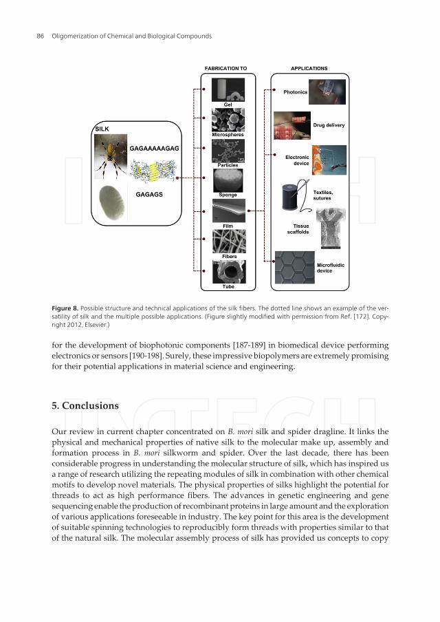
Silks are biocompatible, biodegradable and have implant ability, as well as morphologicflexibility. Silk fiber has been used as extremely thin suture for eye or nerve surgery for longhistory [170]. Nowadays, one attractive application of silk fibers is act as a source of novelbiomaterials. Recent progress with processing of silk fibers into various material forms, usuallyvia the formation of the fibroin/spidroin solution, including thread, hydrogels, tubes, sponges,microspheres, particles and films [9, 171], promotes the field of applications for silk fibers ingeneral (Figure 8) [172]. Silk protein can be modified by chemical treatment or used incombination with other materials and the silk-based biomaterials have been transformed forhigh-technology uses, with promising futures in the fields of biomedicine and materialengineering. Numerous studies have demonstrated that fibroin supports cell attachment andproliferation for a variety of cell types [173-178]. Studies have established a potential for silk-based biomaterials use as tissue engineering scaffolds, such as skeletal tissue like bone [179],ligaments [180], and cartilage [181, 182], as well as skin [183], blood vessels [184] and nerve[185]. Silks can be designed and offer another biomedical applications, such as delivery of smallmolecule drugs, proteins and genes [186]. Silk fibroin possesses remarkable optical properties,such as near-perfect transparency in a visible range. It has been identified as a suitable material
Silk Fiber — Molecular Formation Mechanism, Structure-Property Relationship and Advanced Applicationshttp://dx.doi.org/10.5772/57611
85
for the development of biophotonic components [187-189] in biomedical device performingelectronics or sensors [190-198]. Surely, these impressive biopolymers are extremely promisingfor their potential applications in material science and engineering.
5. Conclusions
Our review in current chapter concentrated on B. mori silk and spider dragline. It links thephysical and mechanical properties of native silk to the molecular make up, assembly andformation process in B. mori silkworm and spider. Over the last decade, there has beenconsiderable progress in understanding the molecular structure of silk, which has inspired usa range of research utilizing the repeating modules of silk in combination with other chemicalmotifs to develop novel materials. The physical properties of silks highlight the potential forthreads to act as high performance fibers. The advances in genetic engineering and genesequencing enable the production of recombinant proteins in large amount and the explorationof various applications foreseeable in industry. The key point for this area is the developmentof suitable spinning technologies to reproducibly form threads with properties similar to thatof the natural silk. The molecular assembly process of silk has provided us concepts to copy
Figure 8. Possible structure and technical applications of the silk fibers. The dotted line shows an example of the ver‐satility of silk and the multiple possible applications. (Figure slightly modified with permission from Ref. [172]. Copy‐right 2012, Elsevier.)
Oligomerization of Chemical and Biological Compounds86
and mimic the silkworm’s or spider’s ways of making and processing silks with tunableproperties. Such knowledge is beneficial for further improvement of synthetic polymer-basedmaterials. Combined with the discovery of new bio-inspired materials, the future applicationspace seems more and more broad. As a whole, further development of related yield isunderway.
Acknowledgements
The authors thank the financial support from the National Science Foundation of China (NSFC)under Grant 51073113, 91027039 and the Natural Science Foundation of the Jiangsu HigherEducation Institutions of China under Grant 10KJA540046. This work was also supported bythe Priority Academic Program Development of Jiangsu Higher Education Institutions(PAPD). We also acknowledge support from the Priority Academic Program Development ofJiangsu Higher Education Institutions (PAPD), Qing Lan Project for Excellent Scientific andTechnological Innovation Team of Jiangsu Province (2012) and Project for Jiangsu Scientificand Technological Innovation Team (2013). The author, Xinfang Liu, especially thank thesupport of the Postdoctoral Science Foundation of Jiangsu province (No. 1201030B).
Author details
Xinfang Liu and Ke-Qin Zhang*
*Address all correspondence to: [email protected]
National Engineering Laboratory for Modern Silk, College of Textile and ClothingEngineering, Soochow University, Suzhou, China
References
[1] Eisoldt L., Smith A., Scheibel T. Decoding the secrets of spider silk. Materials Today2011; 14(3): 80–86.
[2] Craig C L. Evolution of arthropod silks. Annual Review of Entomology 1997; 42: 231–267.
[3] Heim M., Keerl D., Scheibel T. Spider silk: From soluble protein to extraordinary fi‐ber. Angewandte Chemie, International Edition 2009; 48(20): 3584–3596.
[4] Shao Z., Vollrath F. Surprising strength of silkworm silk. Nature 2002; 418(15): 741–741.
Silk Fiber — Molecular Formation Mechanism, Structure-Property Relationship and Advanced Applicationshttp://dx.doi.org/10.5772/57611
87
[5] Iizuka E. Silk thread: Mechanism of spinning and its mechanical properties. Journalof Applied Polymer Science: Applied Polymer Symposium 1985; 41: 173–185.
[6] Heslot H. Artifical fibrous proteins: A review. Biochimie 1998; 80(1): 19–31.
[7] Asakura T., Kaplan D L. Silk production and processing. Encyclopedia of Agricultur‐al Science 1994; 4: 1–11.
[8] Lewis R. Unraveling the weave of spider silk. Bioscience 1996; 46(9): 636–638.
[9] Rockwood D N., Preda R C., Yücel T., Wang X Q., Lovett M L., Kaplan D L. Materialsfabrication from Bomyx mori silk fibroin. Nature Protocols 2011; 6(10): 1612–1631.
[10] Humenik M., Smith A M., Scheibel T. Recombinant spider silks – Biopolymers withpotential for future applications. Polymer 2011; 3(1): 640–661.
[11] Fu C., Shao Z., Fritz V. Animal silks: their structures, properties and artificial produc‐tion. Chemical Communication 2009; 43: 6515–6529.
[12] Hardy J G., Römer L M., Scheibel T R. Polymeric materials based on silk proteins.Polymer 2008; 49(30): 4309–4327.
[13] Gührs K H., Weisshart K., Grosse F. Lessons from nature – protein fibers. Reviews inMolecular Biotechnology 2000; 74(2): 121–134.
[14] Shen Y., Johnson M A., Martin D C. Microstructural characterization of Bombyx morisilk fibers. Macromolecules 1998; 31(25): 8857–8864.
[15] Li S F., McGhie A J., Tang S L. New internal structure of spider dragline silk revealedby atomic force microscopy. Biophysical Journal 1994; 66(4): 1209–1212.
[16] Oroudjev E., Soares J., Arcdiacono S., Thompson J B., Fossey S A., Hansma H G. Seg‐mented nanofibers of spider dragline silk: Atom force microscopy and single-mole‐cule force spectroscopy. Proceedings of the National Academy of Science of theUnited States of America 2002; 99(9, suppl.2): 6460–6465.
[17] Augsten K., Muehlig P., Hermann C. Glycoproteins and skin-core structure in Nephi‐la clavipes spider silk observed by light and electron microscopy. Scanning 2000;22(1): 12–15.
[18] Putthanarat S., Stribeck N., Fossey S A., Eby R K., Adams W W. Investigation of thenanofibrils of silk fibers. Polymer 2000; 41(21): 7735–7747.
[19] Du N., Liu X Y., Narayanan J., Li L., Lim M L M., Li D. Design of superior spider silk:From nanostructure to mechanical properties. Biophysical Journal 2006; 91(12): 4528–4535.
[20] Padamwar M N., Pawar A P. Silk sericin and its applications: A review. Journal ofScientific & Industrial Research 2004; 63(4): 323–329.
Oligomerization of Chemical and Biological Compounds88

[21] Robson R M. Microvoids in Bombyx mori silk. An electron microscope study. Interna‐tional Journal of Biological Macromolecules 1999; 24(2-3): 145–150.
[22] Frische S., Maunsbach A B., Vollrath F. Elongate cavities and skin-core structure inNephila spider silk observed by electron microscopy. Journal of Microscopy 1998;189(1): 64–70.
[23] Takei F., Kikuchi Y., Kikuchi A., Mizuno S., Shimura K. Further evidence for impor‐tance of the subunit combination of silk fibroin in its efficient secretion from the pos‐terior silk gland cells. The Journal of Cell Biology 1987; 105(1): 175–180.
[24] Tanaka K., Mori K., Mizuno S. Immunological identification of the major disulfide-linked light component of silk fibroin. Journal of Biochemistry (Tokyo) 1993; 114(1):1–4.
[25] Tanaka K., Kajiyama N., Ishikura K., Waga S., Kikuchi A., Ohtomo K., Takagi T.,Mizuno S. Determination of the site of disulfide linkage between heavy and lightchains of silk fibroin produced by Bombyx mori. Biochimica et Biophysica Act (BBA) –Protein Structure and Molecular Enzymology 1999; 1432(1): 92–103.
[26] Xu M., Lewis R V. Structure of a protein superfiber: spider dragline silk. Proceedingsof the National Academy of Science of the United States of America 1990; 87(18):7120–7124.
[27] Hinman M B., Lewis R V. Isolation of a clone encoding a second dragline silk fibroin.Nephila clavipes dragline silk is a two-protein fiber. The Journal of Biological Chemis‐try 1992; 267(27): 19320–19324.
[28] Beckwitt R., Arcidiacono S. Sequence conservation in the C-terminal region of spidersilk proteins (spidroin) from Nephila clavipes (Tetragnathidae) and Araneus bicentenar‐ius (Araneidae). The Journal of Biological Chemistry 1994; 269(9): 6661–6663.
[29] Sponner A., Vater W., Rommerskirch W., Vollrath F., Unger E., Grosse F., WeisshartK. The conserved C-termini contribute to the properties of spider silk fibroins. Bio‐chemical and Biophysical Research Communications 2005; 338(2): 897–902.
[30] Eisoldt L., Thamm C., Scheibel T. The role of terminal domains during storage andassembly of spider silk proteins. Biopolymers 2012; 97(6): 355–361.
[31] Rising A., Hjälm G., Engström W., Johansson J. N-terminal nonrepetitive domaincommon to dragline, flagelliform, and cylindriform spider silk proteins. Biomacro‐molecules 2006; 7(11): 3120–3124.
[32] Motriuk-Smith D., Smith A., Hayashi C Y., Lewis R V. Analysis of the conserved N-terminal domains in major ampullate spider silk proteins. Biomacromolecules 2005;6(6): 3152–3159.
Silk Fiber — Molecular Formation Mechanism, Structure-Property Relationship and Advanced Applicationshttp://dx.doi.org/10.5772/57611
89
[33] Zhou C Z., Confalonieri F., Jacquet M., Perasso R., Li Z G., Janin J. Silk fibroin: struc‐tural implications of a remarkable amino acid sequence. Proteins: Structure, Func‐tion, and Genetics 2001; 44(2): 119–122.
[34] Zhou C Z., Confalonieri F., Medina N., Zivanovic Y., Esnault C., Yang T., Jacquet M.,Janin J., Duguet M., Perasso R., Li Z G. Fine organization of Bombyx mori fibroinheavy chain gene. Nucleic Acid Research 2000; 28(12): 2413–2419.
[35] Askarieh G., Hedhammar M., Nordling K., Saenz A., Casals C., Rising A., JohanssonJ., Knight S D. Self-assembly of spider silk proteins is controlled by a pH-sensitive re‐lay. Nature 2010; 465(13): 236–238.
[36] Hagn F., Eisoldt L., Hardy J G., Vendrely C., Coles M., Scheibel T., Kessler, H. A con‐served spider silk domain acts as a molecular switch that controls fibre assembly.Nature 2010; 465(13): 239–242.
[37] He Y X., Zhang N N., Li W F., Jia N., Chen B Y., Zhou K., Zhang J., Chen Y., Zhou CZ. N-terminal domain of Bombyx mori fibroin mediates the assembly of silk in re‐sponse to pH decrease. Journal of Molecular Biology 2012; 418(3-4): 197–207.
[38] Hagn F. A structural view on spider silk proteins and their role in fiber assembly.Journal of Peptide Science 2012; 18(6): 357–365.
[39] Ittah S., Cohen S., Garty S., Cohn D., Gat U. An essential role for the C-terminal do‐main of a dragline spider silk protein in directing fiber formation. Biomacromole‐cules 2006; 7(6): 1790–1795.
[40] Bini E., Knight D P., Kaplan D L. Mapping domain structures in silks from insectsand spiders related to protein assembly. Journal of Molecular Biology 2004; 335(1-2):27–40.
[41] Simmons A., Ray E., Jelinski L. W. Solid-state 13C NMR of Nephila clavipes draglinesilk establishes structure and identity of crystalline regions. Macromolecules 1994;27(18): 5235–5237.
[42] Asakura T., Yao J. 13C CP/MAS NMR study on structural heterogeneity in Bombyxmori silk fiber and their generation by stretching. Protein Science 2002; 11(11): 2706–2713.
[43] Boulet-Audet M., Lefèvre T., Buffeteau T., Pézolet M. Attenuated total reflection in‐frared spectroscopy: An efficient technique to quantitatively determine the orienta‐tion and conformation of proteins in single silk fibers. Applied Spectroscopy 2008;62(9): 956–962.
[44] Holland G P., Creager M S., Jenkins J E., Lewis R V., Yarger J L. Determining secon‐dary structure in spider dragline silk by carbon-carbon correlation solid-state NMRspectroscopy. Journal of the American Chemistry Society 2008; 130(30): 9871–9877.
[45] Colomban P., Dinh H M., Riand J., Prinsloo L C., Mauchamp B. Nanomechanics ofsingle silkworm and spider fibres: a Raman and micro-mechanical in situ study of the
Oligomerization of Chemical and Biological Compounds90
conformation change with stress. Journal of Raman Spectroscopy 2008; 39(12): 1749–1764.
[46] Lefèvre T., Rousseau M E., Pézolet M. Protein secondary structure and orientation insilk as revealed by Raman spectromicroscopy. Biophysical Journal 2007; 92(8): 2885–2895.
[47] Holland G P., Jenkins J E., Creager M S., Lewis R V., Yarger J L. Quantifying the frac‐tion of glycine and alanine in β-sheet and helical conformations in spider draglinesilk using solid-state NMR. Chemical Communication 2008; 43: 5568–5570.
[48] Jenkins J E., Creager M S., Lewis R V., Holland G P., Yarger J L. Quantitative correla‐tion between the protein primary sequences and secondary structures in spider drag‐line silks. Biomacromolecules 2010; 11(1): 192–200.
[49] Ling S., Qi Z., Knight D P., Shao Z., Chen X. Synchrotron FTIR microspectroscopy ofsingle natural silk fibers. Biomacromolecules 2011; 12(9): 3344–3349.
[50] Dicko C., Knight D., Kenney J M., Vollrath F. Structural conformation of spidroin insolution: A synchrotron radiation circular dichroism study. Biomacromolecules 2004;5(3): 758–767.
[51] Lefèvre T., Paquet-Mercier F., Rioux-Dubé J F., Pézolet M. Structure of silk by Ramanspectromicroscopy: From the spinning glands to the fibers. Biopolymers 2012; 97(6):322–336.
[52] Kaplan D.; Adams W W., Farmer B., Viney C., editors. Silk polymers: materials sci‐ence and biotechnology. American Chemical Society Symposium Series 1994.
[53] Marsh R E., Corey R B., Pauling L. An investigation of the structure of silk fibroin.Biochimica et Biophysica Acta 1955; 16: 1–34.
[54] Warwicker J O. Comparative studies of fibroins: II. The crystal structures of variousfibroins. Journal of Molecular Biology 1960; 2(6): 350–362.
[55] Dekker M. In: Porter D. (ed.) Group interaction modeling of polymer properties.New York. 1995. P499.
[56] Porter D., Vollrath F., Shao Z. Predicting the mechanical properties of spider silk as amodel nanostructured polymer. The European Physical Journal E 2005; 16(2): 199–206.
[57] Rousseau M E., Lefèvre T., Beaulieu L., Asakura L., Pézolet M. Study of protein con‐formation and orientation in silkworm and spider silk fibers using Raman micro‐spectroscopy. Biomacromolecules 2004; 5(6): 2247–2257.
[58] Krimm S., Bandekar J. Vibrational Spectroscopy and conformation of peptides, poly‐peptides, and proteins. Advances in Protein Chemistry 1986; 38: 181–364.
Silk Fiber — Molecular Formation Mechanism, Structure-Property Relationship and Advanced Applicationshttp://dx.doi.org/10.5772/57611
91
[59] Dong J., Wan Z., Popov M., Carey P R., Weiss M. A. Insulin assembly damps confor‐mational fluctuations: Raman analysis of amide I line-widths in native states and fi‐brils. Journal of Molecular Biology 2003; 330(2): 431–442.
[60] Zheng S., Li G., Yao W., Yu T. Raman spectroscopic investigation of the denaturationprocess of silk fibroin. Applied Spectroscopy 1989; 43(7): 1269–1272.
[61] Simmons A H., Michal C A., Jelinski L W. Molecular orientation and two-componentnature of the crystalline fraction of spider dragline silk. Science 1996; 271(5245): 84–87.
[62] Takahashi Y. Crystal structure of silk of Bombyx mori. In: Kaplan D.; Adams W W.,Farmer B., Viney C. (eds.) Silk polymers: materials science and biotechnology. Amer‐ican Chemical Society Symposium Series 1994. 544: P168–175.
[63] Drummy L F., Farmer B L., Naik R R. Correlation of the β-sheet crystal size in silkfibers with the protein amino acid sequence. Soft Matter 2007; 3(7): 877–882.
[64] Termonia Y. Molecular modeling of spider silk elasticity. Macromolecules 1994;27(25): 7378–7381.
[65] Kümmerlen J., van Beek J D., Vollrath F., Meier B H. Local structure in spider drag‐line silk investigated by two-dimensional spin-diffusion nuclear magnetic resonance.Macromolecules 1996; 29(8): 2920–2928.
[66] Becker M A., Tuross N. Initial degradation changes found in Bombyx mori silk fibroin.In: Kaplan D.; Adams W W., Farmer B., Viney C. (eds.) Silk polymers: materials sci‐ence and biotechnology. American Chemical Society Symposium Series 1994. 544:P252–269.
[67] Yang Z., Grubb D T., Jelinski L W. Small-angle X-ray scattering of spider draglinesilk. Macromolecules 1997; 30(26): 8254–8261.
[68] Plaza G R., Pérez-Rigueiro J., Riekel C., Perea G B., Agulló-Rueda F., BurghammerM., Guinea G V., Elices M. Relationship between microstructure and mechanicalproperties in spider silk fibers: identification of two regimes in the microstructuralchanges. Soft Matter 2012; 8(22): 6015–6020.
[69] Sampath S., Isdebski T., Jenkins J E., Ayon J V., Henning R W., Orgel J P R O., Anti‐poa O., Yarger J L. X-ray diffraction study of nanocrystalline and amorphous struc‐ture within major and minor ampullate dragline spider silks. Soft Matter 2012; 8(25):6713–6722.
[70] Grubb D T., Jelinski L W. Fiber morphology of spider silk: The effects of tensile de‐formation. Macromolecules 1997; 30(10): 2860–2867.
[71] Grubb D T., Ji G. Molecular chain orientation in supercontracted and re-extendedspider silk. International Journal of Biological Macromolecules 1999; 24(2-3): 203–210.
Oligomerization of Chemical and Biological Compounds92

[72] Cruz D H., Rousseau M E., West M M., Pézolet M., Hitchcock A P. Quantitative map‐ping of the orientation of fibroin β-sheets in B. mori cocoon fibers by scanning trans‐mission X-ray microscopy. Biomacromolecules 2006; 7(3): 836–843.
[73] van Beek J D., Hess S., Vollrath F., Meier B H. The molecular structure of spider drag‐line silk: Folding and orientation of the protein backbone. Proceedings of the Nation‐al Academy of Science of the United States of America 2002; 99(16): 10266–10271.
[74] Marcotte I., van Beek J D., Meier B H. Molecular disorder and structure of spiderdragline silk investigated by two-dimensional solid-state NMR spectroscopy. Macro‐molecules 2007; 40(6): 1995–2001.
[75] Asakura T., Yao J., Yamane T., Umemura K., Ulrich A S. Heterogeneous structure ofsilk fibers from Bombyx mori resolved by 13C solid-state NMR spectroscopy. Journalof the American Chemical Society 2002; 124(30): 8794–8795.
[76] Gosline J M., Denny M W., DeMont M E. Spider silk as rubber. Nature 1984;309(5968): 551–552.
[77] Gatesy J., Hayashi C., Motriuk D., Woods J., Lewis R. Extreme diversity, conserva‐tion, and convergence of spider silk fibroin sequence. Science 2001; 291(5513): 2603–2605.
[78] Ma B Y., Nussinov R. Molecular dynamics simulations of alanine rich β-sheet oligom‐ers: Insight into amyloid formation. Protein Science 2002; 11(10): 2335–2350.
[79] Keten S., Buehler M J. Atomistic model of the spider silk nanostructure. AppliedPhysics Letters 2010; 96(15): 153701–153703.
[80] Keten S., Buehler M J. Nanostructure and molecular mechanics of spider dragline silkprotein assemblies. Journal of the Royal Society Interface 2010; 7(53): 1709–1721.
[81] Du N., Yang Z., Liu X Y., Li Y., Xu H Y. Structural origin of the strain-hardening ofspider silk. Advanced Functional Materials 2011; 21(4): 772–778.
[82] Li X., Eles P T., Michal C A. Water permeability of spider dragline silk. Biomacromo‐lecules 2009; 10(5): 1270–1275.
[83] Ene R., Papadopoulos P., Kremer F. Partial deuteration probing structural changes insupercontracted spider silk. Polymer 2010; 51(21): 4784–4789.
[84] Paquet-Mercier F., Lefèvre T., Auger M., Pézolet M. Evidence by infrared spectrosco‐py of the presence of two type of β-sheets in major ampullate spider silk and silk‐worm silk. Soft Matter 2013; 9(1): 208–215.
[85] Rousseau M E., Cruz D H., West M M., Hitchcock A P., Pézolet M. Nephila clavipesspider dragline silk microstructure studies by scanning transmission X-ray microsco‐py. Journal of the American Chemical Society 2007; 129(13): 3897–3905.
Silk Fiber — Molecular Formation Mechanism, Structure-Property Relationship and Advanced Applicationshttp://dx.doi.org/10.5772/57611
93
[86] Fossey S A., Tripathy S. Atomistic modeling of interphases in spider silk fibers. Inter‐national Journal of Biological Macromolecules 1999; 24(2-3): 119–125.
[87] Thiel B L., Guess K B., Viney C. Non-periodic lattice crystals in the hierarchical mi‐crostructure of spider (major ampullate) silk. Biopolymers 1997; 41(7): 703–719.
[88] Trancik J E., Czernuszka J T., Bell F I., Viney C. Nanostructural features of a spiderdragline silk as revealed by electron and X-ray diffraction studies. Polymer 2006;47(15): 5633–5642.
[89] Swanson B O., Blackledge T A., Beltran J., Hayashi C Y. Variation in the materialproperties of spider dragline silk across species. Applied Physics A: Materials Sci‐ence & Processing 2006; 82(2): 213–218.
[90] Sirichaisit J., Brookes V L., Young R J., Vollrath F. Analysis of structure/property rela‐tionships in silkworm (Bombyx mori) and spider dragline (Nephila edulis) silks usingRaman spectroscopy. Biomacromolecules 2003; 4(2): 387–394.
[91] Denny M. The physical properties of spider’s silk and their role in the design of orb-webs. The Journal of Experimental Biology 1976; 65(2): 483–506.
[92] Gosline J M., Guerette P A., Ortlepp C S., Savage K N. The mechanical design of spi‐der silks: From fibroin sequence to mechanical function. The Journal of ExperimentalBiology 1999; 202(23): 3295–3303.
[93] Hu X., Vasanthavada K., Kohler K., McNary S., Moore A M F., Vierra C A. Molecularmechanisms of spider silk. Cellular and Molecular Life Sciences 2006; 63(17): 1986–1999.
[94] Ko F K., Jovicic J. Modeling of mechanical properties and structural design of spiderweb. Biomacromolecules 2004; 5(3): 780–785.
[95] Sezutsu H., Yukuhiro K. Dynamic rearrangement within the Antheraea pernyi silk fi‐broin gene is associated with four types of repetitive units. Journal of Molecular Evo‐lution 2000; 51(4): 329–338.
[96] Fu C., Porter D., Chen X., Vollrath F., Shao Z. Understanding the mechanical proper‐ties of Antheraea pernyi silk – From primary structure to condensed structure of theprotein. Advanced Functional Materials 2011; 21(4): 729–737.
[97] Garb J E., Dimauro T., Lewis R V., Hayashi C Y. Expansion and intragenic homogeni‐zation of spider silk since the triassic: Evidence from mygalomorphae (tarantulas andtheir kin) spidroins. Molecular Biology and Evolution 2007; 24(11): 2454–2464.
[98] Brooks A E., Steinkraus H B., Nelson S R., Lewis R V. An investigation of the diver‐gence of major ampullate silk fibers from Nephila clavipes and Argiope aurantia. Bio‐macromolecules 2005; 6(6): 3095–3099.
[99] Liu Y., Sponner A., Porter D., Vollrath F. Proline and processing of spider silks. Bio‐macromolecules 2008; 9(1): 116–121.
Oligomerization of Chemical and Biological Compounds94

[100] Brooks A E., Stricker S M., Joshi S B., Kamerzell T J., Middaugh C R., Lewis R V.Properties of synthetic spider silk fibers based on Argiope aurantia MaSp2. Biomacro‐molecules 2008; 9(6): 1506–1510.
[101] Rauscher S., Baud S., Miao M., Keeley F., Pomes R. Proline and glycine control pro‐tein self-organization into elastomeric or amyloid fibrils. Structure 2006; 14(11): 1667–1676.
[102] Hayashi C Y., Shipley N H., Lewis R V. Hypotheses that correlate the sequence,structure, and mechanical properties of spider silk proteins. International Journal ofBiological Macromolecules 1999; 24(2-3): 271–275.
[103] Shao Z., Vollrath F., Yang Y., Thøgersen H C. Structure and behavior of regeneratedspider silk. Macromolecules 2003; 36(4): 1157–1161.
[104] Lee S M., Pippel E., Göesele U., Dresbach C., Qin Y., Chandran C V., Bräeuniger T.,Hause G., Knez M. Greatly increased toughness of infiltrated spider silk. Science2009; 324(5926): 488–492.
[105] Riekel C., Müller M. In situ X-ray diffraction during forced silking of spider silk.Macromolecules 1999; 32(13): 4464–4466.
[106] Riekel C., Vollrath F. Spider silk fibre extrusion: combined wide-and small-angle X-ray microdiffraction experiments. International Journal of Biological Macromole‐cules. 2001; 29(3): 203–210.
[107] Khan M R., Morikawa H., Gotoh Y., Miura M., Ming Z., Sato Y., Iwasa M. Structuralcharacteristics and properties of Bombyx mori silk fiber obtained by different artificialforcibly silking speeds. Internation Journal of Biological Macromolecules 2008; 42(3):264–270.
[108] Keten S., Xu Z., Ihle B., Buehler M J. Nanoconfinement controls stiffness, strengthand mechanical toughness of β-sheet crystals in silk. Nature Materials 2010; 9(4): 359–367.
[109] Xiao S., Stacklies W., Debes C., Gräter F. Force distribution determines optimallength of β-sheet crystals for mechanical robustness. Soft Matter 2011; 7(4): 1308–1311.
[110] Sinsawat A., Putthanarat S., Magoshi Y., Pachter R., Eby R K. X-ray diffraction andcomputational studies of the modulus of silk (Bombyx mori). Polymer 2002; 43(4):1323–1330.
[111] Krasnov I., Diddens I., Hauptmann N., Helms G., Ogurreck M., Seydel T., Funari SS., Muller M. Mechanical properties of silk: Interplay of deformation on macroscopicand molecular length scales. Physical Review Letters 2008; 100(4): 048104/1–048104/4.
[112] Sinsawat A., Putthanarat S., Magoshi Y., Pachter R., Eby R K. The crystal modulus ofsilk (Bombyx mori). Polymer 2003; 44(3): 909–910.
Silk Fiber — Molecular Formation Mechanism, Structure-Property Relationship and Advanced Applicationshttp://dx.doi.org/10.5772/57611
95
[113] Vollrath F., Porter D. Spider silk as a model biomaterial. Applied Physics A: Materi‐als Science & Processing 2006; 82(2): 205–212.
[114] Nova A., Keten S., Pugno N M., Redaelli A., Buehler M J. Molecular and nanostruc‐tural mechanisms of deformation, strength and toughness of spider silk fibrils. NanoLetters 2010; 10(7): 2626–2634.
[115] Papadopoulos P., Sölter J., Kremer F. Hierarchies in the structural organization ofspider silk – a quantitative model. Colloid and Polymer Science 2009; 287(2): 231–236.
[116] Giesa T., Arslan M., Pugno N M., Buehler M J. Nanoconfinement of spider silk fibrilsbegets superior strength, extensibility, and toughness. Nano Letters 2011; 11(11):5038–5046.
[117] Brown C P., Harnegea C., Gill H S., Price A J., Traversa E., Licoccia S., Rosei F. Roughfibrils provide a toughening mechanism in biological fibers. ACS Nano 2012; 6(3):1961–1969.
[118] Lefèvre T., Paquet-Mercier F., Lesage S., Rousseau M E., Bédard S., Pézolet M. Studyby Raman spectromicroscopy of the effect of tensile deformation on the molecularstructure of Bombyx mori silk. Vibrational Spectroscopy 2009; 51(1): 136–141.
[119] Eles P T., Michal C A. A DECODER NMR study of backbone orientation in Nephilaclavipes dragline silk under varying strain and draw rate. Biomacromolecules 2004;5(3): 661–665.
[120] Seydel T., Kölln K., Krasnov I., Diddens I., Hauptmann N., Helms G., Ogurreck M.,Kang S G., Koza M M., Müller M. Silkworm silk under tensile strain investigated bysynchrotron X-ray diffraction and neutron spectroscopy. Macromolecules 2007, 40(4):1035–1042.
[121] Brookes V L., Young R J., Vollrath F J. Deformation micromechanics of spider silk.Journal of Materials Science 2008; 43(10): 3728–3732.
[122] Wu X., Liu X Y., Du N., Xu G., Li B. Unraveled mechanism in silk engineering: Fastreeling induced silk toughening. Applied Physics Letters 2009; 95: 093703/1–093703/4.
[123] Jelinski L W., Blye A., Liivak O., Michal C., LaVerde G., Seidel A., Shah N., Yang Z.Orientation, structure, wet-spinning, and molecular basis for supercontraction of spi‐der dragline silk. International Journal of Biological Macromolecules 1999; 24(2-3):197–201.
[124] Holland G P., Jenkins J E., Creager M S., Lewis R V., Yarger J L. Solid-state NMR in‐vestigation of major and minor ampullate spider silk in the native and hydratedstates. Biomacromolecules 2008; 9(2): 651–657.
[125] Shao Z., Vollrath F., Sirichaisit J., Young R J. Analysis of spider silk in native and su‐percontracted states using Raman spectroscopy. Polymer 1999; 40(10): 2493–2500.
Oligomerization of Chemical and Biological Compounds96

[126] Savage K N., Gosline J M. The effect of proline on the network structure of major am‐pullate silks as inferred from their mechanical and optical properties. The Journal ofExperimental Biology 2008; 211(12): 1937–1947.
[127] Liu Y., Shao Z., Vollrath F. Relationships between supercontraction and mechanicalproperties of spider silk. Nature Materials 2005; 4(12): 901–905.
[128] Bell F I., McEwen I J., Viney C. Supercontraction stress in wet spider dragline. Nature2002; 416(6876): 37–37.
[129] Papadopoulos P., Ene R., Weidner I., Kremer F. Similarities in the structural organi‐zation of major and minor ampullate spider silk. Macromolecular Rapid Communi‐cations 2009; 30(9-10): 851–857.
[130] Blackledge T A., Boutry C., Wong S C., Baji A., Dhinojwala A., Sahni V., Agnarsson,I. How super is supercontraction? Persistent versus cyclic responses to humidity inspider dragline silk. The Journal of Experimental Biology 2009; 212(13): 1981–1988.
[131] Liu Y., Shao Z., Vollrath F. Elasticity of spider silks. Biomacromolecules 2008; 9(7):1782–1786.
[132] Boutry C., Blackledge T A. Evolution of supercontraction in spider silk: structure −function relationship from tarantulas to orb-weavers. The Journal of Experimental Bi‐ology 2010, 213(20): 3505–3514.
[133] Guan J., Vollrath F., Porter D. Two mechanisms for supercontraction in Nephila spi‐der dragline silk. Biomacromolecules 2011; 12(11): 4030–4035.
[134] Glišović A., Vehoff T., Davies R J., Salditt T. Strain dependent structural changes ofspider dragline silk. Macromolecules 2008; 41(2): 390–398.
[135] Wong Po Foo C., Bini E., Hensman J., Knight D P., Lewis R V., Kaplan D L. Role ofpH and charge on silk protein assembly in insects and spiders. Applied Physics A:Materials Science and Processing 2006; 82(2): 223–233.
[136] Vollrath F., Knight D. P. Liquid crystalline spinning of spider silk. Nature 2001;410(6828): 541–548.
[137] Knight D P., Vollrath F. Liquid crystals and flow elongation in a spider’s silk produc‐tion line. Proceedings of the Royal Society B: Biological Science 1999; 266(1418): 519–523.
[138] Kerkam K., Viney C., Kaplan D L., Lombardi S. Liquid crystallinity of natural silk se‐cretions. Nature 1991; 349(6310): 596–598.
[139] Willcox P J., Gido S P. Muller W. Kaplan D L. Evidence of a cholesteric liquid crystal‐line phase in natural silk spinning processes. Macromolecules 1996; 29(15): 5106–5110.
Silk Fiber — Molecular Formation Mechanism, Structure-Property Relationship and Advanced Applicationshttp://dx.doi.org/10.5772/57611
97

[140] Lefevre T, Boudreault S, Cloutier C, Pezolet M. Conformational and orientationaltransformation of silk proteins in the major ampullate gland of Nephila clavipes spi‐ders. Biomacromolecules 2008; 9(9): 2399–2407.
[141] Vollrath F., Knight D P. Structure and function of the silk production pathway in thespider Nephila edulis. International Journal of Biological Macromolecules 1999;24(2-3): 243–249.
[142] Knight D P., Vollrath F. Changes in element composition along the spinning duct in aNephila spider. Naturwissenschaften 2001; 88(4): 179–182.
[143] Terry A E., Knight D P., Porter D., Vollrath F. pH induced changes in the rheology ofsilk fibroin solution from the middle division of Bombyx mori silkworm. Biomacromo‐lecules 2004; 5(3): 768–772.
[144] Jin H J., Kaplan D L. Mechanism of silk processing in insects and spiders. Nature2003; 424(28): 1057–1061.
[145] Monti P., Taddei P., Freddi G., Asakura T., Tsukada M. Raman spectroscopic charac‐terization of Bombyx mori silk fibroin: Raman spectrum of silk I. Journal of RamanSpectroscopy 2001; 32(2): 103–107.
[146] Asakura T., Yamane T., Nakazawa Y., Kameda T., Ando K. Structure of Bombyx morisilk fibroin before spinning in solid state studied with wide angle X-ray scatteringand 13C cross-polarization/magic angle spinning NMR. Biopolymers 2001; 58(5): 521–525.
[147] Taddei P., Asakura T., Yao J., Monti P. Raman study of poly(alanine-glycine)-basedpeptides containing tyrosine, valine, and serine as model for the semicrystalline do‐mains of Bombyx mori silk fibroin. Biopolymers 2004; 75(4): 314–324.
[148] Asakura T., Ashida, J., Yamane T., kameda T., Nakazawa, Y., Ohgo K., Komatsu K. Arepeated β-turn structure in poly(Ala-Gly) as a model for silk I of Bombyx mori silkfibroin studied with two-dimensional spin-diffusion NMR under off magic anglespinning and rotational echo double resonance. Journal of Molecular Biology 2001;306(2): 291–305.
[149] Asakura T., Ohgo K., Komatsu K., Kanenari M., Okuyama K. Refinement of repeatedβ-turn structure for silk I conformation of Bombyx mori silk fibroin using 13C solid-state NMR and X-ray methods. Macromolecules 2005; 38(17): 7397–7403.
[150] Hirijida D H., Do K G., Wong S., Zax D., Jelinski L W. 13C NMR of Nephila clavipesmajor ampullate silk gland. Biophysical Journal 1996; 71(6): 3442–3447.
[151] Hronska M., van Beek J D., Willimason P T F., Vollrath F., Meier B H. NMR charac‐terization of native liquid spider dragline silk from Nephila edulis. Biomacromolecules2004; 5(3): 834–839.
[152] Lefèvre T., Leclerc J., Rioux-Dubé J F., Buffeteau T., Paquin M C., Rousseau M E.,Cloutier I., Auger M., Gagné S M., Boudreault S., Cloutier C., Pézolet M. Conforma‐
Oligomerization of Chemical and Biological Compounds98
tion of spider silk proteins in situ in the intact major ampullate gland and in solution.Biomacromolecules 2007; 8(8): 2342–2344.
[153] Roemer L., Scheibel T. In: Scheibel T. (ed.) Fibrous proteins. Austin: Landes Bio‐science; 2008. P137–151.
[154] Exler J H., Hummerich D., Scheibel T. The amphiphilic properties of spider silks areimportant for spinning. Angewandte Chemie International Edition 2007; 46(19):3559–3562.
[155] Magoshi J., Magoshi Y., Becker M A., Nakamura S. Biospinning (silk fiber formation,multiple spinning mechanisms). In: Salamone J C. (ed.) Polymeric Materials Encyclo‐pedia. CRC Press: New York. 1996. vol. 1: P667–679.
[156] Vollrath F., Knight D P., Hu X W. Silk production in a spider involves acid bath treat‐ment. Proceedings of the Royal Society B: Biological Science 1998; 265(1398): 817–820.
[157] Hardy J G., Scheibel T R. Silk-inspired polymers and proteins. Biochemical SocietyTransactions 2009; 37(4): 677–681.
[158] Chen P., Kim H S., Park C Y., Kim H S., Chin I J., Jin H J. pH-triggered transition ofsilk fibroin from spherical micelles to nanofibrils in water. Macromolecular Research2008; 16(6): 539–543.
[159] Dicko C., Kenney J M., Knight D., Vollrath F. Transition to a β-sheet-rich structure inspidroin in vitro: The effects of pH and cations. Biochemistry 2004; 43(44): 14080–14087.
[160] Greving I., Cai M., Vollrath F., Schniepp H C. Shear-induced self-assembly of nativesilk proteins into fibrils studied by atomic force microscopy. Biomacromolecules2012; 13(3): 676–682.
[161] Xie F., Zhang H., Shao H., Hu X. Effect of shearing on formation of silk fibers fromregenerated Bombyx mori silk fibroin aqueous solution. International Journal of Bio‐logical Macromolecules 2006; 38(3-5): 284–288.
[162] Kinahan M E., Filippidi E., Köster S., Hu X., Evans H M., Pfohl T., Kaplan D L., WongJ. Tunable silk: Using microfluidics to fabricate silk fibers with controllable proper‐ties. Biomacromolecules 2011; 12(5): 1504–1511.
[163] Rammensee S., Slotta U., Scheibel T., Bausch A R. Assembly mechanism of recombi‐nant spider silk proteins. Proceedings of the National Academy of Science of theUnited States of America 2008; 105(18): 6590–6595.
[164] Gronau G., Qin Z., Buehler M J. Effect of sodium chloride on the structure and stabil‐ity of spider silk’s N-terminal protein domain. Biomaterial Science 2013; 1(3): 276–284.
Silk Fiber — Molecular Formation Mechanism, Structure-Property Relationship and Advanced Applicationshttp://dx.doi.org/10.5772/57611
99

[165] Hagn F., Thamm C., Scheibel T., Kessler H. pH-Dependent dimerization and salt-de‐pendent stabilization of the N-terminal domain of spider dragline silk − implicationsfor fiber formation. Angewandte Chemie International Edition 2011; 50(1): 310–313.
[166] Landreh M., Askarieh G., Nordling K., Hedhammar M., Rising A., Casals C., Astor‐ga-Wells J., Alvelius G., Knight S D., Johansson J., Jornvall H., Bergman T. A pH-de‐pendent dimer lock in spider silk protein. Journal of Molecular Biology 2010; 404(2):328–336.
[167] Dandin S B., Nirmal Kumar S. Bio-medical uses of silk and its derivative. Indian Silk2006; 45(9):5–8.
[168] Manohar Reddy R. Value addition span of silkworm cocoon – Time for utility opti‐mization. International Journal of Industrial Entomology 2008; 17(1): 109–113.
[169] Michael B. Food management. Proquest Agriculture Journals 2004; 60: 39.
[170] Altman G H., Diaz F., Jakuba C., Calabro T., Horan R L., Chen J., Lu H., Richmond J.,Kaplan D L. Silk-based biomaterials. Biomaterials 2003; 24(3): 401–416.
[171] Vepari C., Kaplan D L. Silk as a biomaterial. Progress in Polymer Science 2007;32(8-9): 991–1007.
[172] Gronau G., Krishnaji S T., Kinahan M E., Giesa T., Wong J Y., Kaplan D L., Buehler MJ. A review of combined experimental and computational procedures for assessingbiopolymer structure-process-property relationships. Biomaterials 2012; 33(33): 8240–8255.
[173] Meinel L., Hofmann S., Karageorgiou V., Kirker-Head C., McCool J., Gronowicz G.,Zichner L., Langer R., Vunjak-Novakovic G., Kaplan D L. The inflammatory respons‐es to silk films in vitro and in vivo. Biomaterials 2005; 26(2): 147–155.
[174] Gupta M K., Khokhar S K., Phillips D M., Sowards L A., Drummy L F., Kadakia M P.,Naik R R. Patterned silk films cast from ionic liquid solubilized fibroin as scaffoldsfor cell growth. Langmuir 2007; 23(3): 1315–1319.
[175] Servoli E., Maniglio D., Motta A., Predazzer R., Migliaresi C. Surface properties ofsilk fibroin films and their interaction with fibroblasts. Macromolecular Bioscience2005; 5(12): 1175–1183.
[176] Min B M., Jeong L., Lee K Y., Park W H. Regenerated silk fibroin nanofibers: watervapor-induced structural changes and their effects on the behavior of normal humancells. Macromolecular Bioscience 2006; 6(4): 285–292.
[177] Unger R E., Wolf M., Peters K., Motta A., Migliaresi C., James Kirkpatrick C. Growthof human cells on a non-woven silk fibroin net: a potential for use in tissue engineer‐ing. Biomaterials 2004; 25(6): 1069–1075.
Oligomerization of Chemical and Biological Compounds100
[178] Jin H J., Chen J., Karageorgiou V., Altman G H., Kaplan D L. Human bone marrowstromal cell responses on electrospun silk fibroin mats. Biomaterials 2004; 25(6):1039–1047.
[179] Wharram S E., Zhang X., Kaplan D L., McCarthy S P. Electrospun silk material sys‐tems for wound healing. Macromolecular Bioscience 2010; 10(3): 246–257.
[180] Fan H., Liu H., Wong E J W., Toh S L., Goh J C H. In vivo study of anterior cruciateligament regeneration using mesenchymal stem cells and silk scaffold. Biomaterials2008; 29(23): 3324–3337.
[181] Mandal B B., Priya A S., Kundu S C. Novel silk sericin/gelation 3-D scaffolds and 2-Dfilms: Fabrication and characterization for potential tissue engineering applications.Acta Biomaterialia 2009; 5(8): 3007–3020.
[182] Kasoju N., Bhonde R R., Bora U. Preparation and characterization of Antheraea assamasilk fibroin based novel non-woven scaffolds for tissue engineering applications.Journal of Tissue Engineering and Regenerative Medicine 2009; 3(7): 539–552.
[183] Sugihara A., Sugiura K., Morita H., Ninagawa T., Tubouchi K., Tobe R., Izumiya M.,Horio T., Abraham N G., Ikehara S. Promotive effects of a silk film on epidermal re‐covery from full-thickness skin wounds. Proceedings of the Society for ExperimentalBiology and Medicine 2000; 225(1): 58–64.
[184] Unger R E., Sartoris A., Peters K., Motta A., Migliaresi C., Kunkel M., Bulnheim U.,Rychly J., Kirkpatrick C J. Tissue-like self-assembly in cocultures of endothelial cellsand osteoblasts and the formation of microcapillary-like structures on three-dimen‐sional porous biomaterials. Biomaterials 2007; 28(27): 3965–3976.
[185] Yang Y., Chen X., Ding F., Zhang P., Liu J., Gu X. Biocompatibility evaluation of silkfibroin with peripheral nerve tissues and cells in vitro. Biomaterials 2007; 28(9): 1643–1652.
[186] Numata K., Kaplan D L., Silk-based delivery systems of bioactive molecules. Ad‐vanced Drug Delivery Reviews 2010; 62(15): 1497–1508.
[187] Lawrence B D., Cronin-Golomb M., Georgakoudi I., Kaplan D L., Omenetto F G. Bio‐active silk protein biomaterial systems for optical devices. Biomacromolecules 2008;9(4): 1214–1220.
[188] Perry H., Gopinath A., Kaplan D L., Negro L D., Omenetto F G. Nano- and micropat‐terning of optically transparent, mechanically robust, biocompatible silk fibroinfilms. Advanced Materials 2008; 20(16): 3070–3072.
[189] Omenetto F G., Kaplan D L. A new route for silk. Nature Photonics 2008; 2(11): 641–643.
Silk Fiber — Molecular Formation Mechanism, Structure-Property Relationship and Advanced Applicationshttp://dx.doi.org/10.5772/57611
101

[190] Amsden J J., Perry H., Boriskina S V., Gopinath A., Kaplan D L., Negro L D., Omenet‐to F G. Spectral analysis of induced color change on periodically nanopatterned silkfilms. Optics Express 2009; 17(23): 21271–21279.
[191] Kim D H., Kim Y S., Amsden J., Panilaitis B., Kaplan D L., Omenetto F G., Zakin MR., Rogers J A. Silicon electronics on silk as a path to bioresorbable, implantable devi‐ces. Applied Physics Letters 2009; 95(13): 133701/1–133701/3.
[192] Kim D H., Viventi J., Amsden J. J., Xiao J., Vigeland L., Kim Y S., Blanco J A., Panilai‐tis B., Frechette E. S., Contreras D., Kaplan D L., Omenetto F G., Huang Y., Hwang KC., Zakin M R., Litt B., Rogers J A. Dissolvable films of silk fibroin for ultrathin con‐formal bio-integrated electronics. Nature Materials 2010; 9(6): 511–517.
[193] Parker S T., Domachuk P., Amsden J., Bressner J., Lewis J A., Kaplan D L., OmenettoF G. Biocompatible silk printed optical waveguides. Advanced Materials 2009;21(13): 2411–2415.
[194] Kharlampieva E., Kozlovskaya V., Wallet B., Shevchenko V V., Naik R R., Vaia R.,Kaplan D L., Tsukruk V V. Co-cross-linking silk matrices with silica nanostructuresfor robust ultrathin nanocomposities. ACS Nano 2010; 4(12): 7053–7063.
[195] Steven E., Park J G., Paravastu A., Lopes E B., Brooks J S., Englander O., Siegrist T.,Kaner P., Alamo R G. Physical characterization of functionalized spider silk: elec‐tronic and sensing properties. Science and Technology of Advanced Materials 2011;12(5): 0055002/1–005002/13.
[196] Huby N., Vié V., Renault A., Beaufils S., Lefèvre T., Paquet-Mercier F., Pézolet M.,Bêche B. Native spider silk as a biological optical fiber. Applied Physics Letters 2013;102(12): 123702/1–123702/3.
[197] Kim S., Mitropoulos A N., Spitzberg J D., Kaplan D L., Omenetto F G. Silk proteinbased hybrid photonic-plasmatic crystal. Optics Express 2013; 21(7): 8897–8903.
[198] Huang X., Liu G., Wang X. New secrets of spider silk: Exceptionally high thermalconductivity and its abnormal change under stretching. Advanced Materials 2012;24(11): 1482–1486.
Oligomerization of Chemical and Biological Compounds102